Embed Size (px)
Citation preview
2566
Vascular occlusion initiates mechanisms to reestablish local blood supply. Angiogenesis, the sprouting of
capillaries, is distinguished from arteriogenesis, the growth of collateral arteries from preexisting arteriolar anastomo-ses. During arteriogenesis, CD11b+ myeloid cells, neu-trophils, monocytes, and macrophages are recruited after endothelial cell activation by hemodynamic alterations.1–3 They extravasate, accumulate in the adventitia and perivas-cular space, and trigger collateral growth by liberation or secretion of growth factors and matrix metalloproteinases.1 Recently, Kim et al4 demonstrated enhanced neovascu-larization by local injection of adductor-derived granulo-cyte receptor-1 (Gr-1)dim/CD11b+ cells in murine hindlimb ischemia.
The proangiogenic role of endothelial carcinoembryonic antigen−related cell adhesion molecule 1 (CEACAM1) has been described.5–8 CEACAM1 is a highly glycosylated transmembrane protein of the immunoglobulin superfamily and is expressed on endothelia, epithelia, and leukocytes, including macrophages and granulocytes.9 Its endothelial overexpression correlated with increased tumor angiogenesis and vessel maturation in mammary carcinogenesis,8 whereas in CEACAM1 null hosts, tumor vessels were instable, leaky, and facilitated pulmonary metastasis. In a model for chronic cutaneous inflammation,5 CEACAM1+ myeloid cells support angiogenesis and wound healing. Therefore, the present study was conducted to investigate whether CEACAM1+ myeloid cells have a proarteriogenic function.
Received on: July 3, 2012; final version accepted on: August 21, 2012.From the Institute of Clinical Chemistry, Diagnostic Center, University Medical Center Hamburg-Eppendorf, Hamburg, Germany (T.B., P.L., C.W.,
A.K.H.); Institute of Osteology and Biomechanics, Center for Experimental Medicine, University Medical Center Hamburg-Eppendorf, Hamburg, Germany (R.P.M., M.A.); Department of Oncology and Hematology, BMT with Section Pneumology, University Cancer Center Hamburg, University Medical Center Hamburg-Eppendorf, Hubertus Wald Tumorzentrum, Hamburg, Germany (M.B.); Department of General and Internal Cardiology, University Heart Center Hamburg, Hamburg, Germany (A.K.); Cardiovascular Center Oberallgäu-Kempten, Academic Teaching Hospital, University of Ulm, Immenstadt, Germany (W.D.I.); Department of Internal Medicine II, University Medical Center Ulm, Ulm, Germany (W.R.); Department of Orthopedics, Trauma, and Hand Surgery Division, Tongji Hospital, Tongji Medical College, Huazhong University of Science and Technology, Wuhan, Hubei, P.R. China (Z.Z.); and Department of Plastic and Hand Surgery, Faculty of Medicine, Klinikum rechts der Isar, Technische Universität München, Munich, Germany (Z.Z.).
*These authors have contributed equally to this work.Correspondence to Andrea Kristina Horst, PhD, Institute of Clinical Chemistry, University Medical Center Hamburg-Eppendorf, Martinistraße 52,
D-20246 Hamburg, Germany. E-mail [email protected]© 2012 American Heart Association, Inc.
Arterioscler Thromb Vasc Biol is available at http://atvb.ahajournals.org DOI: 10.1161/ATVBAHA.112.300015
Objective—Previously, we demonstrated the relevance for endothelial carcinoembryonic antigen−related cell adhesion molecule 1 (CEACAM1) expression in collateral formation. However, a proarteriogenic role for CEACAM1+ myeloid cells is unknown. Here, we investigated the contribution of CEACAM1+ myeloid cells on collateral formation.
Methods and Results—Collateral growth and vascular remodeling were analyzed in CEACAM1-competent and CEACAM1 null mice after femoral artery ligation in hindlimb ischemia. Reperfusion of the adductor muscles was evaluated by Laser Doppler measurements and microcomputed tomography imaging. In CEACAM1 null mice, poor reperfusion and reduced collateral formation were observed, accompanied by reduction in arterial diameters. Using flow cytometry, we identified an increase of the muscle-resident CD11b+/granulocyte receptor-1+ (Gr-1+) population in CEACAM1 null mice only, pointing toward a CEACAM1-dependent functional deviation. Direct and reciprocal bone marrow transplantations between CEACAM1-competent and CEACAM1 null mice, and antibody-mediated depletion of the CD11b+/Gr-1+ population, confirmed the requirement of CEACAM1 expression on the CD11b+/Gr-1+ population for reestablishment of perfusion after arterial occlusion.
Conclusion—CEACAM1 expression on CD11b+/Gr-1+ myeloid cells is a prerequisite for adequate collateral formation. (Arterioscler Thromb Vasc Biol. 2012;32:2566-2568.)
Key Words: arteriogenesis ◼ collateral growth ◼ carcinoembryonic antigen−related cell adhesion molecule 1 ◼ Gr-1 ◼ myeloid cells
Acceleration of Collateral Development by Carcinoembryonic Antigen-Related Cell Adhesion Molecule 1
Expression on CD11b+/Gr-1+ Myeloid Cells—Brief ReportThomas Bickert, Robert Percy Marshall, Ziyang Zhang, Peter Ludewig, Mascha Binder, Anna Klinke,
Wolfgang Rottbauer, Michael Amling, Christoph Wagener, Wulf D. Ito,* Andrea Kristina Horst*
Nov
embe
r 20
12 Integrative Physiology/Experimental Medicine
by guest on July 12, 2018http://atvb.ahajournals.org/
Dow
nloaded from
by guest on July 12, 2018http://atvb.ahajournals.org/
Dow
nloaded from
by guest on July 12, 2018http://atvb.ahajournals.org/
Dow
nloaded from
by guest on July 12, 2018http://atvb.ahajournals.org/
Dow
nloaded from
by guest on July 12, 2018http://atvb.ahajournals.org/
Dow
nloaded from
by guest on July 12, 2018http://atvb.ahajournals.org/
Dow
nloaded from
by guest on July 12, 2018http://atvb.ahajournals.org/
Dow
nloaded from
by guest on July 12, 2018http://atvb.ahajournals.org/
Dow
nloaded from

Bickert et al CEACAM1+/CD11b+/Gr-1+ Cells Support Arteriogenesis 2567
Materials and MethodsAnimal ExperimentsMale mice (C57BL/6J, 8–12 weeks old) were maintained according to the Federation of European Laboratory Animal Science Associations (FELASA) guidelines for the care and use of experimental animals. The Materials and Methods are detailed in the online-only Data Supplement.
ResultsWe observed increased collateral formation in CEACAM1-competent mice 6 days after surgical occlusion of the femoral artery (Figure 1A and 1B). Immediately after ligation, perfu-sion rates dropped to ≈6% in both mouse lines. Three days after occlusion, reestablishment of perfusion in the adductor muscles was observed in CEACAM1-competent mice and to a significantly lesser extent, in CEACAM1 null (Ceacam1−/−) mice (22.70±2.29% versus 15.58±1.56%, respectively; Figure 1A and 1C). On day 6, acceleration of recovery and reestab-lishment of perfusion in CEACAM1-competent mice over Ceacam1−/− mice became even more evident (50.62±1.37% versus 33.07±2.28%; Figure 1C). Additionally, the lumi-nal diameters of arteries were significantly increased in the adductor muscles of CEACAM1-competent mice compared with Ceacam1−/− mice (Figure 1D and 1E).
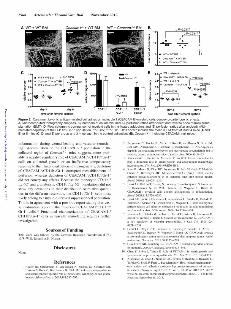
Using direct and reverse bone marrow transplantation between CEACAM1-competent donors and Ceacam1−/− recipients, we demonstrate that transfer of CEACAM1+ BM reconstituted adequate collateral formation, irrespective of the Ceacam1 genotype of the recipient. On the contrary, introduction of CEACAM1 null grafts into wild-type (WT) mice compromised arteriogenesis, and the number of collater-als was decreased opposed to WT or Ceacam1−/− mice that
received WT BM (WT+WT BM, 9.8±0.5; Ceacam1−/−+WT BM, 9.8±0.7; WT+Ceacam1−/− BM, 6.4±1.0; Figure 2A−2C). Thus, we characterized myeloid and hematopoietic cell popu-lations in peripheral blood, BM, and adductors of the different mouse lines.
In the adductors of Ceacam1−/− mice, 7 days after liga-tion of the femoral artery, only the CD11b+/Gr-1+ population exhibited a relative local increase (Figure 2D; Figure I in the online-only Data Supplement). Antibody-mediated deple-tion of this population conveyed proof that perfusion recov-ery is sensitive toward elimination of CEACAM1-expressing CD11b+Gr-1+ cells but not CEACAM1-negative CD11b+Gr-1+ cells (Figure 2E).
DiscussionWe describe for the first time that CEACAM1+ myeloid cells are essential for adequate arteriogenesis in a model of murine hindlimb ischemia and that their presence increases the number of collateral arteries and collateral vessel cali-bers. Preexisting collateral diameters in naïve mice were not altered in CEACAM1 null mice, immediately after ves-sel occlusion, which is different from platelet endothelial cell adhesion molecule 1−knockout mice compared with wild-type littermates.10 Therefore, the immunoglobulin fam-ily members platelet endothelial cell adhesion molecule and CEACAM1 mediate their proarteriogenic effects via differ-ent mechanisms or differentially modulate the arteriogenic activity of myeloid cells. Because only transplantation of WT BM into Ceacam1−/− mice reintroduced complete recov-ery in the arteriogenic response, we confirmed the require-ment for CEACAM1+ BM−derived cells for resolution of
Figure 1. Carcinoembryonic antigen−related cell adhesion molecule 1 (CEACAM1) deficiency impairs collateral formation. A, Microcom-puted tomography analyses, (B) numbers of collaterals, (C) perfusion recovery, (D) arterial diameters, and (E) endothelial (CD31, green) and pericyte staining (neuron-glial antigen 2 [NG2], red) of arteries in the adductors of CEACAM1-competent and Ceacam1−/− mice at indi-cated time points (C) or 7 days after femoral artery ligation. The blue bars in A indicate the location of femoral artery ligation, whereas the arrows point to collateral vessels. Data shown include the mean±SEM from at least 6 mice (A, B, and C) or 3 mice (D and E) per group. Ceacam1−/− indicates CEACAM1 null mice.
by guest on July 12, 2018http://atvb.ahajournals.org/
Dow
nloaded from
2568 Arterioscler Thromb Vasc Biol November 2012
inflammation during wound healing and vascular remodel-ing5: Accumulation of the CD11b+/Gr-1+ population in the collateral region of Ceacam1−/− mice suggests, more prob-ably, a negative regulatory role of CEACAM1−/CD11b+/Gr-1+ cells on collateral growth or an ineffective compensatory response to their functional deficiency. Congruently, depletion of CEACAM1+/CD11b+/Gr-1+ corrupted reestablishment of perfusion, whereas depletion of CEACAM1−/CD11b+/Gr-1+ did not convey any effects. Because the monocytic CD11b+/Ly-6C+ and granulocytic CD11b+/Ly-6G+ populations did not show any deviations in their distribution or relative quanti-ties in the different reservoirs, the CD11b+/Gr-1+ cells most likely belong to a myeloid-derived suppressor cell population. This is in agreement with a previous report stating that ves-sel maturation is poor in the presence of CEACAM1−CD11b+/Gr-1+ cells.11 Functional characterization of CEACAM1+/CD11b+/Gr-1+ cells in vascular remodeling requires further investigation.
Sources of FundingThis work was funded by the German Research Foundation (DFG 13/3; W.D. Ito and A.K. Horst).
DisclosuresNone.
References 1. Hoefer IE, Grundmann S, van Royen N, Voskuil M, Schirmer SH,
Ulusans S, Bode C, Buschmann IR, Piek JJ. Leukocyte subpopulations and arteriogenesis: specific role of monocytes, lymphocytes and granu-locytes. Atherosclerosis. 2005;181:285–293.
2. Bergmann CE, Hoefer IE, Meder B, Roth H, van Royen N, Breit SM, Jost MM, Aharinejad S, Hartmann S, Buschmann IR. Arteriogenesis depends on circulating monocytes and macrophage accumulation and is severely depressed in op/op mice. J Leukoc Biol. 2006;80:59–65.
3. Khmelewski E, Becker A, Meinertz T, Ito WD. Tissue resident cells play a dominant role in arteriogenesis and concomitant macrophage accumulation. Circ Res. 2004;95:E56–E64.
4. Kim JA, March K, Chae HD, Johnstone B, Park SJ, Cook T, Merfeld-Clauss S, Broxmeyer HE. Muscle-derived Gr1(dim)CD11b(+) cells enhance neovascularization in an ischemic hind limb mouse model. Blood. 2010;116:1623–1626.
5. Horst AK, Bickert T, Brewig N, Ludewig P, van Rooijen N, Schumacher U, Beauchemin N, Ito WD, Fleischer B, Wagener C, Ritter U. CEACAM1+ myeloid cells control angiogenesis in inflammation. Blood. 2009;113:6726–6736.
6. Horst AK, Ito WD, Dabelstein J, Schumacher U, Sander H, Turbide C, Brümmer J, Meinertz T, Beauchemin N, Wagener C. Carcinoembryonic antigen-related cell adhesion molecule 1 modulates vascular remodeling in vitro and in vivo. J Clin Invest. 2006;116:1596–1605.
7. Nouvion AL, Oubaha M, Leblanc S, Davis EC, Jastrow H, Kammerer R, Breton V, Turbide C, Ergun S, Gratton JP, Beauchemin N. CEACAM1: a key regulator of vascular permeability. J Cell Sci. 2010;123: 4221–4230.
8. Gerstel D, Wegwitz F, Jannasch K, Ludewig P, Scheike K, Alves F, Beauchemin N, Deppert W, Wagener C, Horst AK. CEACAM1 creates a pro-angiogenic tumor microenvironment that supports tumor vessel maturation. Oncogene. 2011;30:4275–4288.
9. Gray-Owen SD, Blumberg RS. CEACAM1: contact-dependent control of immunity. Nat Rev Immunol. 2006;6:433–446.
10. Chen Z, Rubin J, Tzima E. Role of PECAM-1 in arteriogenesis and specification of preexisting collaterals. Circ Res. 2010;107:1355–1363.
11. Arabzadeh A, Chan C, Nouvion AL, Breton V, Benlolo S, Demarte L, Turbide C, Brodt P, Ferri L, Beauchemin N. Host-related carcinoembry-onic antigen cell adhesion molecule 1 promotes metastasis of colorec-tal cancer. Oncogene. April 2, 2012: doi: 10.1038/onc.2012.112. http://www.nature.com/onc/journal/vaop/ncurrent/full/onc2012112a.html. Accessed September 18, 2012.
Figure 2. Carcinoembryonic antigen−related cell adhesion molecule 1 (CEACAM1)+-myeloid cells convey proarteriogenic effects. A, Microcomputed tomography analyses, (B) numbers of collaterals, and (C) perfusion ratios after direct and reverse bone marrow trans-plantation (BMT). D, Flow cytometric comparison of myeloid cells in the ligated adductors and (E) perfusion ratios after antibody (Ab)-mediated depletion of the CD11b+/Gr-1+ population. *P<0.05; ** P<0.01. Data shown include the mean±SEM from at least 4 mice (A and B) or 5 mice (C, D, and E) per group and 3 mice each in the control collectives (E). Ceacam1−/− indicates CEACAM1 null mice.
by guest on July 12, 2018http://atvb.ahajournals.org/
Dow
nloaded from
Kristina HorstKlinke, Wolfgang Rottbauer, Michael Amling, Christoph Wagener, Wulf D. Ito and Andrea
Thomas Bickert, Robert Percy Marshall, Ziyang Zhang, Peter Ludewig, Mascha Binder, AnnaBrief Report−− Myeloid Cells+/Gr-1+Adhesion Molecule 1 Expression on CD11b
Acceleration of Collateral Development by Carcinoembryonic Antigen-Related Cell
Print ISSN: 1079-5642. Online ISSN: 1524-4636 Copyright © 2012 American Heart Association, Inc. All rights reserved.
Greenville Avenue, Dallas, TX 75231is published by the American Heart Association, 7272Arteriosclerosis, Thrombosis, and Vascular Biology
doi: 10.1161/ATVBAHA.112.3000152012;
2012;32:2566-2568; originally published online September 6,Arterioscler Thromb Vasc Biol.
http://atvb.ahajournals.org/content/32/11/2566World Wide Web at:
The online version of this article, along with updated information and services, is located on the
http://atvb.ahajournals.org/content/suppl/2013/08/21/ATVBAHA.112.300015.DC1Data Supplement (unedited) at:
http://atvb.ahajournals.org//subscriptions/
at: is onlineArteriosclerosis, Thrombosis, and Vascular Biology Information about subscribing to Subscriptions:
http://www.lww.com/reprints
Information about reprints can be found online at: Reprints:
document. Question and AnswerPermissions and Rightspage under Services. Further information about this process is available in the
which permission is being requested is located, click Request Permissions in the middle column of the WebCopyright Clearance Center, not the Editorial Office. Once the online version of the published article for
can be obtained via RightsLink, a service of theArteriosclerosis, Thrombosis, and Vascular Biologyin Requests for permissions to reproduce figures, tables, or portions of articles originally publishedPermissions:
by guest on July 12, 2018http://atvb.ahajournals.org/
Dow
nloaded from

1
Supplemental Material
Supplemental Methods.
Generation of Ceacam1-/- mice
Ceacam1-/- mice were generated by homologous recombination-assisted deletion of exons 1-4
and insertion of a Neo cassette using Sv129 ES cells (Genoway, Lyon, France). Backcrossing
was performed for 12 generations onto C57BL/6J background. Mice were genotyped by
polymerase chain reaction using a four-primer PCR using primers Ex5zuHorN1(5'-GGT CAC
AGA GTC TAG TTC TT-3') and HorN1 (5'-CTA AAG CGC ATG CTC CAG ACT GCC-3')
to identify the knockout allele (430 bp), and PN5 (5'-TAC ATG AAA TYG CAC CAG TCG
C-3') and PN8 (5'-CTG CCC CTG GCG CTT GGA-3') to identify the wild type allele6. The
construct for the targeted deletion is shown in Supplementary Figure II.
Induction of arteriogenesis and perfusion measurement
Hind limb ischemia was induced in mice by femoral artery ligation as previously described6.
Briefly, mice were anesthetized with 8 mg/kg body weight xylazine and 100 mg/kg ketamine
intraperitoneally and the right femoral artery was ligated twice proximal to the superficial
epigastric artery using 7-0 silk sutures (Ethicon, Livingston, Scotland). Before and after as
well on day three and six blood flow was measured using the Laser Doppler O2C (Oxygen to
See; LEA Medizintechnik, Gießen, Germany). Therefore mice were anesthetized with
isoflurane inhalation and kept on a warming plate with 37 °C. The shallow well LF-2 was
fixed with tape on the footpad of the mice. Blood flow was measured three times alternately
on the right and left footpad as arbitrary units [AU]. Perfusion was expressed as ratio of the
flow of the occluded and non-occluded hind limb.
Evaluation of collateral growth with µCT analysis
Seven days after femoral artery occlusion, mice were anesthetized, perfused first with pre-
warmed saline containing heparin via the left ventricle and subsequently with 45 % barium
sulfate in 9 % galantine. After hardening of the galantine on ice the legs were dissected, fixed
overnight in 4 % paraformaldehyde and transferred in 70 % ethanol. Using Micro Computer
Tomography (µCT; 1CT40, Scanco, Bassersdorf, Switzerland), lower limbs were scanned (55
kV/145 µA, voxel size 15 µm) and a three-dimensional reconstruction of the blood vessel
microstructure was performed. Finally corkscrew collaterals were calculated blinded, without
2
knowledge of the type of mice investigated.
Immunohistology of arteries in the adductor muscle
Seven days after femoral artery ligation of WT and Ceacam1-/- mice animals were euthanized
and perfused with saline. The adductor muscles were dissected, placed in O.C.T. compound
(Sakura, Zoeterwoude, Netherlands) and snapfrozen in liquid nitrogen. 7-µm cryosections
were fixed with ice-cold acetone and stained with antibodies binding PECAM1 (rat anti-
CD31, unlabeled, Acris, Herford, Germany) and NG2 (rabbit anti-NG2, unlabeled, Millipore,
Schwalbach, Germany). As secondary antibodies, goat anti-rat Alexa488 (Invitrogen, Life
technologies, Carlsbad, CA) and swine anti-rabbit TRITC (DAKO, Glostrup, Denmark) were
used. Counterstaining of nuclei was performed with DAPI (Invitrogen). Slides were mounted
with Aqua PolyMount (Polysciences, Warrington, PA) and visualised with the Leica
microscope DM5000 B connected with the camera DFC 360 FX (Leica, Wetzlar, Germany).
Pictures were taken with Leica software AF 6000 and statistic calculations of capillary
densities or vessel diameters larger than 15µm were performed with ADOBE photoshop
software. For assessment of vessel wall thickness, the CD31-positive endothelial as well
NG2-positive pericyte cell layers were included. To determine vessel lumen dimension and
sizes, the vessel lumens were highlighted in ADOBE photoshop software, and the diameters
of the marked vessels were approximated by calculation of a size-matched circle. Only
vessels with a diameter larger than 15 µm were considered, since smaller vessels are pre-
capillaries and not arterioles.
Flow cytometric analyses
Single cell suspensions from peripheral blood, BM and the adductor muscles at the site of
femoral artery ligation were analysed by flow cytometry seven days after ligation. Femurs
were flushed and passed through a 70-µm cell strainer (BD, Heidelberg, Germany) to obtain
single cell suspensions. Adductor muscles were resected, weighed and digested using a
cocktail of 10 U/ml dispase (BD Pharmingen), 0.2 mg/ml ColIV (Worthington, Lakewood,
NJ) and 0.1 mg/ml DNAse I (Roche Diagnostics, Mannheim, Germany) in DMEM for 1 hour
at 37 °C with constant shaking. The suspension was passed through a 70-µm cell strainer.
Unspecific binding of antibodies was blocked using Cohn-II (Sigma-Aldrich, Steinheim,
Germany). The following monoclonal antibodies were used: FITC-labeled anti-CD11b (BD),
PE-labeled anti-Gr-1 (BD), Alexa647-labeled anti-F4/80 (BioLegend, San Diego, CA),
Alexa647-labeled anti-CEACAM1 (CC1, a kind gift from K. Holmes), PE-labeled anti-Ly-6C
3
(BD), PE-labeled anti-Ly-6G (BD), PE-labeled anti-CCR2 (R&D, Minneapolis, MN),
Alexa647-labeled anti-CD206 (eBioscience, San Diego, CA), PE-labeled anti-sca-1 (BD),
FITC-labeled anti-CD31 (BD), APC-labeled anti-VEGFR1 (R&D) and PE-labeled anti-
CXCR4 (eBioscience). As controls the appropriate isotype antibodies were used. Dead cells
were excluded by 7-AAD (BD) staining. Analyses were performed with a FACScalibur (BD)
flow cytometer using CellQuestProTM (BD) software. To quantitate CD11b+/Gr-1+ and
CD11b+/Ly-6C+ populations, all live cells were included in the gate; in double labelings for
CD11b+/Gr-1+ or CD11b+/Ly-6C+, these double positive cells were quantified in dot plot
analyses as calculated by the CellQuestProTM software.
Bone marrow transfer
BM chimeras were generated as previously described5. Briefly, WT and Ceacam1-/- mice
were irradiated with 8 Gy. One day after, mice received 1x10⁷ BM cells from either WT or
Ceacam1-/- mice. Transfer efficiency was checked 60 days thereafter by flow cytometric
analyses. To distinguish between donor and recipient cells the two allelic forms CD45.1 and
CD45.2 were used.
Ablation of Gr-1+ cells
To systemically deplete Gr-1+ cells in vivo, 150 µg of the monoclonal antibody RB6-8C5 (Gr-
1, eBioscience) were injected intraperitoneally one day before and two and five days after
femoral artery ligation. As a control, the appropriate isotype antibody was used. To monitor
the depletion of Gr-1 positive cells, polymorphonuclear cells from blood smears one day after
each bolus were counted.
Statistical analyses
Statistical analyses were carried out following F-test analyses with the Mann-Whitney U test.
P values less than 0.05 were considered to be statistically significant.
4
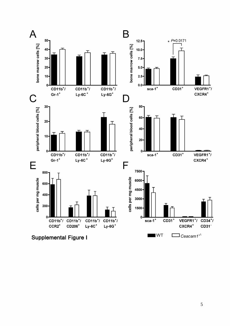
Supplemental Figure Legends. Supplemental Figure I: Analyses of myeloid cells and hemangiogenic progenitor
populations. Flow cytometric analyses of BM, peripheral blood and muscles seven days after
femoral artery ligation of WT and Ceacam1-/- mice. Myeloid cells and endothelial progenitors
did not reveal differential amounts or distribution of CD11b+/Ly-6C+ and CD11b+/Ly-6G+
cells (A, C, E). No quantitative differences in the presence of type 1 macrophages (identified
by CD11b and CCR2) or type 2 macrophages (identified by CD11b and CD206) were found
(E). CD31+ cells were only increased in BM (p=0.0219) (B) from Ceacam1-/- mice but not in
peripheral blood (D) or adductor muscles (F). Likewise, no spatio-quantitative differences
were found for sca-1+ and VEGFR1+/CXCR4+ populations. Data shown include the mean ±
SEM from at least 4 mice per group.
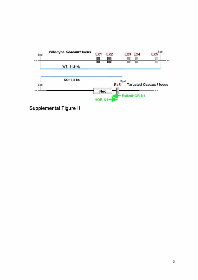
Supplemental Figure II: Transgenic construct for the generation of CEACAM1-
knockout mice. Two SpeI restriction sites are located upstream and downstream of exons 1-5
from the murine Ceacam1 gene (upper panel). They comprise 11.9 kb. The recombinant allele
with insertion of the Neo cassette and deletions of exons 1-4 can be identified by a 6.8 kb Spe
I restriction fragment. Recombinant alleles were identified by primers HOR-N1 and
Ex5zuHOR-N1, encompassing the overlapping fragment between the Neo cassette and exon 5
of the Ceacam1 gene. The location of the primers is indicated by green arrows (lower panel).
5
6