Embed Size (px)
Citation preview

Endocrine
hypertension
Justyna Kuliczkowska-Płaksej
Department of Endocrinology, Diabetes and Isotope Therapy
Wroclaw Medical University

Idiopathic hypertension (primary or essential) - 85%
Secondary hypertension - identifiable conditions - 15%:
- primary renal disease
- oral contraceptive use
- sleep apnea syndrome
- congenital or acquired cardiovascular disease (i.e. coarctation of the aorta)
- excess hormonal secretion
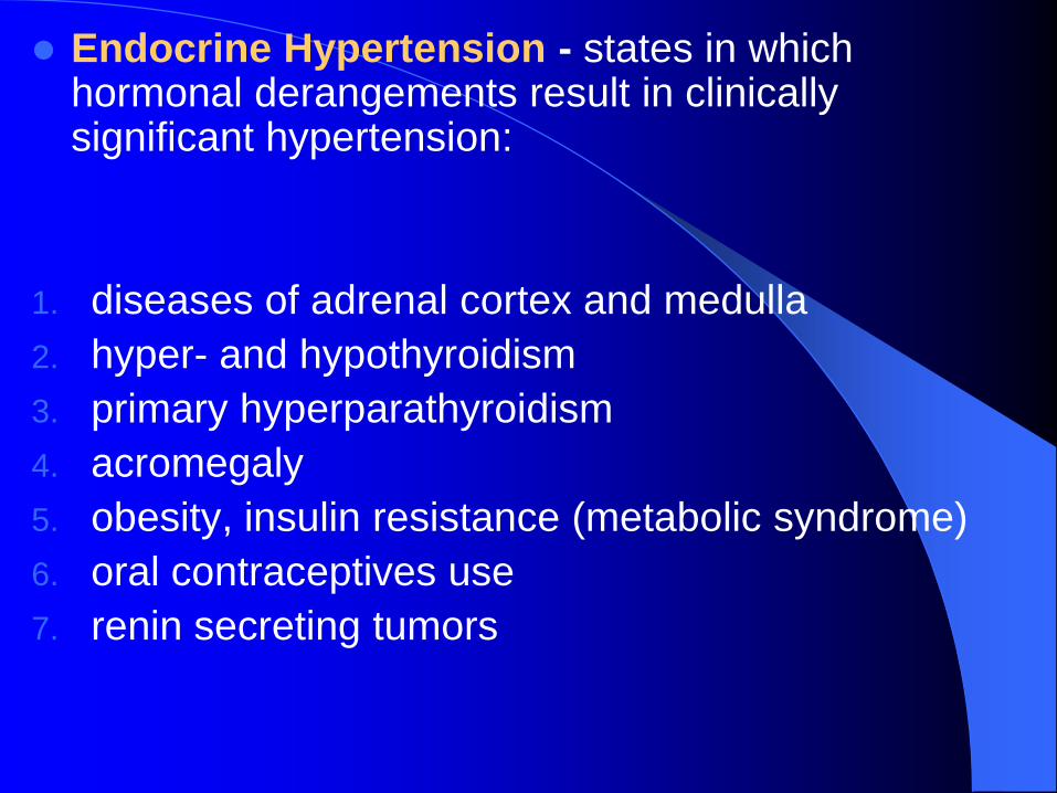
Endocrine Hypertension - states in which hormonal derangements result in clinically significant hypertension:
1. diseases of adrenal cortex and medulla
2. hyper- and hypothyroidism
3. primary hyperparathyroidism
4. acromegaly
5. obesity, insulin resistance (metabolic syndrome)
6. oral contraceptives use
7. renin secreting tumors
Hypertension of adrenal origin
Primary aldosteronism
Cushing’s syndrome
Pheochromocytoma
Syndromes due to excess deoxycorticosterone
production
Type II pseudohypoaldosteronism (Arnold-Healy-
Gordon Syndrome)
Androgen- & estrogen-producing adrenal tumors
Primary cortisol resistance

Primary aldosteronism
prevalence: previously - 1-2%, recent data – 4-10%
sporadic or familial
adenoma (Conn’s syndrome): 30-40%
hyperplasia (uni- or bilateral) of zona glomerulosa
tissue: 60%
aldosterone-producing adrenocortical carcinoma: 0.5-1%
dexamethasone (glucocorticoid)-remediable aldosteronism: 1-3%
ectopic secretion (ovarian, renal and intestinal carcinoma) < 1%

1. Aldosterone-producing adenoma (APA)- small (usually < 2 cm), more common in women
2. Bilateral adrenal hyperplasia (BAH) –idiopathic, milder extent of hyperaldosteronism, degree of hypokalemia and suppression of PRA compared to APA
3. Unilateral adrenal hyperplasia (UAH) - hypertension and biochemical abnormalities may be cured or ameliorated by unilateral adrenalectomy (similar to APA)
4. Adrenal carcinomas – rare, usually large tumor (> 5 cm), the diagnosis based upon evidence of extension of the tumor through the adrenal capsule or a high mitotic index on histologic examination
Primary aldosteronism - causes

Familial aldosteronism (2%)
familial hyperaldosteronism type 1: glucocorticoid-remediable (GRA), autosomal dominant disorder - chimeric gene duplication: 5’-promotor region of the 11 β-hydroxylase gene (regulated by ACTH) + coding sequences of the aldosterone synthase gene
regulation of aldosterone synthesis by ACTH
Primary aldosteronism - causes

should be considered in patients with:
• early-onset hypertension
• suppressed PRA
• strong family history of early cerebral hemorrhage (< 35 yr)
diagnosis of GRA:
suppression of serum aldosterone (dexamethasone 0.5 mg each 6 h for 48 h - reduction of aldosterone to undetectable levels (< 4 ng/dl))
genetic testing
treatment - dexamethasone
Primary aldosteronism - causes
familial hyperaldosteronism type 2:
not glucocorticoid remediable
responsible gene has been linked to chromosome
7p22 but has not yet been identified
present in at least two relatives

ALDOSTERONE
Potassium depletion
Hypokalemia
Vasoconstriction
HYPERTENSION
+ inotropic effect
Cardiac output
Sodium retension Fluid retension
ECFV expansion
Plasma volume
Intracellular Na+
Ca2+ efflux
Vascular
smooth muscle
contraction
Total peripheral
resistance
Renin suppression

Hyperaldosteronism - clinical features
hypertension (severe)
hypokalemia (< 3.5 mEq/l), alkalosis
fatigue, weakness
thirst
polyuria (especially nocturnal)
paresthesias
headaches
Symptoms of
K+ depletion

Primary hyperaldosteronism - diagnosis
biochemical assays: K+ (< 3.5 mEq/l), K+ in urine
(> 50 mmol/24h)
up to 20% of patients may have normal or low-
normal serum K+ concentrations!!!
hormonal assays: aldosterone and plasma renin
activity before and after 2 hours in the upright
posture (activation of renin system with
aldosterone)
in case of adenoma – no significant change or
aldosterone

24-hour urine collection for aldosterone (>20 g/24h)
aldosterone (PA, ng/dL) to renin activity (PRA, ng/ml/h) ratio:
< 20 - normotensive or essential hypertension
> 20 - suspicion for primary aldosteronism
> 30 (with a PA > 15 ng/dl) - 90% sensitivity and 91% specificity for the diagnosis of primary aldosteronism
50 - diagnostic of primary aldosteronism
the best radiographic procedure: adrenal CT
scanning
adrenal vein sampling -‘gold standard’ in diagnosis of
unilateral versus bilateral aldosterone hypersecretion
US – less sensitive
MRI, adrenal scintigraphy – second choice diagnostic
tools

Primary hyperaldosteronism - treatment
adenoma - surgery (often laporoscopic) resection of
APA may cure or ameliorate hypertension in APA
patients and reverse the hypokalemia
bilateral adrenal hyperplasia - mineralocorticoid
receptor antagonist – eplerenone (2 x 25 mg) or
spironolactone (100-500 mg)

spironolactone: 50-400 mg/day, usually 2x/day,
side effects (at doses >100 mg/day) - especially in
males (gynecomastia and erectile dysfunction) and
females (menstrual dysfunction)
eplerenone - no anti-androgen activity
dihydropyridine calcium channel antagonists -
effectively blood pressure and may aldosterone
secretion
dietary sodium restriction (<100 mmol/day), regular
aerobic exercise, maintenance of ideal body weight


Pheochromocytoma
catecholamine-producing tumors of chromaffin cells
WHO classification:
intra-adrenal paragangliomas (85%) -
pheochromocytomas
extra-adrenal paragangliomas (15%)
from sympathetic nervous system-associated
chromaffin tissue
from parasympathetic nervous system-associated
chromaffin tissue

present at any age (more commonly in the 4-5th
decades)
located in adrenals - 90% in adults, 70% in children
unilateral - 90% – more frequent in the right adrenal
(65% vs. 35%)
bilateral – 10% in adults, 35% in children, (common
in familial syndromes)
Pheochromocytoma

Chromaffin paragangliomas
extra-adrenal tumors arising from sympathetic
ganglia
10% of chromaffin-tissue tumors in adults, 30% in
children
36-60% - functional, secreting norepinephrine,
normetanephrine
70% - intra-abdominal (juxtarenal, para-aortic
region, bladder)
30% - chest (anterior or posterior mediastinum,
heart)

Pheochromocytoma - clinical presentation
Hypertension >98%
Hypertension sustained 50-60%
Hypertension paroxysmal 50%
Headache 70-90%
Palpitations+tachycardia 50-70%
Diaphoresis 60-70%
Fever >66%
Pallor 30-60%
Hyperglycemia 42%
Abdominal/chest pain 20-50%
Nervousness 35-40%
Anxiety 20%
Nausea 26-43%
Vomiting 26-43%
Fatigue 15-40%
Flushing 18%
Orthostatic hypotension 12%

Headaches + palpitations + sweating in patient with hypertension – suspicion for a pheochromocytoma
Attacks of signs precipitated by: palpation of the
tumor, postural changes, exertion, anxiety, trauma,
pain, ingestion of foods or beverages containing
tyramine (certain cheese, beer, wine), use of certain
drugs (histamine, glucagon, phenothiazine,
metoclopramide), intubation, induction of
anesthesia, chemotherapy, endoscopy,
catheterization, micturition or bladder distension (in
case of bladder tumors)

Pheochromocytoma/paraganglioma - secreted
substances
adrenal pheochromocytomas - mainly epinephrine
extra-adrenal paragangliomas - norepinephrine
serum level of catecholamines does not correlate
with tumor size!!!
neuropeptide Y – very potent vasoconstrictor
neuron-specific enolase (NSE) – elevated in about
50% of patients with malignant pheochromocytoma
other peptides: PTHrP, ACTH, erythropoietin, IL-6,
CgA, adrenomedullin
Malignant pheochromocytoma
15% of pheochromocytomas
functional metastases:
to the skull – common, palpable
to the ribs – chest pain
to the spine – back pain, neurologic symptoms
pulmonary and mediastinal metastases – dyspnea, hemoptysis, pleural effusion, Horner’s syndrome
to the thoracic duct – chylothorax
to the liver – hepatomegaly
in mesenteric nodes

Genetic syndromes associated with
pheochromocytomas & paragangliomas
20-30% of pheochromocytomas or paragangliomas
recommendations for genetic screening:
extraadrenal paragangliomas
multifocal tumors
onset of symptoms at a young age (< 50)
family history of such tumors
patients with other manifestations of familial
syndromes

Multiple endocrine neoplasia type 2 (MEN2)
autosomal dominant
mutations in RET protooncogene on chromosome
10 (encoding a transmembrane receptor tyrosine
kinase)
MEN2A (90%)
MEN2B (10%)
in both types – pheochromocytomas develop in
adrenals, 4% - metastatic, usually bilateral

MEN2A (Sipple’s syndrome)
medullary thyroid carcinoma (95-100%)
hyperparathyroidism (multiglandular hyperplasia –
35%)
pheochromocytoma (50%) – often in middle age
high incidence of lichen planus amyloidosis and
Hirschprung’s disease

MEN2B
medullary thyroid carcinoma (more aggressive and
at earlier age than in MEN2A)
mucosal neuromas
intestinal ganglioneuromas
marfanoid habitus
pheochromocytoma
hyperparathyroidism – absent!
Von Hippel-Lindau Disease (VHL)
autosomal dominant
tumors in multiple tissues:
pheochromocytomas (only in type 2 VHL)
hemangioblastomas in the retina, cerebellum, spinal cord
renal cysts
renal clear-cell carcinoma
cysts in pancreas
endolymphatic sac tumors (vertigo, hearing loss, ataxia)
adnexal cystadenomas
epididymal cystadenomas

pheochromocytomas in VHL2: at an early age (mean 28 years), bilateral, produce only epinephrine
type 2A VHL:
hemangioblastomas + pheochromocytomas, low risk for developing renal cell carcinomas
type 2B VHL:
hemangioblastomas + pheochromocytomas, high risk for developing renal cell carcinomas
type 2C VHL:
pheochromocytomas, no hemangioblastomas or renal cell carcinomas

von Recklinghausen’s neurofibromatosis type 1 (NF-1)
autosomal dominant
mutation in the NF-1 tumor suppressor gene
optic gliomas, plexiform neurofibromas, subcutaneous neurofibromas, schwannomas of cranial and vertebral nerve roots, hypothalamic hamartomas, skeletal abnormalities
high risk of developing malignant peripheral nerve sheath tumors and leukemia
multiple cutaneous pigmented cafe au lait spots (usually more then 6 spots > 1.5 cm)

Pheochromocytomas in NF-1
0.1-5.7% of patients
similar to sporadic pheochromocytomas:
84% - solitary adrenal tumors
10% - bilateral
6% - extra-adrenal paragangliomas
12% - metastases or local invasion
mean age at diagnosis – 42 years

Familial paraganglioma/pheochromocytoma
syndrome
autosomal dominant
mutations in genes encoding mitochondrial complex II
multicentric head/neck paragangliomas, sympathetic
paragangliomas, adrenal pheochromocytomas

Pheochromocytoma - diagnosis
24-h urine collection for metanephrines
metanephrines in plasma
plasma or urine free catecholamines (less sensitive
and specific)
ultrasound scanning
metaiodobenzylguanidine (MIBG) scintigraphy
CT and MRI imaging
positron emission tomography (PET) scanning
somatostatin receptor imaging with 111In-Octreotide
venous sampling for catecholamines

Pheochromocytoma - treatment
preoperative management:
calcium channel blockers
alpha-adrenergic blockers
ACE inhibitors
beta-adrenergic blockers (not prescribed without
alpha-blockers)
metyrosine

Pheochromocytoma - surgical management
patients should be normotensive, well-hydrated
monitoring during surgery: blood pressure, ECG
laparoscopy
needlescopic adrenalectomy
adrenal cortex-sparing surgery
open laparotomy
Syndromes due to excess deoxycorticosterone
(DOC) production
DOC – second most important
mineralocorticosteroid hormone
excess DOC should be suspected in any
hypertensive patient with hypokalemia and
suppression of renin and aldosterone production

Syndromes due to excess DOC production
17-hydroxylase deficiency
11-hydroxylase deficiency
androgen- and estrogen-producing adrenal tumors
primary cortisol resistance

17-hydroxylase deficiency
single gene mutation
impaired synthesis of cortisol, sex steroids
secretion of large amounts of deoxycorticosterone
(DOC) and corticosterone
decreased aldosterone secretion, suppression of
renin

17α-hydroxylase deficiency
recognized at the time of puberty in young adults
presenting symptoms:
hypertension ( DOC, low renin, low aldosterone)
hypokalemia
primary amenorrhea
pseudohermaphroditism
cortisol deficiency
no virilization or retarded growth
11-hydroxylase deficiency
mutations of genes on chromosome 8
recognized in newborns and infants
classic, mild, late-onset forms (usually mild, with
partial defect of cortisol production)
androgens, 11-deoxycortisol, DOC,
17-hydroxyprogesterone, urinary 17-ketosteroids
and 17-hydroxycorticosteroids
cortisol

11-hydroxylase deficiency
virilization
hypertension ( DOC, hypokalemia, low renin)
diagnosis: 11-deoxycortisol, DOC, urinary
excretion of their metabolites

Androgen and estrogen producing adrenal tumors
malignancies originating in the zona reticularis
features of mineralocorticoid excess: hypertension,
hypokalemia, renin supression
inhibition of 11-hydroxylase with increased
production of androgens, DOC, 11-deoxycortisol
enzymatic inhibition probably is due to high intra-
adrenal concentration of androgens
(pseudosubstrate for the reaction)

Syndrome of apparent mineralocorticoid excess (11β-hydroxysteroid dehydrogenase deficiency)
rare disorder
mutation in gene encoding 11-hydroxysteroid
dehydrogenase type 2, which is present in the renal
tubule
periperal metabolism of cortisol: impaired
conversion to cortisone in the renal tubule
accumulation of cortisol and occupancy of
mineralocorticosteroid receptor
diagnosis: free steroids in urine (cortisol/cortisone)
or steroid metabolites
treatment: spironolactone, eplerenone, triamterene,
amiloride

Chronic ingestion of Licorice
glycyrrhizic acid in Licorice and its metabolite –
glycyrrhetinic acid – inhibit 11-hydroxysteroid
dehydrogenase in the kidney (syndrome of
apparent mineralocorticoid excess)
derivative - carbenoxolone (an antigastric ulcer
drug)
symptoms: hypertension, hypokalemia, renal Na
retension, volume expansion, suppressed plasma
renin activity, metabolic alkalosis

Liddle’s syndrome
familial disorder, autosomal dominant pattern of
inheritance
defect in the epithelial Na channel resulting in
constitutive activation
symptoms: hypertension, hypokalemia, renal K
wasting, metabolic alkalosis, suppressed plasma
renin activity, low aldosterone
treatment: administration of amiloride or triamteren
(inhibitors of Na channel), salt restriction

Mutations of mineralocorticoid receptor
(Geller’s syndrome)
autosomal dominant form of hypertension caused
by a mutation in ligand binding domain – partial
activation in the absence of aldosterone
hypertension before the age of 20

Type II pseudohypoaldosteronism
(Arnold-Healy-Gordon Syndrome)
autosomal dominant disorder
hypertension+hyperkalemia
low renin, low aldosteron
impairment of renal K+ excretion
mutations in genes encoding kinases that regulate Na+, Cl-, K+ pathways in distal nephron segments
increased renal NaCl reabsorption with inhibited K+ secretion
treatment: severe dietary salt restriction, antihypertensives (especially thiazide diuretics)

Glucocorticoid resistance
autosomal recessive or dominant
inactivating mutations of the glucocorticoid receptor
gene
cortisol and ACTH
no clinical features of Cushing’s syndrome
permanent ACTH production of compounds
with mineralocorticoid activity
cortisol stimulates mineralocorticoid receptor

Renin-secreting tumors
very rare
usually hemangiopericytomas containing
juxtaglomerular cells
other tumors secreting renin: Wilms’ tumor,
pulmonary tumors
hypertension, hypokalemia
high renin+high aldosterone
diagnosis: biochemical testing, CT, venous
sampling

Cushing’s syndrome
chronic glucocorticoid excess
ACTH-dependent (90%)
pituitary adenoma (Cushing’s disease) – 80%
nonpituitary neoplasm (ectopic ACTH secretion) –
10%
ACTH-independent (10%)
iatrogenic (glucocorticoids)
adrenal neoplasm (adenoma, carcinoma)
nodular adrenal hyperplasia
Cushing’s syndrome - clinical features
obesity – central, mainly affecting the face („moon
like face”), neck („buffalo hump”), trunk, abdomen,
with relative sparing of the extremities
skin changes – easy bruising, striae (red to purple,
most commonly abdominal), acne, slow healing of
minor wounds, mucocutaneous fungal infections,
hyperpigmentation in case of ectopic ACTH
secretion
Cushing’s syndrome – clinical features
hirsutism
gonadal dysfunction
psychologic disturbances – emotional lability,
anxiety, depression, poor concentration, psychosis
muscle weakness (more often proximal)
osteoporosis
diabetes mellitus
hypokalemia
hypertension

Cushing’s syndrome – associated hypertension
hypertension in approximately 80% of cases of endogenous hypercortisolemia (only in 10-20% in case of exogenous)
night-time RR decline is significantly lower than that in patients with essential hypertension

Cushing’s syndrome – associated hypertension
mechanisms of RR elevation:
hepatic production of angiotensinogene
cardiac output
production of prostaglandins (inhibition of phospholipase A)
insulin resistance
oversaturation of 11-HSD activity with increased mineralocorticoid effect
vascular sensitivity to catecholamines
extra- and intravasular volume
ectopic ACTH production – frequently associated with DOC
and corticosterone – mineralcorticosteroid excess state

Cushing’s syndrome - diagnosis
diurnal rhythm of cortisol secretion
urinary free cortisol (24 h urine collection)
plasma ACTH
dexamethasone suppression test:
screening test (short test) with 1 mg at bedtime (determination of cortisol at 8.00 a.m. – in healthy subjects < 1.8 µg/dl)
low- and high-dose
pituitary MRI
inferior pertosal sinus sampling
CT and MRI of adrenal glands

Cushing’s syndrome - treatment
pituitary adenoma: surgery (transsphenoidal or transcranial), radiation
ectopic ACTH syndrome: surgery (if possible), drugs that block steroid synthesis (aminoglutethimide, ketoconazole, metyrapone), bilateral adrenalectomy in very severe cases
adrenal tumors: surgery, mitotane (the drug of choice in case of adrenal carcinoma)

Metabolic syndrome
characterized by:
hypertension
abdominal obesity
dyslipidemia
insulin resistance
insulin resistance - significantly associated with
hypertension
patients with essential hypertension - often insulin
resistant

Metabolic syndrome and hypertension
insulin - direct stimulation of the calcium pump
calcium loss from the cell (in a cell resistant to
insulin the insulin-induced calcium loss is
decreased)
increased intracellular calcium increased
vascular smooth muscle cells vasoconstriction
sodium retention
activity of the adrenergic nervous system
obesity is associated with production of
adipokines that have impact on blood pressure

Hyperparathyroidism and hypertension
hypercalcemia - associated with incidence of
hypertension
in patients with primary hyperparathyroidism,
hypertension is observed in approximately 40% of
cases
the mechanisms of these associations are unclear
(hypercalcemia with response to
catecholamines???)
hypertension is not cured or better controlled after
parathyroidectomy!

Hyperthyroidism and hypertension
systolic blood pressure by:
heart rate (tachycardia)
systemic vascular resistance
cardiac output
stroke volume

Hypothyroidism and hypertension
positive association between serum TSH and blood pressure
hypothyroid patients often have:
diastolic blood pressure
impaired endothelial function
systemic vascular resistance
extracellular volume expansion
subclinical hypothyroidism seems not to be associated with hypertension

Acromegaly and hypertension
prevalence of hypertension- 46%
GH - antinatriuretic actions, may lead to sodium retention and volume expansion
systolic output and high heart rate (may lead to congestive heart failure)
the RAAS system appears to be implicated in the pathogenesis of hypertension in patients with acromegaly ( PRA, disturbances in dopaminergic regulation of aldosterone secretion)