Embed Size (px)
Citation preview

Abstract
Objectives: Advancements in nanotechnology have led to nanoparticle (NP) use in various fields of medicine. Although the potential of NPs is promising, the lack of documented evidence on the toxicological effects of NPs is concerning. A few studies have documented that homeopathy uses NPs. Unfortunately, very few sound scientific studies have explored the toxic effects of ho-meopathic drugs. Citing this lack of high-quality scien-tific evidence, regulatory agencies have been reluctant to endorse homeopathic treatment as an alternative or adjunct treatment. This study aimed to enhance our insight into the impact of commercially-available ho-meopathic drugs, to study the presence of NPs in those drugs and any deleterious effects they might have, and to determine the distribution pattern of NPs in zebraf-ish embryos (Danio rerio).
Methods: Homeopathic dilutions were studied using high-resolution transmission electron microscopy with selected area electron diffraction (SAED). For the toxicity assessment on Zebrafish, embryos were ex-posed to a test solution from 4 - 6 hours post-fertiliza-tion, and embryos/larvae were assessed up to 5 days post-fertilization (dpf) for viability and morphology. Toxicity was recorded in terms of mortality, hatching delay, phenotypic defects and metal accumulation. Around 5 dpf was found to be the optimum develop-mental stage for evaluation.
Results: The present study aimed to conclusively prove the presence of NPs in all high dilutions of ho-meopathic drugs. Embryonic zebrafish were exposed to three homeopathic drugs with two potencies (30CH, 200CH) during early embryogenesis. The resulting morphological and cellular responses were observed. Exposure to these potencies produced no visibly sig-nificant malformations, pericardial edema, and mor-tality and no necrotic and apoptotic cellular death.
Conclusion: Our findings clearly demonstrate that no toxic effects were observed for these three homeopath-ic drugs at the potencies and exposure times used in this study. The embryonic zebrafish model is recom-mended as a well-established method for rapidly as-sessing the toxicity of homeopathic drugs.
ISSN 2093-6966 [Print], ISSN 2234-6856 [Online]Journal of Pharmacopuncture 2016;19[4]:319-328DOI: https://doi.org/10.3831/KPI.2016.19.033
This is an Open-Access article distributed under the terms of the Creative CommonsAttribution Non-Commercial License (http://creativecommons.org/licenses/by-nc/4.0/)which permits unrestricted noncommercial use, distribution, and reproduction in anymedium, provided the original work is properly cited.
This paper meets the requirements of KS X ISO 9706, ISO 9706-1994 and ANSI/NISOZ39.48-1992 (Permanence of Paper).
*Corresponding AuthorMansee Thakur. Department of Biotechnology and CRL, MGMIHS and MGMCET, Sector-1, Kamothe, Navi Mumbai 410209, Maharashtra, India. Tel: +91-022-2743-2890 Fax: +91-022-2743-1094E-mail: [email protected]
ⓒ 2016 Korean Pharmacopuncture Institute http://www.journal.ac
Embryonic Zebrafish Model - A Well-Established Method for Rapidly Assessing the Toxicity of Homeopathic Drugs- Toxicity Evaluation of Homeopathic Drugs Using Zebrafish Embryo Model -
Himanshu R Gupta1, Yogesh Patil2, Dipty Singh3, Mansee Thakur4*
Original article
Received: Sep 07, 2016 Reviewed: Dec 01, 2016 Accepted: Dec 01, 2016
Key Wordshomeopathic medicine, homeopathy, traditional medicine, therapeutic use, transmission electron microscope, zebraf-ish toxicity
1 Department of Medical Genetics, MGMIUDBS, Mahatma Gandhi Mission Institute of Health Sciences, Kamothe, Navi Mumbai, India2 MGMCET & Central Research Laboratory, Mahatma Gandhi Mission Institute for Health Sciences, Kamothe, Navi Mumbai, India3 National Centre for Preclinical Reproductive and Genetic Toxicology (NIRRH), National Institute of Research in Reproductive Health (ICMR), Jehangir Merwanji Street, Parel, Mumbai, India4 Mahatma Gandhi Mission College of Engineering and Technology & Department of Medical Biotechnology, Central Research Laboratory, MGM Medical College, MGMIHS, Kamothe, Navi Mumbai, India
1. Introduction
Although in recent years we have witnessed a new begin-ning of interest in homeopathy, the reliability of its main principles, the ‘simile’ and the ‘dilution’ of medicines, is yet to be demonstrated on experimentally, and few studies have been conducted to understand its underlying mech-anism(s) [1-3]. These few efforts have borne fruit in that they have generated several hypotheses, but none of them have been sufficiently validated empirically, with results ranging from the scientifically preposterous “memory of water” to the prodigious hand waving around quantum physics [4]. With the emergence of new tools provided by nanoscience and nanotechnology, new research op-tions have been opened. The latest such attempts using these new tools found that extremely diluted homeopathic solutions did not contain the solute, but in fact contained the starting material in the forms of nanoparticles (NPs) and aggregates that were detectable by using electron microscopy, atomic spectroscopy, fluorescence spectros-copy, and cyclic voltammetry [5, 6]. Based on the results of the above studies, homeopathy is, in fact, a form of na-nopharmacology [7]. Through a specific pharmacological process of potentization/tituration, medicinal substances are broken down in a grinding machine into NPs. Howev-er, whether the homeopathic effect depends on the pres-ence of the NPs is still not known. Recent research has shown that remedies can carry numerous NPs, even up to a 200CH potency, and that homeopathy is not “just water”. Another hypothesis says that solutions of homeopathic drugs are ultra-molecular dilutions of the starting material (above Avogadro’s number). However, as above, whether these NPs are completely responsible for homeopathy’s therapeutic effects is still unknown. Perhaps, they are. Although many people believe that highly diluted home-opathic medicines, which are recommended by profes-sionals, may be safe and unlikely to produce any adverse action, drawing precise conclusions from the reported literature in this field is difficult. This could be due to the low methodological quality of the reports claiming possi-ble adverse effects [8] or to the many comments coming from the uproar over a critical controversy [9]. That the raw materials for many homeo¬pathic medicines are known to be poisons and that other studies have found that homeo-pathic drugs containing NPs exert biological effects on the body are both true. Even though homeopathic medicines are generally not likely to cause or lead to harm when dis-pensed appropriately, the toxicological outlook should not be ignored, specifically when using low concentrations of a risky starting material. Every toxicologist knows that a substance is poisonous only in a certain dose.
In the past few years, the number of in vitro and in vivo studies to evaluate the pharmacological kinetics or effi-cacy of some homeopathic remedies has been increasing [10, 11]. Despite the increasing popularity of homeopa-thy in biological applications, in vitro and in vivo toxici-ty studies on homeopathic drugs are still lacking. So far, no studies have investigated the toxicity of homeopathic drugs by using the zebrafish model. Our general working hypothesis is that using the zebrafish model should help to clarify some of the mechanisms of action of these tradi-
tional medicines.Over the last decade, the zebrafish (Danio rerio) model
has come to be recognized as a unique vertebrate mod-el for assessing drugs in vivo with respect to a wide range of toxicological and safety pharmacological evaluation, because of its sequenced genome and highly-conserved genetic pathway between zebrafish and human [12-14]. This model is utilized for high turnout toxicity screening of diverse compounds. This whole-animal model may be useful as a rapid, high-throughput, low-cost assay for tox-icity screening of compounds in the early developmental stages of Zebrafish [15]. Recent studies reported that the zebrafish embryo model has good predictivity for the tox-icity and the teratogenicity of compounds in rodents [16-19].
The aim of this study was to investigate the possible ex-istence of NPs in homeopathic medicines, and if possible, to examine whether this suffices to qualify homeopathy as a form of nanomedicine. Another aim was to illustrate the embryonic toxicity in zebrafish embryos exposed to homeopathy drugs for 4 - 96 hours post fertilization (hpf). To investigate the toxic effects in zebrafish embryos ex-posed to homeopathy drugs, we conducted a sequence of assessments, including embryonic mortality, the hatching rate, the presence of malformations, and whole-embryo cellular death. Taking embryonic toxicity as the indicator for evaluating homeopathic toxicity will be beneficial for studies on homeopathy and safety evaluations.
2. Materials and Methods
Medicines (potentized hydro alcoholic solutions) with different potencies (30CH and 200CH) based on Calcar-ea carbonica, Argenticum metallicum and Sepia were ob-tained from Schwabe India (authorized producing house in Mumbai certified by Good Manufacturing Practices (GMP) and International Organizarion for Standardiza-tion (ISO). The procured drugs were colorless, odorless, pre-sterilized and endotoxin free. The remedies were stored in brown-colored glass containers at room temper-ature away from sunlight.
For the ultraviolet-visible spectrometry (UV-Vis) charac-terization of the homeopathic drugs, their particle sizes were evaluated by using an Epoch Microplate Spectro-photometer (BioTek-SN-141215F) to monitor the visible absorption band. Data reported encompassed the wave-length range from 190 to 1100 nanometers (nm) [20]. For the nanoform characterization of the homeopathic drugs, the sizes and the morphologies of the homeopathic drugs were determined by using field emission gun - transmis-sion electron microscopy (FEG-TEM) (Tecnai G2, F30 magnification 58 X 1 million X (300 kV)). Carbon-coated grids were immersed in the sample and left to dry prior to observation. The selected area electron diffraction (SAED) ring patterns were examined using high-resolution TEM (HR-TEM).
All animal experiments were conducted with prior ap-proval from Mahatma Gandhi Missions’s Medical Col-lege’s Institutional Animal Ethics Committee. All proce-dures for the maintenance and care of the zebrafish were
http://www.journal.ac320 Journal of Pharmacopuncture 2016;19[4]:319-328

http://www.journal.ac 321Journal of Pharmacopuncture 2016;19[4]:319-328
in accordance with procedures described in the zebrafish book [21, 22]. Zebrafish embryos were collected 4 - 5 hpf from the zebrafish aquarium at the zebrafish facility of MGM Central Research Laboratory, MGMIHS, Navi Mum-bai, and were staged according to standard procedures. For the toxicity studies, six groups were homeopathically treated as follows: Calcarea carbonica at 30CH and 200CH, Argenticum metallicum at 30CH and 200CH, and Sepia at 30CH and 200CH. An potentized hydro alcoholic solution and an untreated control group of fertilized eggs were used as a control.
For the treatments with the above drugs, three sets of freshly prepared fertilized eggs were used, with 10 eggs being placed in ten individual wells of a 24-well plate. The homeopathic drugs treatment began at 24, 48 and 72 hpf for all 3 sets of embryos respectively. For the 24 hpf expo-sure set, eggs in the wells were exposed to the above-men-tioned drugs at 24 hpf when the eggs had developed nor-mally through different stages like blastula, gastrula and segmentation. For the 48 hpf and the 72 hpf exposure sets, the embryos in individual wells were exposed to the afore-said concentrations of the homeopathic drugs at 48 and 72 hpf when the eggs/embryos had developed normally past the pharyngula stage and hatching period, respec-tively. As a control, an untreated control group of 30 fer-tilized eggs and potentized hydro-alcoholic solution were examined in parallel. Observations for any adverse effects due to treatments with the homeopathic drugs were made on zebrafish embryos and larvae and were documented at specified time points (t = 6, 12, 24, 48, 60, 72, 84 and 96 hours). The endpoints used to assess developmental tox-icity comprised embryo/larva survival and hatching rate. Malformations among the embryos and the larvae from both the control and the treated groups were described and documented using a stereo microscope (Motic SM3-168 series). Dead embryos/larvae were removed immedi-
ately once they had been observed. Tests were performed in triplicate.
To investigate the role of apoptosis in the toxicity of ho-meopathy drugs at different strengths, we used acridine orange (AO) staining of the drug-treated an -untreated embryos [23]. AO stains cells with disturbed plasma mem-brane permeability, so it preferentially stains necrotic or late apoptotic cells whereas normal cells are impermeable to AO. Then, embryos were rinsed three times with phos-phate buffer solution (PBS) and incubated in a solution of AO at 5 µg/mL for 30 minutes in darkness at 28°C, which was followed by three rinses in PBS. Embryos were exam-ined using fluorescence microscopy (EVOL FL Auto-Life Technologies, AMAFP1000).
3. Results
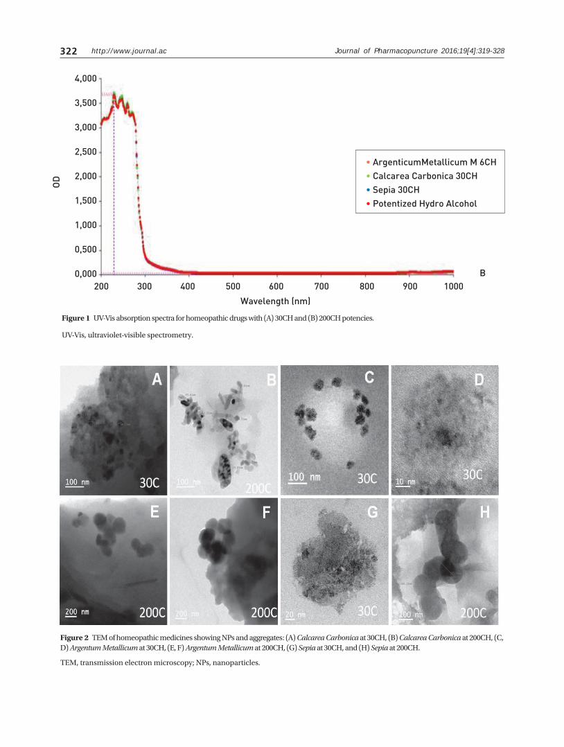
The properties of NPs change, depending upon their size. Thus, the size-dependent evolution of semiconducting NPs must be known to evaluate the properties of the ma-terial properly. UV-visible absorption spectroscopy is ex-tensively used to observe the optical properties of nano-sized particles, and as a result, new evidence is emerging on the quality and the properties of homeopathic medi-cines or their remedies. For these reasons, we determined the UV-V absorption spectra for three homeopathic drugs with different potencies (30CH, and 200CH), and the spec-tra are shown in Fig. 1. Specifically, the UV-Vis spectral graphs revealed a large peak absorbance signal close to a wavelength of 230 nm, with smaller peaks through approx-imately 350 nm in wavelength, which were notable in all samples, as well as in the potentized hydro alcohol control.
Calcarea carbonica, Argentum metallicum, and Sepia at dilutions of 30CH and 200CH were analyzed using bright-field TEM. The electron micrographs clearly showed the
4,000
3,500
3,000
2,500
2,000
1,500
1,000
0,500
0,000
OD
200 300 400 500 600 700 800 900 1000
Wavelength (nm)
• ArgenticumMetallicum M 6CH• Calcarea Carbonica 30CH• Sepia 30CH• Potentized Hydro Alcohol
A
(Continued)
http://www.journal.ac322
UV-Vis, ultraviolet-visible spectrometry.
TEM, transmission electron microscopy; NPs, nanoparticles.
Figure 1 UV-Vis absorption spectra for homeopathic drugs with (A) 30CH and (B) 200CH potencies.
Figure 2 TEM of homeopathic medicines showing NPs and aggregates: (A) Calcarea Carbonica at 30CH, (B) Calcarea Carbonica at 200CH, (C, D) Argentum Metallicum at 30CH, (E, F) Argentum Metallicum at 200CH, (G) Sepia at 30CH, and (H) Sepia at 200CH.
4,000
3,500
3,000
2,500
2,000
1,500
1,000
0,500
0,000
OD
200 300 400 500 600 700 800 900 1000
Wavelength (nm)
• ArgenticumMetallicum M 6CH• Calcarea Carbonica 30CH• Sepia 30CH• Potentized Hydro Alcohol
Journal of Pharmacopuncture 2016;19[4]:319-328
B

http://www.journal.ac 323Journal of Pharmacopuncture 2016;19[4]:319-328
TEM, transmission electron microscopy; SAED, selected area electron diffraction; NPs, nanoparticles.
HR-TEM, high-resolution transmission electron microscopy.
Figure 3 Bright-field TEM image and corresponding SAED patterns of NPs and conglomerates: (A, a) Calcarea Carbonica at 30CH, (B, b) Calcarea Carbonica at 200CH, (C, c) Argentum Metallicum at 30CH, (D, d) Argentum Metallicum at 200CH, (E ,e) Sepia at 30CH, and (F, f) Sepia 200CH.
Figure 4 HR-TEM images of the homeopathic medicines showing various degrees of crystallinity: (A, B) Calcarea Carbonica, (C) Argentum Metallicum, and (D, E) Sepia.

http://www.journal.ac324
Figure 5 (A) Survival and (B) hatching rate of zebrafish embryos exposed to different potencies of three homeopathic drugs at 96 hours post-fertilization.
Hatching Rate
B
Survival Rate
A
Journal of Pharmacopuncture 2016;19[4]:319-328

http://www.journal.ac 325Journal of Pharmacopuncture 2016;19[4]:319-328
Figure 6 Phenotypic observation and apoptotic pattern seen over the time course of exposures to homeopathic drugs at different potencies from 4 hour to 96 hour post-fertilization.
Bright field images of (A) an untreated embryo at 24 hour (B) untreated larvae at 96 hour and (C) a potentized hydro alcoholic solution at 24 hours; (D) larvae exposed to a potentized hydro alcoholic solution observed after 96 hour; embryos treated with 30CH potency of (E) Calcaria Carb, (G) Argenticum Metallicum, and (I) Sepia observed after 24 hour; (F, H, J) larvae post 30 CH exposure at 96 hpf (K, M, O) embryos treated with 200CH potency post 24 hours. (L, N, P) larvae post 200 CH exposure at 96 hpf. Fluorescent images of an untreated embryo at (a) 24 hour and (b) 96 hour. untreated embryo and larvae. Fluorescent potentized images of the hydro alcoholic solution at (c) 24 hour and (d) 96 hour. Fluorescent images of embryos treated with a 30CH potency of (e) Calcaria Carb, (g) Argenticum Metallicum, and (i) Sepia observed after 24 hour and (f, h, j) larvae at 96 hpf, embryos treated with 200CH potency of (k) Calcaria Carb, (m) Argenticum Metallicum, and (o) Sepia observed at 24 hours and (l, n, p) larvae at 96 hpf.
hpf , hours post fertilization.

326 http://www.journal.ac
presence of NPs and conglomerates in all three homeo-pathic medicines at both dilutions. We also observed di-versity in the shapes and the sizes of the NPs in the various medicines analyzed as well as in their different potencies. TEM analysis of Calcarea carbonica showed spherically shaped NPs of 20 - 40 nm in diameter at 30CH whereas at 200CH, it showed elliptical NPs with sizes in the range from 20 to 50 nm (Figs. 2A, 2B). In the case of Argentum metalli-cum, the shapes appeared to be spherical for both poten-cies, but the sizes varied (Figs. 2C-2F). The lower dilution of Argentum metallicum had smaller particles (30 - 60 nm) as compared to the higher dilution (50 - 100 nm). Similarly, Sepia at a dilution of 200C showed the presence of bigger spherical NPs of 100 – 200 nm in diameter (Figs. 2G, 2H).
The bright field TEM images confirmed the presence of NPs and conglomerates in all three homeopathic medi-cines. The conglomerates appeared as NPs clustered in the mesh of some matrix, which was further analyzed by using SAED to confirm the elemental composition. The SAED patterns of Calcarea carbonica and Sepia clearly revealed their polycrystalline nature (Figs. 3A, 3B, 3E, 3F) whereas the SAED pattern of the Argentum metallicum nanocluster consistently showed a crystalline pattern (Figs. 3C, 3D). The polycrystalline and the crystalline properties of Calcarea carbonica, Argentum metallicum and Sepia were found to be consistent for the two potencies that were studied. The crystalline lattices of these homeopathic medicines were further analyzed using HRTEM to study their atomic struc-tures. HRTEM images of these medicines showed various degrees of crystallinity from nanograins, dot and line ar-ray patterns, to lattice fringes (Figs. 4A-4D). This evidence indicates that the science of homeopathy is a form of NP nanomedicine [20]. To assess the toxicity of the above-mentioned homeo-pathic drugs at dilutions of 30CH and 200CH, we used the zebrafish embryo model to determine whether and how homeopathic medicines, which contain NPs, influence embryonic development. In this study, zebrafish embryos were exposed to homeopathic drugs for 96 hours, as shown in Fig. 5A. Here, drugs with 30CH and 200CH potencies had no toxic effects on zebrafish embryos or larvae. This result suggests that the development of toxicity in zebraf-ish due to homeopathic drugs is neither time nor dose de-pendent. The hatching rates among surviving embryos exposed to different potencies of the homeopathic drugs in each of the three sets showed no significant differences; at high dilution, as well as less dilution, these three homeopathic drugs with different CH values had no toxic effects on the zebrafish embryos. As shown in Fig. 5B, no toxic effect of the homeopathic drugs was detected when observing the mortality among zebrafish embryos. Mortality was found to be neither dose nor time dependent (data not shown). As compared with the control group, none of the three ho-meopathic drugs had any significant effect on mortality in the zebrafish embryos. All the embryos survived after ex-posure to the homeopathic drugs at dilutions of 30CH and 200CH for 96 hours. Zebrafish embryos exposed to homeopathic drugs devel-oped normal morphologies, and zebrafish larvae showed a range of phenotypic effects after exposure. Uninflated
swim bladder, yolk sac oedema, and dispersed pigment cells were the phenotypes most commonly observed. Bent tail, brachial arch hypoplasia, and bent body axis were the least recorded phenotypes. The six compounds did not produce any significant effects possibly because these compounds are not teratogenic according to the criteria used here. Simultaneously, we determined whether these homeopathic drugs induced apoptosis in zebrafish em-bryos and larvae by performing acridine orange staining to detect cellular death in live embryos. In the control group, a low, natural fluorescence was observed, which was sim-ilarly observed in the treated samples, as shown in Fig. 6.
4. Discussion
Surely, homeopathy must be free of undesirable effects. The typical homeopathic medicines at higher dilutions contain no active molecules, so they cannot cause any un-desirable effects. The raw materials for most homeopathic medicines are known to be poisons. Still, every toxicologist knows that a substance is poisonous only in a certain dose. One could even go one step further and argue that the generally acknowledged absence of side effects has made homeopathy as popular as it is today. In recent decades, homeopathy has developed a growing basic science and is providing clinical and health services, with the research literature showing unique physicochemical properties, biological activities in vitro and in vivo and exceptional safety.
The physicochemical properties of homeopathic reme-dies show that they contain low, but measurable, amounts of NPs [5, 6, 20, 24]. Advancements in the fields of nano-science and nanotechnology have resulted in countless possibilities for consumer product applications, many of which have already migrated from laboratory benches onto store shelves. From the results of the present study, the correlation of the UV-VIS spectral results with those derived from the analysis of the HR-TEM images provides further insights into the morphologies of homeopathic drugs. Clearly, the FEG-TEM images of the three different homeopathy preparations with two potencies showed var-ying size distributions of NPs. Interestingly, they contained significant proportions of NPs. Furthermore, despite the wide differences regarding the amounts of dilution- from 30CH to 200CH, no significant differences in the shapes and the sizes of the particles from those in the original ma-terial were observed. In addition, recent studies indicated that homeopathically-prepared medicines (HMs) contain source NPs, silicates [5, 6, 11, 20, 24-28], and other less well-characterized nanostructures. However, the TEM im-ages in this study did not confirm that these NPs were NPs of the starting material. In this study, we only investigated the possibility of NPs existing in homeopathic medicines and examined whether homeopathy as a form of nano-medicine could cause any adverse effects or toxicity.
Over 100 studies have evaluated the protective and the therapeutic effects of homeopathic doses of normally toxic substances [29]. A team of doctors from German research institutions and America’s Walter Reed Hospital performed
Journal of Pharmacopuncture 2016;19[4]:319-328

http://www.journal.ac 327Journal of Pharmacopuncture 2016;19[4]:319-328
Bellavite P, Andrioli G, Lussignoli S, Signorini A, Orto-lani R, Conforti A. A scientific reappraisal of the ‘prin-ciple of similarity’. Med Hypotheses. 1997;49(3):203-12.Bellavite P, Signorini A. The emerging science of home-opathy. Berkeley (CA): North Atlantic; 2002. 424 p.Teixeira MZ. ‘Paradoxical strategy for treating chronic diseases’: a therapeutic model used in homeopathy for more than two centuries, Homeopathy. 2005;94(4):265-6.Roduner E. Size matters: why nanomaterials are differ-ent. Chem Soc Rev. 2006;35(7):583-92.Chikramane PS, Suresh AK, Bellare JR, Kane SG. Ex-treme homeopathic dilutions retain starting mate-rials: a nanoparticulate perspective. Homeopathy. 2010;99(4):231-42.Sharma A, Purkait B. Identification of medicinally active ingredient in ultradiluted Digitalis purpurea: fluorescence spectroscopic and cyclic-voltammet-ric study. J Anal Methods Chem. 2012;2012:DOI: 10.1155/2012/109058.Bellavite P, Signorini A. The emerging science of home-opathy: complexity, biodynamics and nanopharma-cology. California: North Atlantic Books; 1995. 424 p.Dantas F, Rampes H. Do homeopathic medicines pro-voke adverse effects? a systematic review. Br Homeo-path J. 2000;89(S1):S35-8.Posadzki P, Alotaibi A, Ernst E. Adverse effects of home-opathy: a systematic review of published case reports and case series. Int J Clin Pract. 2012;66(12):1178-88.Vickers AJ. Independent replication of pre-clinical re-search in homoeopathy: a systematic review. Forsch Komplementarmed. 2004;6(6):311-20.Barve R, Chaughule R. Size-dependent in vivo/in vitro results of homoeopathic herbal extracts. J Nanostruct Chem. 2013;3(1):18.Goldsmith P. Zebrafish as a pharmacological tool: the how, why and when. Curr Opin Pharmacol. 2004;4(5):504-12.Rubinstein AL. Zebrafish assays for drug toxicity screen-ing. Expert Opin Drug Metab Toxicol. 2006;2(2):231-40.Winter MJ, Redfern WS, Hayfield AJ, Owen SF, Valentin JP, Hutchinson TH. Validation of a larval zebrafish lo-comotor assay for assessing the seizure liability of early-stage development drugs. J Pharmacol Toxicol Methods. 2008;57(3):176-87.Postlethwait JH, Woods IG, Ngo-Hazelett P, Yan YL, Kel-ly PD, Chu F, et al. Zebrafish comparative genomics and the origins of vertebrate chromosomes. Genome Res. 2000;10(12):1890-902.Redfern WS, Waldron G, Winter MJ, Butler P, Holbrook N, Wallis R, et al. Zebrafish assays as early safety phar-macology screens: paradigm shift or red herring?. J Pharmacol Toxicol Methods. 2008;58(2):110-7.
1.
2.
3.
4.
5.
6.
7.
8.
9.
10.
11.
12.
13.
14.
15.
16.
a meta-analysis of this study [31]. The zebrafish model is a vertebrate model that is gaining attention for preclinical drug-toxicity and drug-screening applications. Zebrafish embryos develop most of the major organ systems present in mammals, including the cardiovascular, nervous and digestive systems, in less than one week. Their early stages of life are susceptible to the adverse effects of drugs and chemicals [30]. In the present study, categorical lethal and sub-lethal endpoints were recorded from 24 hpf onwards for the three homeopathic drugs at the two different po-tencies (30CH and 200CH) that were tested. No delayed (72 hpf or 96 hpf) lethal endpoints were recorded for any of the three drugs at either of the two potencies. The observa-tions recorded in this study were in accordance with those in the zebrafish embryo toxicity test described earlier [21, 22]. We should note that the proposed zebrafish embryo model for toxicity testing is easy to perform and that the data obtained from this study will be helpful in assessing the potential risk of various homeopathic drugs having dif-ferent components such as organic, metallic, plant-based components.
5. Conclusion
Therapeutically, homeopathic medicines can cause clini-cal toxicity if the medications are improperly used. The few available clinical studies showed insufficient evidence of toxicity. However, insufficient clinical evidence proves nei-ther a lack of toxicity assessment nor a lack of a scientific basis for treatment. The scientific evidence presented here has shown that homeopathic drugs have no proven toxic-ity in zebrafish embryos (in vivo) at either lower or higher potencies. Most conventional treatments continue to be associated with severe and sometimes fatal adverse effects while homeopathic treatments have been found to be free from such adverse effects.
Acknowledgment
The authors gratefully acknowledge the technical support from Bhabha Atomic Research Centre (BARC) and Indian Institute of Technology (IIT), Mumbai. We also wish to thank MGM Institue of Health Sciences for providing the facility to carry out the research work.
Authors’ contributions
HG and YR performed the experiments and helped in data acquisition. DS interpreted the electron microscopy results. MT designed the experiments and gave intellec-tual input. HG, DS, and MT wrote the manuscript. All au-thors read and approved the final manuscript.
ORCID
Mansee Thakur. http://orcid.org/0000-0003-4743-1476.
Conflict of interest
The authors declare that there are no conflicts of interest.
References

http://www.journal.ac328
17.
18.
19.
20.
21.
22.
23.
24.
25.
26.
27.
28.
29.
30.
31.
Parng C, Seng WL, Semino C, McGrath P. Zebrafish: a preclinical model for drug screening. Assay Drug Dev Technol. 2002;1(1):41-8.Ali S, van Mil HGJ, Richardson MK. Large-scale assess-ment of the zebrafish embryo as a possible predictive model in toxicity testing. Plos one. 2011;6(6):DOI: 10.1371/journal.pone.0021076.McGrath P, Li CQ. Zebrafish: a predictive model for assessing drug-induced toxicity. Drug Discov Today. 2008;13(9-10):394-401.Bell IR, Muralidharan S, Schwartz GE. Nanoparti-cle characterization of traditional homeopathical-ly-manufactured silver (Argentum metallicum) medi-cines and placebo controls. J Nanomed Nanotechnol. 2015;6(4):DOI: 10.4172/2157-7439.1000311.Westerfield M. A guide for the laboratory use of zebraf-ish danio (brachydanio) rerio. Eugene:University of Or-egon Press; 2000.Test no. 236: fish embryo acute toxicity (FET) test [internet]. OECD; 2013 [Nov, 2016]. Available from: http://www.oecd-ilibrary.org/docserver/down-load/9713161e.pdf?expires=1481098481&id=id&ac-cname=guest&checksum=0EFE0447C6B022B5CA-F2AAEB18B9F3CC.Hu YL, Qi W, Han F, Shao JZ, Gao JQ. Toxicity evalu-ation of biodegradable chitosan nanoparticles us-ing a zebrafish embryo model. Int J Nanomedicine. 2011;6:3351-9.Chikramane PS, Kalita D, Suresh AK, Kane SG, Bellare JR. Why extreme dilutions reach non-zero asymptotes: a nanoparticulate hypothesis based on froth flotation. Langmuir. 2012;28(45):15864-75.Upadhyay RP, Nayak C. Homeopathy emerging as na-nomedicine. International Journal of High Dilution Re-search. 2011;10(37):299-310.Demangeat JL. NMR relaxation evidence for sol-ute-induced nanosized superstructures in ultramo-lecular aqueous dilutions of silica–lactose. J Mol Liq. 2010;155(2-3):71-9.Rajendran ES. Field emission scanning electron mi-croscopic (FESEM) and energy dispersive spectro-scopic (EDS) studies of centesimal scale potencies of the homeopathic drug Lycopodium clavatum. AJHM. 2015;108(1):9-18.Konovalov A, Ryzhkina I. Highly diluted aqueous solu-tions: formation of nano-sized molecular assemblies (nanoassociates). Geochem Int. 2014;52(13):1207-26.Linde K, Jonas WB, Melchart D, Worku F, Wagner H, Ei-tel F. Critical review and meta-analysis of serial agitated dilutions in experimental toxicology. Hum Exp Toxicol. 1994;13(7):481-92.Makri A, Goveia M, Balbus J, Parkin R. Children’s sus-ceptibility to chemicals: a review by developmental stage. J Toxicol Environ Health, Part B. 2004;7(6):417-35.Fun with homeopaths and meta-analyses of homeop-athy trials [internet]. Science Based Medicine; 2008. Available from: https://www.sciencebasedmedicine.org/contact-us/.
Journal of Pharmacopuncture 2016;19[4]:319-328