Embed Size (px)
Citation preview

Embryological Features of Tofieldia glutinosa and Their Bearing on the EarlyDiversification of Monocotyledonous Plants
SAMUEL J. HOLLOWAY and WILLIAM E. FRIEDMAN*
Department of Ecology and Evolutionary Biology, University of Colorado, Boulder, CO 80309, USA
Received: 14 March 2008 Returned for revision: 23 April 2008 Accepted: 29 April 2008 Published electronically: 29 May 2008
† Background and Aims Although much is known about the vegetative traits associated with early monocot evol-ution, less is known about the reproductive features of early monocotyledonous lineages. A study was made ofthe embryology of Tofieldia glutinosa, a member of an early divergent monocot clade (Tofieldiaceae), andaspects of its development were compared with the development of other early divergent monocots in order togain insight into defining reproductive features of early monocots.† Methods Field-collected developing gynoecial tissues of Tofieldia glutinosa were prepared for histologicalexamination. Over 600 ovules were sectioned and studied using brightfield, differential interference contrast,and fluorescence microscopy. High-resolution digital imaging was used to document important stages ofmegasporogenesis, megagametogenesis and early endosperm development.† Key Results Development of the female gametophyte in T. glutinosa is of a modified Polygonum-type. At maturitythe female gametophyte is seven-celled and 11-nucleate with a standard three-celled egg apparatus, a binucleatecentral cell (where ultimately, the two polar nuclei will fuse into a diploid secondary nucleus) and three binucleateantipodal cells. The antipodal nuclei persist past fertilization, and the process of double fertilization appears to yielda diploid zygote and triploid primary endosperm cell, as is characteristic of plants with Polygonum-type femalegametophytes. Endosperm development is helobial, and free-nuclear growth initially proceeds at equal rates inboth the micropylar and chalazal endosperm chambers.† Conclusions The analysis suggests that the shared common ancestor of monocots possessed persistent and proli-ferating antipodals similar to those found in T. glutinosa and other early-divergent monocots (e.g. Acorus andmembers of the Araceae). Helobial endosperm among monocots evolved once in the common ancestor of all mono-cots excluding Acorus. Thus, the analysis further suggests that helobial endosperm in monocots is homoplasiouswith those helobial endosperms that are present in water lilies and eudicot angiosperms.
Key words: Tofieldia, Tofieldiaceae, Alismatales, monocots, embryology, female gametophyte, antipodals, development,endosperm.
INTRODUCTION
Collectively, monocots are a remarkably diverse (Cronquist,1988; Davis et al., 2004, 2006; Soltis et al., 2005, 2007)and ancient clade of flowering plants (Dahlgren et al.,1985, Cronquist, 1988; Friis et al., 2004; Soltis et al.,2005). From this perspective, reconstructing the biologicalfeatures that defined the first or ancestral monocots mightbe viewed as a daunting task. Nevertheless, probable syna-pomorphies of monocots include a long list of vegetativefeatures: the presence of a single cotyledon in the matureembryo, rhizomatous growth, shoot-borne roots, loss of avascular cambium (and secondary growth), loss of vesselelements in stems and leaves, a dissected stele (atactostele)and parallel leaf venation (Arber, 1925; Dahlgren et al.,1985; Cronquist, 1988; Stevens, 2001; although seeStevens, 2006, for discussion of some disputed synapo-morphic characters).
In contrast to the numerous vegetative synapomorphiesof monocots, far less is known about the reproductive fea-tures that defined the first monocots. Helobial endospermdevelopment is more common in monocots than in‘dicots’ (Palser, 1975; Dahlgren et al., 1985) but does notoccur in the shared common ancestor of all monocots
(Floyd and Friedman, 2000; Stevens, 2001). Septal nec-taries are restricted to the monocots, and appear early inthe evolution of this clade (Igersheim et al., 2001).Successive wall formation during microsporogenesis canbe traced back to the shared common ancestor of monocots(Furness et al., 2002), and early monocots (like the majorityof angiosperms) inherited Polygonum-type female gameto-phyte development from their shared common ancestorwith eudicots, magnoliids and Chloranthaceae (Dahlgrenet al., 1985; Williams and Friedman, 2004).
To shed further light on the origin, early evolution anddiversification of embryological characters within mono-cots, Tofieldia glutinosa (Tofieldiaceae) was chosen forthe present examination. Recent phylogenetic analysesplace Tofieldia in or closely allied with the Alismatales,an early-diverging clade within monocots (Soltis et al.,2000, 2007; APG II, 2003; Davis et al., 2004, 2006;Graham et al., 2006), with a fossil record extending back110 to 120 million years (Friis et al., 2004). WithinAlismatales, Graham et al. (2006) place Tofieldiaceae assister to all remaining alismatids, and Soltis et al. (2007)place Tofieldiaceae as sister to all alismatids except theAraceae. Davis et al. (2004) provide the most detailed phy-logeny of the Alismatales (sensu lato), placingTofieldiaceae as sister to all alismatids except the* For correspondence. E-mail [email protected]
# The Author 2008. Published by Oxford University Press on behalf of the Annals of Botany Company. All rights reserved.
For Permissions, please email: [email protected]
Annals of Botany 102: 167–182, 2008
doi:10.1093/aob/mcn084, available online at www.aob.oxfordjournals.org
Dow
nloaded from https://academ
ic.oup.com/aob/article/102/2/167/184782 by guest on 17 D
ecember 2021

Araceae, while Davis et al. (2006) present an unresolvedpolytomy involving Tofieldiaceae, Araceae, and a cladethat includes Acorales and the remaining alismatids (seeGraham et al., 2006, for discussion of conflicting phyloge-netic signals in the rbcL and atpA data sets of Davis et al.,
2004, 2006). These molecular phylogenetic accounts standin contrast to previous (morphologically based) classifi-cations that placed Tofieldia within the Melanthiaceae(now Liliales; Dahlgren et al., 1985) and Petrosaviaceae(now Petrosaviales; Tamura, 1998). The currently accepted
A
D E F
B C
FI G. 1. Megasporogenesis in Tofieldia glutinosa produces a single functional megaspore. (A) Megasporocyte surrounded by two cell layers of nucellus.(B) First meiotic division of megasporocyte. Arrows indicate nuclei derived from meiosis I. (C) Dyad stage. The chalazal dyad cell, with nucleus in pro-phase (arrow) is significantly larger than the micropylar dyad cell. (D) Completion of meiosis II in the chalazal dyad to produce a functional megaspore.The micropylar dyad cell is arrested in prophase II. (E) Tetrad stage in which meiosis II was completed in both the micropylar and chalazal dyad cells. Thechalazal-most cell is the functional megaspore. (F) Functional megaspore with three crushed and degenerated megaspores (arrows). All figures areoriented such that the micropylar pole is at the top and the chalazal pole is at the bottom. All sections were stained with the DNA fluorochromeDAPI and visualized with differential interference contrast (DIC) and fluorescence optics. Images are composites of two micrographs of the samesection (one with fluorescence and one with DIC). cdc, Chalazal dyad cell; fm, functional megaspore; mdc, micropylar dyad cell; msc, megasporocyte;
nfm, nonfunctional megaspore; nu, nucellus. Scale bars ¼ 10 mm.
Holloway and Friedman — Embryological Features of Tofieldia glutinosa168
Dow
nloaded from https://academ
ic.oup.com/aob/article/102/2/167/184782 by guest on 17 D
ecember 2021

phylogenetic position of Tofieldia makes it ideal for theexamination of evolutionary patterns among early divergentmonocots and the reconstruction of defining characteristicsof the first monocots.
In this report, an account of female gametophytedevelopment in Tofieldia glutinosa is provided, withspecial attention given to megasporogenesis, the behaviourof the antipodals, the fertilization process, and early endo-sperm development. Tofieldia had been studied previouslyby Seelieb (1924) and Sokoloska-Kulczycka (1980,1985), but as will be demonstrated, some features of itsembryology may have been mischaracterized. In additionto the embryological examination of Tofieldia, severalreproductive features that are likely to have characterizedthe common ancestor of the Alismatales, as well as otherearly-divergent clades of monocots, are reconstructed.The overriding goal in this phylogenetically based com-parative analysis of monocot female gametophyte andendosperm patterns of development is to identify theembryological features that defined and shaped the earlyevolution and diversification of this remarkable clade.
MATERIALS AND METHODS
Tofieldia glutinosa is a small, perennial herb native to NorthAmerica from Alaska south to Oregon and Wyoming(Hitchcock, 1944a, b; Packer, 1993). It is synonymouswith Narthecium glutinosum Michx. and Triantha glutinosa(Michx.) Baker. Tofieldia species have hermaphroditic
flowers with three partially fused carpels (carpels are post-genitally united, and congentially united only at the base;Igersheim et al., 2001), each containing numerous ovules,surrounded by a whorl of anthers. Flowers and buds atvarious stages of development were collected in July2006, June 2007 and July 2007 from South Prairie Bog(Gifford Pinchot National Forest) in Washington (USDepartment of Agriculture – Forest Service permitnumber 42-M03966-G).
Flowers were fixed for 24 h in 3 : 1 (95 % ethanol:aceticacid) and stored in 70 % ethanol or fixed in ‘triple fix’ (2 %formaldehyde, 1 % glutaraldehyde, 2 % acrolein in Pipesbuffer), rinsed, and stored in Pipes buffer (pH 6.8, with5 mM EGTA and 1 M MgSO4). Gynoecial tissues weredehydrated through a graded ethanol series, then infiltratedand embedded with glycol methacrylate (JB-4 embeddingkit; Electron Microscopy Sciences, Hatfield, PA, USA).Embedded tissue was serially sectioned into 4-mm-thickribbons and mounted onto slides. Slides were stained witheither 0.1 % toluidine blue or 0.25 mg mL21 of DAPI(40,6-diamidino-2-phenylindole) in 0.05 M Tris (pH 7.2).A Zeiss Axiophot microscope (Carl Ziess, Oberkochen,Germany) equipped with a Zeiss Axiocam digital camerawas used for digital imaging using brightfield, DIC and flu-orescence optics. Fluorescence was visualized with an HBO100-W burner with excitation filter (365 nm, band pass12 nm), dichroic mirror (FT395; Zeiss) and barrier filter(LP397; Zeiss). Images were processed with AdobePhotoshop CS2 (Adobe Systems, San Jose, CA, USA).
A B C D
FI G. 2. Syncytial development of the female gametophyte in Tofieldia glutinosa. (A) Two-nucleate stage with nuclei (arrows) located at each pole of thefemale gametophyte and separated by a large central vacuole. Degenerated megaspores are still visible, but not discernable from one another. (B)Transition to a four-nucleate female gametophyte. The two nuclei (arrows) at the micropylar pole are emerging from telophase while the two sets ofchromosomes (arrows) at the chalazal pole are still in telophase. Note that the mitotic divisions are perpendicular to one another. (C) Four-nucleate(arrows) female gametophyte. (D) Eight-nucleate female gametophyte prior to cellularization and migration of polar nuclei. All sections were stainedwith toluidine blue. The red box indicates digital superposition of the nucleus from the adjacent histological section. cv, Central vacuole; dm, degenerate
megaspores; int, integument; nu, nucellus; pn, polar nucleus. Scale bars ¼ 10 mm.
Holloway and Friedman — Embryological Features of Tofieldia glutinosa 169
Dow
nloaded from https://academ
ic.oup.com/aob/article/102/2/167/184782 by guest on 17 D
ecember 2021
All Photoshop operations were applied to the entire imageexcept as noted in figure captions.
Parsimony analyses were performed using the programMacClade (Sinauer, Sunderland, MA, USA). The phylo-genies used in the present analyses are based on publi-cations by Graham et al. (2006) and Soltis et al. (2007).
RESULTS
Megasporogenesis
Previous studies suggested that megasporogenesis inTofieldia calyculata is variable, yielding either one ortwo functional megaspores (Sokolowska-Kulczycka, 1980,1985). Consequently, the female gametophytes of
T. calyculata were reported to be of the bisporicAllium-type, the Veratrum Lobelianum-type (a variant ofthe Allium-type), or the monosporic Polygonum-type(seven-celled, eight-nucleate). Bisporic types of femalegametophytes were reported to develop approximatelytwice as frequently as the monosporic Polygonum-type(Sokolowska-Kulczycka, 1980, 1985). However, no evi-dence of bisporic female gametophyte development in therelated species T. glutinosa was found after examining110 ovules in various stages of megasporogenesis.
The megasporocyte of T. glutinosa contains a large, con-spicuous nucleus (Fig. 1A). Meiosis I produces two dyadcells (Fig. 1B, C). In the majority of cases, the micropylardyad cell is smaller from its inception (Fig. 1C). Meiosis IIproceeds normally in the chalazal dyad cell, producing two
A
B
C
D F
GE
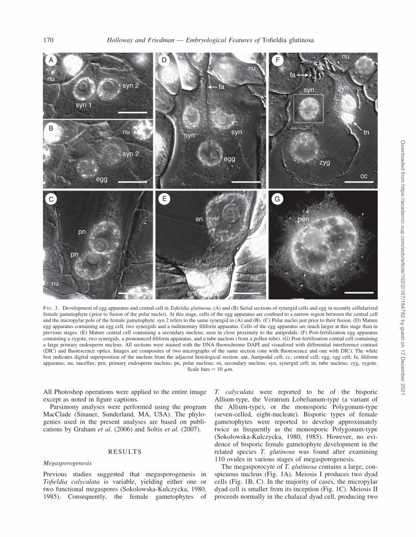
FI G. 3. Development of egg apparatus and central cell in Tofieldia glutinosa. (A) and (B) Serial sections of synergid cells and egg in recently cellularizedfemale gametophyte (prior to fusion of the polar nuclei). At this stage, cells of the egg apparatus are confined to a narrow region between the central celland the micropylar pole of the female gametophyte. syn 2 refers to the same synergid in (A) and (B). (C) Polar nuclei just prior to their fusion. (D) Matureegg apparatus containing an egg cell, two synergids and a rudimentary filiform apparatus. Cells of the egg apparatus are much larger at this stage than inprevious stages. (E) Mature central cell containing a secondary nucleus, seen in close proximity to the antipodals. (F) Post-fertilization egg apparatuscontaining a zygote, two synergids, a pronounced filiform apparatus, and a tube nucleus (from a pollen tube). (G) Post-fertilization central cell containinga large primary endosperm nucleus. All sections were stained with the DNA fluorochrome DAPI and visualized with differential interference contrast(DIC) and fluorescence optics. Images are composites of two micrographs of the same section (one with fluorescence and one with DIC). The whitebox indicates digital superposition of the nucleus from the adjacent histological section. ant, Antipodal cell; cc, central cell; egg, egg cell; fa, filiformapparatus; nu, nucellus; pen, primary endosperm nucleus; pn, polar nucleus; sn, secondary nucleus; syn, synergid cell; tn, tube nucleus; zyg, zygote.
Scale bars ¼ 10 mm.
Holloway and Friedman — Embryological Features of Tofieldia glutinosa170
Dow
nloaded from https://academ
ic.oup.com/aob/article/102/2/167/184782 by guest on 17 D
ecember 2021

unequally sized megaspores, of which the chalazal-most cellis larger (Fig. 1D). In the micropylar dyad, meiosis IIfrequently aborts during prophase II. In these instances,
condensed chromosomes can be seen as long as the cellremains distinctly visible, up to and through the earlieststages of female gametophyte development (Fig. 1D).When meiosis II is completed in both the micropylar and cha-lazal dyads, a tetrad of megaspores is produced. Of these onlythe chalazal-most megaspore is functional (Fig. 1E). Thus,the development of the female gametophyte of T. glutinosais strictly monosporic. The remaining three (or two, ifmeiosis II aborts in the micropylar dyad) nonfunctionalmegaspores (Fig. 1F) are only visible as distinct cells untilthe first free-nuclear mitotic division of the functional mega-spore, at which point the degenerating megaspores becomeindistinguishable from one another and appear only as a dark-staining mass (Fig. 2A). The functional megaspore is approx.35–50 mm long and 12–20 mm wide.
Female gametophyte development
Syncytial development of the female gametophyte ofT. glutinosa is fundamentally similar to that of most angio-sperms with a Polygonum-type female gametophyte. Thefemale gametophyte becomes polarized following the first
A B C D
FI G. 4. Pollen tube growth in Tofieldia glutinosa. (A) Pollen tube growing out of a pollen grain on the stigma. (B) Numerous pollen tubes grow throughthe stylar canal. (C) Pollen tube growth through the micropyle. This image is a composite of three serial histological sections of the same ovule. Red boxesindicate digital superposition of pollen tube and surrounding tissue from adjacent histological sections. (D) Penetration of the nucellus by a pollen tube.Pollen tube growth through the nucellus is intercellular. (E) Pollen tube growth through the nucellus and into the filiform apparatus of the female game-tophyte. The red box indicates digital superposition of the pollen tube and surrounding tissue from the adjacent histological section. All sections werestained with toluidine blue. fa, Filiform apparatus; int, integument; nu, nucellus; pg, pollen grain; pt, pollen tube; st, style; stc, stylar canal; stg,
stigma. Scale bars ¼ 10 mm.
A B
FI G. 5. Post-fertilization egg apparatus in Tofieldia glutinosa. (A)Remnants of the pollen tube are visible at the micropylar pole of thefemale gametophyte. (B) A well-developed filiform apparatus inside thedegenerated synergid cell. The tube nucleus is still visible at this stage.Sections were stained with toluidine blue and visualized with DICoptics. cc, Central cell; fa, filiform apparatus; nu, nucellus; syn, synergid
cell; tn, pollen tube nucleus; zyg, zygote. Scale bars ¼ 10 mm.
Holloway and Friedman — Embryological Features of Tofieldia glutinosa 171
Dow
nloaded from https://academ
ic.oup.com/aob/article/102/2/167/184782 by guest on 17 D
ecember 2021

free nuclear mitotic division of the functional megaspore,with one nucleus situated at the micropylar pole of thefemale gametophyte, and the other at the chalazal pole(Fig. 2A). The two nuclei border a large central vacuolethat fills virtually all of the space between them(Fig. 2A). At this stage, the female gametophyte isapprox. 50–65 mm in length and 20–30 mm in width. Asecond wave of free nuclear mitosis (Fig. 2B) yields apair of nuclei at each pole of the female gametophyte(Fig. 2C). These mitotic divisions occur perpendicular toeach other (Fig. 2B). At the four-nucleate stage, thefemale gametophyte ranges from 65 mm to 85 mm inlength and 25 to 35 mm in width. All four nuclei undergoa third mitotic division to produce four nuclei at eachpole of the female gametophyte (Fig. 2D).
Upon cellularization, the egg apparatus, which is com-prised of a single egg cell and a pair of synergids, is con-fined to a relatively small space between the central celland the nucellus at the micropylar pole of the female game-tophyte (Fig. 3A, B). All three cells of the egg apparatus aredensely cytoplasmic and each has a wall in contact withthe micropylar boundary of the female gametophyte(Fig. 3A, B), although in non-median sections, the eggcell often appears to be positioned below the synergids.The two polar nuclei, which are initially positioned at oppo-site poles, migrate toward the centre of the female gameto-phyte and meet along the periphery of the central cell,approximately one-half to two-thirds of the distance fromthe micropylar end of the female gametophyte (Fig. 3C).When the polar nuclei meet, the female gametophyte isapprox. 80–100 mm long and 30–40 mm wide. As thefemale gametophyte grows, the egg apparatus alsoexpands, protruding into the central cell (Fig. 3D). A rudi-mentary filiform apparatus is recognizable inside the syner-gids at this stage (Fig. 3D). The polar nuclei fuse prior tofertilization to form a large secondary nucleus with asingle large nucleolus (Fig. 3E). This fusion occurs nearthe spot where the polar nuclei initially come in contact
with each other, and is followed by the migration of the sec-ondary nucleus towards the chalazal end of the central cell.
At maturity (as denoted by a secondary nucleus in thecentral cell), the egg cell is highly vacuolate comparedwith the synergids, which remain densely cytoplasmic(Fig. 3D, F). The egg nucleus, which is initially positionedin the centre of the egg cell (Fig. 3D), occupies the periph-ery of the egg cell by the time fertilization occurs. Thezygote nucleus is also seen in this position, close tothe cell wall (Fig. 3F). Following fertilization (see below)the primary endosperm nucleus (the result of a fusionevent involving the secondary nucleus and a secondsperm nucleus; Fig. 3G) is located in close proximityto the antipodal cells at the extreme chalazal end ofthe central cell. At the time of fertilization, the femalegametophyte measures as much as 205 mm long and110 mm wide.
Double fertilization
Following pollination, numerous pollen tubes can be seenpenetrating the stigma (Fig. 4A) and growing through thestylar canal (Fig. 4B). Upon entering the ovary, pollentubes radiate in all directions until they reach receptiveovules, at which point individual pollen tubes growthrough the micropyle (Fig. 4C) and penetrate the nucellusof ovules (Fig. 4D). Pollen tube growth through the nucellusis intercellular (Fig. 4E). The pollen tube enters the femalegametophyte through one of the two synergids, where itdeposits two sperm. The remnants of the pollen tube, asingle tube nucleus, and the filiform apparatus (whichbecomes much more elaborate in advance of fertilization)remain visible inside the degenerating synergid for sometime after fertilization (Fig. 5A, B; also see Fig. 3F). Thezygote nucleus typically contains two nucleoli (Fig. 5B)and remains undivided during the early stages of endospermdevelopment (see below).
A B C
FI G. 6. Double fertilization in Tofieldia glutinosa. (A) Fertilization of the egg nucleus. The sperm nucleus is still visible within the egg nucleus and thetube nucleus is present in the adjacent synergid. (B) Fertilization of the secondary nucleus by the second sperm nucleus. The sperm nucleus is still visible(with nucleolus) within the secondary nucleus. (C) Second fertilization event involving two unfused polar nuclei and a sperm nucleus. All sections werestained with the DNA fluorochrome DAPI and visualized with differential interference contrast (DIC) and fluorescence optics. Images are composites oftwo micrographs of the same section (one with fluorescence and one with DIC). egg, Egg cell; pn, polar nucleus; sn, secondary nucleus; sp, sperm
nucleus; syn, synergid cell; tn, tube nucleus. Scale bars ¼ 10 mm.
Holloway and Friedman — Embryological Features of Tofieldia glutinosa172
Dow
nloaded from https://academ
ic.oup.com/aob/article/102/2/167/184782 by guest on 17 D
ecember 2021

Three fertilization events and over 100 fertilized femalegametophytes were observed. One sperm nucleus fuseswith the egg nucleus (Fig. 6A) and the second spermnucleus fuses with the secondary nucleus (Fig. 6B).Double fertilization appears to be synchronous, since inthose three fertilization events neither the central cell northe egg cell was found to be fertilized when the otherwas not. Although the polar nuclei usually fuse prior to fer-tilization, one instance was observed in which the secondfertilization event involved the triple fusion of twounfused polar nuclei and a single sperm nucleus (Fig. 6C).
Early endosperm development
After the first mitotic division of the primary endospermnucleus (Fig. 7A), a wall forms between the two daughter
nuclei and partitions the former central cell into twochambers: a large micropylar chamber and a significantlysmaller chalazal chamber (Fig. 7B, C). Free-nuclearmitosis is initiated at near-equal rates in each chamber(Fig. 8). This differs slightly from endosperm developmentin T. calyculata, where free-nuclear development in thechalazal chamber is limited from the onset in the vastmajority of cases (Sokolowska-Kulczycka, 1980). Despiteinitially near-equal rates of free-nuclear development, thechalazal chamber, which is more densely cytoplasmic,remains the smaller of the two chambers. Endospermscontaining up to 64 nuclei (32 in each chamber) wereobserved in this study, and at that stage, the zygote hadnot yet divided.
Antipodal proliferation
At the end of the syncytial phase of female gametophytedevelopment, three antipodal nuclei occupy a narrow,densely cytoplasmic pocket at the chalazal end of thefemale gametophyte (Fig. 9A). Their nuclei stain darklyand each contains a pronounced nucleolus (Fig. 9).Cellularization of the chalazal region yields three uninucle-ate antipodal cells (Fig. 9B). As the female gametophytematures, each of the three antipodal cells undergoes asingle free-nuclear division (Fig. 9C–E). These nucleardivisions occur prior to fertilization and are usually asyn-chronous. The order in which antipodal nuclei divideseems to be irregular, and on only one occasion were allthree antipodal nuclei seen dividing simultaneously.Additional cell divisions do not occur, so the numberof antipodal cells remains fixed at three. Of the 396mature prefertilization female gametophytes examined, 90
A
B
C
FI G. 7. Helobial endosperm development in Tofieldia glutinosa. (A) Thefirst mitotic division of the primary endosperm nucleus occurs in thebottom half of the central cell. (B) Two-celled, two-nucleate endosperm.A wall (arrow) forms between the two daughter nuclei of the primary endo-sperm nucleus partitioning the central cell into a large micropylar chamberand a smaller chalazal chamber. (C) Post-fertilization female gametophytewith binucleate chalazal endosperm chamber and binucleate (one nucleusis visible in this section) micropylar endosperm chamber. The zygote isvisible at the micropylar pole. One synergid, the filiform apparatus, andfour of six antipodal nuclei are also visible in this section. All sectionswere stained with DAPI and visualized with DIC and fluorescenceoptics. All images are composites of two micrographs of the samesection (one utilizing fluorescence optics and one utilizing DIC optics).an, Antipodal nuclei; chc, chalazal chamber of endosperm; en, endospermnucleus; fa, filiform apparatus; mic, micropylar chamber of endosperm;
nu, nucellus; syn, synergid cell; zyg, zygote. Scale bars ¼ 10 mm.
36
32
28
24
20
Num
ber
of n
ucle
i in
the
chal
azal
cha
mbe
r
Number of nuclei in the micropylar chamber
16
12
8
4
00 4 8 12 16 20 24 28 32 36
FI G. 8. Relative rates of syncytial development in the micropylar and cha-lazal endosperm chambers in Tofieldia glutinosa. Endosperm nuclear pro-liferation occurs at roughly equal rates in each chamber. Endospermbeyond the 64-nucleate stage was not observed. The size of each datapoint is proportional to the number of times the specific endosperm con-
figuration was observed.
Holloway and Friedman — Embryological Features of Tofieldia glutinosa 173
Dow
nloaded from https://academ
ic.oup.com/aob/article/102/2/167/184782 by guest on 17 D
ecember 2021

contained three antipodal nuclei, 39 contained four nuclei,58 contained five nuclei, and 208 contained a full comp-lement of six antipodal nuclei. Just prior to fertilization,the female gametophyte of T. glutinosa is seven-celledand ten-nucleate (the two polar nuclei in the central cellhaving fused to yield a single secondary nucleus). Thebinucleate antipodal cells (Fig. 9F) persist past fertilizationand are still visible when endosperm begins to develop(Fig. 7). On one occasion, eight antipodal nuclei wereobserved, and, in this instance, there were two binucleatecells and a single cell containing four nuclei.
After the endosperm proceeds through three or fourrounds of mitosis, the antipodal nuclei become less discern-able from one another, and are often small and irregularlyshaped. Degenerate antipodals are visible in the femalegametophyte for some time after fertilization. Very littleis left of the antipodals by the time endosperm developmentreaches the two-celled, 64-nucleate stage.
Female gametophyte length directly correlates with thenumber of antipodal nuclei found inside the gametophyte(r2 ¼ 0.7381; Fig. 10). In addition, there is a burst offemale gametophyte growth associated with the fertilizationprocess (Fig. 10). In several instances however, large
female gametophytes with only three antipodal nucleiwere found (Fig. 10). It is unlikely, however, that antipodalnuclei in these female gametophytes did not proliferatesince gametophytes with three antipodal nuclei occur intwo significantly different size ranges (t-test; P ¼ 4.6 �10222; Fig. 10). Thus, the presence of three antipodalnuclei in large female gametophytes is likely to be theresult of either nuclear fusion within the antipodal cellsor a complete degeneration of one nucleus in each cell.
DISCUSSION
Female gametophyte development in Tofieldia
The mature female gametophyte is seven-celled and11-nucleate, although after the two polar nuclei fuse toproduce a diploid secondary nucleus, the gametophyte con-tains ten nuclei. Nevertheless, development of the femalegametophyte of T. glutinosa is essentially Polygonum-type with subsequent amplification of the nuclear contentsof the three antipodals. The present results are largely inagreement with Seelieb’s embryological study ofT. calyculata (Seelieb, 1924), but contrast with the studies
A
D E F
B C
FI G. 9. Proliferation of antipodal nuclei in Tofieldia glutinosa. (A) Three antipodal nuclei (arrows) prior to cellularization of the female gametophyte.(B) Three uninucleate antipodal cells (arrows) following cellularization of the female gametophyte. (C) Antipodal nucleus in mitosis (arrow). Mitoticdivisions of the antipodals are usually asynchronous. (D) Single binucleate antipodal and two uninucleate antipodals. (E) Two binucleate antipodalsand a single uninucleate antipodal. (F) Three binucleate antipodal cells. The secondary nucleus is also visible in this section. All sections werestained with toluidine blue. Red boxes indicate digital superposition of nuclei from adjacent histological sections. cc, Central cell; cv, central
vacuole; nu, nucellus; sn, secondary nucleus. Scale bars ¼ 10 mm.
Holloway and Friedman — Embryological Features of Tofieldia glutinosa174
Dow
nloaded from https://academ
ic.oup.com/aob/article/102/2/167/184782 by guest on 17 D
ecember 2021

of Sokolowska-Kulczycka (1980, 1985) who reported con-siderable variation in female gametophyte developmentof T. calyculata during megasporogenesis. Accordingto Sokolowska-Kulczycka (1980, 1985), monosporicPolygonum-type female gametophytes occur in slightlyless than one-third of all ovules, while the majority offemale gametophytes are bisporic, and develop accordingto either the Allium-type or a variant referred to as theVeratrum Lobelianum-type (seven-celled, eight-nucleate,derived from the chalazal dyad in both types). However,no evidence of bisporic female gametophyte developmentwas found in T. glutinosa.
Sokolowska-Kulczycka (1980) did not directly observeseveral key stages of megasporogenesis. Instead, the dis-tinction between monospory and bispory was made on thebasis of the number of degenerated megaspores identifiedadjacent to the developing female gametophyte (two orthree). This approach can be problematic, since fullydegenerated megaspores can be difficult to discern fromone another, and often appear simply as a crushed necro-tic mass of tissue (Fig. 2A). Furthermore, in many taxawith Polygonum-type female gametophytes, includingT. glutinosa, the micropylar dyad cell may abort priorto or during the second meiotic division process, suchthat only two degenerated cells are observable uponinitiation of the functional megaspore (Maheshwari,1950; Bouman, 1984; Johri et al., 1992; Russell, 2001).The present data show that in T. glutinosa early degener-ation of the micropylar dyad cell occurs in approx. 30 %of ovules. Thus, a monosporic female gametophytecould easily be mistaken for a bisporic female gameto-phyte if the distinction is made solely on the basis ofthe number of degenerated cells formed duringmegasporogenesis.
Reports of bisporic female gametophyte development arefrequently inaccurate. Maheshwari (1955) reviewed 58families in which bispory was reported to occur and con-cluded that, in at least 29 of these families, findings of bis-poric female gametophyte development were not justified.Maheshwari (1955) attributed many of these inaccuraciesto incomplete observations during megasporogenesis, andto misinterpretation of the triad stage (in which thechalazal-most cell – the functional megaspore – is mista-ken for an undivided chalazal dyad cell). More recently,reports of bispory have been refuted in Illicium (Williamsand Friedman, 2004), Kadsura (Battaglia, 1986; Friedmanet al., 2003) and Epipactis (Fredrikson, 1992). While it ispossible that interspecific (and intraspecific, in the case ofT. calyculata) variation in female gametophyte develop-ment exists in Tofieldiaceae, this is unlikely to be thecase. Far more probable is that there is variation withinTofieldia species with respect to the completion ofmeiosis II in the micropylar dyad cell.
Antipodal proliferation in Tofieldia and other monocots
Proliferation of antipodal nuclei during female gameto-phyte maturation is the only deviation from the plesio-morphic Polygonum-type developmental pattern inT. glutinosa. It is unclear at this point whether the behaviourof the antipodals in T. glutinosa is anomalous or represen-tative of Tofieldiaceae. Seelieb (1924) describes and illus-trates a normal Polygonum-type female gametophyte inT. calyculata and does not suggest that the antipodals pro-liferate. Sokolowska-Kulczycka (1980) specifically reportsthat increased numbers of antipodal nuclei or cells werenot found in T. calyculata, although no images of maturefemale gametophytes were published. Clearly, variationin antipodal behaviour in different species of Tofieldia ispossible. However, the preponderance of antipodal prolifer-ation within other members of the Alismatales as well asin Acorus (Table 1) suggests that antipodal proliferationis probably plesiomorphic in Tofieldiaceae based oncurrent interpretations of angiosperm phylogeny (Fig. 11).Thus, the reported lack of antipodal proliferation inT. calyculata could represent a character reversion orsimply have resulted from inadequate sampling of develop-mental stages.
In many angiosperm lineages, especially within themonocots, antipodals, once formed, continue to develop(Table 1; see also Williams and Friedman, 2004) througha free nuclear proliferation (as is the case in T. glutinosa),a cellular proliferation, or both. Among monocots, antipo-dal proliferation has been reported in Acorus andmembers of the Alismatales, Arecales, Asparagales,Liliales, Pandanales, Poales and Zingiberales (for a com-plete list of references, see Table 1). In extreme cases (asin the Poaceae or Pandanaceae), several hundred antipodalcells may be formed (Cheah and Stone, 1975; Anton andCocucci, 1984; Johri et al., 1992). In other lineages, suchas Acorus calamus, only five antipodal cells are produced(Buell, 1938).
Williams and Friedman (2004) discussed variations inantipodal development in early divergent monocots,
220
200
180
160
140
120
100
80
60
40
20
Ave
rage
leng
th (
mm)
Number of antipodal nucleiPre-fertilization Post-fertilization
03 4 5 6U 6F 3R
FI G. 10. Female gametophyte length relative to the number of antipodalnuclei present in Tofieldia glutinosa. 6U refers to unfertilized femalegametophytes with six antipodal nuclei. 6F refers to fertilized femalegametophytes with six antipodal nuclei measured before division of theprimary endosperm nucleus. 3R refers to fertilized female gametophytesin which the number of antipodal nuclei has been reduced from six to three.
Holloway and Friedman — Embryological Features of Tofieldia glutinosa 175
Dow
nloaded from https://academ
ic.oup.com/aob/article/102/2/167/184782 by guest on 17 D
ecember 2021

eudicots, magnoliids and Chloranthaceae. They were not,however, able to trace the evolutionary transition betweennon-proliferating and proliferating antipodals to a specificnode on the angiosperm phylogeny (Williams andFriedman, 2004). Parsimony analysis reveals that antipodalproliferation is likely to be the ancestral condition formonocots (Fig. 12). Depending on the phylogenetic pos-ition of Chloranthaceae, which remains uncertain (Qiuet al., 1999; Graham and Olmstead, 2000; Graham et al.,2000; Soltis et al., 2000; APG II, 2003; Hilu et al., 2003;Davis et al., 2004; Graham et al., 2006; Qiu et al., 2006;Moore et al., 2007), antipodal proliferation may even be asynapomorphy of the common ancestor of monocots, mag-noliids, eudicots and Chloranthaceae, although this wouldrequire a reversion in magnoliids and early eudicots
(Fig. 12A). Alternatively, if Chloranthaceae is sister to themagnoliid clade as some recent studies suggest (Stevens,2001; Hansen et al., 2007), antipodal proliferation inChloranthaceae could be homoplasious with the similarphenomenon in monocots (Fig. 12B). The present analysisfurther suggests that modest proliferation of the sort seen inearly-divergent monocots like Tofieldia, Acorus (Buell,1938) and members of the Araceae (Campbell, 1903,1905; Gow, 1908, 1913; Coulter, 1908), in which only afew supernumerary antipodals or antipodal nuclei areformed, is the ancestral condition among monocotyledo-nous plants.
In angiosperms, the antipodals are perhaps the most vari-able component of the mature female gametophyte (Johriet al., 1992). They may be ephemeral, degenerating soon
TABLE 1. Reports of antipodal proliferation in basal eudicots, magnoliids and monocots
Clade FamilyAntipodal
proliferation type Sources
Unplaced angiosperms Chloranthaceae Cellular Yoshida (1957); Johri et al. (1992)Basal eudicots Buxaceae, Cellular and nuclear Wiger (1935), Johri et al. (1992)
Menispermaceae, Nuclear Joshi (1939); Johri et al. (1992)Nelumbonaceae, Cellular and nuclear Batygina et al. (1982)Papaveraceae, Cellular Berg (1967); Johri et al. (1992)Ranunculaceae Cellular and nuclear Mottier (1895); Coulter (1898); Tilton and Lersten (1981)
Magnoliids Lauraceae Cellular Bambacioni-Mezzetti (1935); Sastri (1963); Johri et al. (1992)Monimiaceae Celluar Davis (1966); Johri et al. (1992)Piperaceae Cellular Kanta (1962); Johri et al. (1992)
Alismatales sensu lato(Monocots)
Acoraceae Cellular Buell (1938)
Araceae Cellular Campbell (1903), (1905); Gow (1908, 1913); Coulter (1908)Hydrocharitaceae Cellular and nuclear Wylie (1904)*; Johri et al. (1992)†
Juncaginaceae Cellular Hill (1900); Johri et al. (1992)Potamogetonaceae Cellular Tilton and Lersten (1981); Johri et al. (1992)†
Tofieldiaceae Nuclear Reported hereArecales (Monocots) Arecaceae Nuclear Rao (1959); Johri et al. (1992; under ‘Palmae’)Asparagales(Monocots)
Anthericaceae Nuclear Cave (1948)
Amaryllidaceae Cellular Johri et al. (1992)Convallariaceae Cellular McAllister (1909)Hypoxidaceae Cellular Johri et al. (1992)Iridaceae Cellular Haeckel (1930); Rudall et al. (1984)Orchidaceae Cellular Proddubnaya-Arnoldi (1967); Johri et al. (1992)
Liliales (Monocots) Colchicaceae Cellular and nuclear Sulbha (1954); Johri et al. (1992; under ‘Liliaceae’)Liliaceae Cellular and nuclear Schnarf (1931); Davis (1966); Johri et al. (1992)Melanthiaceae Cellular and nuclear Eunus (1951); Sokolowska-Kulczycka (1973); Tilton and Lersten (1981)
Pandanales(Monocots)
Pandanaceae Cellular and nuclear Campbell (1909, 1910); Cheah and Stone (1975); Johri et al. (1992)
Poales (Monocots) Anarthriaceae Nuclear Linder and Rudall (1993)Bromeliaceae Cellular Lakshmanan (1967); Johri et al. (1992)Centrolepidaceae Cellular Hamann (1962); Johri et al. (1992)Poaceae Cellular and nuclear Weatherwax (1926); Yamaura (1933); Sass (1946); Cass and Jensen (1970);
Tilton and Lersten (1981); Rao et al. (1983); Saini et al. (1983); Anton andCocucci (1984); Aulbach-Smith and Herr (1984); Crane and Carman (1987);Johri et al. (1992); Jane (1999)
Rapateaceae Cellular Johri et al. (1992)Restionaceae Cellular Borwein et al. (1949); Kircher (1986); Rudall and Linder (1988)
Zingiberales(Monocots)
Marantaceae Cellular and nuclear Mauritzon (1936); Johri et al. (1992)
‘Antipodal proliferation type’ refers to either nuclear proliferation (resulting in extra antipodal nuclei) or cellular proliferation (resulting in extraantipodal cells). All family/ordinal assignments conform to the recommendations of APG II (2003). NB In most cases, only a few taxa within a givenlineage have been examined. Taxa not mentioned here either have not been studied, or are reported to lack proliferating antipodals.
* The extra antipodal reported by Wylie (1904) is more likely to be the chalazal endosperm chamber.† Johri et al.’s (1992) report of no antipodal proliferation conflicts with other reports cited here.
Holloway and Friedman — Embryological Features of Tofieldia glutinosa176
Dow
nloaded from https://academ
ic.oup.com/aob/article/102/2/167/184782 by guest on 17 D
ecember 2021

after their formation, as in Arabidopsis (Murgia et al., 1993;Rojek et al., 2005) and many other taxa, or may persist toand even well beyond fertilization (Johri et al., 1992;Williams and Friedman, 2004). Parsimony analyses havedemonstrated that in those lineages diverging above theparaphyletic basal grade of Amborella, Nymphaeales andAustrobaileyales, persistence of the antipodal cells is theancestral condition (Williams and Friedman, 2004). Thepresent parsimony analysis shows this to be the case formonocots as well (Fig. 13). Ephemeral antipodals appearto be a derived condition most commonly found in highereudicots (e.g. Violaceae, Cucurbitaceae, Solanaceae,Rutaceae, Malpighiaceae, etc.; Johri et al., 1992).
The physiological role (or roles) of antipodals has notbeen definitively demonstrated, but there is reasonablesupport for the idea that antipodals may be important inthe transfer of nutrients from the sporophyte to the femalegametophyte and endosperm (Westermaier, 1890; Riddle,1898; Ikeda, 1902; Lloyd, 1902; Schnarf, 1929; Brinkand Cooper, 1944; Coe, 1954; Kapil and Bhatnagar,1981; Mogensen, 1981; Johri et al., 1992). The positionof antipodals at the interface of the sporophytic tissue andthe female gametophyte and endosperm is certainly congru-ent with this view, as is the presence of wall ingrowths inthe antipodals of certain taxa (Westermaier, 1890;Mogensen, 1981). Nevertheless, for now it is unclearwhether modest levels of antipodal proliferation (as inTofieldia) are adaptive (e.g. represent a functional benefitto the seed) or are a neutral variation of female gameto-phyte structure.
Helobial endosperm
Helobial endosperm development is common in mono-cots (Table 2; see also Swamy and Parameswaran, 1963;
Palser, 1975), but ab initio cellular endosperm is the ances-tral condition of this clade, a feature most likely inheritedfrom the shared common ancestor of all angiosperms(Floyd and Friedman, 2000). Endosperm development inAcorus, Amborella, most Nymphaeales andAustrobaileyales, and early-divergent eudicots is of the cel-lular type (Buell, 1938; Johri et al., 1992; Floyd andFriedman, 2000). However, parsimony analysis revealsthat in monocots the transition from cellular to helobialendosperm occurred after the divergence of Acorus, assum-ing that Acorus is sister to all other monocots (Chase et al.,1993, 1995, 2000; Duvall et al., 1993; Davis et al., 1998;Doyle and Endress, 2000; Graham and Olmstead, 2000;Graham et al., 2000, 2006; Hilu et al., 2003; Soltis et al.,
Ara
ceae
Ara
ceae
Sch
euch
eria
ceae
But
omac
eae
Alis
mat
acea
e
Tofie
ldia
ceae
Antipodal proliferation
A
No antipodal proliferation
Apo
noge
tona
ceae
Hyd
roch
arita
ceae
Zos
tera
ceae
Tofie
ldia
ceae
B
FI G. 11. Evolution of antipodal proliferation in Alismatales. (A)Phylogeny of Soltis et al. (2007). Antipodal proliferation has been reportedin Hydrocharitaceae (Wylie, 1904), but more recent studies suggest thatthis is not actually the case (Johri et al., 1992). (B) Phylogeny ofGraham et al. (2006). Parsimony analysis reveals that antipodal prolifer-
ation is the ancestral condition for Alismatales.A
mbo
rella
Nym
phae
ales
Aus
trob
ailia
les
Mag
nolii
ds
Chl
oran
thac
eae
Cer
atop
yllu
m
Aco
rale
s
Alis
mat
ales
Pan
dana
les
Dio
scor
eale
s
Lilia
les
Asp
arag
ales
Poa
les
Are
cale
s
Zin
gibe
rale
s
Com
mel
inal
es
Eud
icot
s
Am
bore
lla
Nym
phae
ales
Aus
trob
ailia
les
Eud
icot
s
Cer
atop
yllu
m
Chl
oran
thac
eae
Aco
rale
s
Alis
mat
ales
Lilia
les
Pan
dana
les
Asp
arag
ales
Dio
scor
eale
s
Poa
les
Monocots
Are
cale
s
Zin
gibe
rale
s
Com
mel
inal
es
Mag
nolii
ds
Monocots
Antipodal proliferationNo antipodal proliferationNo antipodalsUnresolved
A
B
FI G. 12. Evolution of antipodal proliferation in monocots. In all casesexcept for the Poales, the character state assigned to the group has beenresolved as the ancestral condition for that group based on the presentanalysis. The ancestral condition within the Poales is unresolved, and isclassified as polymorphic. (A) Phylogeny of Soltis et al. (2007). (B)Phylogeny of Graham et al. (2006). For both of these phylogenetic hypoth-eses of angiosperm and monocot relationships, parsimony analysis revealsthat antipodal proliferation either evolved in or was present in the common
ancestor of all monocots.
Holloway and Friedman — Embryological Features of Tofieldia glutinosa 177
Dow
nloaded from https://academ
ic.oup.com/aob/article/102/2/167/184782 by guest on 17 D
ecember 2021

2007), and helobial endosperm should be considered theancestral condition for all monocots excluding Acorus(Fig. 14).
Floyd and Friedman (2000) recognized that the cellularendosperm of Acorus was unique among early-divergentmonocots. However, the angiosperm phylogeny reliedupon in their analysis (Qiu et al., 1999) has undergonerevision, and they did not pinpoint the transition fromcellular to helobial endosperm to such an early phase ofmonocot evolution. In monocots there is likely to be onlya single evolutionary origin of helobial endosperm(Fig. 14), and occurrences of helobial endosperms insome eudicots and magnoliids, and also in Cabomba are
certainly homoplasious with those of monocots (Floydand Friedman, 2000).
CONCLUSIONS
A detailed account of female gametophyte development hasbeen provided in Tofieldia glutinosa from the initiation ofmegasporogenesis through fertilization and the early
Am
bore
lla
Nym
phae
ales
Aus
trob
ailia
les
Mag
nolii
ds
Chl
oran
thac
eae
Cer
atop
yllu
m
Aco
rale
s
Alis
mat
ales
Pan
dana
les
Dio
scor
eale
s
Lilia
les
Asp
arag
ales
Poa
les
Are
cale
s
Zin
gibe
rale
s
Com
mel
inal
es
Eud
icot
s
Am
bore
lla
Nym
phae
ales
Aus
trob
ailia
les
Eud
icot
s
Cer
atop
yllu
m
Chl
oran
thac
eae
Aco
rale
s
Alis
mat
ales
Lilia
les
Pan
dana
les
Asp
arag
ales
Dio
scor
eale
s
Poa
les
Monocots
Are
cale
s
Zin
gibe
rale
s
Com
mel
inal
es
Mag
nolii
ds
Monocots
Persistent antipodalsEphemeral antipodalsNo antipodalsUnresolved
A
B
FI G. 13. Evolution of persistence of antipodals in monocots. In all casesexcept for the Pandanales and the Arecales, the character state assigned tothe group has been resolved as the ancestral condition for that group basedon our analysis. The ancestral conditions within the Pandanales and theArecales are unresolved, and are classified as polymorphic. (A)Phylogeny of Soltis et al. (2007). (B) Phylogeny of Graham et al.(2006). Parsimony analysis reveals that the common ancestor of all angios-perms exclusive of Amborella, Nymphaeales and Austrobaileyales had per-sistent antipodals. It is unclear whether the persistent antipodals inAmborella evolved independently or were inherited from the common
ancestor of all angiosperms.
TABLE 2. Reports of helobial endosperm development inmonocots
Clade Family Source
Alismatales Alismataceae* Johri et al. (1992)Aponogetonaceae Johri et al. (1992)Araceae Grayum (1987, 1991)Butomaceae Johri et al. (1992); Fernando and
Cass (1996)Hydrocharitaceae Johri et al. (1992)Tofieldiaceae Seelieb (1924);
Sokolowska-Kulczycka (1980);reported here
Zosteraceae Dahlgren (1939); Johri et al.(1992)
Asparagales Agavaceae Johri et al. (1992)Alliaceae Johri et al. (1992); Berg (1996)Anthericaceae Cave (1948)Amaryllidaceae Coe (1953); Werker and Fahn
(1975); Johri et al. (1992)Asparagaceae Lazarte and Palser (1979); Tilton
and Lersten (1981); Berg (2003);Halada and Erdelska (2005)
Asphodelaceae Johri et al. (1992)Blandfordiaceae Johri et al. (1992)Hesperocallidaceae Cave (1948)Hyacinthaceae Johri et al. (1992; under
‘Liliaceae’)Hypoxidaceae Geerinck (1969); Johri et al.
(1992)Iridaceae* Goldblatt (1990)Ixiolirionaceae Johri et al. (1992)Themidaceae Berg (2003)
Commelinales Haemodoraceae Browne (1961); Geerinck (1969);Simpson (1988)
Philydraceae Johri et al. (1992)Pontederiaceae Johri et al. (1992)
Dioscorleales Burmanniaceae Johri et al. (1992)Nartheciaceae Browne (1961)
Liliales Campynemataceae* Patterson and Givnish (2002)Melanthiaceae Johri et al. (1992); Patterson and
Givnish (2002)Trilliaceae Swamy (1948); Johri et al. (1992)
Pandanales Cyclanthaceae Johri et al. (1992)Poales Anarthriaceae Linder and Rudall (1993)
Bromeliaceae Rao and Wee (1979); Johri et al.(1992)
Juncaceae Munro and Linder (1997)Sparganiaceae Asplund (1973); Johri et al.
(1992)Typhaceae Asplund (1972); Johri et al.
(1992)Zingiberales Costaceae Kress (1990)
Zingiberaceae Sachar and Arora (1963); Kress(1990)
All family/ordinal assignments conform to the recommendations ofAPG II (2003).
* Nuclear endosperm has also been reported in these families
Holloway and Friedman — Embryological Features of Tofieldia glutinosa178
Dow
nloaded from https://academ
ic.oup.com/aob/article/102/2/167/184782 by guest on 17 D
ecember 2021

stages of endosperm development. It can be said with cer-tainty that the female gametophyte of T. glutinosa is mono-sporic (Fig. 1), and produces seven cells and 11 nuclei (oneegg, two synergids, an initially binucleate central cell, andthree binucleate antipodals) (Figs 2, 3 and 9). Tofieldiaglutinosa produces a helobial endosperm (Fig. 7), whoseearly growth occurs at equal rates in both the micropylarand chalazal endosperm chambers (Fig. 8).
Based on the present study of T. glutinosa and the analy-sis of the embryological literature pertaining to early-divergent monocots, two important conclusions can bedrawn about the reproductive characters of ancestral mono-cots. First, the ancestral monocot female gametophyte hadpersistent antipodal cells that underwent a modest prolifer-ation (Figs 12 and 13). Whether this proliferation was
nuclear or cellular in nature is unclear, but it was almostcertainly simple, producing only a few supernumerary anti-podal cells or nuclei in contrast with the elaborate prolifer-ation found in some highly derived monocot lineages.Second, endosperm development in the first monocotswas of the cellular type (Floyd and Friedman, 2000), buttransitioned once to the helobial type after the divergenceof Acorus from the remainder of the monocot clade(Fig. 14). Finally, we suggest that because of its prevalenceand early appearance within this clade, helobial endospermshould be considered one of the more salient features ofearly monocots.
ACKNOWLEDGEMENTS
We thank Kirsten Ryerson for her assistance with field col-lections and histological preparations, Andrea Ruchty andthe staff of the Mount Adams Ranger District (GiffordPinchot National Forest, Trout Lake, WA, USA) for theirassistance with collection permits and field collections,and the two anonymous reviewers for their helpful sugges-tions towards the improvement of the manuscript. We grate-fully acknowledge funding from the National ScienceFoundation (IOB-0446191 to W.E.F.).
LITERATURE CITED
Anton AM, Cocucci AE. 1984. The grass megagametophyte and its poss-ible phylogenetic implications. Plant Sytematics and Evolution 146:117–121.
APG II. 2003. An update of the Angiosperm Phylogeny Group classifi-cation for the orders and families of flowering plants. BotanicalJournal of the Linnean Society 141: 399–436.
Arber A. 1925. Monocotyledons: a morphological study. Cambridge:Cambridge University Press.
Asplund I. 1972. Embryological studies in the genus Typha. SvenskBotanisk Tidskrift 66: 1–17.
Asplund I. 1973. Embryological studies in the genus Sparganium. SvenskBotanisk Tidskrift 67: 177–200.
Aulbach-Smith CA, Herr JM Jr. 1984. Development of the ovule andfemale gametophyte in Eustachys petraea and E. glauca (Poaceae).American Journal of Botany 71: 427–438.
Bambacioni-Mezzetti V. 1935. Ricerche morfologiche sulle Lauracee, Losviluppo dell’ovulo e dei sacchi pollinici nel Laurus nobilis L. Annalidi Botanica 21: 1–19.
Battaglia E. 1986. Embryological questions. 7. Do new types of embryosac occur in Schisandra? Annals of Botany 44: 69–82.
Batygina TB, Shamrov II, Kolesova GE. 1982. Embryology of theNymphaeales and Nelumbonales. II. The development of the femaleembryonic structures. Botanicheskii Zhurnal 67: 1179–1195.
Berg RY. 1967. Megagametogenesis and seed development inDendromecon rigida (Papaveraceae). Phytomorphology 17: 223–233.
Berg RY. 1996. Development of ovule, embryo sac, and endosperm inDipterostemon and Dichelostemma (Alliaceae) relative to taxonomy.American Journal of Botany 83: 790–801.
Berg RY. 2003. Development of ovule, embryo sac, and endosperm inTriteleia (Themidaceae) relative to taxonomy. American Journal ofBotany 90: 937–948.
Borwein B, Coetsee ML, Krupko S. 1949. Development of the embryosac of Restio dodii and Elegia racemosa. Journal of South AfricanBotany 15: 1–11.
Bouman F. 1984. The ovule. In: Johri BM, ed. Embryology of angios-perms. Berlin: Springer-Verlag.
Brink RA, Cooper DC. 1944. The antipodals in relation to abnormalendosperm behavior in Hordeum jubatun � Secale cereale hybridseeds. Genetics 29: 391–406.
Am
bore
lla
Nym
phae
ales
Aus
trob
ailia
les
Mag
nolii
ds
Chl
oran
thac
eae
Cer
atop
yllu
m
Aco
rale
s
Alis
mat
ales
Pan
dana
les
Dio
scor
eale
s
Lilia
les
Asp
arag
ales
Poa
les
Are
cale
s
Zin
gibe
rale
s
Com
mel
inal
es
Eud
icot
s
Am
bore
lla
Nym
phae
ales
Aus
trob
ailia
les
Eud
icot
s
Cer
atop
yllu
m
Chl
oran
thac
eae
Aco
rale
s
Alis
mat
ales
Lilia
les
Pan
dana
les
Asp
arag
ales
Dio
scor
eale
s
Poa
les
Monocots
Are
cale
s
Zin
gibe
rale
s
Com
mel
inal
es
Mag
nolii
ds
Monocots
Helobial endospermNuclear endospermCellular endospermUnresolved
A
B
FI G. 14. Evolution of helobial endosperm development in angiosperms.In all cases except for the Commelinales and eudicots, the characterstate assigned to the group has been resolved as the ancestral conditionfor that group based on the present analysis. The ancestral conditionswithin Commelinales and eudicots are unresolved, and are classified hereas polymorphic. (A) Phylogeny of Soltis et al. (2007). (B) Phylogeny ofGraham et al. (2006). Parsimony analysis reveals that helobial endospermdevelopment evolved in the common ancestor of all monocots excluding
the Acorales.
Holloway and Friedman — Embryological Features of Tofieldia glutinosa 179
Dow
nloaded from https://academ
ic.oup.com/aob/article/102/2/167/184782 by guest on 17 D
ecember 2021

Browne ET. 1961. Morphological studies in Aletris. I. Development of theovule, megaspores and megagametophyte of A. aurea and their con-nection with the systematics of the genus. American Journal ofBotany 48: 143–147.
Buell MF. 1938. Embryogeny of Acorus calamus. Botanical Gazette 99:556–568.
Campbell DH. 1903. Studies on the Araceae: the embryo-sac and theembryo of Aglaonema and Spathicarpa. Annals of Botany 17:665–687.
Campbell DH. 1905. Studies on Araceae III. Annals of Botany 19:329–349.
Campbell DH. 1909. The embryo-sac of Pandanus. Bulletin of the TorreyBotanical Club 36: 205–220.
Campbell DH. 1910. The embryo-sac of Pandanus coronatus. Bulletin ofthe Torrey Botanical Club 37: 293–295.
Cass DD, Jensen WA. 1970. Fertilization in barley. American Journal ofBotany 57: 62–70.
Cave MS. 1948. Sporogenesis and embryo sac development ofHesperocallis and Leucocrinum in relation to their systematic pos-ition. American Journal of Botany 35: 343–349.
Chase MW, Soltis DE, Olmstead RG, Morgan D, Les DH, Mishler BD,et al. 1993. Phylogenetics of seed plants: an analysis of nucleotidesequences from the plastid gene rbcL. Annals of the MissouriBotanical Garden 80: 528–580.
Chase MW, Stevenson DW, Wilkin P, Rudall PJ. 1995. Monocot sys-tematics: a combined analysis. In: Rudall PJ, Cribb PJ, Cutler DF,Humphries CJeds. Monocotyledons: systematics and evolution.London: Royal Botanic Gardens, Kew, 685–730.
Chase MW, Soltis DE, Soltis PS, Rudall PJ, Fay MF, Hahn WH, et al.2000. Higher-level systematics of the monocotyledons: an assessmentof current knowledge and a new classification. In: Wilson KL,Morrison DAeds. Monocots: systematics and evolution.Collingwood: CSIRO, 3–16.
Cheah CH, Stone BC. 1975. Embryo sac and microsporangium develop-ment in Pandanus (Pandanaceae). Phytomorphology 25: 228–238.
Coe GE. 1953. Cytology of reproduction in Cooperia pedunculata.American Journal of Botany 40: 335–343.
Coe GE. 1954. Distribution of Carbon 14 in ovules of Zephyranthes drum-mondii. Botanical Gazette 115: 342–346.
Coulter JM. 1898. Contribution to the life history of Ranunculus.Botanical Gazette 25: 73–88.
Coulter JM. 1908. Relation of megaspores to embryo sacs in angiosperms.Botanical Gazette 45: 361–366.
Crane CF, Carman JG. 1987. Mechanisms of apomixis in Elymus recti-setus from Eastern Australia and New Zealand. American Journal ofBotany 74: 477–496.
Cronquist A. 1988. The evolution and classification of flowering plants,2nd edn. New York, NY: New York Botanical Garden.
Dahlgren KVO. 1939. Endosperm- und Embryobildung bei Zosteramarina. Botaniska Notiser 1939: 607–615.
Dahlgren RMT, Clifford HT, Yeo PF. 1985. The families of the monoco-tyledons: structure evolution and taxonomy. Berlin: Springer-Verlag.
Davis GL. 1966. Systematic embryology of the angiosperms. New York,NY: John Wiley & Sons.
Davis JI, Simmons MP, Stevenson DW, Wendel JF. 1998. Data decisive-ness, data quality, and incongruence in phylogenetic analysis: anexample from monocotyledons using mitochondrial atpA sequences.Systematic Biology 47: 282–310.
Davis JI, Stevenson DW, Petersen G, Seberg O, Campbell LM,Freudenstein JV, et al. 2004. Phylogeny of the monocots, as inferredfrom rbcL and atpA sequence variation, and a comparison of methodsfor calculating jackknife and bootstrap values. Systematic Botany 29:467–510.
Davis JI, Petersen G, Seberg O, Stevenson DW, Hardy CR, Simmons MP,et al. 2006. Are mitochondrial genes useful for the analysis of monocotrelationships? Taxon 55: 857–870.
Doyle JA, Endress PK. 2000. Morphological phylogenetic analysis ofbasal angiosperms: comparison and combination with moleculardata. International Journal of Plant Sciences 161: S121–S153.
Duvall MR, Clegg MT, Chase MW, Clark WD, Kress WJ, Hills HG,et al. 1993. Phylogenetic hypotheses for the monocotyledons con-structed from rbcL sequence data. Annals of the Missouri BotanicalGarden 80: 607–619.
Eunus AM. 1951. Contributions to the embryology of theLiliaceae. 5. Life history of Amianthium muscae-toxicum Walt.Phytomorphology 1: 73–79.
Fernando DD, Cass DD. 1996. Development and structure of ovuleembryo sac, embryo, and endosperm in Butomus umbellatus(Butomaceae). International Journal of Plant Sciences 157: 269–279.
Floyd SK, Friedman WE. 2000. Evolution of endosperm developmentalpatterns among basal flowering plants. International Journal of PlantSciences 161: S57–S81.
Fredrikson M. 1992. The development of the female gametophyte ofEpipactis (Orchidaceae) and its inference for reproductive ecology.American Journal of Botany 79: 63–68.
Friedman WE. 2006. Embryological evidence for developmental labilityduring early angiosperm evolution. Nature 441: 337–340.
Friedman WE, Gallup WN, Williams JH. 2003. Female gametophytedevelopment in Kadsura: implications for Schisandraceae,Austrobaileyales, and the early evolution of flowering plants.International Journal of Plant Sciences 164: S293–S305.
Friis EM, Pedersen KR, Crane PR. 2004. Araceae from the earlyCretaceous of Portugal: evidence on the emergence of monocotyle-dons. Proceedings of the National Academy of Sciences of the USA101: 16565–16570.
Furness CA, Rudall PJ, Sampson FB. 2002. Evolution of microsporo-genesis in angiosperms. International Journal of Plant Sciences163: 235–260.
Geerinck D. 1969. Genera des Haemodoraceae et des Hypoxidaceae.Bulletin van National Plantentium van Belgie 39: 47–82.
Goldblatt P. 1990. Phylogeny and classification of Iridaceae. Annals of theMissouri Botanical Garden 77: 607–627.
Gow JE. 1908. Studies in Araceae. Botanical Gazette 46: 35–42.Gow JE. 1913. Observation on the morphology of the aroids. Botanical
Gazette 56: 127–142.Graham SW, Olmstead RG. 2000. Utility of 17 chloroplast genes for
inferring the phylogeny of the basal angiosperms. American Journalof Botany 87: 1712–1730.
Graham SW, Reeves PA, Burns ACE, Olmstead RG. 2000.Microstructural changes in noncoding chloroplast DNA: interpret-ation, evaluation, and utility of indels and inversions in basal angio-sperm phylogenetic inference. International Journal of PlantSciences 161: S83–S96.
Graham SW, Zgurski JM, McPherson MA, Cherniawsky DM, SaarelaJM, Horne ESC, et al. 2006. Robust inference of monocot deep phy-logeny using an expanded multigene plastid data set. Aliso 22: 3–20.
Grayum MH. 1987. A summary of evidence and arguments supportingthe removal of Acorus from the Araceae. Taxon 36: 723–729.
Grayum MH. 1991. Systematic embryology of the Araceae. The BotanicalReview 57: 167–203.
Haeckel I. 1930. Uber Iridaceen. Flora 125: 1–82.Halada L, Erdelska O. 2005. Reproductive biology of Ruscus hypoglos-
sum L. in Slovakia. Acta Biologica Cracovienia, Serie Botanique 47:213–217.
Hamann U. 1962. Beitrag zur Embryologie der Centrolepidaceae mitBemerkungen uber den bau der Bluten und Blutenstande und die sys-tematische Stellung der familie. Berichte der Deutschen BotanischenGesellschaft 75: 153–171.
Hansen DR, Dastidar SG, Cai Z, Penaflor C, Kuehl JV, Boore JL,Jansen RK. 2007. Phylogenetic and evolutionary implications ofcomplete chloroplast genome sequences of four early-divergingangiosperms: Buxus (Buxaceae), Chloranthus (Chloranthaceae),Dioscorea (Dioscoreaceae), and Illicium (Schisandraceae).Molecular Phylogenetics and Evolution 45: 547–563.
Hill TG. 1900. The structure and development of Triglochin maritimum.Annals of Botany 14: 83–107.
Hilu KW, Borsch T, Muller K, Soltis DE, Soltis PS, Savolainen V, et al.2003. Angiosperm phylogeny based on matK sequence information.American Journal of Botany 90: 1758–1776.
Hitchcock CL. 1944a. The Tofieldia glutinosa complex of western NorthAmerica. American Midland Naturalist 31: 487–498.
Hitchcock CL. 1944b. Correction: the Tofieldia glutinosa complex ofwestern North America. American Midland Naturalist 32: 784.
Igersheim A, Buzgo M, Endress PK. 2001. Gynoecium diversity and sys-tematics in basal monocots. Botanical Journal of the Linnean Society136: 1–65.
Holloway and Friedman — Embryological Features of Tofieldia glutinosa180
Dow
nloaded from https://academ
ic.oup.com/aob/article/102/2/167/184782 by guest on 17 D
ecember 2021

Ikeda T. 1902. Studies in the physiological functions of antipodals and thephenomena of fertilization in Liliaceae. I. Tricyrtis hirta. Bulletin ofthe College of Agriculture, Tokyo 5: 41–72.
Jane W.-N. 1999. Ultrastructure of embryo development in Arundo formo-sana Hack. (Poaceae). International Journal of Plant Sciences 160:46–63.
Johri BM, Ambegaokar KB, Srivastava PS. 1992. Comparative embry-ology of angiosperms. Berlin: Springer-Verlag.
Joshi AC. 1939. Morphology of Tinospora cordifolia, with some obser-vations on the origin of the single integument, nature of synergidae,and affinities of the Menispermaceae. American Journal of Botany26: 433–439.
Kanta K. 1962. Morphology and embryology of Piper nigrum L.Phytomorphology 12: 207–221.
Kapil RN, Bhatnagar AK. 1981. Ultrastructure and biology of the femalegametophyte in flowering plants. International Review of Cytology70: 291–337.
Kircher P. 1986. Untersuchungen zur Bluten- und Infloreszenzmorphologie,Embryologie und Systemaik der Restionaceen im Vergleich mitGramineen und verwandten Familien. Dissertationes Botanicae 94:1–219.
Kress WJ. 1990. The phylogeny and classification of the Zingiberales.Annals of the Missouri Botanical Garden 77: 698–721.
Lakshmanan KK. 1967. Embryological studies in the Bromeliaceae.1. Lindmania penduliflora (C.H. Wright) Stapf. Proceedings of theIndian Academy of Science B 65: 49–55.
Lazarte JE, Palser BF. 1979. Morphology, vascular anatomy and embryo-logy of pistillate and staminate flowers of Asparagus officinalis.American Journal of Botany 66: 753–764.
Linder HP, Rudall PJ. 1993. The megagametophyte in Anarthria(Anarthriaceae, Poales) and its implications for the phylogeny ofthe Poales. American Journal of Botany 80: 1455–1464.
Lloyd FE. 1902. The comparative embryology of the Rubiaceae. Memoirsof the Torrey Botanical Club 8: 27–112.
McAllister F. 1909. The development of the embryo sac of Smilacinastellata. Botanical Gazette 48: 200–215.
Maddison WP, Maddison DR. 1992. MacClade. Sinauer, Sunderland,Massachusetts, USA.
Maheshwari P. 1950. An introduction to the embryology of angiosperms,1st edn. New York, NY: McGraw-Hill Book Co.
Maheshwari SC. 1955. The occurrence of bisporic embryo sacs inangiosperms – a critical review. Phytomorphology 5: 67–99.
Mauritzon J. 1936. Samenbau und Embryologie einiger Scitamineen.Acta Universitatis Lundensis 30: 1–31.
Mogensen HL. 1981. Ultrastructural localization of adenosine tripho-sphate in the ovules of Saintpaulia ionantha (Gesneriaceae) and itsrelation to synergid function and embryo sac nutrition. AmericanJournal of Botany 68: 183–194.
Moore MJ, Bell CD, Soltis PS, Soltis DE. 2007. Using plastidgenome-scale data to resolve enigmatic relationships among basalangiosperms. Proceedings of the National Academy of Sciences ofthe USA 104: 19363–19368.
Mottier DM. 1895. Contributions to the embryology of Ranunculaceae.Botanical Gazette 20: 241–248, 296–304.
Munro SL, Linder HP. 1997. The embryology and systematic relation-ships of Prionium serratum (Juncaceae: Juncales). AmericanJournal of Botany 84: 850–860.
Murgia M, Huang B.-Q, Tucker SC, Musgrave ME. 1993. Embryo saclacking antipodal cells in Arabidopsis thaliana (Brassicaceae).American Journal of Botany 80: 824–838.
Packer JG. 1993. Two new combinations in Triantha (Liliaceae). Novon3: 278–279.
Palser BF. 1975. The bases of the angiosperm phylogeny: embryology.Annals of the Missouri Botanical Garden 62: 621–646.
Patterson TB, Givnish TJ. 2002. Phylogeny, concerted convergence, andphylogenetic niche conservatism in the core Liliales: insights fromrbcL and ndhF sequence data. Evolution 56: 233–252.
Proddubnaya-Arnoldi VA. 1967. Comparative embryology of theOrchidaceae. Phytomorphology 17: 312–320.
Qiu Y-L, Lee J, Bernasconi-Quadroni F, Soltis DE, Soltis PS, Zanis M,et al. 1999. The earliest angiosperms: evidence from mitochondrial,plastid and nuclear genomes. Nature 402: 404–407.
Qiu Y-L, Li L, Hendry TA, Li R, Taylor DW, Issa MJ, et al. 2006.Reconstructing the basal angiosperm phylogeny: evaluating infor-mation content of mitochondrial genes. Taxon 55: 837–856.
Rao AN, Wee YC. 1979. Embryology of the pineapple, Ananas comosus(L.) Merr. New Phytologist 83: 485–497.
Rao CV. 1959. Contributions to the embryology of Palmae. 2. Ceroxylinae.Journal of the Indian Botanical Society 38: 46–75.
Rao MK, Kumari KA, Grace JR. 1983. Cytology of antipodals cells withsome observations on the male and female gametophyte developmentin pearl millet, Pennisetum americanum (L.) Leeke. BotanicalGazette 144: 201–206.
Riddle LC. 1898. The embryology of Alyssum. Botanical Gazette 26:314–324.
Rojek J, Kuta E, Bohdanowicz J. 2005. In vitro culture promotes partialautonomous endosperm development in unfertilized ovules of wild-type Arabidopsis thaliana var. Columbia. Sexual PlantReproduction 18: 29–36.
Rudall PJ, Linder HP. 1988. Megagametophyte and nucellus inRestionaceae and Flagellariaceae. American Journal of Botany 75:1777–1786.
Rudall PJ, Owens SJ, Kenton AY. 1984. Embryology and breedingsystems in Crocus (Iridaceae) – a study in causes of chromosomevariation. Plant Systematics and Evolution 148: 119–134.
Russell SD. 2001. Female gametogenesis. In: Bhojwani SS, Soh WYeds.Current trends in the embryology of angiosperms. Dordrecht:Kulwer Academic Publishers, 67–88.
Sachar RC, Arora U. 1963. Some embryological aspects of Amomumdealbatum and Hedychium acuminatum. Botanical Gazette 124:353–360.
Saini HS, Sedgley M, Aspinall D. 1983. Effects of heat stress during floraldevelopment on pollen tube growth and ovary anatomy in wheat(Triticum aestivum L.). Australian Journal of Plant Physiology 10:137–144.
Sass JE. 1946. The development of endosperm and antipodal tissue inArgentine waxy maize. American Journal of Botany 33: 791–795.
Sastri RLN. 1963. Studies in the Lauraceae. VI. Comparative embryologyand phylogeny. American Journal of Botany 27: 425–433.
Schnarf K. 1929. Embryologie der Angiospermen. Berlin: BorntragerSchnarf K. 1931. Vergleichende Embryologie der Angiospermen. Berlin:
Borntrager.Seelieb W. 1924. Beitrage zur Entwicklungsgeschichte von Tofieldia caly-
culata (L.) Wahlenb. Botaniska Notiser 77: 172–178.Simpson MG. 1988. Embryological development of Lachnanthes carolini-
ana (Haemodoraceae). American Journal of Botany 75: 1394–1408.Sokolowska-Kulczycka A. 1973. Development of bisporic embryo sacs in
Veratrum lobelianum Bernh. ActaBiologica Cracoviensia, SerieBotanique 14: 85–98.
Sokolowska-Kulczycka A. 1980. Embryological studies of Tofieldia caly-culata (L.) Whlb. Acta Biologica Cracoviensia, Serie Botanique 22:113–128.
Sokolowska-Kulczycka A. 1985. The influence of temperature on the fre-quency of developmental types of ES in Tofieldia calyculata (L.)Whlb. Acta Biologica Cracoviensia, Serie Botanique 27: 1–7.
Soltis DE, Soltis PS, Chase MW, Mort ME, Albach DC, Zannis M,et al. 2000. Angiosperm phylogeny inferred from 18S rDNA, rbcL,and atpB sequences. Botanical Journal of the Linnean Society 133:381–461.
Soltis DE, Soltis PS, Endress PK, Chase MW. 2005. Phylogeny and evol-ution of angiosperms. Sunderland, MA: Sinauer Associates.
Soltis DE, Gitzendanner MA, Soltis PS. 2007. A 567-taxon data set forangiosperms: the challenges posed by Baysian analyses of large datasets. International Journal of Plant Sciences 168: 137–157.
Stevens PF. 2001 onwards. Angiosperm Phylogeny Website. http://www.mobot.org/MOBOT/research/APweb/. 24 April 2007.
Stevens PF. 2006. Book review: Phylogeny and evolution of angiosperms.International Journal of Plant Sciences 167: 607–611.
Sulbha K. 1954. The embryology of Iphigenia indica Kunth.Phytomorphology 4: 180–191.
Swamy BGL. 1948. On the post-fertilization development of Trilliumundulatum. Cellule 52: 7–14.
Swamy BGL, Parameswaran N. 1963. The helobial endosperm.Biological Review 38: 1–50.
Holloway and Friedman — Embryological Features of Tofieldia glutinosa 181
Dow
nloaded from https://academ
ic.oup.com/aob/article/102/2/167/184782 by guest on 17 D
ecember 2021

Tamura MN. 1998. Nartheciaceae. In: Kubitski Ked. The families andgenera of vascular plants. Vol. III. Liliinae. Berlin: Springer,381–392.
Tilton VR, Lersten NR. 1981. Ovule development in Ornithogalum cau-datum (Liliaceae) with a review of selected papers on angiospermreproduction. III. Nucellus and megagametophyte. New Phytologist88: 477–504.
Weatherwax P. 1926. Persistence of the antipodal tissue in the develop-ment of the seed of maize. Bulletin of the Torrey Botanical Club53: 381–384.
Werker E, Fahn A. 1975. Seed anatomy of Pancratium species from threedifferent habitats. Botanical Gazette 136: 396–403.
Westermaier M. 1890. Zur Embryologie der Phanerogamen, insbesondereuber die sogenannten Antipoden. Acta Nova der KaiserlichLeopoldinisch – Carolinisch Deutschen Akademie der Naturforscher57: 1–39.
Wiger J. 1935. Embryological studies on the families Buxaceae,Meliaceae, Simaroubaceae and Burseraceae. Dissertation, LundUniversity, Sweden.
Williams JH, Friedman WE. 2004. The four-celled female gametophyteof Illicium (Illiciaceae; Austrobaileyales): implications for under-standing the origin and early evolution of monocots, eumagnoliids,and eudicots. American Journal of Botany 91: 332–351.
Wylie RB. 1904. The morphology of Elodea canadensis: contributionsfrom the Hull Botanical Laboratory LII. Botanical Gazette 37: 1–22.
Yamaura A. 1933. Karyologische und embryologische Studien uber einigeBambusa-Arten. (Vorlaufige Mitteilung). Botanical Magazine, Tokyo47: 551–555.
Yoshida O. 1957. Embryologische Studien uber die Ordnung Piperales.1. Embryologie von Chloranthus japonicus Sieb. Journal of theCollege of Arts and Sciences, Chiba University (Natural Science) 2:172–178.
Holloway and Friedman — Embryological Features of Tofieldia glutinosa182
Dow
nloaded from https://academ
ic.oup.com/aob/article/102/2/167/184782 by guest on 17 D
ecember 2021