Embed Size (px)
Citation preview
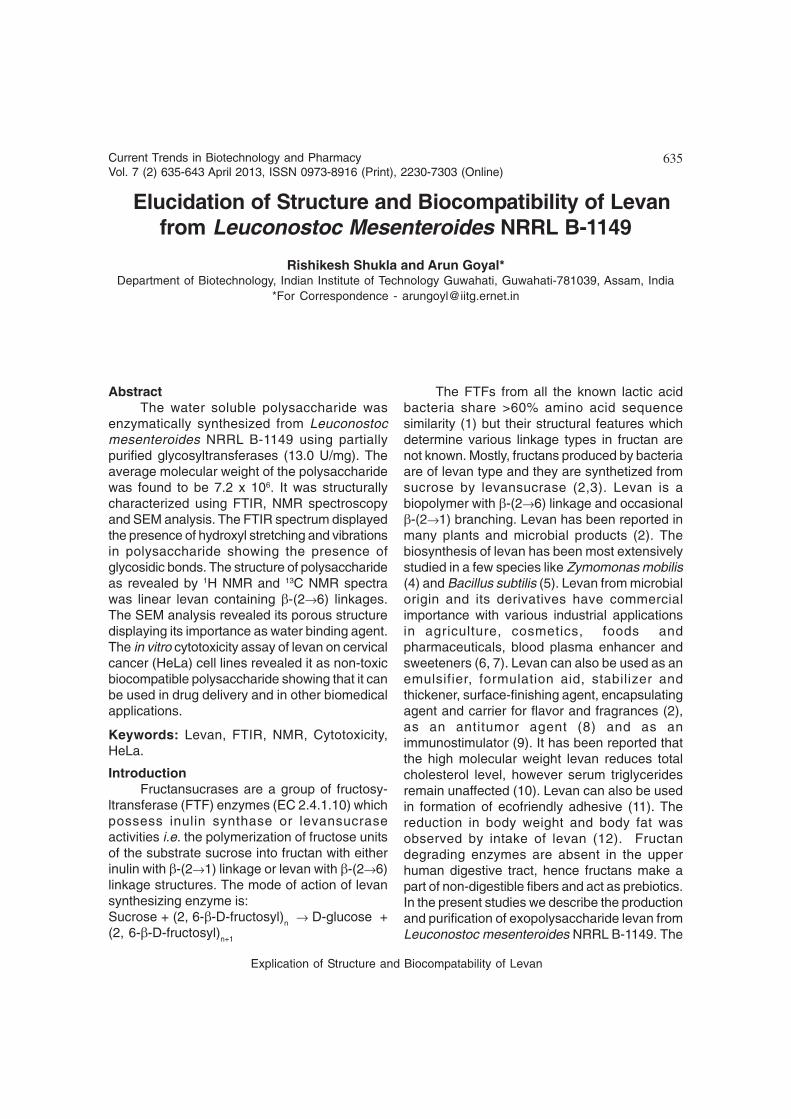
635Current Trends in Biotechnology and PharmacyVol. 7 (2) 635-643 April 2013, ISSN 0973-8916 (Print), 2230-7303 (Online)
AbstractThe water soluble polysaccharide was
enzymatically synthesized from Leuconostocmesenteroides NRRL B-1149 using partiallypurified glycosyltransferases (13.0 U/mg). Theaverage molecular weight of the polysaccharidewas found to be 7.2 x 10⁶. It was structurallycharacterized using FTIR, NMR spectroscopyand SEM analysis. The FTIR spectrum displayedthe presence of hydroxyl stretching and vibrationsin polysaccharide showing the presence ofglycosidic bonds. The structure of polysaccharideas revealed by 1H NMR and 13C NMR spectrawas linear levan containing β-(2→6) linkages.The SEM analysis revealed its porous structuredisplaying its importance as water binding agent.The in vitro cytotoxicity assay of levan on cervicalcancer (HeLa) cell lines revealed it as non-toxicbiocompatible polysaccharide showing that it canbe used in drug delivery and in other biomedicalapplications.
Keywords: Levan, FTIR, NMR, Cytotoxicity,HeLa.
IntroductionFructansucrases are a group of fructosy-
ltransferase (FTF) enzymes (EC 2.4.1.10) whichpossess inulin synthase or levansucraseactivities i.e. the polymerization of fructose unitsof the substrate sucrose into fructan with eitherinulin with β-(2→1) linkage or levan with β-(2→6)linkage structures. The mode of action of levansynthesizing enzyme is:Sucrose + (2, 6-β-D-fructosyl)
n → D-glucose +
(2, 6-β-D-fructosyl)n+1
The FTFs from all the known lactic acidbacteria share >60% amino acid sequencesimilarity (1) but their structural features whichdetermine various linkage types in fructan arenot known. Mostly, fructans produced by bacteriaare of levan type and they are synthetized fromsucrose by levansucrase (2,3). Levan is abiopolymer with β-(2→6) linkage and occasionalβ-(2→1) branching. Levan has been reported inmany plants and microbial products (2). Thebiosynthesis of levan has been most extensivelystudied in a few species like Zymomonas mobilis(4) and Bacillus subtilis (5). Levan from microbialorigin and its derivatives have commercialimportance with various industrial applicationsin agriculture, cosmetics, foods andpharmaceuticals, blood plasma enhancer andsweeteners (6, 7). Levan can also be used as anemulsifier, formulation aid, stabilizer andthickener, surface-finishing agent, encapsulatingagent and carrier for flavor and fragrances (2),as an antitumor agent (8) and as animmunostimulator (9). It has been reported thatthe high molecular weight levan reduces totalcholesterol level, however serum triglyceridesremain unaffected (10). Levan can also be usedin formation of ecofriendly adhesive (11). Thereduction in body weight and body fat wasobserved by intake of levan (12). Fructandegrading enzymes are absent in the upperhuman digestive tract, hence fructans make apart of non-digestible fibers and act as prebiotics.In the present studies we describe the productionand purification of exopolysaccharide levan fromLeuconostoc mesenteroides NRRL B-1149. The
Elucidation of Structure and Biocompatibility of Levanfrom Leuconostoc Mesenteroides NRRL B-1149
Rishikesh Shukla and Arun Goyal*Department of Biotechnology, Indian Institute of Technology Guwahati, Guwahati-781039, Assam, India
*For Correspondence - [email protected]
Explication of Structure and Biocompatability of Levan

636Current Trends in Biotechnology and PharmacyVol. 7 (2) 635-643 April 2013, ISSN 0973-8916 (Print), 2230-7303 (Online)
polysaccharide produced was structurallycharacterized using Fourier Transform Infrared(FTIR) spectrometry, Nuclear MagneticResonance (NMR) spectroscopy and ScanningElectron Microscopy (SEM). The polysaccharidewas further analyzed for its biocompatibilityassay.
Materials and MethodsBacterial Strain, medium and growthconditions : The strain Leuconostoc mesenter-oides NRRL B-1149 was obtained from ARSCulture Collection, National Centre forAgricultural Utilization Research, Peoria, USA.The culture was maintained in modified MRSagar medium (13) as a stab at 4°C and subcultured every 15 days. A loopful culture from anagar stab was transferred to 5 ml of sterilemedium described by Tsuchiya et al., 1952 (14)and incubated at 28°C and 180 rpm for 6-8 h.
Production and purification of levan : Thelevan was enzymatically synthesized using themethod as described earlier (17). To 30 ml of 20mM sodium acetate buffer (pH 5.4) containing0.1% (w/v) sodium azide, 10% (w/v) sucrose and5% (w/v) maltose using 600 µl of purified enzyme(13 U/mg, 0.2 mg/ml) was added. The reactionmixture was then incubated at 28°C and 180 rpmfor 24 h. After incubation, the mixture was put ina boiling water bath for 10 min and thencentrifuged at 8,000 g for 10 min. The pelletcontaining insoluble dextran was purified andcharacterized in our study as reported earlier(15). The supernatant containing solublepolysaccharide levan was precipitated using 65%(v/v) final concentration of ethanol. This wasrepeated two times and finally the pelletcontaining soluble polysaccharide was re-suspended in distilled water and lyophilized forfurther studies.
Structural characterization of levan : Thereducing value of purified levan was determinedby copper (Cu) bicinchoninate method (16). Thenumber-average degree of polymerization (DPn)and averagre molecular weight (MW) weredetermined by following equations described byFox and Robyt, 1991 (16).
DPn = (Cc/Cm) x 1.9
where, Cc and Cm are Concentrations ofcarbohydrate sample and maltose as determinedby reducing value (mg/ml)
MW = [DPn x 162] + 18
The purified levan was structurallycharacterized using FTIR, NMR and SEMtechniques. The FTIR spectrum was recordedin spectrometer (Spectrum One FTIRspectrometer, PerkinElmer Instruments, SanJose, CA, USA) for purified fructan in a KBr pellet.Nuclear magnetic resonance (NMR)spectroscopic analysis was performed in a VarianAS400 spectrometer (Agilent Technologies, PaloAlto, CA, USA). The levan was vacuum dried andthen exchanged with deuterium by successivelyophilization steps in D
2O (99.6% atom 2H,
Sigma-Aldrich, St. Louis, MO, USA). 15 mg levansample was dissolved in 0.4 ml of D
2O for 1H
NMR and 30 mg was dissolved in 0.4 ml of D2O
for 13C NMR. Tetramethyl silane (TMS) was usedas an internal reference. The SEM analysis ofdried levan was done by fixing it to the SEM stubwith a double-sided carbon tape. The sample wascoated with ~10 nm Au in a sputter coater(Polaron, Model SC7620). The surface of the drylevan powder was viewed in Field EmissionScanning Electron Microscope (Carl Zeiss, ModelSigma) operated at 10.0 kV.
Cytotoxicity and biocompatibility analysis oflevan : The lyophilized powder of levan from L.mesenteroides NRRL B-1149 was used for invitro cytotoxicity assay. The effect of levan onHeLa cells was determined using the colorimetric3-(4,5-dimethylthiazolyl-2)-2,5-diphenyltetrazolium bromide (MTT) assay as reportedearlier (17). HeLa cells at a density of 1.2x10⁴
cells/well in 100 µl medium were seeded in 96-well plate and allowed to attach at 37oC in 5%CO
2 atmosphere for 12-16 h. After incubation the
medium was removed from plate and the levanat different concentrations (1-1000 µg/ml) wasadded in each well. The medium without levanwas used as negative control. The plate wasincubated at 37oC in 5% CO
2 atmosphere for 48
h. After 48 h the medium were removed, 100 µl
Rishikesh Shukla and Arun Goyal

637Current Trends in Biotechnology and PharmacyVol. 7 (2) 635-643 April 2013, ISSN 0973-8916 (Print), 2230-7303 (Online)
MTT (500 µg/ml) was added to each well andincubated at 37oC for 4 h. The supernatant wasremoved and 100 µl DMSO was added to eachwell. Absorbance was measured at 570 nm by a96-well microplate reader (Tecan, Infinite 200Pro).
Results and DiscussionProduction and purification of levan from L.mesenteroides NRRL B1149 : The soluble levanpurified by ethanol precipitation was lyophilizedand the powdered levan was structurallycharacterized using FTIR, NMR and SEManalysis and was used for biocompatibilitystudies.
Structural characterization of water solublelevan Number average molecular weight : Thenumber average degree of polymerization (DPn)of purified levan sample as determined by Copperbicinchoninate method was 44887. The number
average molecular weight of levan from L.mesenteroides NRRL B-1149 calculated usingDPn was 7.2x10⁶. The water soluble levan fromBacillus polymyxa also showed similar molecularweight of about 2x10⁶ (18). The high molecularweight levans (>107) are reported to haveapplications in reduction of total cholesterol level(10) and in tumor therapy (19).
FTIR analysis of soluble levan : The FTIR dataof soluble levan from L. mesenteroides NRRLB-1149 is shown in Fig. 1. The band in region of3422 cm–1 showed the presence of hydroxylstretching vibration of polysaccharide while theband in 2926 cm–1 region was due to C-Hstretching vibration and the presence of boundwater was confirmed by the band in region of1640 cm–1. The results were supported by Caoet al., 2006 (20) and Liu et al., 2007 (21). Thestrong complex absorption at 1122 and 1063cm–1 signified the stretching vibrations of C-O-C
Fig. 1. FTIR spectrum of levan from Leuconostoc mesenteroides NRRL B-1149.
Explication of Structure and Biocompatability of Levan
638Current Trends in Biotechnology and PharmacyVol. 7 (2) 635-643 April 2013, ISSN 0973-8916 (Print), 2230-7303 (Online)
groups and ring vibrational modes in thecomposition of cyclic structures. The band at1122 cm–1 was assigned to valent vibrations ofC–O–C bond and glycosidic bridge. The
presence of a peak at 1063 cm–1 was due to thegreat chain flexibility present in polysaccharidearound the glycosidic bonds. The similar resultswere also described by Shingel, 2002 (22). These
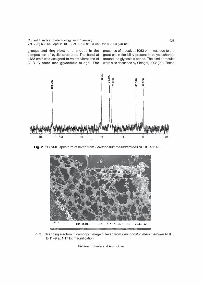
Fig. 2. 13C NMR spectrum of levan from Leuconostoc mesenteroides NRRL B-1149.
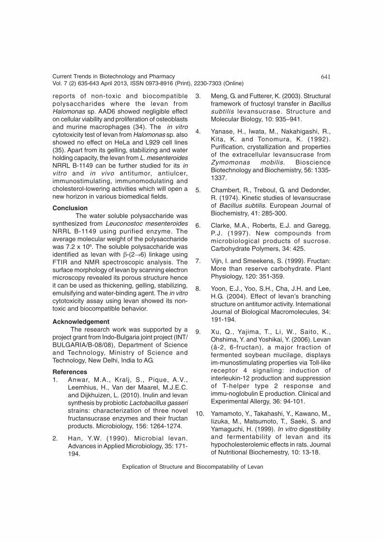
Fig. 3. Scanning electron microscopic image of levan from Leuconostoc mesenteroides NRRLB-1149 at 1.17 kx magnification.
Rishikesh Shukla and Arun Goyal

639Current Trends in Biotechnology and PharmacyVol. 7 (2) 635-643 April 2013, ISSN 0973-8916 (Print), 2230-7303 (Online)
linkages and branching in soluble polysaccharidewas analyzed by 1H NMR and 13C NMRspectroscopy.
1H NMR analysis of soluble levan : The 1H NMRspectrum of soluble levan from L. mesenteroidesNRRL B-1149 showed seven signals in the regionof skeletal protons (4.25-3.10 ppm region) dueto â-fructofuranoside (bFruf) units. The signalsin the regions 3.79, 3.69, 4.18, 4.11, 3.95, 3.84and 3.58 ppm were assigned to H1a, H1b, H3,H4, H5, H6a and H6b, respectively (Table 1). Theassignments for different resonances of 1H and13C NMR of levan from L. mesenteroides NRRLB-1149 is shown in Table 1.The similar resultswere observed in spectra of fructans fromLactobacillus reuteri LB 121 (23) and Bacillus sp.3B6 (24). In 1D-1H NMR spectrum of levan the
Table 1. 1H NMR and 13C NMR chemical shifts of levan from L. mesenteroides NRRL B-1149
1H shifts (in ppm)
H-1a H-1b H-3 H-4 H-5 H-6a H-6b
3.79 3.69 4.18 4.11 3.95 3.84 3.5813C shifts (in ppm)
C-1 C-2 C-3 C-4 C-5 C-6
104.29 80.39 76.40 75.30 63.52 59.96
Table 2. Comparison of 13C NMR chemical shifts of levan from L. mesenteroides NRRL B-1149with other reported strains.
Carbon Levan Levan LevS polymeratom L. mesenteroides Levan L. mesenteroides L. mesenteroides
B-1149 (present study) S. mutans26 B-512FMC27 B-512F36
Chemical shift (ppm)
C-1 59.96 60.7 60.5 60.145
C-2 104.29 104.2 109.1 104.3
C-3 76.40 76.3 76.5 76.523
C-4 75.30 75.2 75.4 75.391
C-5 80.39 80.3 80.7 80.453
C-6 63.52 63.4 63.6 63.57
absence of peak in anomeric region (4.9-5.3ppm) were found, which confirmed the absenceof any anomeric proton in it (Table 1). No signalsin the region downfield to 4.91 ppm confirmedthe absence of branching points. The observedpeak pattern fitted the fructofuranosideconfiguration.
13C NMR analysis of soluble levan : Thestructure of soluble levan produced from L.mesenteroides NRRL B-1149 was determined by13C NMR spectroscopy. The 13C NMR spectrumshowed six major resonances in the region of104.29, 80.39, 76.40, 75.30, 63.52 and 59.96ppm (Fig. 2). The keto-anomeric signal C2 dueto bFruf appeared at 104.29 ppm while C1 andC6 signals were detected at 59.96 and 63.52ppm, respectively. These data were in
Explication of Structure and Biocompatability of Levan

640Current Trends in Biotechnology and PharmacyVol. 7 (2) 635-643 April 2013, ISSN 0973-8916 (Print), 2230-7303 (Online)
accordance with previous reports (25). Thechemical shifts obtained for the polymer werealso similar to those observed in levan from S.mutans (26) but differed from levan produced byL. mesenteroides NRRL B-512 FMC (27), wherea difference in the C2 shift was found (109.1 ppminstead of 104.3 ppm) (Table 2). The 13C-NMRspectrum of levan from L. mesenteroides NRRLB-1149 was also comparable with the spectrumof fructan from Lactobacillus fermentum (AKJ15)having β-(2-1) and β-(2-6) linkages (28). Theabsence of corresponding signal of C2 in 1H NMRspectrum suggested that the glycosidic carbon(104.29 ppm) has no linkage with the hydrogen.The peaks at 80.39, 76.40, and 75.37 ppm weredue to -CH carbons while those at 63.52 and59.96 ppm corresponded to the -CH
2 carbon. The
1H and 13C NMR spectra of solublepolysaccharide from L. mesenteroides NRRL B-1149 showed its nature to be levan with (→6)-β-D-Fruf-(2→)
n structure. This was in agreement
with the previous reports (29,30).
Scanning electron microscopy of levan : Thesurface morphology of dried and powderedsoluble levan as analyzed by scanning electronmicroscopy (SEM) at 1.17 kx, is shown in Fig. 3.The SEM analysis revealed its porous structure.In the present study, the levan was more porousas compared to the insoluble dextran producedby the same strain. Due to the porous structurethe polysaccharides can be used in foods asthickening, gelling, stabilizing, emulsifying, andwater-binding agents (31-33).
Cytotoxicity and biocompatibility analysis oflevan : The effect of levan on viability of HeLacells is shown in Fig. 4. The results showed thatthere was no effect of levan on the viability ofcells up to 48 h even at higher concentration of1000 µg/ml. The levan from L. mesenteroidesNRRL B-1149 in current study was proved to benon-toxic and biocompatible and hence can beused as a biomaterial for biomedical applications.The results were in accordance with previous
Fig. 4. In vitro cytotoxicity assay showing the viability of HeLa cells after treatment with differentconcentrations of levan (1-1000 µg/ml) from Leuconostoc mesenteroides NRRL B-1149 up to 48 hincubation.
Rishikesh Shukla and Arun Goyal
641Current Trends in Biotechnology and PharmacyVol. 7 (2) 635-643 April 2013, ISSN 0973-8916 (Print), 2230-7303 (Online)
reports of non-toxic and biocompatiblepolysaccharides where the levan fromHalomonas sp. AAD6 showed negligible effecton cellular viability and proliferation of osteoblastsand murine macrophages (34). The in vitrocytotoxicity test of levan from Halomonas sp. alsoshowed no effect on HeLa and L929 cell lines(35). Apart from its gelling, stabilizing and waterholding capacity, the levan from L. mesenteroidesNRRL B-1149 can be further studied for its invitro and in vivo antitumor, antiulcer,immunostimulating, immunomodulating andcholesterol-lowering activities which will open anew horizon in various biomedical fields.
ConclusionThe water soluble polysaccharide was
synthesized from Leuconostoc mesenteroidesNRRL B-1149 using purified enzyme. Theaverage molecular weight of the polysaccharidewas 7.2 x 10⁶. The soluble polysaccharide wasidentified as levan with β-(2→6) linkage usingFTIR and NMR spectroscopic analysis. Thesurface morphology of levan by scanning electronmicroscopy revealed its porous structure henceit can be used as thickening, gelling, stabilizing,emulsifying and water-binding agent. The in vitrocytotoxicity assay using levan showed its non-toxic and biocompatible behavior.
AcknowledgementThe research work was supported by a
project grant from Indo-Bulgaria joint project (INT/BULGARIA/B-08/08), Department of Scienceand Technology, Ministry of Science andTechnology, New Delhi, India to AG.
References1. Anwar, M.A., Kralj, S., Pique, A.V.,
Leemhius, H., Van der Maarel, M.J.E.C.and Dijkhuizen, L. (2010). Inulin and levansynthesis by probiotic Lactobacillus gasseristrains: characterization of three novelfructansucrase enzymes and their fructanproducts. Microbiology, 156: 1264-1274.
2. Han, Y.W. (1990). Microbial levan.Advances in Applied Microbiology, 35: 171-194.
3. Meng, G. and Futterer, K. (2003). Structuralframework of fructosyl transfer in Bacillussubtilis levansucrase. Structure andMolecular Biology, 10: 935–941.
4. Yanase, H., Iwata, M., Nakahigashi, R.,Kita, K. and Tonomura, K. (1992).Purification, crystallization and propertiesof the extracellular levansucrase fromZymomonas mobilis. BioscienceBiotechnology and Biochemistry, 56: 1335-1337.
5. Chambert, R., Treboul, G. and Dedonder,R. (1974). Kinetic studies of levansucraseof Bacillus subtilis. European Journal ofBiochemistry, 41: 285-300.
6. Clarke, M.A., Roberts, E.J. and Garegg,P.J. (1997). New compounds frommicrobiological products of sucrose.Carbohydrate Polymers, 34: 425.
7. Vijn, I. and Smeekens, S. (1999). Fructan:More than reserve carbohydrate. PlantPhysiology, 120: 351-359.
8. Yoon, E.J., Yoo, S.H., Cha, J.H. and Lee,H.G. (2004). Effect of levan’s branchingstructure on antitumor activity. InternationalJournal of Biological Macromolecules, 34:191-194.
9. Xu, Q., Yajima, T., Li, W., Saito, K.,Ohshima, Y. and Yoshikai, Y. (2006). Levan(â-2, 6-fructan), a major fraction offermented soybean mucilage, displaysim-munostimulating properties via Toll-likereceptor 4 signaling: induction ofinterleukin-12 production and suppressionof T-helper type 2 response andimmu-noglobulin E production. Clinical andExperimental Allergy, 36: 94-101.
10. Yamamoto, Y., Takahashi, Y., Kawano, M.,Iizuka, M., Matsumoto, T., Saeki, S. andYamaguchi, H. (1999). In vitro digestibilityand fermentability of levan and itshypocholesterolemic effects in rats. Journalof Nutritional Biochemestry, 10: 13-18.
Explication of Structure and Biocompatability of Levan
642Current Trends in Biotechnology and PharmacyVol. 7 (2) 635-643 April 2013, ISSN 0973-8916 (Print), 2230-7303 (Online)
11. Combie, J., Steel, A. and Sweitzer, R.(2004). Adhesive designed by nature (andtested at Redstone Arsenal). CleanTechnologies and Environmental Policy, 6:258-262.
12. Kang, S.A., Jang, K.H., Lee, J.C., Chang,B.I., Lim, Y.A. and Song, B.C. (2003). Theeffects of fructose polymer levan on thebody fat accumulation and serum lipidprofiles of Korean women. Korean Journalof Community Nutrition. 8: 986-992.
13. Goyal, A. and Katiyar, S.S. (1996).Regulation of dextransucrase productivityfrom Leuconostoc mesenteroides NRRL B-512F by the maintenance media. Journalof General and Applied Microbiology, 42:81-85.
14. Tsuchiya, H.M., Koepsell, H.J., Corman, J.,Bryant, G., Bogard, M.O., Feger, V.H. andJackson, R.W. (1952). The effect of certaincultural factors on production ofdextransucrase by Leuconostocmesenteroides. Journal of Bacteriology, 64:521-527.
15. Shukla, R., Shukla, S., Bivolarski, V., Iliev,I., Ivanova, I. and Goyal, A. (2011).Production and structural characterizationof insoluble dextran produced in thepresence of maltose from Leuconostocmesenteroides NRRL B-1149. FoodTechnology and Biotechnology, 49(3): 291-296.
16. Fox, J.D. and Robyt, J.F. (1991).Miniaturization of three carbohydrateanalyses using a microsample plate reader.Analytical Biochemistry, 195: 93-96.
17. Mosmann, T. (1983). Rapid colorimetricassay for cellular growth and survival:Application to proliferation and cytotoxicityassays. Journal of Immunological Methods,65: 55-63.
18. Han, Y.W. and Clarke, M.A. (1990).Production and characterization of
microbial levan. Journal of Agricultural andFood Chemistry, 38: 393-396.
19. Calazans, G.M.T., Lima, R.C., de Franca,F.P. and Lopes, C.E. (2000). Molecularweight and antitumor activity ofZymomonas mobilis levans. InternationalJournal of Biological Macromolecules, 27:245- 247.
20. Cao, W., Li, X.Q., Liu, L., Yang, T.H., Li, C.,Fan, H.T., Jia, M., Lu, Z.G. and Mei, Q.B.(2006). Structure of antitumorpolysaccharide from Angelica sinensis(Oliv.) Diels. Carbohydrate Polymers, 66:149-159.
21. Liu, C., Lin, Q., Gao, Y., Ye, L., Xing, Y. andXi, T. (2007). Characterization andantitumor activity of polysaccharide fromStrongylocentrotus nudus eggs.Carbohydrate polymers, 67: 313-318.
22. Shingel, K.I. (2002). Determination ofstructural peculiarities of dextran, pullulanand c-irradiated pullulan by Fourier-transform IR spectroscopy. CarbohydrateResearch, 337: 1445-1451.
23. Van Geel-Schutten, G.H., Faber, E.J., Smit,E., Bonting, K., Smith, M.R., Ten Brink, B.,Kamerling, J.P., Vliegenthart, J.F.G. andDijkhuizen L. (1999). Biochemical andstructural characterization of the glucan andfructan exopolysaccharides synthesized bythe Lactobacillus reuteri wild-type strain andby mutant strains. Applied andEnvironmental Microbiology, 65: 3008-3014
24. Matulova, M., Husarová, S., Capek P.,Sancelme, M. and Delort, A.M. (2011). NMRstructural study of fructans produced byBacillus sp. 3B6, bacterium isolated in cloudwater. Carbohydrate Research, 346: 501-507.
25. Van Hijum, S., van Greel-Schutten, H.,Rahaoui, H., van der Maarel, M. andDijkhuizen, L. (2002). Characterization ofa novel fructosyltransferase from
Rishikesh Shukla and Arun Goyal
643Current Trends in Biotechnology and PharmacyVol. 7 (2) 635-643 April 2013, ISSN 0973-8916 (Print), 2230-7303 (Online)
Lactobacillus reuteri that synthesizes highmolecular weight inulin and inulinoligosaccharides. Applied andEnvironmental Microbiology, 68: 4390-4398.
26. Shimamura, A., Tsuboi, K., Nagase, T., Ito,M., Tsumori, H. and Mukasa, H. (1987).Structural determination of D-fructans fromStreptococcus mutans, serotype b, c, e, andf strains, by 13C-N.M.R. spectroscopy.Carbohydrate Research, 165: 150-154.
27. Kang, H.K., Seo, M.Y., Seo, E.S., Kim, D.,Chung, S.Y., Kimura, A., Day, D.F. andRobyt, J.F. (2005). Cloning and expressionof levansucrase from Leuconostocmesenteroides B-512 FMC in Escherichiacoli. Biochimica et Biophysica Acta,1727(1): 5-15.
28. Dutta, A., Das, D. and Goyal, A. (2012).Purification and characterization of fructanand fructansucrase from Lactobacillusfermentum (AKJ15) isolated from Kodo koJaanr, a fermented beverage from NorthEastern Himalayas. International Journal ofFood Science and Nutrition, 63(2): 216-224.
29. Angyal, S.J. and Bethell G.S. (1976).Conformational analysis in carbohydratechemistry. III. The 13C N.M.R. spectra of thehexuloses. Australian Journal of Chemistry,29(6): 1249-1265.
30. Barrow, K.D., Collins, J.G., Rogers, P.L. andSmith, G.M. (1984). The structure of a novelpolysaccharide isolated from Zymomonasmobilis determined by nuclear magneticresonance spectroscopy. European Journalof Biochemistry, 145(1): 173-179.
31. Kirk, R.E., Othmer, D.F., Kroschwitz, J.I.and Howe-Grant, H. (1991). Kirk-OthmerEncyclopedia of Chemical Technology. Vol.16, Wiley Inc., New York.
32. Khan, T., Park, J.K. and Kwon, J.H. (2007).Functional biopolymers produced bybiochemical technology consideringapplications in food engineering. KoreanJournal Chemical Engineering, 24: 816-826.
33. Shih, I.L., Yu, Y.T., Shieh, C.J. and Hsieh,C.Y. (2005). Selective production andcharacterization of levan by Bacillus subtilis(Natto) Takahashi. Journal of Agriculturaland Food Chemistry, 53: 8211 -8215.
34. Poli, A., Kazak, H., Gürleyendað, B.,Tommonaro, G., Pieretti, G., Toksoy Öner,E. and Nicolaus, B. (2009). High levelsynthesis of levan by a novel Halomonasspecies growing on defined media.Carbohydrate Polymer, 78: 651-657.
35. Küçükaºik, F., Kazak, H., Güney, D., Finore,I., Poli, A., Yenigün, O., Nicolaus, B. andToksoy Öner, E. (2011). Molasses asfermentation substrate for levan productionby Halomonas sp. Applied Microbiology andBiotechnology, 89:1729-1740.
36. Sandra, M.A., Maria, E.R., Lorenzo S.,Agustín, L.M. and Clarita, O.C. (2006).Identification and functionalcharacterization of levS, a gene encodingfor a levansucrase from Leuconostocmesenteroides NRRL B-512F. Gene, 376:59-67.
Explication of Structure and Biocompatability of Levan