Embed Size (px)
Citation preview
RESEARCH ARTICLE
Elucidation of the compatible interaction
between banana and Meloidogyne incognita via
high-throughput proteome profiling
Aisyafaznim Al-Idrus1,2*, Sebastien Christian Carpentier3,4, Mohamad Taufiq Ahmad1,
Bart Panis5, Zulqarnain Mohamed1,2
1 Genetics and Molecular Biology Division, Institute of Biological Sciences, Faculty of Science, University of
Malaya, Kuala Lumpur, Malaysia, 2 Center for Research in Biotechnology for Agriculture (CEBAR),
University of Malaya, Kuala Lumpur, Malaysia, 3 Laboratory of Tropical Crop Improvement, Division of Crop
Biotechnics, Faculty of Bioscience Engineering, Katholieke Universiteit Leuven, Leuven, Belgium,
4 SYBIOMA: Facility for SYstems BIOlogy based MAss spectrometry, Leuven, Belgium, 5 Bioversity
International, Belgian Office at KU Leuven, Leuven, Belgium
Abstract
With a diverse host range, Meloidogyne incognita (root-knot nematode) is listed as one of
the most economically important obligate parasites of agriculture. This nematode species
establishes permanent feeding sites in plant root systems soon after infestation. A compati-
ble host-nematode interaction triggers a cascade of morphological and physiological pro-
cess disruptions of the host, leading to pathogenesis. Such disruption is reflected by altered
gene expression in affected cells, detectable using molecular approaches. We employed a
high-throughput proteomics approach to elucidate the events involved in a compatible
banana- M. incognita interaction. This study serves as the first crucial step in developing nat-
ural banana resistance for the purpose of biological-based nematode management pro-
gramme. We successfully profiled 114 Grand naine root proteins involved in the interaction
with M. incognita at the 30th- and 60th- day after inoculation (dai). The abundance of proteins
involved in fundamental biological processes, cellular component organisation and stress
responses were significantly altered in inoculated root samples. In addition, the abundance
of proteins in pathways associated with defence and giant cell maintenance in plants such
as phenylpropanoid biosynthesis, glycolysis and citrate cycle were also implicated by the
infestation.
Introduction
Plants are constantly exposed to a range of pathogenic organisms inhabiting the soil. Amongst
these, plant-parasitic nematodes (PPN) are documented as soil pathogens of economic impor-
tance incurring approximately US$100 billion worth annual crop losses [1]. Amongst the
PPN, sedentary root-knot nematodes (RKN; Meloidogyne spp.) are one of nature’s most suc-
cessful obligate parasites. Meloidogyne incognita was reported to be the most widely distributed
PLOS ONE | https://doi.org/10.1371/journal.pone.0178438 June 2, 2017 1 / 25
a1111111111
a1111111111
a1111111111
a1111111111
a1111111111
OPENACCESS
Citation: Al-Idrus A, Carpentier SC, Ahmad MT,
Panis B, Mohamed Z (2017) Elucidation of the
compatible interaction between banana and
Meloidogyne incognita via high-throughput
proteome profiling. PLoS ONE 12(6): e0178438.
https://doi.org/10.1371/journal.pone.0178438
Editor: T. R. Ganapathi, Bhabha Atomic Research
Centre, INDIA
Received: January 3, 2017
Accepted: May 12, 2017
Published: June 2, 2017
Copyright: © 2017 Al-Idrus et al. This is an open
access article distributed under the terms of the
Creative Commons Attribution License, which
permits unrestricted use, distribution, and
reproduction in any medium, provided the original
author and source are credited.
Data Availability Statement: All relevant data are
within the paper and its Supporting Information
file.
Funding: This work was supported by the Ministry
of Science, Technology and Innovation Malaysia,
under Top-down Grant [number 53-02-03-1069,
2010];University of Malaya, Kuala Lumpur,
Malaysia under Postgraduate research Grant
[number PG194-2014B, 2014], and UMRG grant
[number RP005E-13BIO, 2013]. We would also
like to acknowledge University of Malaya, for
species in this genus attacking more than 3000 plant species including agricultural crops [2, 3,
4, 5, 6] such as bananas (Musa spp.). Banana production was considerably affected by RKN
infestations particularly in the absence of Radopholus similis [7, 8]. RKN are a problem for
bananas in the tropics especially in Asian countries [8] and in dry sub-tropical countries [9].
At least five RKN species infect Musa withM. incognita andM. javanica being the more com-
monly found [10, 11, 12].
Similar to other RKN species, Meloidogyne incognita is a sedentary plant-parasite with
evolved strategies to infest plant species by manipulating fundamental key elements of plant
cell development [13]. During a compatible interaction, this parasite induces re-differentiation
of root cells into multinucleated nematode feeding cells (giant cells). These giant cells operate
as nutrient sinks that meet the nutritional demands of developing nematode individuals. Syn-
chronously, hyperplasia and hypertrophy of the surrounding cells lead to the formation of a
root gall [13, 14], thus affecting plant growth and its anchorage system. Detailed infection
mechanism and feeding site establishment process have been reported/reviewed in [15, 16, 17,
18, 19]. Wilting and stunted growth are distinctive symptoms for infected banana plants [20],
leading further reduction in bunch weight and fruit production. Jonathan et al. [21] reported
that banana fruit size, total carbohydrate, total soluble sugars and ascorbic acid were reduced
as a result of M. incognita infestation. As such, there is a dire need for effective control of this
nematode in banana farms.
Control measures at the macro level such as crop rotation and using clean planting materi-
als are inefficient while the only effective solution using nematicides is undesirable due to its
detrimental effect to the humankind and the environment [22]. Therefore, alternative manage-
ment strategies such as crop genetic improvement are gaining new interest worldwide [8]. Sev-
eral nematode resistance (Nem-R) genes expressing NBS-LRR proteins [23] have been isolated
from various plants [24, 25], all conferring resistance against sedentary endoparasites [24]. The
first nematode resistance gene cloned was Hs1pro-1 from sugar beet conferring resistance
against sugar beet cyst nematodes [26, 27]. Four other genes namely Mi-1, Hero A, Gpa2 and
Gro1-4 were cloned from tomato and potato relatives [24]. However since a gene transfer
attempt from tomato into tobacco using the cloned Mi gene was unsuccessful [27], efforts
were focused more on searching for plants’ own natural tolerance/resistance gene(s) against
the pathogen. Therefore the quest for new mechanisms acting against M. incognita relies pri-
marily on the knowledge garnered from plant-pathogen interaction studies. Since there is, as
yet, no resistant banana cultivar reported against M. incognita, the current work describes our
attempt to model the compatible interaction withM. incognita using the commercial dessert
cultivar, Grand naine.
Parasite survival strategies and host defence mechanisms are the key factors governing the
complex host-nematode relationship [18]. For the past 18 years, many studies utilising differ-
ent approaches have been conducted to obtain deeper understanding of plant-pathogen inter-
action in order to formulate efficient control strategies [1, 18, 28, 29]. In the advent of the
‘omics’ era, proteomics is seen as a potential platform to ‘decode’ the communication used
between the two organisms due to the fact that proteins are the most immediate macromole-
cules involved in biological actions and responses. A comprehensive review on proteomic
analysis of plant response to nematode infection can be obtained in Escobar et al. [30]. Echoing
that of mentioned in Palomares-Rius et al. [31] and Quirino et al. [32], we found that most
proteomic analyses of plants subjected to biotic and abiotic treatments were conducted on
plant model species such as Arabidopsis and rice. Besides, amongst a handful of proteomics
studies on plant-pathogen interactions, most of them were carried out to address bacterial and
fungal diseases of foliar tissues and not many of root tissues.
M. incognita-banana interaction
PLOS ONE | https://doi.org/10.1371/journal.pone.0178438 June 2, 2017 2 / 25
funding the first author’s stay in the Laboratory of
Tropical Crop Improvement, Leuven, Belgium
through the Flagship programme [number FL002-
2010, 2010].
Competing interests: The authors have declared
that no competing interests exist.
The difficulty in carrying out proteomics studies on root-pathogen interactions has been
reported in a review [33]. Recently, Palomares-Rius et al. [31] and Li et al. [34] reported a com-
mendable 2D-based proteomic study of tripartite interaction on chickpea and Fusarium-
banana root interaction, respectively. However, the throughput of gel-based systems is limited
and only a scarce amount of interesting proteins has been discovered. Therefore we adopted
the gel-free, Liquid Chromatography-Mass Spectrometry (LC-MS)—based Proteomics plat-
form to elucidate proteins involved in banana-M. incognita interaction at the 30th- and 60th—
day after inoculation (dai). The two time points were chosen in order to obtain an increased
number of galled cells for increased peptide abundance detection. Gel- and label-free LC-MS
methods represent attractive alternatives since they are robust and amenable to all types of bio-
logical samples, despite being cost effective. This is the first high throughput report to reveal
the molecular profile and dynamic changes occurring in banana roots inoculated withM.
incognita. Our report adds new insights to plant reactions during nematode infestation and
new knowledge on the weak points in plant defence mechanisms which can serve as the basis
towards developing a molecular-based nematode combating regime.
Materials and methods
Plant materials and M. incognita culture
Banana cv. Grand naine (ITC 1256) plantlets were obtained from the International Transit
Centre (ITC) of Musa collection at Katholieke Universitiet Leuven, Belgium. The plantlets
were propagated and maintained in Murashige and Skoog (MS) basal media (pH 5.8) supple-
mented with 4.1 μM biotin, 5.7 μM indoleacetic acid (IAA), 5.4 μM naphtaleneacetic acid
(NAA), 87 μM sucrose and 2 g/L gelrite with 3 mg/L 6-Bensylaminopurine (BAP) prior to
transfer onto rooting media (MS including vitamins, 1 mL/L ascorbic acid, 30 g/L sucrose, 2 g/
L gelrite and 0.5 g/L active charcoal, pH 6.15 at 60˚C). Banana plantlets were left in the rooting
media for a month until reaching a four or five-leaf-stage prior to transplantation. Transplan-
tation and acclimatisation processes were carried out as described in [35]. Briefly, the plantlets
were transplanted into a 200 mL plastic pots containing sand: peat (2:1) soil mixture and left to
acclimatise under greenhouse conditions for 8 weeks. Fertiliser was applied twice a week start-
ing from the second week post transplantation. The plants were later transferred into 1 L pots
for inoculation procedure. Meloidogyne incognita (Malaysian population) culture was obtained
from Malaysian Agricultural Research and Development Institute (MARDI). This culture was
then propagated and maintained in Grand naine host plants until future use.
Nematode inoculation
Collection of RKN egg masses from plant hosts and hatching of juvenile stage two (J2) nema-
todes were carried out as described in [35]. To estimate the number of J2 used for treatments,
J2 individuals obtained per mL were counted in triplicates and later averaged. dH2O was used
to adjust to 1000 J2 per treatment. Treatment was carried out using a single inoculation proto-
col as we previously described [35]. Briefly, plants transplanted into 1 L pots were left to accli-
matise for a week. Following that, one of the primary roots was selected as an inoculation
target. The selected root was placed across a 3.5 cm diameter plastic Petri dish (the target root
fragment) and left in soil for three days prior to inoculation. One thousand J2 nematodes were
inoculated onto the targeted root fragment and the target site was left in soil until the experi-
ment was terminated at either 30- or 60-days after inoculation (dai). Each time point consisted
of three nematode-inoculated banana plantlets and three control plantlets (three biological
replicates for each treatment). Upon harvesting, the targeted inoculation site was excised from
the root system (one root fragment for each banana plant), thoroughly washed under running
M. incognita-banana interaction
PLOS ONE | https://doi.org/10.1371/journal.pone.0178438 June 2, 2017 3 / 25
tap water, air-dried and weighed. The weighed root fragment was placed into a fresh 1.5 mL
microcentrifuge tube, snap-frozen in liquid nitrogen and then stored in -80˚C until further
lyophilisation.
Root tissue lyophilisation and protein isolation
Stored banana root sample was lyophilised for 48 hours at -100 C˚ according to [36]. Banana
root proteins were then extracted using phenol extraction methanol/ammonium acetate pre-
cipitation-based protocol as described in [37] with minor modifications suitable for LC-MS
shotgun platform. Protein extraction procedure was conducted at 4˚C unless mentioned oth-
erwise. Briefly, 60 mg lyophilised banana root tissue were ground using cooled mortar and pes-
tle in the presence of liquid nitrogen and later suspended in a fresh 2 mL microcentrifuge tube
containing 750 μL Extraction buffer [100 mM Tris-HCl pH 8.3, 5 mM EDTA, 100 mM KCl,
1% w/v DTT, 30% w/v sucrose; complete protease inhibitor cocktail (Roche Applied Science)
and mixed. Subsequently, 750 μL buffered phenol were added to the sample and vortexed for
10 min. The mixture was then centrifuged (12,000 rpm, 10 min) and the resulting phenolic
phase was transferred into a fresh microcentrifuge tube and protein was re-extracted using
equal volume of extraction buffer. Sample was then centrifuged (12, 000 rpm, 5 min) and
resulting phenolic phase was transferred into a fresh tube. Proteins were precipitated overnight
in 5 volumes methanol (100mM ammonium acetate) at -20˚C. Following this, the sample was
then centrifuged (13,000 rpm, 60 min) to collect the pellet and later rinsed twice in 2 mL rins-
ing solution (cold acetone/0.2% DTT). Note that for the first rinse, the pellet was left in the
rinsing solution for 1 hr at -20˚C and centrifuged (13, 000 rpm, 30 min). Cleaned pellet was
left air-dried and suspended in 100 μL Lysis buffer (8M urea, 5mM DTT, 30 mM Tris). Protein
quantification was carried out using 2-D Quant Kit (Amersham, UK) following manufactur-
er’s protocol.
Peptide digestion and separation
Twenty micrograms of protein sample was incubated in 0.02 M DTT for 15 min. This mixture
was then mixed with 0.05 M iodoacetamide and incubated in the dark for 30 min prior to dilu-
tion in 0.05 M ammonium bicarbonate. An aliquot of 20 μg of protein was then digested with
0.2 μg/μL trypsin and incubated overnight at 37˚C. Samples were acidified with trifluoroacetic
acid (0.1% final concentration) and de-salted using C18 solid phase extraction according to
the manufacturer (Pierce™ C18 Spin Columns, Thermo Fisher Scientific, Ghent, Belgium).
Peptides were eluted with 40 μL 70% acetonitrile (ACN), after which solvents were evaporated
using a speedvac and dissolved in 5% ACN, 0.1% formic acid.
Peptide separation and MS analysis
The UPLC—MS/MS analysis was performed on a Q Exactive Orbitrap mass spectrometer
(Thermo Scientific, USA) as described by [38]. The samples (5 μL containing 1 μg peptides)
were injected and separated on an Ultimate 3000 UPLC system (Dionex, Thermo Scientific)
equipped with a C18 PepMap100 pre-column (5 μm, 300 μm × 5 mm, Thermo Scientific) and
an EasySpray C18 column (3 μm, 75 μm × 15 cm, Thermo Scientific) using a gradient of 5% to
20% ACN in 0.1% formic acid (FA) in 10 min followed by a gradient of 10% to 35% ACN in
0.1% FA in 4 min and then a final gradient from 35% to 95% ACN in 0.1% FA in 2.5 min. The
flow-rate was set at 250 μL/ min. The mass spectrometer was operated in a positive ion mode
with a nanospray voltage of 1.5 kV and a source temperature of 250˚C. ProteoMass LTQ/
FT-Hybrid ESI Pos. Mode CalMix (MSCAL5-1EA SUPELCO, Sigma-Aldrich) was used as an
external calibrant and the lock mass 445.12003 as an internal calibrant. The instrument was
M. incognita-banana interaction
PLOS ONE | https://doi.org/10.1371/journal.pone.0178438 June 2, 2017 4 / 25
operated in a data-dependent acquisition (DDA) mode with a survey MS scan at a resolution
of 70,000 (FWHM at m/z 200) for the mass range of m/z 350–1800 for precursor ions, followed
by MS/MS scans of the top 10 most intense peaks with + 2, + 3 and + 4 charged ions above a
threshold ion count of 16,000 at a 35,000 resolution using a normalised collision energy (NCE)
of 29 eV with an isolation window of 3.0 m/z and dynamic exclusion of 10 s. All data were
acquired with Xcalibur 2.2 software (Thermo Scientific).
Protein identification
All raw data were converted into mgf files using Progenesis v4.1 (Nonlinear Dynamics, UK).
The spectra were searched using Mascot (Version 2.2.06; Matrix science, London, England)
against our in-house Musa database (76,220 sequences) containing all protein sequences of
published A and B genome along with contaminant sequences (trypsin and keratin). The fol-
lowing parameters were used: the enzyme was trypsin and one miscleavage was allowed,
cystein-carbamidomethylation was chosen as a fixed modification and methionine-oxidation
as a variable one. Precursor peptide charge state was 2+ and 3+, error window on experimental
peptide mass values was 10 ppm and 20 absolute milli-mass units were chosen for fragment
ion mass tolerance. Peptides assigned to keratin or trypsin were not taken into account.
For data validation, the false discovery rate (FDR) was calculated using Scaffold (Version:
Scaffold_3.6.3; Proteome Software Inc., Portland, OR, USA). An integrated version of X! Tan-
dem (Version: CYCLONE, 2010.12.01.1) in Scaffold was used for an additional database
searching. For FDR calculation, the search results from both Mascot and X! Tandem were
combined automatically by Scaffold with the following settings: a peptide confidence level of
95%, a protein confidence level of 80%, a minimum peptide number of 1, and the thresholds
of each search engines separately.
Peptide quantification and data analysis
Quantitative analysis was performed using Progenesis LC—MS version 4.1 (Nonlinear
Dynamics) as described by [39] with some modifications. For alignment, a reference run was
selected, after which the files were aligned automatically, manual landmark vectors were not
necessary. The sensitivity of the peak picking limits was put to four. With these settings, Pro-
genesis LC—MS generates an aggregate run that contains all ions from the analysed runs. Pep-
tides with charges two to five were retained in the filter step, and then the data were
normalised by calculating abundance ratios to a reference run. Peptides with p-value< 0.05
(Progenesis) were retained for further analysis and abundances of all corresponding significant
peptides were summed per protein accession number. By this, protein abundances were calcu-
lated based on peptide abundances. A list of identified proteins was then generated and sub-
jected to Principal Component Analysis (PCA) to assess whether or not proteins from similar
experimental condition cluster with each other. The software STATISTICA (Version 8.0;
Tulsa, OK, USA) was used for data analysis. Statistical evaluation started with a principal com-
ponent analysis (PCA) according to [40]. Only proteins (ANOVA: P�0.05) with more than
1.5 times fold change and showing maximum ion score of more than 30 were deemed signifi-
cant and selected for further analysis. For ease of analysis, pairwise comparison matrices were
used to compare between treatments i.e 60-dai inoculated vs. 60-dai control samples; and
30-dai inoculated vs. 30-dai control samples in order to profile proteins involved during the
interaction. For these matrices, protein abundance in control samples were used as the baseline
to obtain protein fold changes. These proteins were then subjected to Gene Ontology (GO)
analysis using BLAST2GO and grouped based on GO levels obtained from this software. In
cases where a protein was present with more than one GO level, the most general level that
M. incognita-banana interaction
PLOS ONE | https://doi.org/10.1371/journal.pone.0178438 June 2, 2017 5 / 25
described its function was chosen. Protein-GO interactions were mapped using Cytoscape
(available for free at http://www.cytoscape.org/download.php).
Results
Banana root proteins showed significant abundance difference upon
interaction with Meloidogyne incognita
In this study, we observed that galls were more visible to the naked eye as swellings on inocu-
lated banana roots at 60-dai compared to root fragments inoculated for 30 days (Fig 1). No
galls were present in all control samples. A total of 408 proteins were subjected to Principal
Component Analysis (PCA). We found that principal component one (PC1) separated 60-dai
control samples from 60-dai inoculated samples. However, definitive clustering was not
obtained for samples harvested at 30-dai (Fig 2). On the other hand, all 30-dai samples were
separated from the 60-dai samples by PC2. We identified 114 root proteins showing significant
differential abundance levels between nematode-inoculated root tissues and control root tis-
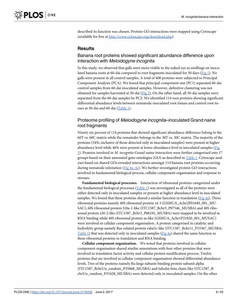
sues at 30-dai and 60-dai (Table 1).
Proteome profiling of Meloidogyne incognita–inoculated Grand naine
root fragments
Ninety-six percent of 114 proteins that showed significant abundance difference belong to the
60T vs. 60C matrix while the remainder belongs to the 30T vs. 30C matrix. The majority of the
proteins (54%; inclusive of those detected only in inoculated samples) were present at higher
abundance level while 46% were present at lower abundance level in inoculated samples (Fig
3). Proteins involved in M. incognita-Grand naine interaction were further categorised into 17
groups based on their annotated gene ontologies (GO) as described in Table 1. Cytoscape anal-
yses based on shared GOs revealed interactions amongst 114 banana root proteins occurring
during nematode infestation (Fig 4a–4c). We further investigated protein-GO interactions
involved in fundamental biological process, cellular component organisation and response to
stresses.
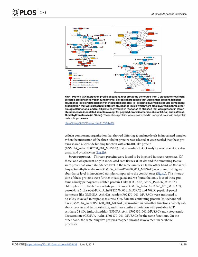
Fundamental biological processes. Interaction of ribosomal proteins categorised under
the fundamental biological processes (Table 1) was investigated as all of the proteins were
either detected only in inoculated samples or present at higher abundance level in inoculated
samples. We found that these proteins shared a similar function in translation (Fig 4a). Three
ribosomal proteins namely 40S ribosomal protein s4-3 (GSMUA_Achr2P01640_001_MU-
SAC), 60S ribosomal protein I10a-1-like (ITC1587_Bchr3_P07546_MUSBA) and 40S ribo-
somal protein s20-2-like (ITC1587_Bchr3_P06192_MUSBA) were mapped to be involved in
RNA binding while 40S ribosomal protein sa-like (GSMUA_Achr1P21820_001_MUSAC)
were involved in cellular component organisation. A protein categorised in catalytic and
hydrolytic group namely Ras-related protein raba5c-like (ITC1587_Bchr11_P33367_MUSBA;
Table 1) that was detected only in inoculated samples (Fig 4a) shared the same function as
these ribosomal proteins in translation and RNA binding.
Cellular component organization. We noted that proteins involved in cellular
component organisation shared similar annotations with four other proteins that were
involved in translation factor activity and cellular protein modification process. Twelve
proteins that are involved in cellular component organisation showed differential abundance
levels. Two of the proteins namely Ru large subunit-binding protein subunit alpha
(ITC1587_BchrUn_random_P35868_MUSBA) and tubulin beta chain-like (ITC1587_B-
chrUn_random_P35428_MUSBA) were detected only in inoculated samples. On the other
M. incognita-banana interaction
PLOS ONE | https://doi.org/10.1371/journal.pone.0178438 June 2, 2017 6 / 25

hand, alpha-glucan-protein synthase (ITC1587_Bchr4_P10810_MUSBA), tubulin alpha-
3-chain-like (ITC1587_Bchr6_P17875_MUSBA) and 40S ribosomal protein Sa-like
protein (GSMUA_Achr1P21820_001_MUSAC) showed higher abundance levels in
inoculated samples while other proteins were present in lower abundances in inoculated
samples (Fig 4b). Interestingly, we had profiled two tubulin beta chain protein species
(ITC1587_Bchr6_P16601_MUSBA and GSMUA_Achr6P04600_001_MUSAC) involved in
Fig 1. An example of severity of galls observed on the 60th-dai compared to the 30th-dai that were
visible to the naked eye, indicated by the arrows. No galls formed in control root fragments for both time
points.
https://doi.org/10.1371/journal.pone.0178438.g001
Fig 2. Principle Component Analysis (PCA) conducted on 408 proteins showed PC1 separates 60-dai
inoculated samples (I60) from 60-dai control samples (C60) while definitive separation was not
obtained for 30-dai inoculated samples (I30) and 30-dai control samples (C30). On the other hand, PC2
separates 30-dai samples (C30 and I30) from 60-dai samples (C60 and I60).
https://doi.org/10.1371/journal.pone.0178438.g002
M. incognita-banana interaction
PLOS ONE | https://doi.org/10.1371/journal.pone.0178438 June 2, 2017 7 / 25

Table 1. List of proteins showing significant abundance difference (together with their accession numbers) in inoculated samples at 30- and 60-
dai after inoculation with 1000J2 M. incognita. The list is sorted according to the fold change of each protein.
Protein Reference No. Fold change AnovaP
�0.05
Max
ion
score
No. of
peptides
Fundamental Biological Processes
60-dai
40s ribosomal protein s2-3-like GSMUA_AchrUn_randomP09450_001_MUSAC Detected only
in inoculated
0.01 34.37 1
60s ribosomal protein I22-2-like GSMUA_Achr3P00720_001_MUSAC Detected only
in inoculated
0.01 54.86 1
60s ribosomal protein l10a-1-like ITC1587_Bchr3_P07546_MUSBA Detected only
in inoculated
0.01 58.35 3
Adenosine kinase 2-like GSMUA_Achr2P00250_001_MUSAC Detected only
in inoculated
0 51.3 3
Cinnamyl alcohol dehydrogenase GSMUA_Achr4P06150_001_MUSAC Detected only
in inoculated
0 32.51 5
60s ribosomal protein l9-like ITC1587_Bchr5_P13916_MUSBA 469.7 0.01 47.46 2
40s ribosomal protein s20-2-like ITC1587_Bchr3_P06192_MUSBA 55 0.02 33.43 2
40s ribosomal protein s15a-1 GSMUA_Achr1P17170_001_MUSAC 29.4 0.01 38.51 1
40s ribosomal protein s16-like ITC1587_Bchr6_P15714_MUSBA 27.3 0 34.19 1
40s ribosomal protein s14 GSMUA_Achr2P20380_001_MUSAC 22 0.01 53.54 2
S-adenosylmethionine synthase ITC1587_Bchr7_P18740_MUSBA 17 0.03 30.77 2
V-type proton ATPase catalytic subunit a-like GSMUA_Achr11P08060_001_MUSAC 3.5 0.05 79.24 1
40s ribosomal protein s4-3 GSMUA_Achr2P01640_001_MUSAC 3.4 0.03 46.34 2
S-adenosylmethionine synthase 2 ITC1587_Bchr1_P01149_MUSBA 3.4 0.01 34.37 1
60s ribosomal protein l4-like GSMUA_Achr5P03060_001_MUSAC 3.2 0.01 43.69 5
40s ribosomal protein sa-like GSMUA_Achr1P21820_001_MUSAC 2.4 0.02 33.36 3
ATP synthase f0 subunit 1 GSMUA_AchrUn_randomP15230_001_MUSAC -1.5 0.03 55.78 6
ATP synthase subunit GSMUA_Achr10P27350_001_MUSAC -1.6 0.01 74.99 1
ATP synthase subunit GSMUA_Achr9P21710_001_MUSAC -2 0.01 82.42 8
Probable ATP synthase 24 kda (mitochondrial) GSMUA_Achr6P02850_001_MUSAC -2.2 0 54.84 2
ATP synthase subunit mitochondrial ITC1587_Bchr10_P31293_MUSBA -2.6 0.01 31.84 1
Aspartate (mitochondrial) ITC1587_Bchr6_P16093_MUSBA -2.4 0 87.65 2
Phosphoenolpyruvate carboxylase GSMUA_Achr6P26850_001_MUSAC -2.6 0 33.06 1
Aspartate-semialdehyde dehydrogenase GSMUA_Achr10P18110_001_MUSAC -3.6 0.022 89.03 1
Pyruvate dehydrogenase e1 component subunit
beta- (mitochondrial)
GSMUA_Achr5P25000_001_MUSAC -3.7 0.02 74.19 4
Minor allergen alt a 7-like GSMUA_Achr5P26440_001_MUSAC -6.1 0.02 34.57 2
Binding
60-dai
General
Probable calcium-binding protein cml7 ITC1587_Bchr9_P27746_MUSBA 5.2 0.05 40.13 1
Germin-like protein 5–1 GSMUA_Achr5P18440_001_MUSAC -7.5 0.01 32.5 1
RNA
Elongation factor 1-alpha ITC1587_Bchr6_P15150_MUSBA Detected only
in inoculated
0 51.4 4
Elongation factor 2 GSMUA_Achr4P01020_001_MUSAC Detected only
in inoculated
0 38.36 5
Protein
O-methyltransferase ITC1587_Bchr3_P07963_MUSBA Detected only
in inoculated
0.04 35.39 2
(Continued )
M. incognita-banana interaction
PLOS ONE | https://doi.org/10.1371/journal.pone.0178438 June 2, 2017 8 / 25

Table 1. (Continued)
Protein Reference No. Fold change AnovaP
�0.05
Max
ion
score
No. of
peptides
Carbohydrate
Protein gos9-like ITC1587_Bchr9_P25965_MUSBA 73.6 0.02 37.89 1
Small molecule
Succinate dehydrogenase GSMUA_Achr6P31640_001_MUSAC -2.8 0.01 48.27 2
Succinate dehydrogenase ITC1587_Bchr7_P18621_MUSBA -2 0.01 60.71 5
Biosynthetic and primary metabolic process
30-dai
Biosynthetic and primary metabolic process
Caffeoyl- O-methyltransferase GSMUA_Achr6P36400_001_MUSAC 5.3 0.04 33.56 2
60-dai
Biosynthetic process only
Alpha-glucan-protein synthase ITC1587_Bchr4_P10810_MUSBA 3.7 0.03 52.64 5
Biosynthetic and primary metabolic process
Aspartate (cytoplasmic) GSMUA_Achr4P08110_001_MUSAC Detected only
in inoculated
0.05 61.97 2
5-methyltetrahydropteroyltriglutamate—
homocysteine methyltransferase 1
ITC1587_Bchr5_P11892_MUSBA 19.8 0.03 39.61 3
5-methyltetrahydropteroyltriglutamate-
homocysteine expressed
ITC1587_Bchr4_P10741_MUSBA 16.3 0 72.27 2
5-methyltetrahydropteroyltriglutamate—
homocysteine methyltransferase
GSMUA_Achr7P01530_001_MUSAC 5.4 0 48.51 8
5-methyltetrahydropteroyltriglutamate-
homocysteine expressed
GSMUA_Achr4P22700_001_MUSAC 3.2 0 73.39 5
5-methyltetrahydropteroyltriglutamate—
homocysteine methyltransferase
GSMUA_Achr4P21470_001_MUSAC 2.9 0.03 57.16 3
Methylthioribose kinase-like GSMUA_Achr7P05460_001_MUSAC -4.4 0.05 57.09 2
Cysteine synthase ITC1587_Bchr4_P10620_MUSBA -5.2 0.01 46.61 2
Catabolic process
30-dai
Peroxidase 4-like ITC1587_Bchr11_P34142_MUSBA -3.9 0.04 58.08 1
60-dai
Methylmalonate-semialdehyde dehydrogenase GSMUA_Achr4P22360_001_MUSAC -3.3 0.04 59.01 1
Probable aldehyde dehydrogenase isoform x1 GSMUA_AchrUn_randomP11080_001_MUSAC -5.8 0.01 43.62 3
Monodehydroascorbate (chloroplastic) GSMUA_Achr5P17510_001_MUSAC -15.3 0 30.18 3
Catalytic and hydrolytic activity
60-dai
Ras-related protein raba5c-like ITC1587_Bchr11_P33367_MUSBA Detected only
in inoculated
0 48.91 4
UDP-glucuronic acid decarboxylase 6-like GSMUA_Achr6P05080_001_MUSAC 4.4 0 46.94 3
Lignin-forming anionic peroxidase-like GSMUA_Achr4P05250_001_MUSAC -4.1 0.01 95.8 7
Biotin carboxylase (chloroplastic) ITC1587_Bchr8_P24200_MUSBA 3 0.04 47.45 1
Heat shock protein 70 GSMUA_Achr2P16250_001_MUSAC -4.9 0.02 57.06 4
Probable plastid-lipid-associated protein
(chloroplastic)
GSMUA_Achr4P20110_001_MUSAC -6 0 36.97 1
Cellular component organization
60-dai
(Continued )
M. incognita-banana interaction
PLOS ONE | https://doi.org/10.1371/journal.pone.0178438 June 2, 2017 9 / 25
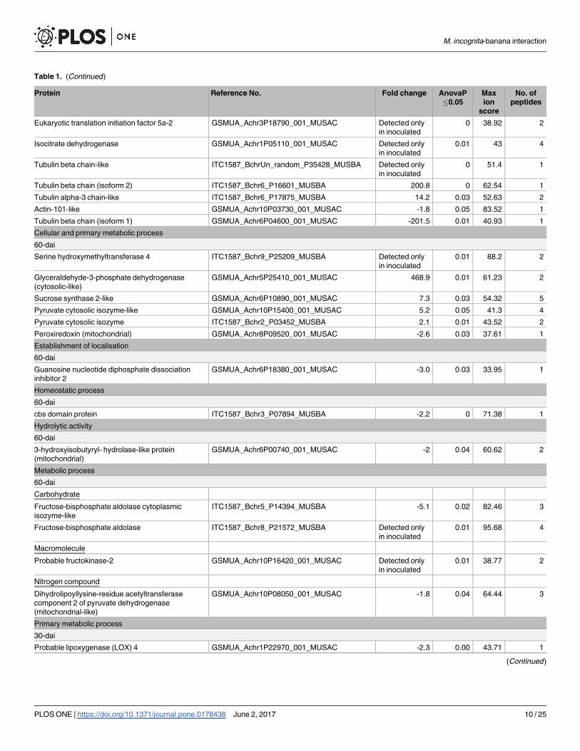
Table 1. (Continued)
Protein Reference No. Fold change AnovaP
�0.05
Max
ion
score
No. of
peptides
Eukaryotic translation initiation factor 5a-2 GSMUA_Achr3P18790_001_MUSAC Detected only
in inoculated
0 38.92 2
Isocitrate dehydrogenase GSMUA_Achr1P05110_001_MUSAC Detected only
in inoculated
0.01 43 4
Tubulin beta chain-like ITC1587_BchrUn_random_P35428_MUSBA Detected only
in inoculated
0 51.4 1
Tubulin beta chain (isoform 2) ITC1587_Bchr6_P16601_MUSBA 200.8 0 62.54 1
Tubulin alpha-3 chain-like ITC1587_Bchr6_P17875_MUSBA 14.2 0.03 52.63 2
Actin-101-like GSMUA_Achr10P03730_001_MUSAC -1.8 0.05 83.52 1
Tubulin beta chain (isoform 1) GSMUA_Achr6P04600_001_MUSAC -201.5 0.01 40.93 1
Cellular and primary metabolic process
60-dai
Serine hydroxymethyltransferase 4 ITC1587_Bchr9_P25209_MUSBA Detected only
in inoculated
0.01 88.2 2
Glyceraldehyde-3-phosphate dehydrogenase
(cytosolic-like)
GSMUA_Achr5P25410_001_MUSAC 468.9 0.01 61.23 2
Sucrose synthase 2-like GSMUA_Achr6P10890_001_MUSAC 7.3 0.03 54.32 5
Pyruvate cytosolic isozyme-like GSMUA_Achr10P15400_001_MUSAC 5.2 0.05 41.3 4
Pyruvate cytosolic isozyme ITC1587_Bchr2_P03452_MUSBA 2.1 0.01 43.52 2
Peroxiredoxin (mitochondrial) GSMUA_Achr8P09520_001_MUSAC -2.6 0.03 37.61 1
Establishment of localisation
60-dai
Guanosine nucleotide diphosphate dissociation
inhibitor 2
GSMUA_Achr6P18380_001_MUSAC -3.0 0.03 33.95 1
Homeostatic process
60-dai
cbs domain protein ITC1587_Bchr3_P07894_MUSBA -2.2 0 71.38 1
Hydrolytic activity
60-dai
3-hydroxyisobutyryl- hydrolase-like protein
(mitochondrial)
GSMUA_Achr6P00740_001_MUSAC -2 0.04 60.62 2
Metabolic process
60-dai
Carbohydrate
Fructose-bisphosphate aldolase cytoplasmic
isozyme-like
ITC1587_Bchr5_P14394_MUSBA -5.1 0.02 82.46 3
Fructose-bisphosphate aldolase ITC1587_Bchr8_P21572_MUSBA Detected only
in inoculated
0.01 95.68 4
Macromolecule
Probable fructokinase-2 GSMUA_Achr10P16420_001_MUSAC Detected only
in inoculated
0.01 38.77 2
Nitrogen compound
Dihydrolipoyllysine-residue acetyltransferase
component 2 of pyruvate dehydrogenase
(mitochondrial-like)
GSMUA_Achr10P08050_001_MUSAC -1.8 0.04 64.44 3
Primary metabolic process
30-dai
Probable lipoxygenase (LOX) 4 GSMUA_Achr1P22970_001_MUSAC -2.3 0.00 43.71 1
(Continued )
M. incognita-banana interaction
PLOS ONE | https://doi.org/10.1371/journal.pone.0178438 June 2, 2017 10 / 25
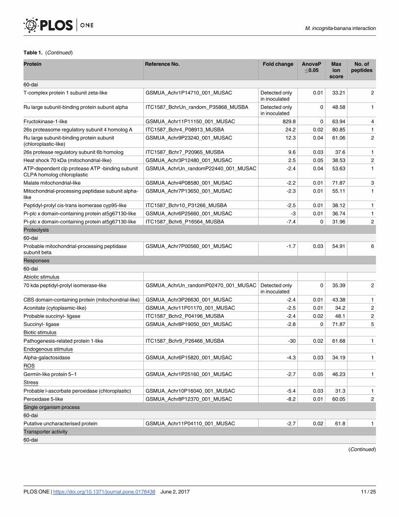
Table 1. (Continued)
Protein Reference No. Fold change AnovaP
�0.05
Max
ion
score
No. of
peptides
60-dai
T-complex protein 1 subunit zeta-like GSMUA_Achr1P14710_001_MUSAC Detected only
in inoculated
0.01 33.21 2
Ru large subunit-binding protein subunit alpha ITC1587_BchrUn_random_P35868_MUSBA Detected only
in inoculated
0 48.58 1
Fructokinase-1-like GSMUA_Achr11P11150_001_MUSAC 829.8 0 63.94 4
26s proteasome regulatory subunit 4 homolog A ITC1587_Bchr4_P08913_MUSBA 24.2 0.02 80.85 1
Ru large subunit-binding protein subunit
(chloroplastic-like)
GSMUA_Achr9P23240_001_MUSAC 12.3 0.04 61.06 2
26s protease regulatory subunit 6b homolog ITC1587_Bchr7_P20965_MUSBA 9.6 0.03 37.6 1
Heat shock 70 kDa (mitochondrial-like) GSMUA_Achr3P12480_001_MUSAC 2.5 0.05 38.53 2
ATP-dependent clp protease ATP -binding subunit
CLPA homolog chloroplastic
GSMUA_AchrUn_randomP22440_001_MUSAC -2.4 0.04 53.63 1
Malate mitochondrial-like GSMUA_Achr4P08580_001_MUSAC -2.2 0.01 71.87 3
Mitochondrial-processing peptidase subunit alpha-
like
GSMUA_Achr7P13650_001_MUSAC -2.3 0.01 55.11 1
Peptidyl-prolyl cis-trans isomerase cyp95-like ITC1587_Bchr10_P31266_MUSBA -2.5 0.01 38.12 1
Pi-plc x domain-containing protein at5g67130-like GSMUA_Achr6P25660_001_MUSAC -3 0.01 36.74 1
Pi-plc x domain-containing protein at5g67130-like ITC1587_Bchr6_P16564_MUSBA -7.4 0 31.96 2
Proteolysis
60-dai
Probable mitochondrial-processing peptidase
subunit beta
GSMUA_Achr7P00560_001_MUSAC -1.7 0.03 54.91 6
Responses
60-dai
Abiotic stimulus
70 kda peptidyl-prolyl isomerase-like GSMUA_AchrUn_randomP02470_001_MUSAC Detected only
in inoculated
0 35.39 2
CBS domain-containing protein (mitochondrial-like) GSMUA_Achr3P26630_001_MUSAC -2.4 0.01 43.38 1
Aconitate (cytoplasmic-like) GSMUA_Achr11P01170_001_MUSAC -2.5 0.01 34.2 2
Probable succinyl- ligase ITC1587_Bchr2_P04196_MUSBA -2.4 0.02 48.1 2
Succinyl- ligase GSMUA_Achr8P19050_001_MUSAC -2.8 0 71.87 5
Biotic stimulus
Pathogenesis-related protein 1-like ITC1587_Bchr9_P26466_MUSBA -30 0.02 61.68 1
Endogenous stimulus
Alpha-galactosidase GSMUA_Achr6P15820_001_MUSAC -4.3 0.03 34.19 1
ROS
Germin-like protein 5–1 GSMUA_Achr1P25160_001_MUSAC -2.7 0.05 46.23 1
Stress
Probable l-ascorbate peroxidase (chloroplastic) GSMUA_Achr10P16040_001_MUSAC -5.4 0.03 31.3 1
Peroxidase 5-like GSMUA_Achr8P12370_001_MUSAC -8.2 0.01 60.05 2
Single organism process
60-dai
Putative uncharacterised protein GSMUA_Achr11P04110_001_MUSAC -2.7 0.02 61.8 1
Transporter activity
60-dai
(Continued )
M. incognita-banana interaction
PLOS ONE | https://doi.org/10.1371/journal.pone.0178438 June 2, 2017 11 / 25
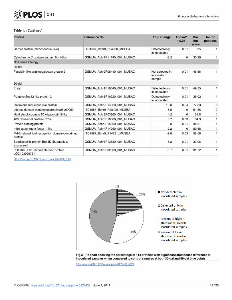
Table 1. (Continued)
Protein Reference No. Fold change AnovaP
�0.05
Max
ion
score
No. of
peptides
Carrier protein (mitochondrial-like) ITC1587_Bchr8_P24300_MUSBA Detected only
in inoculated
0.01 35 1
Cytochrome C oxidase subunit 6b-1-like GSMUA_Achr7P11740_001_MUSAC -2.2 0 30.35 1
No Gene Ontology
30-dai
Fasciclin-like arabinogalactan protein 2 GSMUA_Achr2P05440_001_MUSAC Not detected in
inoculated
sample
0.01 45.86 1
60-dai
Enoyl GSMUA_Achr1P19640_001_MUSAC Detected only
in inoculated
0.01 49.35 1
Putative Sec12-like protein 2 GSMUA_Achr6P10220_001_MUSAC Detected only
in inoculated
0.01 38.02 1
Isoflavone reductase-like protein GSMUA_Achr2P14320_001_MUSAC 10.3 0.04 77.33 6
btb poz domain-containing protein at5g60050 ITC1587_Bchr9_P28128_MUSBA 6.5 0 51.86 2
Heat shock cognate 70 kda protein 2-like GSMUA_Achr9P03960_001_MUSAC 4.2 0 31.8 1
40S ribosomal protein S27-2 GSMUA_Achr3P18600_001_MUSAC 3.7 0.03 34.9 1
Protein binding protein GSMUA_Achr8P15600_001_MUSAC 3 0.01 34.51 1
mfp1 attachment factor 1-like GSMUA_Achr4P14260_001_MUSAC -2.2 0 50.88 1
Md-2-related lipid recognition domain-containing
protein
ITC1587_Bchr5_P14231_MUSBA -2.8 0.03 58.58 1
Seed specific protein Bn15D1B, putative,
expressed
GSMUA_Achr8P13580_001_MUSAC -5.2 0.01 37.06 1
PREDICTED: uncharacterised protein
LOC103996731
GSMUA_Achr9P02000_001_MUSAC -5.7 0.01 31.72 1
https://doi.org/10.1371/journal.pone.0178438.t001
Fig 3. Pie chart showing the percentage of 114 proteins with significant abundance difference in
inoculated samples when compared to control samples at both 30-dai and 60-dai time points.
https://doi.org/10.1371/journal.pone.0178438.g003
M. incognita-banana interaction
PLOS ONE | https://doi.org/10.1371/journal.pone.0178438 June 2, 2017 12 / 25
cellular component organisation that showed differing abundance levels in inoculated samples.
When the interaction of the three tubulin proteins was selected, it was revealed that these pro-
teins shared nucleotide binding function with actin101-like protein
(GSMUA_Achr10P03730_001_MUSAC) that, according to GO analysis, was present in cyto-
plasm and cytoskeleton (Fig 4b).
Stress responses. Thirteen proteins were found to be involved in stress responses. Of
these, one was present only in inoculated root tissues at 60-dai and the remaining twelve
were present at lower abundance level in the same samples. On the other hand, at 30-dai caf-
feoyl-O-methyltransferase (GSMUA_Achr6P36400_001_MUSAC) was present at higher
abundance level in inoculated samples compared to the control ones (Fig 4c). The interac-
tion of these proteins were further investigated and we found that only four of these pro-
teins namely pathogenesis related protein 1-like (ITC1587_Bchr9_P26466_MUSBA),
chloroplastic probable 1-ascorbate peroxidase (GSMUA_Achr10P16040_001_MUSAC),
peroxidase 5-like (GSMUA_Achr8P12370_001_MUSAC) and 70kDa peptidyl-prolyl
isomerase-like (GSMUA_AchrUn_randomP02470_001_MUSAC) were annotated to
be solely involved in response to stress. CBS domain-containing protein (mitochondrial-
like) (GSMUA_Achr3P26630_001_MUSAC) is involved in two other functions namely cat-
abolic process and transportation, and share similar annotation with probable ATP
synthase 24 kDa (mitochondrial; GSMUA_Achr6P02850_001_MUSAC) and cytoplasmic-
like aconitate (GSMUA_Achr11P01170_001_MUSAC) for the same functions. On the
other hand, the remaining five proteins mapped showed involvement in catabolic
processes.
Fig 4. Protein-GO interaction profile of banana root proteome generated from Cytoscape showing (a)
selected proteins involved in fundamental biological processes that were either present at higher
abundance level or detected only in inoculated samples, (b) proteins involved in cellular component
organisation that were present at different abundance levels which were also involved in three other
biological functions, and (c) all proteins involved in response to stresses that were present in lower
abundances in inoculated samples except for peptidyl-prolyl isomerase-like (at 60-dai) and caffeoyl-
O-methyltransferase (at 30-dai). These stress proteins were also involved in transport, catabolic and protein
metabolic processes.
https://doi.org/10.1371/journal.pone.0178438.g004
M. incognita-banana interaction
PLOS ONE | https://doi.org/10.1371/journal.pone.0178438 June 2, 2017 13 / 25

KEGG pathways associated with nematode infection in banana root
samples
We investigated three KEGG pathways that were associated with defence and giant cell main-
tenance in bananas which include lignification and energy metabolism namely phenylpropa-
noid biosynthesis, glycolysis and citrate cycle pathways.
Lignification. Lignification process is greatly influenced by enzyme activities in phenyl-
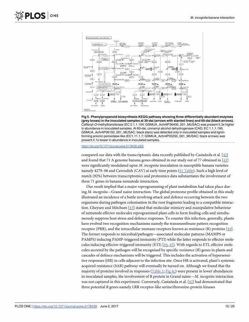
propanoid biosynthesis and three enzymes were found to be implicated in this pathway (Fig 5).
Here, we found that caffeoyl-O-methyltransferase (EC 2.1.1.104; GSMUA_Achr6P36400_001_
MUSAC) was present at an increased abundance level (5.3-fold) at 30-dai in inoculated sam-
ples. Cinnamyl alcohol dehydrogenase (CAD; EC 1.1.1.195; GSMUA_Achr4P06150_001_MU-
SAC), on the other hand, was detected only in inoculated samples at 60-dai. At the same time
point, lignin-forming anionic peroxidase-like enzyme (EC 1.11.1.7; GSMUA_Achr4P05
250_001_MUSAC) was found present at low abundance level (4.1-fold) in inoculated samples.
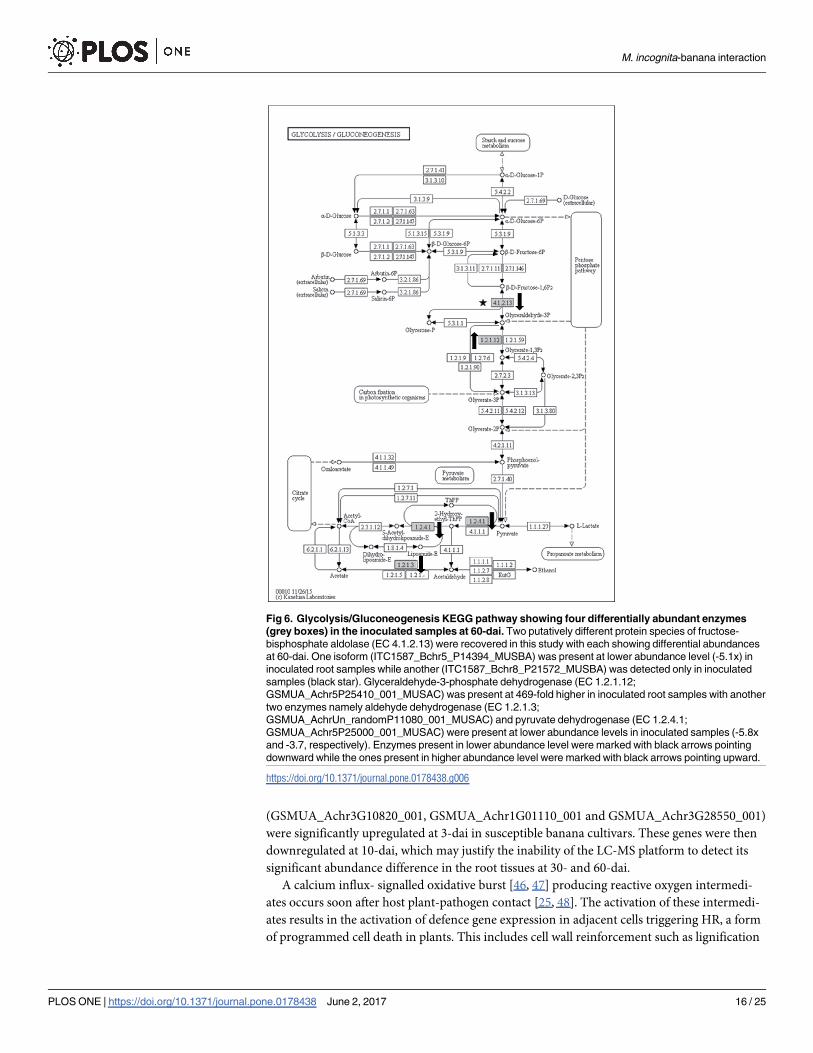
Carbohydrate catabolism. Four proteins were differentially abundant in the glycolysis
pathway (Fig 6). Interestingly, two putative protein species of fructose-bisphosphate aldolase (EC
4.1.2.13; ITC1587_Bchr5_P14394_MUSBA and ITC1587_Bchr8_P21572_MUSBA) were recov-
ered in this study with each showing differing abundance levels at 60-dai. The putative isoform
with the accession number ITC1587_Bchr5_P14394_MUSBA was present at lower abundance
level (5.1-fold) while another putative isoform (ITC1587_Bchr8_P21572_MUSBA) was detected
only in inoculated samples. This enzyme together with glyceraldehyde-3-phosphate dehydroge-
nase (EC 1.2.1.12; GSMUA_Achr5P25410_001_MUSAC) that showed significantly increased
abundance level (468.9-fold) are involved in the production of glycerate-3-phosphate. On the
other hand, aldehyde dehydrogenase (EC 1.2.1.3; GSMUA_AchrUn_randomP11080_001_MU-
SAC) and pyruvate dehydrogenase (EC 1.2.4.1; GSMUA_Achr5P25000_001_MUSAC) were
present at lower abundance levels in inoculated samples at 5.8-fold and 3.7-fold, respectively.
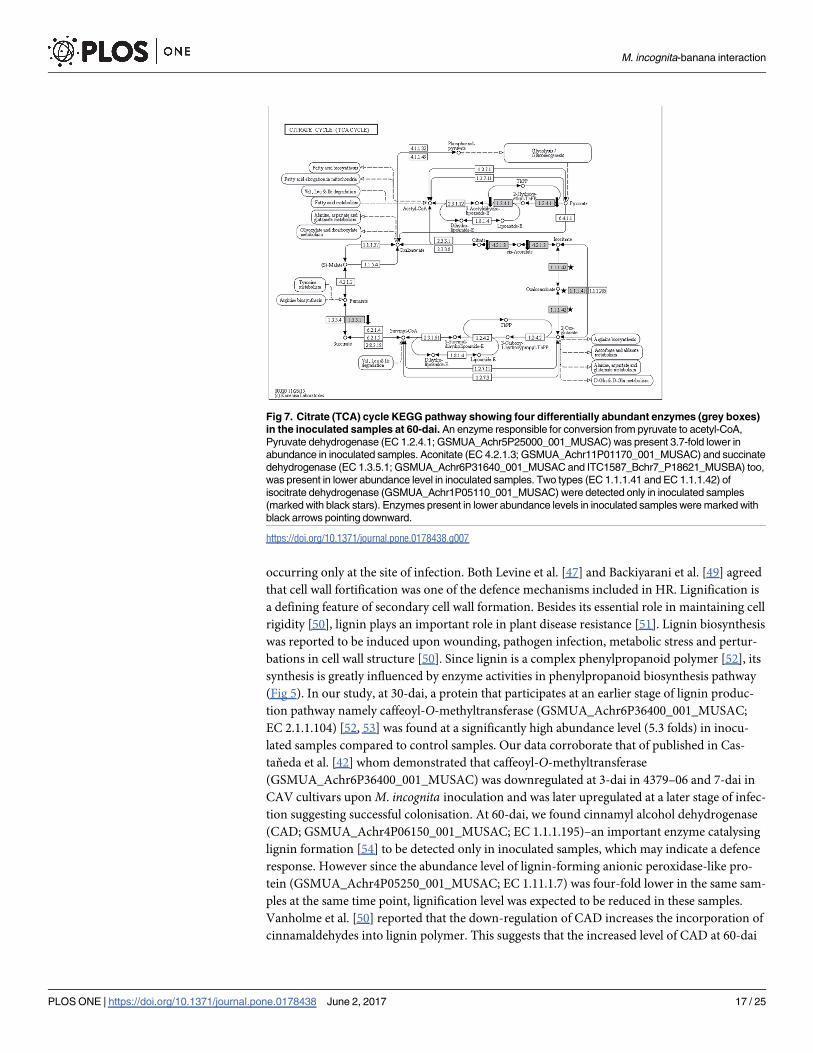
Energy metabolism. Four enzymes were present at differential abundances in the Citrate
cycle pathway (Fig 7). From the four, three were present at lower abundance level in inoculated
samples. The three enzymes are aconitate (EC 4.2.1.3; GSMUA_Achr11P01170_001_MUSAC),
pyruvate dehydrogenase e1 component subunit beta (EC 1.2.4.1; GSMUA_Achr5P25000_001_
MUSAC) and succinate dehydrogenase (EC 1.3.5.1; GSMUA_Achr6P31640_001_MUSAC;
ITC1587_Bchr7_P18621_MUSBA). Two types (EC 1.1.1.41 and EC 1.1.1.42) of isocitrate dehy-
drogenase (GSMUA_Achr1P05110_001_MUSAC) were detected only in inoculated samples.
Discussion
In this study, we investigated banana responses at the 30th—and 60th- day after inoculation
(dai) with Meloidogyne incognita using a Liquid Chromatography-Mass Spectrometry
(LC-MS) shotgun approach. We opted to use our in-house-designed single inoculation site
approach [35] due to our past failures in obtaining consistent data using a general inoculation
approach described by [41]. We inoculated 1000 J2 M. incognita on approximately 50 mg root
fragment in order to increase the number of infected cells per 50 mg root fragment (wet weight
used in our protein isolation procedure). By doing so, we hypothesised that the chance to
detect protein abundance changes in two sample types (inoculated vs. control) will also be
increased. This approach was deemed efficient since galls were visible to the naked eye as
swellings on inoculated banana roots for both time points i.e. 30- and 60-dai (Fig 1). It is note-
worthy that galls were more visible on inoculated samples at 60-dai compared to 30-dai. This
may be due to nematode population build-up in 60-day old root fragments. In addition, we
have also successfully identified 114 proteins (from A and B genomes) showing significant
abundance difference (p� 0.05) between inoculated and non-inoculated samples. We
M. incognita-banana interaction
PLOS ONE | https://doi.org/10.1371/journal.pone.0178438 June 2, 2017 14 / 25
compared our data with the transcriptomic data recently published by Castaňeda et al. [42]
and found that 71 A genome banana genes obtained in our study out of 77 obtained in [42]
were significantly modulated upon M. incognita inoculation in susceptible banana varieties
namely 4279–06 and Cavendish (CAV) at early time points (S1 Table). Such a high level of
match (92%) between transcriptomics and proteomics data substantiates the involvement of
these 71 genes in banana-nematode interaction.
Our result implied that a major reprogramming of plant metabolism had taken place dur-
ing M. incognita—Grand naine interaction. The global proteome profile obtained in this study
illustrated an incidence of a battle involving attack and defence occurring between the two
organisms during pathogen colonisation in the root fragments leading to a compatible interac-
tion. Gheysen and Mitchum [43] stated that molecular mimicry and manipulative behaviour
of nematode effector molecules reprogrammed plant cells to form feeding cells and simulta-
neously suppress host stress and defence responses. To counter this infection, generally, plants
have evolved two recognition mechanisms namely the transmembrane pattern recognition
receptor (PRR), and the intracellular immune receptors known as resistance (R) proteins [44].
The former responds to microbial/pathogen—associated molecular patterns (MAMPS or
PAMPS) inducing PAMP-triggered immunity (PTI) while the latter responds to effector mole-
cules inducing effector-triggered immunity (ETI) [44, 45]. With regards to ETI, effector mole-
cules secreted by the pathogen will be recognised by specific resistance (R) genes in plants and
cascades of defence mechanisms will be triggered. This includes the activation of hypersensi-
tive responses (HR) in cells adjacent to the infection site. Once HR is activated, plant’s systemic
acquired resistance (SAR) pathway will eventually be turned on. Although we found that the
majority of proteins involved in responses (Table 1; Fig 4c) were present in lower abundances
in inoculated samples, the involvement of R protein in Grand naine—M. incognita interaction
was not captured in this experiment. Conversely, Castaňeda et al. [42] had demonstrated that
three potential R genes namely LRR receptor-like serine/threonine-protein kinases
Fig 5. Phenylpropanoid biosynthesis KEGG pathway showing three differentially abundant enzymes
(grey boxes) in the inoculated samples at 30-dai (arrows with slanted lines) and 60-dai (black arrows).
Caffeoyl-O-methyltransferase (EC 2.1.1.104; GSMUA_Achr6P36400_001_MUSAC) was present 5.3x higher
in abundance in inoculated samples. At 60-dai, cinnamyl alcohol dehydrogenase (CAD; EC 1.1.1.195;
GSMUA_Achr4P06150_001_MUSAC; black stars) was detected only in inoculated samples and lignin-
forming anionic peroxidase-like (EC1.11.1.7; GSMUA_Achr4P05250_001_MUSAC; black arrows) was
present 4.1x lesser in abundance in inoculated samples.
https://doi.org/10.1371/journal.pone.0178438.g005
M. incognita-banana interaction
PLOS ONE | https://doi.org/10.1371/journal.pone.0178438 June 2, 2017 15 / 25
(GSMUA_Achr3G10820_001, GSMUA_Achr1G01110_001 and GSMUA_Achr3G28550_001)
were significantly upregulated at 3-dai in susceptible banana cultivars. These genes were then
downregulated at 10-dai, which may justify the inability of the LC-MS platform to detect its
significant abundance difference in the root tissues at 30- and 60-dai.
A calcium influx- signalled oxidative burst [46, 47] producing reactive oxygen intermedi-
ates occurs soon after host plant-pathogen contact [25, 48]. The activation of these intermedi-
ates results in the activation of defence gene expression in adjacent cells triggering HR, a form
of programmed cell death in plants. This includes cell wall reinforcement such as lignification
Fig 6. Glycolysis/Gluconeogenesis KEGG pathway showing four differentially abundant enzymes
(grey boxes) in the inoculated samples at 60-dai. Two putatively different protein species of fructose-
bisphosphate aldolase (EC 4.1.2.13) were recovered in this study with each showing differential abundances
at 60-dai. One isoform (ITC1587_Bchr5_P14394_MUSBA) was present at lower abundance level (-5.1x) in
inoculated root samples while another (ITC1587_Bchr8_P21572_MUSBA) was detected only in inoculated
samples (black star). Glyceraldehyde-3-phosphate dehydrogenase (EC 1.2.1.12;
GSMUA_Achr5P25410_001_MUSAC) was present at 469-fold higher in inoculated root samples with another
two enzymes namely aldehyde dehydrogenase (EC 1.2.1.3;
GSMUA_AchrUn_randomP11080_001_MUSAC) and pyruvate dehydrogenase (EC 1.2.4.1;
GSMUA_Achr5P25000_001_MUSAC) were present at lower abundance levels in inoculated samples (-5.8x
and -3.7, respectively). Enzymes present in lower abundance level were marked with black arrows pointing
downward while the ones present in higher abundance level were marked with black arrows pointing upward.
https://doi.org/10.1371/journal.pone.0178438.g006
M. incognita-banana interaction
PLOS ONE | https://doi.org/10.1371/journal.pone.0178438 June 2, 2017 16 / 25
occurring only at the site of infection. Both Levine et al. [47] and Backiyarani et al. [49] agreed
that cell wall fortification was one of the defence mechanisms included in HR. Lignification is
a defining feature of secondary cell wall formation. Besides its essential role in maintaining cell
rigidity [50], lignin plays an important role in plant disease resistance [51]. Lignin biosynthesis
was reported to be induced upon wounding, pathogen infection, metabolic stress and pertur-
bations in cell wall structure [50]. Since lignin is a complex phenylpropanoid polymer [52], its
synthesis is greatly influenced by enzyme activities in phenylpropanoid biosynthesis pathway
(Fig 5). In our study, at 30-dai, a protein that participates at an earlier stage of lignin produc-
tion pathway namely caffeoyl-O-methyltransferase (GSMUA_Achr6P36400_001_MUSAC;
EC 2.1.1.104) [52, 53] was found at a significantly high abundance level (5.3 folds) in inocu-
lated samples compared to control samples. Our data corroborate that of published in Cas-
taňeda et al. [42] whom demonstrated that caffeoyl-O-methyltransferase
(GSMUA_Achr6P36400_001_MUSAC) was downregulated at 3-dai in 4379–06 and 7-dai in
CAV cultivars uponM. incognita inoculation and was later upregulated at a later stage of infec-
tion suggesting successful colonisation. At 60-dai, we found cinnamyl alcohol dehydrogenase
(CAD; GSMUA_Achr4P06150_001_MUSAC; EC 1.1.1.195)–an important enzyme catalysing
lignin formation [54] to be detected only in inoculated samples, which may indicate a defence
response. However since the abundance level of lignin-forming anionic peroxidase-like pro-
tein (GSMUA_Achr4P05250_001_MUSAC; EC 1.11.1.7) was four-fold lower in the same sam-
ples at the same time point, lignification level was expected to be reduced in these samples.
Vanholme et al. [50] reported that the down-regulation of CAD increases the incorporation of
cinnamaldehydes into lignin polymer. This suggests that the increased level of CAD at 60-dai
Fig 7. Citrate (TCA) cycle KEGG pathway showing four differentially abundant enzymes (grey boxes)
in the inoculated samples at 60-dai. An enzyme responsible for conversion from pyruvate to acetyl-CoA,
Pyruvate dehydrogenase (EC 1.2.4.1; GSMUA_Achr5P25000_001_MUSAC) was present 3.7-fold lower in
abundance in inoculated samples. Aconitate (EC 4.2.1.3; GSMUA_Achr11P01170_001_MUSAC) and succinate
dehydrogenase (EC 1.3.5.1; GSMUA_Achr6P31640_001_MUSAC and ITC1587_Bchr7_P18621_MUSBA) too,
was present in lower abundance level in inoculated samples. Two types (EC 1.1.1.41 and EC 1.1.1.42) of
isocitrate dehydrogenase (GSMUA_Achr1P05110_001_MUSAC) were detected only in inoculated samples
(marked with black stars). Enzymes present in lower abundance levels in inoculated samples were marked with
black arrows pointing downward.
https://doi.org/10.1371/journal.pone.0178438.g007
M. incognita-banana interaction
PLOS ONE | https://doi.org/10.1371/journal.pone.0178438 June 2, 2017 17 / 25
could potentially decreases the incorporation of cinnamaldehydes into the polymer in the
inoculated samples, indicating that lignification was halted at 60-dai. This suggests that at this
time point,M. incognita might have targeted CAD for giant cell formation/maintenance which
involves cell wall dissolution. It is noteworthy that Glazer et al. [55] had reported inhibition of
xylem lignification in Meloidogyne javanica-infected tomato roots at 14- and 28-dai. We
hypothesise that in an interaction involving banana and root-knot nematodes, such an
increase in protein abundance of a host at a later stage of infection (60-dai) may translate to
manipulation of host’s cell system by the nematodes in order to maintain their nutrient sink,
the giant cells. This is in line with that of found in Castaňeda et al. [42] highlighting the
decreased level of expression of cell wall-associated genes in nematode- inoculated root tissues
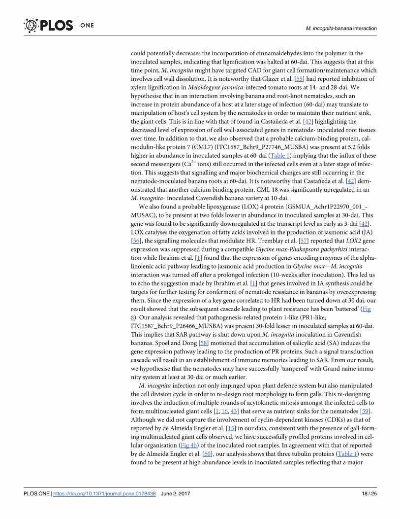
over time. In addition to that, we also observed that a probable calcium-binding protein, cal-
modulin-like protein 7 (CML7) (ITC1587_Bchr9_P27746_MUSBA) was present at 5.2 folds
higher in abundance in inoculated samples at 60-dai (Table 1) implying that the influx of these
second messengers (Ca2+ ions) still occurred in the infected cells even at a later stage of infec-
tion. This suggests that signalling and major biochemical changes are still occurring in the
nematode-inoculated banana roots at 60-dai. It is noteworthy that Castaňeda et al. [42] dem-
onstrated that another calcium binding protein, CML 18 was significantly upregulated in an
M. incognita- inoculated Cavendish banana variety at 10-dai.
We also found a probable lipoxygenase (LOX) 4 protein (GSMUA_Achr1P22970_001_-
MUSAC), to be present at two folds lower in abundance in inoculated samples at 30-dai. This
gene was found to be significantly downregulated at the transcript level as early as 3-dai [42].
LOX catalyses the oxygenation of fatty acids involved in the production of jasmonic acid (JA)
[56], the signalling molecules that modulate HR. Tremblay et al. [57] reported that LOX2 gene
expression was suppressed during a compatible Glycine max-Phakopsora pachyrhizi interac-
tion while Ibrahim et al. [1] found that the expression of genes encoding enzymes of the alpha-
linolenic acid pathway leading to jasmonic acid production in Glycine max—M. incognitainteraction was turned off after a prolonged infection (10-weeks after inoculation). This led us
to echo the suggestion made by Ibrahim et al. [1] that genes involved in JA synthesis could be
targets for further testing for conferment of nematode resistance in bananas by overexpressing
them. Since the expression of a key gene correlated to HR had been turned down at 30 dai, our
result showed that the subsequent cascade leading to plant resistance has been ‘battered’ (Fig
8). Our analysis revealed that pathogenesis-related protein 1-like (PR1-like;
ITC1587_Bchr9_P26466_MUSBA) was present 30-fold lesser in inoculated samples at 60-dai.
This implies that SAR pathway is shut down uponM. incognita inoculation in Cavendish
bananas. Spoel and Dong [58] motioned that accumulation of salicylic acid (SA) induces the
gene expression pathway leading to the production of PR proteins. Such a signal transduction
cascade will result in an establishment of immune memories leading to SAR. From our result,
we hypothesise that the nematodes may have successfully ‘tampered’ with Grand naine immu-
nity system at least at 30-dai or much earlier.
M. incognita infection not only impinged upon plant defence system but also manipulated
the cell division cycle in order to re-design root morphology to form galls. This re-designing
involves the induction of multiple rounds of acytokinetic mitosis amongst the infected cells to
form multinucleated giant cells [1, 16, 43] that serve as nutrient sinks for the nematodes [59].
Although we did not capture the involvement of cyclin-dependent kinases (CDKs) as that of
reported by de Almeida Engler et al. [15] in our data, consistent with the presence of gall-form-
ing multinucleated giant cells observed, we have successfully profiled proteins involved in cel-
lular organisation (Fig 4b) of the inoculated root samples. In agreement with that of reported
by de Almeida Engler et al. [60], our analysis shows that three tubulin proteins (Table 1) were
found to be present at high abundance levels in inoculated samples reflecting that a major
M. incognita-banana interaction
PLOS ONE | https://doi.org/10.1371/journal.pone.0178438 June 2, 2017 18 / 25
cytoskeletal rearrangement had taken place in these samples. Note that both α- and β- tubulin
proteins will polymerise into microtubules—one of the key components of the cytoskeletons
that play an indirect role in maintaining cell shape [61]. Interestingly, one of the tubulin-beta
chain protein species obtained (GSMUA_Achr6P04600_001_MUSAC) was present at lower
abundance in inoculated sample while the other (ITC1587_BchrUn_random_P35428_-
MUSBA) was present at ~200-fold higher in the same tissues suggesting that one of these pro-
tein species is important in microtubule formation of giant cells. In 2004, de Almeida Engler
et al. [60] reported the presence of a large number of unusual and randomly oriented actin
bundles and cables during giant cell development. We found that the abundance of an actin-
101-like protein (GSMUA_Achr10P03730_001_MUSAC) was decreased by almost two-fold in
inoculated samples suggesting abnormal cell development had occurred in inoculated samples.
This may justify the importance of actin for a successful M. incognita colonisation in plant root
tissues. It is noteworthy that the transcriptomic expression profile of both tubulin-beta chain
(GSMUA_Achr6P04600_001_MUSAC) and actin (GSMUA_Achr10P03730_001_MUSAC)
genes obtained in Castaňeda et al. [42] at earlier nematode infection time-points matched our
proteome profiling data.
Fig 8. A hypothetical schematic defence pathway proposed on a compatible interaction between M.
incognita and Grand naine primarily based on Castaňeda et al. [42], Hahlbrock et al. [46], Levine et al.
[47], and Tanaka et al. [71]. Nematode stylet penetration into the plant cell results in production of
extracellular adenosine-5’- triphosphate (eATP) which signals for calcium (Ca2+) influx. Such a signalling
response will trigger the stress and disease resistance pathway leading to incompatibility. Both pathways
were obtained from plant and hormone signal transduction KEGG pathways (http://www.genome.jp/kegg-bin/
show_pathway?ath04075). Note that the pathway leading to cell senescent was abridged for the purpose of
simplicity. Proteins involved in stress response and disease resistance pathways obtained in our study are
mapped here. Mitogen-activated protein kinase (MAPK); Nonexpresser of PR genes 1 (NPR1);
Pathogenesis-related protein 1 (PR-1).
https://doi.org/10.1371/journal.pone.0178438.g008
M. incognita-banana interaction
PLOS ONE | https://doi.org/10.1371/journal.pone.0178438 June 2, 2017 19 / 25

A total of 11 ribosomal proteins were found either at higher abundances or detected only in
inoculated samples. This finding is consistent with that of reported by Jammes et al. [13] where
71 genes encoding 40S and 60S ribosomal proteins were specifically upregulated inM. incog-nita- inoculated Arabidopsis root samples suggesting an increased level of protein synthesis in
giant cells. This justified the notion proposed by Ibrahim et al. [1] that nematodes utilise plant
resources to develop and reproduce. In our study, proteins involved in carbohydrate catabolism
and energy metabolism were differentially abundant in inoculated root samples to either meet
the nematode’s energy and carbon demand, or, evading plant defense response. We found
potentially two fructose-bisphosphate aldolase (4.1.2.13) protein species present at opposing
abundance levels in glycolysis/gluconeogenesis KEGG pathway (Fig 5). This enzyme catalyses
an aldol cleavage of fructose-1, 6-bisphosphate to dihydroxyacetone-phosphate and glyceralde-
hyde 3-phosphate in a reversible aldol condensation [62]. It is noteworthy that, this aldolase
could either be cytosolic or chloroplastic in plants [63]. Although both protein species differ in
their primary structure and intracellular localisation, they both play an essential role in carbo-
hydrate metabolism; hence, differing levels of abundance of both protein species could impli-
cate plant growth [64]. Konishi et al. [64] reported that they observed notable accumulation of
Gibberellic acid-induced aldolase at apical region of rice seedling roots. Interestingly, active cell
division occurred in both apical region and root differentiation zone where the nematodes
reside [16] suggesting that increased levels of aldolase may stimulate root cell growth that medi-
ate energy production. However, in the case of nematode-infested root system, we hypothesise
that cell growth, in this context, reflects giant cell formation, and plant energy production may
be manipulated by the pathogen to sustain their longevity. Another enzyme in the glycolytic
pathway present at significantly high abundance level was glyceraldehyde-3-phosphate dehy-
drogenase (GAPDH; EC 1.2.1.12; GSMUA_Achr5P25410_001_MUSAC). Castaňeda et al. [42]
demonstrated that this gene was upregulated as early as 10-dai in a Cavendish banana upon M.
incognita infestation. This enzyme is responsible to produce energy and supply intermediates
for cellular metabolism [65] and had been implicated in embryo development, root growth [66]
as well as in transducing hydrogen peroxide signals in Arabidopsis response to stress [67]. We
also noted that two enzymes involved in glycolysis namely pyruvate dehydrogenase (EC 1.2.4.1;
GSMUA_Achr5P25000_001_MUSAC) and aldehyde dehydrogenase (EC 1.2.1.3; GSMUA_A-
chrUn_randomP11080_001_MUSAC), responsible in the production of acetyl-CoA, were pres-
ent at lower abundance level in inoculated samples suggesting implicated acetyl-CoA
production in inoculated samples. Acetyl-CoA is an important molecule involves in the Citrate
cycle (TCA cycle) pertinent in ATP production [61]. We found four enzymes involved in the
Citrate cycle pathway to be differentially abundant. Although isocitrate dehydrogenase (EC
1.1.1.41 and EC 1.1.1.42; GSMUA_Achr1P05110_001_MUSAC) was found to be present only
in inoculated sample, an enzyme that was responsible in converting succinate to fumarate
namely succinate dehydrogenase (EC 1.3.5.1; GSMUA_Achr6P31640_001_MUSAC;
ITC1587_Bchr7_P18621_MUSBA) was present twice times lower in abundance in inoculated
samples. This may potentially implicate the production of ATP in the inoculated samples. Our
result showed four ATP synthase subunits (GSMUA_AchrUn_randomP15230_001_MUSAC,
GSMUA_Achr10P27350_001_MUSAC, GSMUA_Achr9P21710_001_MUSAC and
ITC1587_Bchr10_P31293_MUSBA) and one probable mitochondrial ATP synthase
(GSMUA_Achr6P02850_001_MUSAC) to be present in lower abundance levels (1.5 to 2.6
folds lower; Table 1) in inoculated samples. ATP synthase is known as a highly conserved
enzyme that involved in catalysing ATP synthesis from ADP and phosphate during mitochon-
drial respiration [68]. Although it was expected that the abundance level for ATP synthase to be
high in tissues undergoing adverse metabolic reprogramming activity such as the galls, our pro-
filing result proved otherwise. This enzyme is a complicated protein complex and is divided
M. incognita-banana interaction
PLOS ONE | https://doi.org/10.1371/journal.pone.0178438 June 2, 2017 20 / 25
into two sectors namely a soluble globular F1 catalytic sector and a membrane-bound F0 pro-
ton-translocating sector [69]. In our result, one of the proteins of the latter complex was present
at a lower abundance level in inoculated samples suggesting the presence of deformed ATP
synthase complex with an inability to translocate proton in inoculated root cells. Proton trans-
location is important for the cell to generate proton electrochemical gradient that serves as the
driving force for ion and metabolite uptake across the plasma membrane [70]. Therefore the
deformation of ATP synthase complex in the inoculated samples may indicate interference in
such mechanism in nematode-infested banana root cells. This set-back will eventually impair
plant growth and fruit production.
In this study, by using our in-house-designed single inoculation site approach [35] coupled
with an Orbitrap LC-MS platform, we conclude that 114 banana root proteins showed signifi-
cant abundance changes when inoculated with Meloidogyne incognita at 30- and 60-dai. Our
study revealed that these changes affected proteins involved primarily in fundamental biologi-
cal processes, cellular component organisation and stress responses. Our analysis provided a
new spectrum of knowledge especially on plant-parasite interaction from the perspective of a
non-model organism. Corroborating transcriptomic data obtained in [42], we had identified
players in banana defence and response pathway against M. incognita infestation namely caf-
feoyl- O-methyltransferase (GSMUA_Achr6P36400_001_MUSAC) probable lipoxygenase
(LOX) 4 protein (GSMUA_Achr1P22970_001_MUSAC) and pathogenesis-related protein
1-like (PR1-like; ITC1587_Bchr9_P26466_MUSBA) that could serve as targets for further
functional tests in order to develop a tolerant/ resistant banana cultivar against M. incognita.
Here, we propose a hypothetical defence pathway leading to a compatible interaction between
Grand naine—M. incognita primarily based on Castaňeda et al. [42], Hahlbrock et al. [46],
Levine [47], and Tanaka et al. [71] (Fig 8). We also have mapped the stress and defence pro-
teins obtained from our study to this hypothetical pathway.
Supporting information
S1 Table. Comparison between transcriptomic data obtained in [42] and proteomic data
from the current study revealed that 71 A genome banana genes in the current study out
of 77 obtained in [42] were significantly modulated during banana-Meloidogyne incognitainteraction.
(XLSX)
Acknowledgments
We wish to thank Mr. Mohd Nazarudin Anuar from Malaysian Agricultural Research and
Development Institute (MARDI) for supplying the Malaysian population Meloidogyne incog-nita culture for the study. We are also grateful to the anonymous reviewers for their useful
comments on the manuscript.
Author Contributions
Conceptualization: AA ZM.
Data curation: SCC MTA AA.
Formal analysis: SCC AA.
Funding acquisition: ZM AA.
Investigation: AA MTA.
M. incognita-banana interaction
PLOS ONE | https://doi.org/10.1371/journal.pone.0178438 June 2, 2017 21 / 25
Methodology: AA SCC.
Project administration: AA.
Resources: AA ZM SCC BP.
Supervision: AA.
Validation: AA SCC.
Visualization: AA.
Writing – original draft: AA.
Writing – review & editing: AA SCC BP ZM.
References1. Ibrahim HMM, Hosseini P, Alkharouf NW, Hussein EHA, Gamal El-Din AE KY, Aly MAM, et al. Analysis
of gene expression in soybean (Glycine max) roots in response to the root knot nematode Meloidogyne
incognita using microarrays and KEGG pathways. BMC genomics 2011; 12:1471–2164.
2. Sasser JN. Root-knot nematodes: a global menace to crop production. Plant Disease 1980; 64: 36–41.
3. Jepson SB. Identification of root-knot nematodes (Meloidogyne species). Wallingford, UK: CAB Inter-
national: 1987. 265 p.
4. Huang G, Dong R, Allen R, Davis EL, Baum TJ, Hussey RS. A root-knot nematode secretory peptide
functions as a ligand for a plant transcription factor. Molecular Plant-Microbe Interactions 2006; 19:
463–470. https://doi.org/10.1094/MPMI-19-0463 PMID: 16673933
5. Abad P, Gouzy J, Aury J-M, Castagnone-Sereno P, Danchin EGJ, Dleury E, et al. Genome sequence
of the metazoan plant-parasitic nematode Meloidogyne incognita. Nature Biotechnology 2008; 26:
909–915. https://doi.org/10.1038/nbt.1482 PMID: 18660804
6. Niu J-h, Guo Q-x, Jian H, Chen C-l, Yang D, Liu Q, et al. Rapid detection of Meloidogyne spp. by LAMP
assay in soil and roots. Crop Protection 2011; 30: 1063–1069.
7. De Waele D, Davide RD. The root-knot nematodes of banana. Musa Pest Fact sheet No.3. France:
INIBAP; 1998.
8. Queneherve P, Salmon F, Topart P, Horry JP. Nematode resistance in bananas: screening results on
some new Mycosphaerella resistant banana hybrids. Euphytica 2009; 165: 137–143.
9. Jaizme-Vega MC, Tenoury P, Pinochet J, Jaumot M. Interactions between the root-knot nematode
Meloidogyne incognita and Glomus mosseae in banana. Plant and Soil 1997; 196: 27–35.
10. Gowen SR, Queneherve P, Fogain R. Nematode parasites of bananas and plantains. In: Luc M, Sikora
RA, Bridge J, editors. Plant parasitic nematodes in subtropical and tropical agriculture. Wallingford:
CABI Publishing; 2005, p. 611–643.
11. Wang KH, Hooks CRR. Survey of nematodes on banana in Hawai’I, and methods used for their control.
CTAHR Cooperative Extension Service PD-69: 2009. 7 p.
12. Sayed Abdul Rahman SA, Mohd Zain SN, Bilal Mat MZ, Sidam AK, Othman RY, Mohamed Z. Popula-
tion distribution of plant-parasitic nematodes of bananas in Peninsular Malaysia. Sains Malaysiana
2014; 43: 175–183.
13. Jammes F, Lecomte P, de Almeida-Engler J, Bitton F, Martin-Magniette M-L, Renou J. P, et al.
Genome-wide expression profiling of the host response to root-knot nematode infection in Arabidopsis.
The Plant Journal 2005; 44: 447–458. https://doi.org/10.1111/j.1365-313X.2005.02532.x PMID:
16236154
14. Molinari S, Baser N. Induction of resistance to root-knot nematodes by SAR elicitors in tomato. Crop
Protection 2010; 29: 1354–1362.
15. de Almeida Engler J, De Vleesschauwer V, Burssens S, Celenza JL Jr., Inze D, Van Montagu M, et al.
Molecular markers and cell cycle inhibitors show the importance of cell cycle progression in nematode-
induced galls and syncytia. The Plant Cell 1999; 11: 793–807. PMID: 10330466
16. Williamson VM, Gleason CA. Plant-nematode interactions. Current Opinion in Plant Biology 2003; 6:
327–333. PMID: 12873526
17. Vanholme B, De Meutter J, Tytgat T, Van Montagu M, Coomans A, Gheysen G. Secretions of plant-
parasiti nematodes: A molecular update. Gene 2004; 332: 13–27. https://doi.org/10.1016/j.gene.2004.
02.024 PMID: 15145050
M. incognita-banana interaction
PLOS ONE | https://doi.org/10.1371/journal.pone.0178438 June 2, 2017 22 / 25
18. Curtis RH. Plant parasitic nematode proteins and the host-parasite interaction. Briefings in Functional
Genomics and Proteomics 2007; 6: 50–55. https://doi.org/10.1093/bfgp/elm006 PMID: 17525074
19. Caillaud M-C, Dubreuil G, Quentin M, Perfus-Barbeoch L, Lecomte P, de Almeida Engler J, et al. Root-
knot nematodes manipulate plant cell functions during a compatible interaction. Journal of Plant Physi-
ology 2008; 165: 104–113. https://doi.org/10.1016/j.jplph.2007.05.007 PMID: 17681399
20. Williamson VM, Hussey RS. Nematode pathogenesis and resistance in plants. The Plant Cell 1996; 8:
1735–1745. https://doi.org/10.1105/tpc.8.10.1735 PMID: 8914324
21. Jonathan EI, Rajendran G, Padmanabhan D, Ayyamperumal A. Occurrence, crop loss and manage-
ment of root-knot nematode, (Meloidogyne incognita) on banana. In: Sing HP,Chadha KL, editors.
Banana- Improvement, Production and Utilisation, Proceedings of the Conference on ‘Challenges for
Banana Production and Utilization in 21st Century’. Trichy: Association for the Improvement in Produc-
tion and Utilization of Banana; 2000.
22. De Waele D, Elsen A. Challenges in tropical plant Nematology. Annual Review of Phytopathology 2007;
45: 457–485. https://doi.org/10.1146/annurev.phyto.45.062806.094438 PMID: 17489690
23. van der Biezen EA, Jones JDG. Plant disease-resistance proteins and the gene-for-gene concept.
Trends in Biochemical Sciences 1998; 23: 454–456. PMID: 9868361
24. Williamson VM, Kumar A. Nematode resistance in plants: the battle underground. Trends in Genetics
2006; 22: 396–403. https://doi.org/10.1016/j.tig.2006.05.003 PMID: 16723170
25. Mehta A, Brasileiro ACM, Souza DSL, Romano E, Campos MA, Grossi-de-Sa MF, et al. Plant-pathogen
interactions: what is proteomics telling us? The FEBS Journal 2008; 275: 3731–3746. https://doi.org/
10.1111/j.1742-4658.2008.06528.x PMID: 18616468
26. Cai D, Kleine M, Kifle S, Harloff H-J, Sandal NN, Marcker KA, et al. Positional cloning of a gene for a
nematode resistance in sugar beet. Science 1997; 275: 832–834. PMID: 9012350
27. Bird DM, Bird AF. Plant-parasitic nematodes. In: Kennedy MW, Harnett W, editors. Parasitic nema-
todes: Molecular biology, biochemistry and immunology Wallingford: CAB International Publishing;
2001, p. 139–166.
28. Favery B, Lecomte P, Gil N, Bechtold N, Bouchez D, Dalmasso A, et al. RPE, a plant gene involved in
early developmental steps of nematode feeding cells. The EMBO Journal 1998; 17: 6799–6811.
https://doi.org/10.1093/emboj/17.23.6799 PMID: 9843485
29. Jaubert S, Ledger TN, Laffaire JB, Piotte C, Abad P, Rosso M-N. Direct identification of stylet secreted
proteins from root-knot nematodes by a proteomic approach. Molecular & Biochemical Parasitology
2002; 121: 205–211.
30. Escobar C, Brown S, Mitchum MG. Transcriptomic and Proteomic analysis of the plant response to
nematode infection. In: Jones J, Gheysen G, Fenoll C, editors. Genomics and Molecular Genetics of
Plant-nematode Interactions. Netherlands: Springer; 2011, p. 157–173.
31. Palomares-Rius JE, Castillo P, Navas-Cortes JA, Jimenez-Diaz RM, Tena M. A proteomic study of in-
root interactions between chickpea pathogens: the root-knot nematode Meloidogyne artiellia and the
soil-borne fungus Fusarium oxysporum f. sp. Ciceris race 5. Journal of Proteomics 2011; 74: 2035–
2051.
32. Quirino BF, Candido ES, Campos PF, Franco OL, Kruger RH. Proteomic approaches to study plant—
pathogen interactions. Phytochemistry 2010; 71: 351–362. https://doi.org/10.1016/j.phytochem.2009.
11.005 PMID: 20005547
33. Mehta A, Magalhães BS, Souza DSL, Vasconcelos EAR, Silva LP, Grossi-de-Sa MF, et al. Rooteomics:
the challenge of discovering plant defense-related proteins in roots. Current Protein and Peptide Sci-
ence 2008; 9: 108–116. PMID: 18393883
34. Li D, Wei T, Abbott CM, Harrich D. The unexpected roles of eukaryotic translation elongation factors in
RNA virus replication and pathogenesis. Microbiology and Molecular Biology Reviews 2013; 77: 253–
266. https://doi.org/10.1128/MMBR.00059-12 PMID: 23699257
35. Sayed Abdul Rahman SA, Mohamed Z, Othman RY, Swennen R, Panis B, De Waele D, et al. In planta
PCR-based detection of early infection of plant-parasitic nematodes in the roots: a step towards the
understanding of infection and plant defence. European Journal of Plant Pathology 2010; 128: 343–
351.
36. Carpentier S, Dens K, Van den houwe I, Swennen R, Panis B. Lyophilization, a practical way to store
and transport tissues prior to protein extraction for 2DE analysis? Proteomics 2007; 7(S1): 64–69.
37. Carpentier S, Witters E, Laukens K, Deckers P, Swennen R, Panis B. Preparation of protein extracts
from recalcitrant plant tissues: An evaluation of different methods for two-dimensional gel electrophore-
sis analysis. Proteomics 2005; 5: 2497–2507. https://doi.org/10.1002/pmic.200401222 PMID:
15912556
M. incognita-banana interaction
PLOS ONE | https://doi.org/10.1371/journal.pone.0178438 June 2, 2017 23 / 25
38. Vanhove A-C, Vermaelen W, Swennen R, Carpentier SC. A look behind the screens: Characterization
of the HSP70 family during osmotic stress in a non-model crop. Journal of Proteomics 2015; 119: 10–
20. https://doi.org/10.1016/j.jprot.2015.01.014 PMID: 25661040
39. Buts K, Michielssens S, Hertog MLATM, Hayakawa E, Cordewener J, America AHP, et al. Improving
the identification rate of data independent label-free quantitative proteomics experiments on non-model
crops: A case study on apple fruit. Journal of Proteomics 2014; 105: 31–15. https://doi.org/10.1016/j.
jprot.2014.02.015 PMID: 24565695
40. Carpentier S, Witters E, Laukens K, Deckers P, Swennen R, Panis B. Preparation of protein extracts
from recalcitrant plant tissues: An evaluation of different methods for two-dimensional gel electrophore-
sis analysis. Proteomics 2005; 5: 2497–2507. https://doi.org/10.1002/pmic.200401222 PMID:
15912556
41. Speijer PR, De Waele D. Screening of Musa germplasm for resistance and tolerance to nematodes.
International Network for the Improvement of Banana and Plantain (INIBAP) Technical Guidelines 1.
Montpellier: INIBAP; 1997. p. 1–47.
42. Castaňeda NEN, Alves GSC, Almeida RM, Amorim EP, Ferreira CF, Togawa RC, et al. Gene expres-
sion analysis in Musa acuminata during compatible interactions with Meloidogyne incognita. Annals of
Botany 2017; 119: 915–930. https://doi.org/10.1093/aob/mcw272 PMID: 28130221
43. Gheysen G, Mitchum MG. How nematodes manipulate plant development pathways for infection. Cur-
rent Opinion in Plant Biology 2011; 4: 415–421.
44. Jones JDG, Dangl JL The plant immune system. Nature 2006; 444: 323–329. https://doi.org/10.1038/
nature05286 PMID: 17108957
45. Robert-Seilaniantz A, Grant M, Jones JDG. Hormone crosstalk in plant disease and defense: More than
just JASMONATESALICYLATE antagonism. Annual Review of Phytopathology 2011; 49: 317–343.
https://doi.org/10.1146/annurev-phyto-073009-114447 PMID: 21663438
46. Hahlbrock K, Bednarek P, Ciolkowski I, Hamberger B, Heise A, Liedgens H, et al. Non-self recognition,
transcriptional reprogramming, and secondary metabolite accumulation during plant/pathogen interac-
tions. Proceedings of the National Academy of Sciences 2003; 100: 14569–14576.
47. Levine A, Pennell RI, Alvarez ME, Palmer R, Lamb C. Calcium-mediated apoptosis in a plant hypersen-
sitive disease resistance response. Current Biology 1996; 6: 427–437. PMID: 8723347
48. Scheideler M, Schlaich NL, Fellenberg K, Beissbarth T, Hauser NC, Vingron M, et al. Monitoring the
switch from housekeeping to pathogen defense metabolism in Arabidopsis thaliana using cDNA arrays.
The Journal of Biological Chemistry 2002; 277: 10555–10561. https://doi.org/10.1074/jbc.M104863200
PMID: 11748215
49. Backiyarani S, Uma S, Arunkumar G, Saraswathi M, Sundararaju P. Differentially expressed genes in
incompatible interactions of Pratylenchus coffeae with Musa using suppression subtractive hybridiza-
tion. Physiological and Molecular Plant Pathology 2014; 86: 11–18.
50. Vanholme R, Demedts B, Morreel K, Ralph J, Boerjan W. Update on lignin biosynthesis and structure.
Plant Physiology 2010; 153: 895–905.
51. Tobimatsu Y, Wagner A, Donaldson L, Mitra P, Niculaes C, Dima O, et al. Visualization of plant cell wall
lignification using fluorescence-tagged monolignols. The Plant Journal 2013; 76: 357–66. https://doi.
org/10.1111/tpj.12299 PMID: 23889038
52. Zhong R, Morrison WH, Himmelsbach DS, Poole FL, Ye Z-H. Essential role of Caffeoyl coenzyme A O-
Methyltransferase in lignin biosynthesis in woody poplar plants. Plant Physiology 2000; 124: 563–577.
PMID: 11027707
53. Wang Y, Chantreau M, Sibout R, Hawkins S. Plant cell wall lignification and monolignol metabolism.
Frontier of Plant Science 2013; 4: 220–234.
54. Trabucco GM, Matos DA, Lee SJ, Saathoff AJ, Priest HD, Mockler TC, et al. Functional characterization
of cinnamyl alcohol dehydrogenase and caffeic acid O-methyltransferase in Brachypodium distachyon.
BMC Biotechnology 2013; 13: 2–18.
55. Glazer I, Apelbaum A, Orion D. Effect of inhibitors and stimulators of ethylene production on gall devel-
opment in Meloidogyne javanica-infected tomato roots. Journal of Nematology 1985; 17: 145–149.
PMID: 19294073
56. Turner JG, Ellis C, Devoto A. The Jasmonate signal pathway. The Plant Cell 2002; 14: S153–S154.
PMID: 12045275
57. Tremblay A, Hosseini P, Alkharouf NW, Li S, Matthews BF. Transcriptome analysis of a compatible
response by Glycine max to Phakopsora pachyrhizi infection. Plant Science 2010; 179: 183–193.
58. Spoel SH, Dong X. How do plants achieve immunity? Defence without specialized immune cells. Nature
Reviews: Immunology 2012; 12: 90–100.
M. incognita-banana interaction
PLOS ONE | https://doi.org/10.1371/journal.pone.0178438 June 2, 2017 24 / 25
59. de Almeida Engler J, Gheysen G. Nematode-induced endoreduplication in plant host cells: Why and
how? Molecular Plant-Microbe Interactions 2013; 26: 17–24. https://doi.org/10.1094/MPMI-05-12-
0128-CR PMID: 23194340
60. de Almeida Engler J, Van Poucke K, Karimi M, De Groodt R, Gheysen G, Engler G. Dynamic cytoskele-
ton rearrangements in giant cells and syncytia of nematode- infected roots. The Plant Journal 2004; 38:
12–26. https://doi.org/10.1111/j.1365-313X.2004.02019.x PMID: 15053756
61. Karp G. Cell and Molecular Biology ( 6th Edition). John Wiley Publishing; 2010.
62. Rutter, WJ. Evolution of aldolase. Fed. Proc. Fed.1964.
63. Lehberz HG, Leadbetter MM, Bradshaw RA. Isolation and characterisation of the cytosolic and chloro-
plast forms of spinach leaf Fructose diphosphate aldolase. Journal of Biological Chemistry 1984; 259:
1011–1017. PMID: 6420397
64. Konishi H, Yamane H, Maeshima M, Komatsu S. Characterisation of fructose-bisphosphate aldolase
regulated by gibberellin in roots of rice seedling. Plant Molecular Biology 2005; 56: 839–848.
65. Plaxton WC. The organization and regulation of plant glycolysis. Annual Review of Plant Physiology
and Plant Molecular Biology 1996; 47: 185–214. https://doi.org/10.1146/annurev.arplant.47.1.185
PMID: 15012287
66. Muñoz-Bertomeu J, Cascales-Miñana B, Irles-Segura A, Mateu I, Nunes-Nesi A, Fernie AR, et al. The
plastidial glyceraldehyde-3-phosphate dehydrogenase is critical for viable pollen development in Arabi-
dopsis. Plant Physiol. 2010; 152: 1830–1841. https://doi.org/10.1104/pp.109.150458 PMID: 20107025
67. Guo L, Devaiah SP, Narasimhan R, Pan X, Zhang Y, Zhang W, et al. Cytosolic glyceraldehyde-3-phos-
phate dehydrogenases interact with phospholipase Dδ to transduce hydrogen peroxide signals in the
Arabidopsis response to stress. Plant Cell 2012; 24: 2200–2212 https://doi.org/10.1105/tpc.111.
094946 PMID: 22589465
68. Schmelz EA, Carroll MJ, LeClere S, Phipps SM, Meredith J, Chourey PS, et al. Fragments of ATP
synthase mediate plant perception of insect attack. PNAS 2006; 103: 8894–8899. https://doi.org/10.
1073/pnas.0602328103 PMID: 16720701
69. Hong S, Pedersen PL. ATP synthase and the actions of inhibitors utilized to study its roles in human
health, disease, and other scientific areas. Microbiology and Molecular Biology Reviews 2008; 72:
590–641. https://doi.org/10.1128/MMBR.00016-08 PMID: 19052322
70. Sze H, Li X, Palmgren G. Energization of plant cell membranes by H+-pumping ATPases: Regulation
and biosynthesis. The Plant Cell 1999; 11: 677–689 PMID: 10213786
71. Tanaka K, Gilroy S, Jones AM, Stacey G. Extracellular ATP signalling in plants. Trends in Cell Biology
2010; 20: 601–608. https://doi.org/10.1016/j.tcb.2010.07.005 PMID: 20817461
M. incognita-banana interaction
PLOS ONE | https://doi.org/10.1371/journal.pone.0178438 June 2, 2017 25 / 25