Embed Size (px)
Citation preview
Electrochemical sensing of ecstasy with electropolymerized molecularlyimprinted poly(o-phenylenediamine) polymer on the surface of disposablescreen-printed carbon electrodes
Rosa A.S. Coutoa, Séfora S. Costaa, Bassim Mounssef Jr.b, João G. Pachecoc, Eduarda Fernandesa,Félix Carvalhod, Cecília M.P. Rodriguese, Cristina Delerue-Matosc, Ataualpa A.C. Bragab,Luís Moreira Gonçalvesb,⁎, M. Beatriz Quinaza,⁎
a REQUIMTE, LAQV, Laboratory of Applied Chemistry, Department of Chemical Sciences, Faculty of Pharmacy, University of Porto, Porto, PortugalbDepartamento de Química Fundamental, Instituto de Química, Universidade de São Paulo (USP), São Paulo, SP, Brazilc REQUIMTE, LAQV, Instituto Superior de Engenharia do Porto (ISEP), Politécnico do Porto, Porto, Portugald REQUIMTE, UCIBIO, Laboratory of Toxicology, Department of Biological Sciences, Department of Biological Sciences, Faculty of Pharmacy, University of Porto, Porto,Portugale Research Institute for Medicines (iMed.ULisboa), Faculty of Pharmacy, Universidade de Lisboa, Lisbon, Portugal
Keywords:AmphetaminesComputational modellingElectroanalysisModified working electrodeMolecular recognitionNon-labelled sensing of recreational drugs
A B S T R A C T
This study demonstrates the ability of an electrochemical sensor based on molecularly imprinted polymers(MIPs) to selectively quantify 3,4-methylenedioxymethamphetamine (MDMA), also known as ecstasy, in bio-logical samples. The device was constructed using ortho-phenylenediamine (o-PD) as the MIP’s buildingmonomer at the surface of a screen-printed carbon electrode (SPCE). The step-by-step construction of the SPCE-MIP sensor was characterized by cyclic voltammetry (CV) and electrochemical impedance spectroscopy (EIS).Density functional theory (DFT) calculations and modelling were performed not only to understand template-monomer interaction but also to comprehend which possible polymer structure - linear or ramified poly(o-PD) –indeed interacts with the analyte. The prepared sensor worked by directly measuring the MDMA oxidation signalthrough square-wave voltammetry (SWV) after an incubation period of 10 min. Several parameters were opti-mized, such as the monomer/template ratio, the number of electropolymerization scanning cycles, and theincubation period, to obtain the best sensing efficiency. Optimized sensors exhibited suitable selectivity, re-peatability (2.6%), reproducibility (7.7%) and up to one month of stable response. A linear range up to0.2 mmol L−1 was found with an r2 of 0.9990 and a limit of detection (LOD) and quantification (LOQ) of 0.79and 2.6 μmol L−1 (0.15 and 0.51 μg mL−1), respectively. The proposed sensor was successfully applied to humanblood serum and urine samples, showing its potential for application in medicine and in forensic sciences.
1. Introduction
Ecstasy is a term coined by a Californian drug-dealer to boost hissales of 3,4-methylenedioxymethamphetamine (MDMA) in the early1980s [1]. It was at that decade when this compound started to be soldas street-drug that could generate novel sensations that are not easilydescribable: “MDMA represents a novel class of compounds, which arecharacterized by a unique set of effects that are neither hallucinogenicnor stimulant but a combination of both” [2,3]. However, its originalsynthesis can be traced back to 1912, by a German chemist namedAnton Köllisch, who would probably continue to produce fine chemicalwork had not he died young in the midst of the first world war [1]. Up
to the moment, there seems not to be any recognized medical appli-cation of MDMA, but many voices continue to advocate a greater in-vestment in its research as a therapeutic agent [4]. However, its det-rimental health effects (which include enhancing mental disorders likedepression and anxiety, promoting panic attacks, creating adversehealth effects like seizures, arrhythmia, or hyperthermia, producingpermanent damage in brain function, liver, kidney and heart toxicity;and even causing death [5–9]), along with an increase in its recrea-tional consumption by some groups [10], generate fear around itshandling, even if to the best intended purposes.
The metabolism of MDMA is mainly mediated by hepatic CYP450enzymes, originating 3,4-methylenedioxyamphetamine (MDA) by N-
demethylation. Both MDMA and MDA are then O-demethylenated tothe catecholic compounds N-methyl-α-methyldopamine (N-Me-α-MeDA) and α-methyldopamine (α-MeDA), respectively, which canundergo oxidation to the correspondent redox-active ortho-quinones.These ortho-quinones may be conjugated with reduced glutathione(GSH), originating glutathionyl adducts. The systemic formation of 5-(glutathion-S-yl)-N-methyl-α-methyldopamine [5-(GSH)-N-Me-α-MeDA] and 5-(glutathion-S-yl)-α-methyldopamine [5-(GSH)-α-MeDA]may be followed to further metabolism into N-acetyl-cysteine (NAC)conjugates [11]. From these compounds, MDA, N-Me-α-MeDA and α-MeDA can be also present, however MDMA levels in plasma and urineare normally higher than these compounds. Not surprisingly, since thequantification of this drug in biological fluids in an easy and accurateway would be of obvious great interest for health and forensics sci-ences, there is a large number of publications concerning the analyticaldetermination of MDMA, in a wide variety of matrices – both biologicalfluids and tissues (such as plasma, urine, saliva, sweat, and hair), aswell as the source materials (tablets, salts, and crystals) [2,12,13] –making use of a wide variety methods. Capillary electrophoresis (CE)with diode array detection (DAD) [14], gas chromatography (GC) withdetection by flame ionization detection (FID) [15] or by mass spec-trometry (MS) [16], liquid chromatography (LC) with detection byspectrophotometry [17], fluorescence [18] or MS [19], and even directMS [20], among others [2,12,13,21–24] have been used to determineMDMA. One can also find scarce electroanalytical methodologies inliterature, (more about these methods will be debated in the sectionResults and discussion) [25–28].
Considering the huge relevance of sample preparation in analyticalchemistry [29], research efforts have also been dedicated to this matterincluding the use of classical techniques like liquid-liquid extraction(applied in the analysis of oral fluid by ion mobility spectrometry andinfrared spectroscopy [22]) or supported liquid extraction (applied inthe analysis of ecstasy tablets by GC–MS [30]) and less classical tech-niques like suspended droplet microextraction (applied in the analysisof human hair by LC-UV [17]). Molecularly imprinted polymers (MIPs)are ‘chemical tissues’ with selective recognition lacunas for analytes,somehow imitating antibodies [31–34]. These chemical fabrics areobtained by polymerization, a process in which the building blocks(monomers) are entangled around a target template (normally, theaimed analyte). Following polymerization, the template molecule isremoved, and the MIP is manufactured, it now possesses spots com-plementary in shape, size, and functionality to the imprinted molecule.Electrochemical MIPs are a special kind of MIPs, which synergisticallycombine the MIPs’ advantages (in particular their selectivity) with theinherent gains of electrochemical sensors, like simplicity, sensitivity,portability, speed of analysis and user-friendliness, these are importantkey features in forensics [35–37]. Electropolymerization has been thecorrect choice especially in the preparation of MIPs with electro-analytical sensing, it enables a simple way of controlling the polymersthickness and morphology, it is reproducible and permits polymeriza-tion in an aqueous media [38–41].
In the present study, a novel electrochemical sensor for MDMA wasconstructed based on the electrochemical polymerization of o-pheny-lenediamine (o-PD), in the presence of the analyte MDMA, thus forminga MIP, on the surface of a portable screen-printed carbon electrode(SPCE). Subsequently to optimization and analytical validation, it wasapplied in the analysis of biological samples, namely human bloodserum and urine. Although one can find in literature a few manuscriptsdeveloping MIPs for MDMA [42–44] (MIPs were developed by ther-mopolymerization, using methacrylic acid, ethyleneglycoldimethacry-late and 2,2’-azobis(isobutyronitrile) as the building monomer, cross-linker and radical initiator, respectively), to the best of the authors’knowledge, none was developed by electropolymerization.
2. Experimental
2.1. Chemicals
All commercial reagents were of analytical grade and were usedwithout further purification. All aqueous solutions were prepared usingultrapure water with resistivity not less than 18.2 MΩ cm at 298 K.Potassium hexacyanoferrate (III), potassium hexacyanoferrate (II) tri-hydrate, o-PD, methamphetamine, epinephrine, dopamine, caffeine andtyramine were purchased from Sigma-Aldrich, noradrenaline was pur-chased from Fluka and amphetamine from Tocris Bioscience. Acetatebuffer 0.1 mol L−1, pH 5.0 was prepared with sodium acetate and aceticacid both purchased from Sigma-Aldrich.
2.2. Equipment
Voltammetric measurements, such as cyclic voltammetry (CV) andsquare wave voltammetry (SWV), were carried out using an AutolabPGSTAT10 potentiostat/galvanostat controlled by GPES software.Electrochemical impedance spectroscopy (EIS) studies were performedusing an Autolab PGSTAT204 potentiostat/galvanostat expanded witha FRA32M EIS module (Metrohm) and the NOVA 1.10.1.9 software fordata acquisition. Screen-printed carbon electrodes (SPCE, Metrohm, ref.110) with carbon working (d = 4.0 mm) and auxiliary electrodes and asilver (Ag) pseudo-reference electrode were used.
Magnetic resonance spectroscopy (NMR) spectra were recordedwith a Bruker Avance 300 spectrometer (300.13 MHz for 1H and75.47 MHz for 13C). Chemical shifts (δ) are reported in ppm and cou-pling constants (J) in Hz; the internal standard was TMS.
Positive-ion Electrospray ionization (ESI) mass spectra were ac-quired with a QTOF 2 instrument. Nitrogen was used as nebuliser gasand argon as collision gas; the needle voltage was set at 3000 V, withthe ion source at 80 °C and the desolvation temperature at 150 °C; thecone voltage was 35 V.
2.3. MDMA purification and characterization
MDMA (hydrochloride salt) was extracted and purified from highpurity MDMA tablets kindly provided by the Portuguese Criminal PoliceDepartment. Briefly, tablets were crushed in a porcelain mortar, dis-solved in 10% hydrochloric acid and filtered through G4. The filteredsolution was neutralized with 20% sodium hydroxide and extracted 3times with dichloromethane. The organic phase was then washed threetimes with water and dehydrated with anhydrous sodium sulfate.Dichloromethane was evaporated in a rotary evaporator to yield an oil,which was dissolved in methanol. The methanol solution was acidifiedwith 37% hydrochloric acid and MDMA hydrochloride crystals wereobtained by stepwise addition of ethyl ether, following by storage at4 °C overnight. At the end, crystals were filtered and washed with coldethyl ether. The obtained salt was pure and fully characterized by NMRand mass spectrometry (MS) methodologies (NMR and MS spectra areshown in the Supporting information). Obtained results are in ac-cordance with literature [19,45–48].
2.4. Preparation of the SPCE-MIP
Prior to modification, the bare SPCE were rinsed with water anddried under a N2 flow. The MIP-sensor was obtained by electro-polymerization of a solution containing 0.5 mmol L−1 of the monomero-PD and 2.0 mmol L−1 of the template and target molecule MDMA, inacetate buffer pH 5.0. A 20 μL drop of this solution was released at thesurface of the SPCE and then the electropolymerization was achieved,in this solution, by applying 5 CV scans in the potential range between−0.0 V and +1.2 V, at a scan rate of 100 mV s−1. The non-imprintedpolymer (NIP) sensor was prepared using the same polymerizationconditions without the template molecule. After electropolymerization,

electrodes were washed with deionized water and subjected to templateextraction by submersion in a methanol/deionized water (50:50, v/v)solution, under stirring, for 10 min. Electrodes were finally washed withdeionized water and dried using a N2 flow.
2.5. Theoretical studies
The following commercial software were used in the theoreticalstudies: Spartan'16 from Wavefunction and Gaussian09 package [49]from Gaussian. Spartan’s systematic conformational analysis algorithm,mixing Monte Carlo and Molecular Dynamics simulations for the sam-pling of conformers with energies in accordance with the Boltzmannenergy distribution at room temperature[83]. Merck Molecular ForceField (MMFF) was the force field used.
Subsequent geometry optimizations on the most stable conforma-tions, considered good candidates to model the MIP and its interactionwith the MDMA substrate, were performed with Gaussian09 using firstthe Hartree-Fock method with the 6-31G(p) basis set [50,51]. Fol-lowing, B3LYP-D3 dispersion-corrected functional was used, based onDensity Functional Theory (DFT) [52–54] with the 6-31G(d,p) basis set.Finally, the final geometries for the best candidates were evaluated astrue minimum performing frequency calculations, accepting only op-timized geometries with all positive eigenvalues in the Hessian matrix.
2.6. Electrochemical measurements
Electrochemical measurements were performed by SWV after in-cubation of the SPCE-MIP directly on 50 μL of MDMA solutions withdifferent concentrations for 10 min, (non-labelled measurements, i.e. noredox probes were used), supporting electrolyte was acetate buffer,0.1 mol L−1. SWV was performed in the potential range of 0.0 to+1.6 V, at a frequency of 25 Hz, with the pulse amplitude of 19.95 mVand step potential of 5.1 mV.
Additionally, the step-by-step construction of the modified elec-trodes was characterized by CV and EIS. CV measurements were per-formed between −0.5 and 1.0 V at a scan rate of 50 mV s−1 using a2.0 mmol L−1 of [Fe(CN)6)]3−/4− with 0.1 mol L−1 KCl in acetatebuffer solution pH 5 as redox probe. EIS experiments were conductedusing the same probe, a single sine wave type with amplitude of 0.01 Vand applied potential of 0.16 V, in a frequency range from 0.1 Hz to100 kHz.
All measurements were performed at room temperature (ca. 25 °C).
2.7. Biological samples
Drug-free blood plasma and urine samples were collected from ahealthy adult. Both biological samples were spiked with differentamounts of the stock solution of MDMA and the standard additionmethod was used for the quantification of MDMA.
3. Results and discussion
3.1. Sensor development and characterization
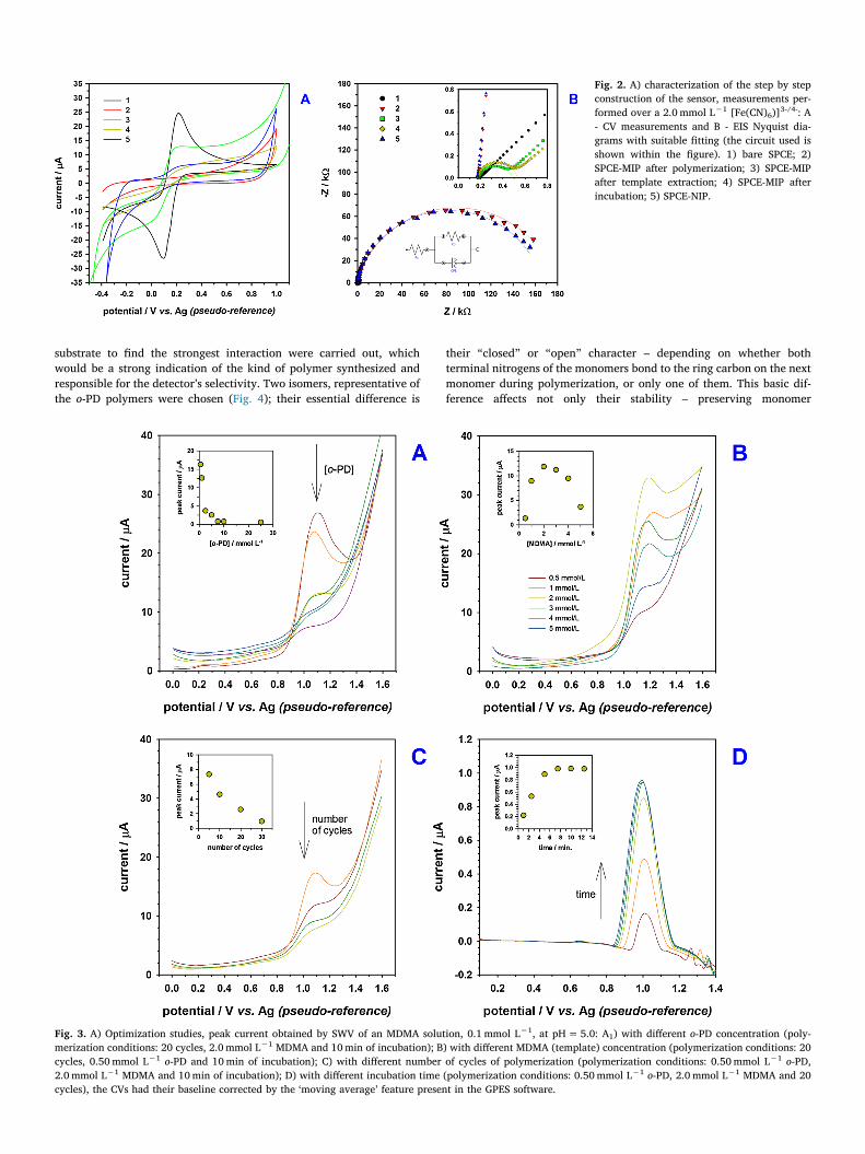
The overall development process of the SPCE-MIP sensor is depictedin Fig. 1. During each step of the construction, the SPCE-MIP sensor was
characterized by CV and EIS (Fig. 2) using 2.0 mmol L−1 [Fe(CN)6)]3−/4−
and 0.1 KCl solution. The CV characterization showed that the bare SPCEallow charge transfer with the typical pair of diffusional reversible peaksin CV [55,56]. When the MIP is formed on the electrode surface, thecurrent decreases in CV and the charge transfer resistance (Rct) in EISclearly increases (the semicircle diameter of Nyquist Plot increased from43.8Ω to 179.3 KΩ), thus both the SPCE-MIP before the removal of thetemplate, and the SPCE-NIP (167.3 KΩ) work as insulators. It is re-markably noticeable the difference between the SPCE-MIP before andafter removing the template (308.0Ω), where the MDMA molecules leftthe cavities and the redox probe obtained easier access to the electrodesurface [57], this being partially reverted when incubating with MDMA(364.1Ω). The removal of the template can also be visualized by SWV(Fig. S1 shown in the Supporting information), since there is not any peakafter the template extraction.
Several information can be obtained from the CVs of the electro-polymerization process (Fig. S2 shown in the Supporting information)[57,58]. Firstly, the lack of any peak is the reverse scan shows that theprocess is irreversible. Then, the current decreases substantially witheach cycle showing that the process is quite efficient. The currentquickly tends to zero showing that the newly formed poly(o-PD) iscovering the electrode´s surface with a non-conductive film [58]. Sev-eral parameters in the preparation of the SPCE-MIP sensor were opti-mized, namely: monomer concentration, template concentration andnumber of cycles in the electropolymerization as well as the time ofincubation with the analyte prior to analysis (Fig. 2). As showed inFig. 3A and B, the best analytical signal was found with 0.50 mmol L−1
o-PD and 2.0 mmol L−1 MDMA, which means a monomer:templateratio of 1:4. The amount of polymer in the electrode surface greatlyinfluences the obtained signal, an excessively thick MIP layer exhibitslow binding capacity, poor site accessibility, and slow absorption ki-netics. The MIP thickness can be adjusted by controlling the number ofcycles of electropolymerization [59], the optimal sensitivity to MDMAwas obtained with 5 scanning cycles (Fig. 3C). Fig. 3D shows the evo-lution of peak current with incubation time at the given conditions,10 min was found to be the optimal analyte-MIP interacting time.
3.2. Theoretical studies
The preparation of electro-polymerized MIPs with o-PD has beenapplied in a variety of analytes ranging from human cardiac troponin T[60], paracetamol (also known as acetaminophen) [61], dopamine [62]to perfluorooctane sulfonate [58]. The produced films are rather ad-vantageous since they can be thin, stable and can be created in-situ onseveral types of substrates [58]. o-PD is one of the most commonmonomers used in electropolymerization, it has good chemical stabilityand the easy polymerization creates a non-conducting compact (rigid)film with hydrophilic and hydrophobic recognition sites. Although o-PDcan easily react using both amine groups simultaneously [63–67] and itis possible to obtain a fully ‘linear’ polymer [68], however that occurs atvery low potentials [69]. Apparently, with this electropolymerization apolymer structure with free amine groups is obtained, which stronglyfacilitates monomer-analyte interaction [66].
In order to further our investigation of the interaction between theo-PD monomers and the MDMA substrate, computational studies of theinteraction between two types of o-PD tetramers and the MDMA
Fig. 1. Schematic illustration of the construction of the MIP-SPCE sensor.
substrate to find the strongest interaction were carried out, whichwould be a strong indication of the kind of polymer synthesized andresponsible for the detector’s selectivity. Two isomers, representative ofthe o-PD polymers were chosen (Fig. 4); their essential difference is
their “closed” or “open” character – depending on whether bothterminal nitrogens of the monomers bond to the ring carbon on the nextmonomer during polymerization, or only one of them. This basic dif-ference affects not only their stability – preserving monomer
Fig. 2. A) characterization of the step by stepconstruction of the sensor, measurements per-formed over a 2.0 mmol L−1 [Fe(CN)6)]3-/4-: A- CV measurements and B - EIS Nyquist dia-grams with suitable fitting (the circuit used isshown within the figure). 1) bare SPCE; 2)SPCE-MIP after polymerization; 3) SPCE-MIPafter template extraction; 4) SPCE-MIP afterincubation; 5) SPCE-NIP.
Fig. 3. A) Optimization studies, peak current obtained by SWV of an MDMA solution, 0.1 mmol L−1, at pH = 5.0: A1) with different o-PD concentration (poly-merization conditions: 20 cycles, 2.0 mmol L−1 MDMA and 10 min of incubation); B) with different MDMA (template) concentration (polymerization conditions: 20cycles, 0.50 mmol L−1 o-PD and 10 min of incubation); C) with different number of cycles of polymerization (polymerization conditions: 0.50 mmol L−1 o-PD,2.0 mmol L−1 MDMA and 10 min of incubation); D) with different incubation time (polymerization conditions: 0.50 mmol L−1 o-PD, 2.0 mmol L−1 MDMA and 20cycles), the CVs had their baseline corrected by the ‘moving average’ feature present in the GPES software.

aromaticity in the first case but not in the second – but also their tor-sional abilities and the availability of terminal nitrogens able to non-covalently interact with the substrate, and thus contribute to the “lockand key” selectivity to MDMA.
Our computational study consisted of, initially, the systematicprospection of the potential energy surface of the many possible con-figurations of non-covalently interacting pairs specified below, o-PDtetramer 1 and MDMA and tetramer 2 and MDMA, and then geometryoptimizations of the most stable conformers with Quantum Mechanicalmethods, in order to find the most favourable interaction in each case.The computational strategy used would be described as a “ladder” ofsuccessive calculation methods, approximating more precisely at eachlevel the correct quantum-mechanical description of the total and in-teraction energy of the studied compounds. Such use of successive ap-proximations is a standard computational protocol necessary to keepthe vast number of calculations necessary for a systematic study of thewide range of possible conformers at feasible computational cost. Asstated earlier, for initial conformer search and geometry optimizations,Spartan’s systematic conformational analysis algorithm, mixing MonteCarlo and Molecular Dynamics was used, and in both cases, ten thou-sand conformers were searched; all with energies low enough to exist ata room-temperature Boltzmann distribution were kept, resulting in5000 and 1100 conformers for the tetramers 1 and 2, respectively.
All of them were then geometry-optimized using Spartan’s im-plementation of the PM6 semi-empirical method [70]. The PM6 methodoffers a more precise description than the pure molecular mechanicsdescription used in the first “rung” of our investigation, the systematicconformer search employing the MMFF. In our second “rung”, thegeometry optimizations using PM6, we were able to investigate withhigher precision the relative energy of the thousands of acceptableconformers, in order to select the most stable among them for furtherinvestigations.
As previously exposed in less detail, the twenty most stable con-formers for each case were then geometry-optimized using theGaussian09 package [49] with a yet more precise level of theory, a third“rung” in our ladder, the Hartree-Fock method. For this third step, the6-31G(p) basis set [50,51] was used, alongside the PCM continuummodel, for the simulation of the dielectric constant of the solvent usedin the synthesis environment, water. Subsequently, the 10 most stablecandidates for each case (the twenty conformers and respective en-ergies are shown in Supporting information) were geometry-optimizedin Gaussian09 using the fourth and highest-precision “rung” in ourladder: the B3LYP exchange-correlation functional with Grimme’s D3correction for dispersion interactions [52–54], a bigger 6-31G(d,p)basis set with polarization functions on hydrogen atoms to capturehydrogen bonding [50,51], and again the PCM model for water. Finally,
bonding and reaction energy for the most stable configuration in eachcase was calculated by geometry-optimizing the interacting moleculesseparately with the same methods, of the last step, that is, of our fourth“rung”, and subtracting their separate energies from the total energy ofthe interacting molecules. Two such calculations were performed. Theformer involves the reaction energy for each tetramer formation. As thetetramer formation involves the condensation of four o-PD monomerswith loss of six hydrogen molecules, both were optimized separately.The later was the separate calculation of both tetramers 1 and 2 in theirfinal conformation in the interaction with the MDMA substrate.
For all the “fourth rung”, B3LYP-D3 calculations, the stability of themolecule, that is, the potential energy surface (PES) minimum characterof the resulting geometry, was confirmed by frequency calculations –that is, calculation of the Hessian matrix for the resulting forces in eachgeometry.
We can see that the so-called ramified tetramer, which we namedtetramer 1, has the strongest interaction with the substrate (Fig. 4). The1.25 kcal mol−1 difference in interaction energies is enough to stronglyindicate that the majority of the MDMA molecules is interacting withthe ramified tetramer, as indicated by previous literature [66]. How-ever, the vast difference in formation energy for both tetramers heavilyfavours the formation of the so-called linear tetramer, which we namedtetramer 2. This indicates that the tetramer 2 has a much larger po-pulation.
We should highlight that a 1.25 kcal mol−1 difference, when un-derstood from the standpoint of Boltzmann populations, which followan exponential trend, indicate a clear selectivity in the MDMA-MIPinteraction favoring the ramified tetramer. While no specific interac-tions – e.g., strong hydrogen bonds – seem responsible for such a se-lectivity, it stands out that the ramified tetramer is able to fold using theMDMA molecule as a template. We are led to ascribe the MDMA-MIPstability to a sum total of non-covalent weak interactions able to shapethe MDMA-MIP complex (see Fig. 4). The linear tetramer is less able toprovide such a stabilization, as we note from the optimized geometriesin Fig. 4. However, it is clearly influenced by the presence of the MDMAmolecule, which breaks the tetramer’s linearity even if that means ap-parently straining it, and despite its aromaticity. In other words, theinteraction of the MDMA with the linear tetramer is also strong, to thepoint of compensating a disturbance of the latter’s aromatic stabiliza-tion. This brings us to a second point of relevance in our results: theintrinsic stability of the linear tetramer, which is much larger than thatof the ramified one, suggests that it is present in a much larger popu-lation. If we take into account that its interaction with MDMA is alsostrong, even if not the most favorable between the two isomers, we arepushed to consider that the linear tetramer also plays an important rolein the electrode's detection ability. Most significantly, however, is the
Fig. 4. Chemical structures of MDMA, ramified and linear tetramers of o-PD (tetramer 1 and 2, respectively). Relative energies and model for the most stable MDMA-tetramer conformer.
discrepancy between intrinsic stability of the tetramers and their ca-pacity to bind MDMA, which follow separate trends. Such a discrepancycould not be captured by an analysis of the interaction of monomersalone with MDMA, which is the most used protocol in theoreticalmodelling of MIPs. The interaction of monomers alone, which does notworry to look at the intrinsic stability of the polymer, may leave thegreatest energetic contributions to the system out of the model, and bethe source of important misevaluations. This would be even more thecase in the present study, in which o-PD monomers are their own cross-linkers, and the stability of the system as a whole is greatly influencedby the intrinsic stability of the polymer chain.
Although this simple model is able to differentiate the interactionenergy between the MDMA and the two tetramers, it is not sufficient toinvestigate the synthesis process, as it is not considering the transitionstates, and the associated activation energy necessary to build a com-plete energy profile of the reaction mechanism. As such, it cannot in-form the rational design of one or another polymerization process, butoffers important insights into the relative weights of the intrinsic sta-bility of the polymer and the strength of its interaction with the analyte.It also throws further light into the nature of o-PD polymers, which hadso far been inconclusive from experimental studies.
Finally, a more complete model, involving the prospection of apotential energy surface for the conformation of o-PD monomersaround MDMA and other tested substrates and the reaction mechanismfor the tetramer formation is under development, and will potentiallyprovide further insight into the process of MIP synthesis and selectivity.
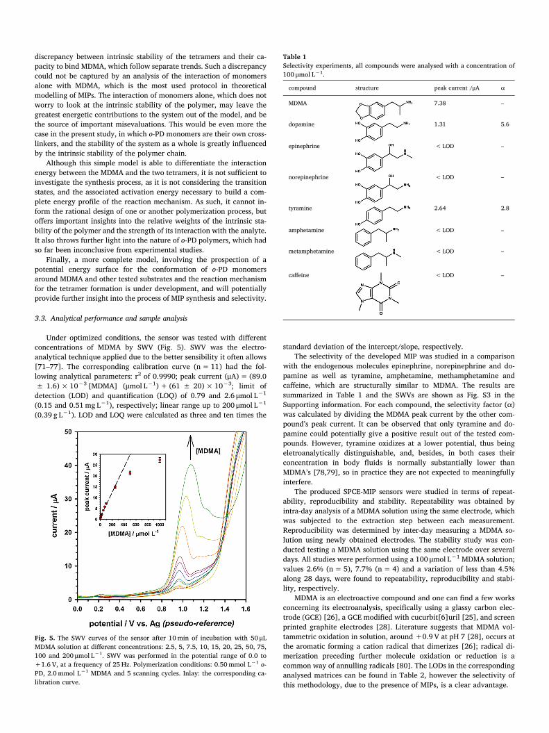
3.3. Analytical performance and sample analysis
Under optimized conditions, the sensor was tested with differentconcentrations of MDMA by SWV (Fig. 5). SWV was the electro-analytical technique applied due to the better sensibility it often allows[71–77]. The corresponding calibration curve (n = 11) had the fol-lowing analytical parameters: r2 of 0.9990; peak current (μA) = (89.0± 1.6) × 10−3 [MDMA] (μmol L−1) + (61 ± 20) × 10−3; limit ofdetection (LOD) and quantification (LOQ) of 0.79 and 2.6 μmol L−1
(0.15 and 0.51 mg L−1), respectively; linear range up to 200 μmol L−1
(0.39 g L−1). LOD and LOQ were calculated as three and ten times the
standard deviation of the intercept/slope, respectively.The selectivity of the developed MIP was studied in a comparison
with the endogenous molecules epinephrine, norepinephrine and do-pamine as well as tyramine, amphetamine, methamphetamine andcaffeine, which are structurally similar to MDMA. The results aresummarized in Table 1 and the SWVs are shown as Fig. S3 in theSupporting information. For each compound, the selectivity factor (α)was calculated by dividing the MDMA peak current by the other com-pound’s peak current. It can be observed that only tyramine and do-pamine could potentially give a positive result out of the tested com-pounds. However, tyramine oxidizes at a lower potential, thus beingeletroanalytically distinguishable, and, besides, in both cases theirconcentration in body fluids is normally substantially lower thanMDMA’s [78,79], so in practice they are not expected to meaningfullyinterfere.
The produced SPCE-MIP sensors were studied in terms of repeat-ability, reproducibility and stability. Repeatability was obtained byintra-day analysis of a MDMA solution using the same electrode, whichwas subjected to the extraction step between each measurement.Reproducibility was determined by inter-day measuring a MDMA so-lution using newly obtained electrodes. The stability study was con-ducted testing a MDMA solution using the same electrode over severaldays. All studies were performed using a 100 μmol L−1 MDMA solution;values 2.6% (n = 5), 7.7% (n = 4) and a variation of less than 4.5%along 28 days, were found to repeatability, reproducibility and stabi-lity, respectively.
MDMA is an electroactive compound and one can find a few worksconcerning its electroanalysis, specifically using a glassy carbon elec-trode (GCE) [26], a GCE modified with cucurbit[6]uril [25], and screenprinted graphite electrodes [28]. Literature suggests that MDMA vol-tammetric oxidation in solution, around +0.9 V at pH 7 [28], occurs atthe aromatic forming a cation radical that dimerizes [26]; radical di-merization preceding further molecule oxidation or reduction is acommon way of annulling radicals [80]. The LODs in the correspondinganalysed matrices can be found in Table 2, however the selectivity ofthis methodology, due to the presence of MIPs, is a clear advantage.
Fig. 5. The SWV curves of the sensor after 10 min of incubation with 50 μLMDMA solution at different concentrations: 2.5, 5, 7.5, 10, 15, 20, 25, 50, 75,100 and 200 μmol L−1. SWV was performed in the potential range of 0.0 to+1.6 V, at a frequency of 25 Hz. Polymerization conditions: 0.50 mmol L−1 o-PD, 2.0 mmol L−1 MDMA and 5 scanning cycles. Inlay: the corresponding ca-libration curve.
Table 1Selectivity experiments, all compounds were analysed with a concentration of100 μmol L−1.
compound structure peak current /μA α
MDMA 7.38 –
dopamine 1.31 5.6
epinephrine < LOD –
norepinephrine < LOD –
tyramine 2.64 2.8
amphetamine < LOD –
metamphetamine < LOD –
caffeine < LOD –

The proposed MIP-SPCE sensor was applied to the quantification ofMDMA in biological matrices, namely human blood serum and urinesamples (calibration curves are shown as Fig. S4 in the Supporting in-formation). Recovery tests experiment was performed by spiking thesamples with MDMA, followed by dilution with acetate buffer solutionpH 5, a recovery of (81.0 ± 2.4) % was obtained with diluted urine(1:10) and (91.4 ± 1.1) % with diluted serum (1:20). Samples were notsubjected to any additional pre-treatment steps. The calibration curves(n =5) had the following analytical parameters: a) urine - r2 of 0.998,peak current (μA)= (100.9 ± 2.7) ×10−3 [MDMA] (μmol L−1
) + (0.16 ± 0.17), LOD and LOQ of 6.4 and 21 μmol L−1 (1.2 and4.1 mg L−1), respectively, with a linear range of up to 100 μmol L−1
(0.19 g L−1); b) blood serum – r2 of 0.9997, peak current(μA)= (42.37 ± 0.95) ×10−3 [MDMA] (μmol L−1)− (73 ± 69)×10−3, LOD and LOQ of 3.9 and 13 μmol L−1 (0.75 and 2.5mg L−1), re-spectively, with a linear range of up to 100 μmol L−1 (0.19 g L−1). BothLODs and LOQs were calculated as three and ten times the standarddeviation of the intercept/slope, respectively.
In fatalities associated with MDMA, levels between 0.6 and4.3 mg L−1, in admission, and post-mortem levels between 0.5 and28.4 mg L−1 were found [81]. Another publication attributes death tothe toxic effects of MDMA alone with a range of 0.5–54 mg L−1 (13cases) [82].
4. Conclusions
In this work, it was successfully shown that it is possible to obtain aselective electrochemical sensor for MDMA using a SCPE-MIP. A poly(o-PD) film was generated at the surface of a SPCE by electro-polymerization of the monomer o-PD as a MDMA recognition element.To the best of our knowledge, electrochemical determination of MDMAbased on an electropolymerized selective sensor has not been reportedbefore. Experimental parameters that affected the performance of theSPCE-MIP sensor were studied and optimized, confirming electro-analytical properties such as linearity, selectivity (in comparison withendogenous and non-endogenous compounds with similar chemicalstructures), stability, repeatability and reproducibility. Furthermore,the sensor was applied in the analysis of MDMA in biological samples,namely human blood serum and urine.
Acknowledgements
This work received financial support from FCT/MCTES throughnational funds and was co-financed by FEDER, under PartnershipAgreement PT2020-UID/QUI/50006/2013-POCI/01/0145/FEDER/007265. RASC wishes to acknowledge FCT for her PhD fellowship (PD/BD/127797/2016) from the PhD Programme in Medicines andPharmaceutical Innovation (i3DU). AACB (grants 2014/25770-6 and
2015/01491-3) and LMG (grant 2018/14425-7) thank the São PauloResearch Foundation (FAPESP) for financial support. AACB (grant309715/2017-2) also thanks the Brazilian National Research Council(CNPq) for financial support and fellowships. This study was financedin part by the Coordenação de Aperfeiçoamento de Pessoal de NívelSuperior - Brasil (CAPES) - Finance Code 001.
References
[1] R.W. Freudenmann, F. Öxler, S. Bernschneider-Reif, The origin of MDMA (ecstasy)revisited: the true story reconstructed from the original documents, Addiction 101(2006) 1241–1245, https://doi.org/10.1111/j.1360-0443.2006.01511.x.
[2] D. Butler, G.G. Guilbault, Analytical techniques for ecstasy, Anal. Lett. 37 (2004)2003–2030, https://doi.org/10.1081/AL-200026665.
[3] D.E. Nichols, Differences between the mechanism of action of MDMA, MBDB, andthe classic hallucinogens. Identification of a new therapeutic class: entactogens, J.Psychoactive Drugs 18 (1986) 305–313, https://doi.org/10.1080/02791072.1986.10472362.
[4] B. Sessa, Why psychiatry needs 3,4-methylenedioxymethamphetamine: a childpsychiatrist’s perspective, Neurotherapeutics 14 (2017) 741–749, https://doi.org/10.1007/s13311-017-0531-1.
[5] K.K. Rigg, A. Sharp, Deaths related to MDMA (ecstasy/molly): prevalence, rootcauses, and harm reduction interventions, J. Substain. Use 23 (2018) 345–352,https://doi.org/10.1080/14659891.2018.1436607.
[6] M. Carvalho, H. Carmo, V.M. Costa, J.P. Capela, H. Pontes, F. Remião, F. Carvalho,M. de L. Bastos, Toxicity of amphetamines: an update, Arch. Toxicol. 86 (2012)1167–1231, https://doi.org/10.1007/s00204-012-0815-5.
[7] J.P. Capela, H. Carmo, F. Remião, M.L. Bastos, A. Meisel, F. Carvalho, Molecularand cellular mechanisms of ecstasy-induced neurotoxicity: an overview, Mol.Neurobiol. 39 (2009) 210–271, https://doi.org/10.1007/s12035-009-8064-1.
[8] M. Carvalho, H. Pontes, F. Remiao, M.L. Bastos, F. Carvalho, Mechanisms under-lying the hepatotoxic effects of ecstasy, Curr. Pharm. Biotechnol. 11 (2010)476–495, https://doi.org/10.2174/138920110791591535.
[9] S.K. Shenouda, F. Carvalho, K.J. Varner, The cardiovascular and cardiac actions ofecstasy and its metabolites, Curr. Pharm. Biotechnol. 11 (2010) 470–475, https://doi.org/10.2174/138920110791591526.
[10] A. Laurin, M. Marinescu, Enquête sur la consommation de MDMA (ecstasy) dans lapopulation homosexuelle masculine française en 2016 : un retour en force, Inf.Psychiatr. 93 (2017) 151–157, https://doi.org/10.1684/ipe.2017.1600.
[11] D.J. Barbosa, R. Serrat, S. Mirra, M. Quevedo, E.G. de Barreda, J. Àvila,L.M. Ferreira, P.S. Branco, E. Fernandes, M. de Lourdes Bastos, J.P. Capela,E. Soriano, F. Carvalho, The mixture of “Ecstasy” and its metabolites impairs mi-tochondrial fusion/fission equilibrium and trafficking in hippocampal neurons, at invivo relevant concentrations, Toxicol. Sci. 139 (2014) 407–420, https://doi.org/10.1093/toxsci/kfu042.
[12] P. Kintz, N. Samyn, Determination of “Ecstasy” components in alternative biologicalspecimens, J. Chromatogr. B: Biomed. Sci. Appl. 733 (1999) 137–143, https://doi.org/10.1016/S0378-4347(98)00521-0.
[13] B. Jamali, M. Torkamanian, N. Badri, B. Sheikholeslami, Y.H. Ardakani, M.-R. Rouini, Assays for MDMA and its metabolites, Neuropathol. Drug Addict. Subst.Misuse, Elsevier, 2016, pp. 503–512, https://doi.org/10.1016/B978-0-12-800212-4.00047-9.
[14] M. Frost, H. Köhler, G. Blaschke, Analysis of “Ecstasy” by capillary electrophoresis,Int. J. Legal Med. 109 (1996) 53–57, https://doi.org/10.1007/BF01355516.
[15] M.C. Lasmar, E.M.A. Leite, Desenvolvimento e validação de um métodocromatográfico em fase gasosa para análise da 3,4-metilenodioximetanfetamina(ecstasy) e outros derivados anfetamínicos em comprimidos, Rev. Bras. CiênciasFarm. 43 (2007) 223–230, https://doi.org/10.1590/S1516-93322007000200008.
[16] M. Pellegrini, F. Rosati, R. Pacifici, P. Zuccaro, F. Romolo, A. Lopez, Rapidscreening method for determination of Ecstasy and amphetamines in urine samplesusing gas chromatography–chemical ionisation mass spectrometry, J. Chromatogr.B 769 (2002) 243–251, https://doi.org/10.1016/S1570-0232(01)00586-4.
Table 2Electroanalytical studies aimed at MDMA determination.
technique LOD/μmol L−1 LOQ/μmol L−1 matrix reference
SWV, using a SPCE-MIP 0.79 2.6 supporting electrolyte This work6.4 21 urine3.9 13 serum
CV, using a GCE modified with cucurbit[6]uril 2.7 9.1 supporting electrolyte [25]SWV, using a GCE 1,2 3.7 supporting electrolyte [26]
2.4 8.3 serumSWV, using a gold electrode 30.9a – urine [27]DPV, using a SPCE 0.21 – supporting electrolyte [28]
a The LOD was not calculated, the value is based in the lower limit of the calibration curve.

[17] Z. Es’haghi, M. Mohtaji, M. Hasanzade-Meidani, M. Masrournia, The measurementof ecstasy in human hair by triple phase directly suspended droplet microextractionprior to HPLC-DAD analysis, J. Chromatogr. B 878 (2010) 903–908, https://doi.org/10.1016/j.jchromb.2010.02.015.
[18] J.L. da Costa, E.R. Pintao, C.M.C. Corrigliano, O. Negrini Neto, Determinação de3,4-metilenodioximetanfetamina (MDMA) em comprimidos de Ecstasy por croma-tografia líquida de alta eficiência com detecção por fluorescência (CLAE-DF), Quim.Nova 32 (2009) 965–969, https://doi.org/10.1590/S0100-40422009000400026.
[19] A.E. Steuer, C. Schmidhauser, M.E. Liechti, T. Kraemer, Development and valida-tion of an LC–MS/MS method after chiral derivatization for the simultaneous ste-reoselective determination of methylenedioxy-methamphetamine (MDMA) and itsphase I and II metabolites in human blood plasma, Drug Test. Anal. 7 (2015)592–602, https://doi.org/10.1002/dta.1740.
[20] M. Poetzsch, A.E. Steuer, C.M. Hysek, M.E. Liechti, T. Kraemer, Development of ahigh-speed MALDI-triple quadrupole mass spectrometric method for the determi-nation of 3,4-methylenedioxymethamphetamine (MDMA) in oral fluid, Drug Test.Anal. 8 (2016) 235–240, https://doi.org/10.1002/dta.1810.
[21] D.T. Burns, R.J. Lewis, P. Stevenson, Determination of 3,4-methylenediox-yamphetamine analogues (“Ecstasy”) by proton nuclear magnetic resonance spec-trometry, Anal. Chim. Acta 339 (1997) 259–263, https://doi.org/10.1016/S0003-2670(96)00485-0.
[22] S. Armenta, S. Garrigues, M. de la Guardia, J. Brassier, M. Alcalà, M. Blanco,Analysis of ecstasy in oral fluid by ion mobility spectrometry and infrared spec-troscopy after liquid–liquid extraction, J. Chromatogr. A 1384 (2015) 1–8, https://doi.org/10.1016/j.chroma.2015.01.036.
[23] H.E. French, M.J. Went, S.J. Gibson, Graphite furnace atomic absorption elementalanalysis of ecstasy tablets, Forensic Sci. Int. 231 (2013) 88–91, https://doi.org/10.1016/j.forsciint.2013.04.021.
[24] B. Lozano-Torres, L. Pascual, A. Bernardos, M.D. Marcos, J.O. Jeppesen, Y. Salinas,R. Martínez-Máñez, F. Sancenón, Pseudorotaxane capped mesoporous silica nano-particles for 3,4-methylenedioxymethamphetamine (MDMA) detection in water,Chem. Commun. 53 (2017) 3559–3562, https://doi.org/10.1039/C7CC00186J.
[25] M.C. Tadini, M.A. Balbino, I.C. Eleoterio, L.S. de Oliveira, L.G. Dias, G. Jean-François Demets, M.F. de Oliveira, Developing electrodes chemically modified withcucurbit[6]uril to detect 3,4-methylenedioxymethamphetamine (MDMA) by vol-tammetry, Electrochim. Acta 121 (2014) 188–193, https://doi.org/10.1016/j.electacta.2013.12.107.
[26] E.M.P.J. Garrido, J.M.P.J. Garrido, N. Milhazes, F. Borges, A.M. Oliveira-Brett,Electrochemical oxidation of amphetamine-like drugs and application to electro-analysis of ecstasy in human serum, Bioelectrochemistry 79 (2010) 77–83, https://doi.org/10.1016/j.bioelechem.2009.12.002.
[27] M. Nevescanin, M. Avramov-Ivic, S. Petrovic, D. Mijin, S. Banovic-Stevic,V. Jovanovic, The use of a gold electrode for the determination of amphetaminederivatives and application to their analysis in human urine, J. Serbian Chem. Soc.78 (2013) 1373–1385, https://doi.org/10.2298/JSC121228032N.
[28] L.R. Cumba, J.P. Smith, K.Y. Zuway, O.B. Sutcliffe, D.R. do Carmo, C.E. Banks,Forensic electrochemistry: simultaneous voltammetric detection of MDMA and itsfatal counterpart “Dr Death” (PMA), Anal. Methods 8 (2016) 142–152, https://doi.org/10.1039/C5AY02924D.
[29] L.M. Gonçalves, I.M. Valente, J.A. Rodrigues, Recent advances in membrane-aidedextraction and separation for analytical purposes, Sep. Purif. Rev. 46 (2017)179–194, https://doi.org/10.1080/15422119.2016.1235050.
[30] A. de Korompay, J.C. Hill, J.F. Carter, N. NicDaeid, R. Sleeman, Supported li-quid–liquid extraction of the active ingredient (3,4-methylenediox-ymethylamphetamine) from ecstasy tablets for isotopic analysis, J. Chromatogr. A1178 (2008) 1–8, https://doi.org/10.1016/j.chroma.2007.11.054.
[31] C.R.T. Tarley, M.D.P.T. Sotomayor, L.T. Kubota, Polímeros biomiméticos emquímica analítica. Parte 1: preparo e aplicações de MIP (“Molecularly ImprintedPolymers”) em técnicas de extração e separação, Quim. Nova 28 (2005) 1076–1086,https://doi.org/10.1590/S0100-40422005000600024.
[32] G.A. Ruiz-Córdova, S. Khan, L.M. Gonçalves, M.I. Pividori, G. Picasso,M.D.P.T. Sotomayor, Electrochemical sensing using magnetic molecularly im-printed polymer particles previously captured by a magneto-sensor, Talanta 181(2018) 19–23, https://doi.org/10.1016/j.talanta.2017.12.085.
[33] S. Khan, S. Hussain, A. Wong, M.V. Foguel, L. Moreira Gonçalves, M.I. PividoriGurgo, M. del P. Taboada, Sotomayor, synthesis and characterization of magnetic-molecularly imprinted polymers for the HPLC-UV analysis of ametryn, React. Funct.Polym. 122 (2018) 175–182, https://doi.org/10.1016/j.reactfunctpolym.2017.11.002.
[34] A.N. Baeza-Fonte, I. Garcés-Lobo, M.D. Luaces-Alberto, L.M. Gonçalves,M.D.P.T. Sotomayor, A.C. Valdés-González, Determination of cephalosporins byUHPLC-DAD using molecularly imprinted polymers, J. Chromatogr. Sci. 56 (2018)187–193, https://doi.org/10.1093/chromsci/bmx099.
[35] W.R. de Araujo, T.M.G. Cardoso, R.G. da Rocha, M.H.P. Santana, R.A.A. Muñoz,E.M. Richter, T.R.L.C. Paixão, W.K.T. Coltro, Portable analytical platforms for for-ensic chemistry: a review, Anal. Chim. Acta 1035 (2018) 1–21, https://doi.org/10.1016/j.aca.2018.06.014.
[36] M.M. Pedroso, M.V. Foguel, D.H.S. Silva, M. del P.T. Sotomayor, H. Yamanaka,Electrochemical sensor for dodecyl gallate determination based on electro-polymerized molecularly imprinted polymer, Sens. Actuators B: Chem. 253 (2017)180–186, https://doi.org/10.1016/j.snb.2017.06.127.
[37] L. Shaw, L. Dennany, Applications of electrochemical sensors: forensic drug ana-lysis, Curr. Opin. Electrochem. 3 (2017) 23–28, https://doi.org/10.1016/j.coelec.2017.05.001.
[38] C. Malitesta, E. Mazzotta, R.A. Picca, A. Poma, I. Chianella, S.A. Piletsky, MIPsensors – the electrochemical approach, Anal. Bioanal. Chem. 402 (2012)1827–1846, https://doi.org/10.1007/s00216-011-5405-5.
[39] P.S. Sharma, A. Pietrzyk-Le, F. D’Souza, W. Kutner, Electrochemically synthesizedpolymers in molecular imprinting for chemical sensing, Anal. Bioanal. Chem. 402(2012) 3177–3204, https://doi.org/10.1007/s00216-011-5696-6.
[40] V. Suryanarayanan, C.-T. Wu, K.-C. Ho, Molecularly imprinted electrochemicalsensors, Electroanalysis 22 (2010) 1795–1811, https://doi.org/10.1002/elan.200900616.
[41] J.G. Pacheco, P. Rebelo, F. Cagide, L.M. Gonçalves, F. Borges, J.A. Rodrigues,C. Delerue-Matos, Electrochemical sensing of the thyroid hormone thyronamine(T0AM) via molecular imprinted polymers (MIPs), Talanta 194 (2019) 689–696,https://doi.org/10.1016/j.talanta.2018.10.090.
[42] F. Ahmadi, J. Ahmadi, M. Rahimi-Nasrabadi, Computational approaches to design amolecular imprinted polymer for high selective extraction of 3,4-methylenediox-ymethamphetamine from plasma, J. Chromatogr. A 1218 (2011) 7739–7747,https://doi.org/10.1016/j.chroma.2011.08.020.
[43] D. Djozan, M.A. Farajzadeh, S.M. Sorouraddin, T. Baheri, Molecularly imprinted-solid phase extraction combined with simultaneous derivatization and dispersiveliquid–liquid microextraction for selective extraction and preconcentration of me-thamphetamine and ecstasy from urine samples followed by gas chromatogra, J.Chromatogr. A 1248 (2012) 24–31, https://doi.org/10.1016/j.chroma.2012.05.085.
[44] D. Djozan, M.A. Farajzadeh, S.M. Sorouraddin, T. Baheri, Determination of me-thamphetamine, amphetamine and ecstasy by inside-needle adsorption trap basedon molecularly imprinted polymer followed by GC-FID determination, Microchim.Acta 179 (2012) 209–217, https://doi.org/10.1007/s00604-012-0879-1.
[45] N. Almeida, L. Benedito, A. Maldaner, A. de Oliveira, A validated NMR approach forMDMA quantification in ecstasy tablets, J. Braz. Chem. Soc. 29 (9) (2018)1944–1950, https://doi.org/10.21577/0103-5053.20180071.
[46] J. Liu, J. Decatur, G. Proni, E. Champeil, Identification and quantitation of 3,4-methylenedioxy-N-methylamphetamine (MDMA, ecstasy) in human urine by 1HNMR spectroscopy. Application to five cases of intoxication, Forensic Sci. Int. 194(2010) 103–107, https://doi.org/10.1016/j.forsciint.2009.10.022.
[47] L. Vlase, D.S. Popa, F. Loghin, S.E. Leucuta, High-throughput toxicological analysisof methamphetamine, MDA and MDMA from human plasma by LC–MS/MS, Rom. J.Leg. Med. 17 (2009), https://doi.org/10.4323/rjlm.2009.213.
[48] M. Concheiro, S.M. dos S.S. Simões, Ó. Quintela, A. de Castro, M.J.R. Dias, A. Cruz,M. López-Rivadulla, Fast LC–MS/MS method for the determination of ampheta-mine, methamphetamine, MDA, MDMA, MDEA, MBDB and PMA in urine, ForensicSci. Int. 171 (2007) 44–51, https://doi.org/10.1016/j.forsciint.2006.10.004.
[49] M.J. Frisch, G.W. Trucks, H.B. Schlegel, G.E. Scuseria, M.A. Robb, J.R. Cheeseman,G. Scalmani, V. Barone, B. Mennucci, G.A. Petersson, H. Nakatsuji, M. Caricato,X. Li, H.P. Hratchian, A.F. Izmaylov, J. Bloino, G. Zheng, J.L. Sonnenberg, M. Hada,M. Ehara, K. Toyota, R. Fukuda, J. Hasegawa, M. Ishida, T. Nakajima, Y. Honda,O. Kitao, H. Nakai, T. Vreven, J.A. Montgomery Jr., J.E. Peralta, F. Ogliaro,M. Bearpark, J.J. Heyd, E. Brothers, K.N. Kudin, V.N. Staroverov, R. Kobayashi,J. Normand, K. Raghavachari, A. Rendell, J.C. Burant, S.S. Iyengar, J. Tomasi,M. Cossi, N. Rega, J.M. Millam, M. Klene, J.E. Knox, J.B. Cross, V. Bakken,C. Adamo, J. Jaramillo, R. Gomperts, R.E. Stratmann, O. Yazyev, A.J. Austin,R. Cammi, C. Pomelli, J.W. Ochterski, R.L. Martin, K. Morokuma, V.G. Zakrzewski,G.A. Voth, P. Salvador, J.J. Dannenberg, S. Dapprich, A.D. Daniels, Ö. Farkas,J.B. Foresman, J.V. Ortiz, J. Cioslowski, D.J. Fox, Gaussian 09, Revision D.01,(2009).
[50] W.J. Hehre, R. Ditchfield, J.A. Pople, Self—consistent molecular orbital methods.XII. Further extensions of gaussian—type basis sets for use in molecular orbitalstudies of organic molecules, J. Chem. Phys. 56 (1972) 2257–2261, https://doi.org/10.1063/1.1677527.
[51] M.M. Francl, W.J. Pietro, W.J. Hehre, J.S. Binkley, M.S. Gordon, D.J. DeFrees,J.A. Pople, Self-consistent molecular orbital methods. XXIII. A polarization-typebasis set for second-row elements, J. Chem. Phys. 77 (1982) 3654–3665, https://doi.org/10.1063/1.444267.
[52] C. Lee, W. Yang, R.G. Parr, Development of the Colle-Salvetti correlation-energyformula into a functional of the electron density, Phys. Rev. B 37 (1988) 785–789,https://doi.org/10.1103/PhysRevB.37.785.
[53] P.J. Stephens, F.J. Devlin, C.F. Chabalowski, M.J. Frisch, Ab initio calculation ofvibrational absorption and circular dichroism spectra using density functional forcefields, J. Phys. Chem. 98 (1994) 11623–11627, https://doi.org/10.1021/j100096a001.
[54] A.D. Becke, Density-functional thermochemistry. III. The role of exact exchange, J.Chem. Phys. 98 (1993) 5648–5652, https://doi.org/10.1063/1.464913.
[55] C. Batchelor-McAuley, L.M. Gonçalves, L. Xiong, A.A. Barros, R.G. Compton,Controlling voltammetric responses by electrode modification; using adsorbedacetone to switch graphite surfaces between adsorptive and diffusive modes, Chem.Commun. 46 (2010) 9037–9039, https://doi.org/10.1039/c0cc03961f.
[56] L.M. Gonçalves, C. Batchelor-McAuley, A.A. Barros, R.G. Compton, Electrochemicaloxidation of adenine: a mixed adsorption and diffusion response on an edge-planepyrolytic graphite electrode, J. Phys. Chem. C 114 (2010) 14213–14219, https://doi.org/10.1021/jp1046672.
[57] X. Li, Y. He, F. Zhao, W. Zhang, Z. Ye, Molecularly imprinted polymer-based sensorsfor atrazine detection by electropolymerization of o-phenylenediamine, RSC Adv. 5(2015) 56534–56540, https://doi.org/10.1039/C5RA09556E.
[58] N. Karimian, A.M. Stortini, L.M. Moretto, C. Costantino, S. Bogialli, P. Ugo,Electrochemosensor for trace analysis of perfluorooctanesulfonate in water basedon a molecularly imprinted poly(o-phenylenediamine) polymer, ACS Sens. 3 (2018)1291–1298, https://doi.org/10.1021/acssensors.8b00154.
[59] X.-C. Fu, X. Chen, Z. Guo, C.-G. Xie, L.-T. Kong, J.-H. Liu, X.-J. Huang, Strippingvoltammetric detection of mercury(II) based on a surface ion imprinting strategy inelectropolymerized microporous poly(2-mercaptobenzothiazole) films modifiedglassy carbon electrode, Anal. Chim. Acta 685 (2011) 21–28, https://doi.org/10.1016/j.aca.2010.11.020.
[60] N. Karimian, M. Vagin, M.H.A. Zavar, M. Chamsaz, A.P.F. Turner, A. Tiwari, Anultrasensitive molecularly-imprinted human cardiac troponin sensor, Biosens.Bioelectron. 50 (2013) 492–498, https://doi.org/10.1016/j.bios.2013.07.013.
[61] Y. Peng, Z. Wu, Z. Liu, An electrochemical sensor for paracetamol based on anelectropolymerized molecularly imprinted o-phenylenediamine film on a multi-

walled carbon nanotube modified glassy carbon electrode, Anal. Methods 6 (2014)5673–5681, https://doi.org/10.1039/C4AY00753K.
[62] D. Wu, H. Li, X. Xue, H. Fan, Q. Xin, Q. Wei, Sensitive and selective determination ofdopamine by electrochemical sensor based on molecularly imprinted electro-polymerization of o-phenylenediamine, Anal. Methods 5 (2013) 1469, https://doi.org/10.1039/c3ay26200f.
[63] J.G. Pacheco, I.M. Valente, L.M. Gonçalves, J.A. Rodrigues, A.A. Barros, Gas-dif-fusion microextraction, J. Sep. Sci. 33 (2010) 3207–3212, https://doi.org/10.1002/jssc.201000351.
[64] R.M. Ramos, L.M. Gonçalves, V. Vyskočil, J.A. Rodrigues, Voltammetric determi-nation of trace amounts of diacetyl at a mercury meniscus modified silver solidamalgam electrode following gas-diffusion microextraction, Talanta 169 (2017),https://doi.org/10.1016/j.talanta.2017.03.077.
[65] J.A. Rodrigues, I.M. Valente, L.M. Gonçalves, J.G. Pacheco, A.A. Barros,Polarographic determination of vitamin C after derivatization with o-phenylene-diamine, Collect. Czechoslov. Chem. Commun. 75 (2010), https://doi.org/10.1135/cccc2010026.
[66] I. Losito, F. Palmisano, P.G. Zambonin, o-Phenylenediamine electropolymerizationby cyclic voltammetry combined with electrospray ionization-ion trap mass spec-trometry, Anal. Chem. 75 (2003) 4988–4995, https://doi.org/10.1021/ac0342424.
[67] W. Zhai, X. Tian, Y. Yan, Y. Xu, Y. Zhao, Y. Liu, Preparation and characterization ofa poly-o-phenylenediamine film modified glassy carbon electrode as a H2O2 sensor,Can. J. Chem. 91 (2013) 1077–1084, https://doi.org/10.1139/cjc-2013-0141.
[68] M. Baibarac, I. Baltog, M. Scocioreanu, B. Ballesteros, J.Y. Mevellec, S. Lefrant, One-dimensional composites based on single walled carbon nanotubes and poly(o-phe-nylenediamine), Synth. Met. 161 (2011) 2344–2354, https://doi.org/10.1016/j.synthmet.2011.09.001.
[69] S. Bilal, R. Holze, Electrochemical copolymerization of o-toluidine and o-phenyle-nediamine, J. Electroanal. Chem. 592 (2006) 1–13, https://doi.org/10.1016/j.jelechem.2006.03.039.
[70] J.J.P. Stewart, Optimization of parameters for semiempirical methods V: mod-ification of NDDO approximations and application to 70 elements, J. Mol. Model.13 (2007) 1173–1213, https://doi.org/10.1007/s00894-007-0233-4.
[71] M. Tefera, A. Geto, M. Tessema, S. Admassie, Simultaneous determination of caf-feine and paracetamol by square wave voltammetry at poly(4-amino-3-hydro-xynaphthalene sulfonic acid)-modified glassy carbon electrode, Food Chem. 210(2016) 156–162, https://doi.org/10.1016/j.foodchem.2016.04.106.
[72] F. Zang, K. Gerasopoulos, X.Z. Fan, A.D. Brown, J.N. Culver, R. Ghodssi, An elec-trochemical sensor for selective TNT sensing based on tobacco mosaic virus-likeparticle binding agents, Chem. Commun. 50 (2014) 12977–12980, https://doi.org/10.1039/C4CC06735E.
[73] A.M. Carvalho, L.M. Gonçalves, I.M. Valente, J.A. Rodrigues, A.A. Barros, Analysisof cardamonin by square wave voltammetry, Phytochem. Anal. 23 (2012) 396–399,https://doi.org/10.1002/pca.1370.
[74] J.G. Silva, E.M. Tavares, L.M. Gonçalves, I.M. Valente, D.O. Carvalho,J.A. Rodrigues, Voltammetric analysis of licochalcone A in licorice, J. Electrochem.Soc. 160 (2013), https://doi.org/10.1149/2.020310jes.
[75] L.M. Goncalves, J. Grosso Pacheco, P. Jorge Magalhaes, J. Antonio Rodrigues,A. Araujo Barros, Determination of free and total sulfites in wine using an automaticflow injection analysis system with voltammetric detection, Food Addit. Contam.Part A: Chem. Anal. Control Expo. Risk Assess. 27 (2010) 175–180, https://doi.org/10.1080/19440040903261547.
[76] D. De Souza, A. Galli, M.L. Calegaro, S.A.S. Machado, R.C. Pires, Utilização damúltipla voltametria de onda quadrada na determinação eletroanalítica de com-postos orgânicos e inorgânicos, Quim. Nova 30 (2007) 458–463, https://doi.org/10.1590/S0100-40422007000200038.
[77] V. Mirceski, R. Gulaboski, M. Lovric, I. Bogeski, R. Kappl, M. Hoth, Square-waveVoltammetry: a review on the recent progress, Electroanalysis. 25 (2013)2411–2422, https://doi.org/10.1002/elan.201300369.
[78] R.C. Causon, M.J. Brown, High-performance liquid chromatography with ampero-metric determination of plasma tyramine, J. Chromatogr. A 317 (1984) 319–325,https://doi.org/10.1016/S0021-9673(01)91670-X.
[79] W. Mäurer, P. Drings, J. Manthey, W. Kübler, Vergleichende Untersuchungen überden Gehalt der Blut-Katecholamine Adrenalin, Noradrenalin und Dopamin inPlasma, Erythrozyten und Thrombozyten, (1975), pp. 245–247, https://doi.org/10.1007/978-3-642-85450-7_54.
[80] E.M. Tavares, A.M. Carvalho, L.M. Gonçalves, I.M. Valente, M.M. Moreira,L.F. Guido, J.A. Rodrigues, T. Doneux, A.A. Barros, Chemical sensing of chalconesby voltammetry: trans-chalcone, cardamonin and xanthohumol, Electrochim. Acta90 (2013) 440–444, https://doi.org/10.1016/j.electacta.2012.12.040.
[81] S.P. Elliott, MDMA and MDA Concentrations in antemortem and postmortem spe-cimens in fatalities following hospital admission, J. Anal. Toxicol. 29 (2005)296–300, https://doi.org/10.1093/jat/29.5.296.
[82] C.M. Milroy, “Ecstasy” associated deaths: what is a fatal concentration? Analysis ofa case series, Forensic Sci. Med. Pathol. 7 (2011) 248–252, https://doi.org/10.1007/s12024-010-9220-7.
[83] Y. Shao, L.F. Molnar, Y. Jung, J. Kussmann, C. Ochsenfeld, S.T. Brown,A.T.B. Gilbert, L.V. Slipchenko, S.V. Levchenko, D.P. O’Neill, R.A. DiStasio Jr.,R.C. Lochan, T. Wang, G.J.O. Beran, N.A. Besley, J.M. Herbert, C.Y. Lin, T. VanVoorhis, S.H. Chien, A. Sodt, R.P. Steele, V.A. Rassolov, P.E. Maslen,P.P. Korambath, R.D. Adamson, B. Austin, J. Baker, E.F.C. Byrd, H. Dachsel,R.J. Doerksen, A. Dreuw, B.D. Dunietz, A.D. Dutoi, T.R. Furlani, S.R. Gwaltney,A. Heyden, S. Hirata, C.-P. Hsu, G. Kedziora, R.Z. Khalliulin, P. Klunzinger,A.M. Lee, M.S. Lee, W.Z. Liang, I. Lotan, N. Nair, B. Peters, E.I. Proynov,P.A. Pieniazek, Y.M. Rhee, J. Ritchie, E. Rosta, C.D. Sherrill, A.C. Simmonett,J.E. Subotnik, H.L. Woodcock III, W. Zhang, A.T. Bell, A.K. Chakraborty,D.M. Chipman, F.J. Keil, A. Warshel, W.J. Hehre, H.F. Schaefer, J. Kong, A.I. Krylov,P.M.W. Gill, M. Head-Gordon, Phys. Chem, Chem. Phys. 8 (2006) 3172, https://doi.org/10.1039/B517914A.
Rosa A.S. Couto, PhD student in Pharmaceutical Sciences in the Faculty of Pharmacy ofthe University of Porto through the PhD Program in Medicines and PharmaceuticalInnovation (i3DU). PharmD from the University of Porto, in 2014. Her research interestscomprise the study of the electrochemical behaviour of pharmaceuticals and the devel-opment of electrochemical portable sensors for clinical and forensic purposes.
Séfora S. Costa, MSc student in Pharmaceutical Sciences in the Faculty of Pharmacy ofthe University of Porto. Her research interests include the study and development ofportable sensors with several medical applications based in electrochemistry.
Bassim Mounssef Júnior is a PhD student at the Chemistry Institute of the University ofSão Paulo (USP) since 2017, studying, from a computational standpoint, zeolite-sup-ported catalysis. He has graduated as a bachelor in Chemistry in 2016 and worked for twoyears as an undergraduate intern at the Applied Physics Department of USP's PhysicsInstitute, working with techniques for the characterization of skutterudites, such as x-raydiffraction and x-ray absorption spectroscopy.
João G. Pacheco obtained his PhD in Chemistry in 2010 (Faculty of Sciences, Universityof Porto, Portugal). Currently he is a postdoctoral research in REQUIMTE and his presentresearch interests include the development of analytical (electrochemical) methodologiesbased on the use of molecularly imprinted polymers for pharmaceutical, clinical, and foodanalysis.
Eduarda Fernandes, PharmD; PhD, is Associate Professor with Aggregation, at theFaculty of Pharmacy, University of Porto, and coordinates the Free Radicals andAntioxidants Unit at LAQV, REQUIMTE. Her research interests include the mechanistictoxicology of drugs of abuse and the development of methodologies for the study of pro-oxidant and pro-inflammatory processes, and related diseases. She is co-author of morethan 170 publications in international peer journals (ca. 5500 citations, h-index 39)(https://sigarra.up.pt/ffup/en/FUNC_GERAL.FORMVIEW?p_codigo=238253).
Félix Carvalho, PharmD, PhD, is Full Professor in the Faculty of Pharmacy, University ofPorto. He is presently Vice-President of the Portuguese Society of Pharmacology,President-Elect of EUROTOX, and Member of the Committee for Evaluation of Medicinesat the National Authority of Medicines and Health Products (INFARMED). His main areaof research is Toxicology, with a special interest in drugs of abuse. During the past 25years, he has published about 300 scientific articles/book chapters (ca. 12,700 citations,h-index 58), and 7 patents (https://sigarra.up.pt/ffup/en/FUNC_GERAL.FORMVIEW?p_codigo=213232).
Cecília M.P. Rodrigues is Director of the Research Institute for Medicines(iMed.ULisboa, http://imed.ulisboa.pt/), Faculty of Pharmacy, University of Lisbon,Portugal. She received a PhD in Pharmacy from the University of Lisbon in 1996, aftertraining at the University of Cincinnati, OH, followed by a postdoc at the University ofMinnesota, MN, USA (1996–99). She is currently Full Professor and Group Leader. Herresearch interests aim at identifying novel mechanism-based molecular targets for ther-apeutic intervention, involved in cell death, differentiation and proliferation. Rodriguesreceived several prizes, including the University of Lisbon Scientific Prize 2016, andpublished 8 book chapters, ca. 200 papers, and 4 global coverage patents (h-index: 58;citations: ca. 19,000) (http://imed.ulisboa.pt/cv/cecilia-maria-pereira-rodrigues/).
Cristina Delerue-Matos obtained her PhD in Chemical-Physics, specialty in electro-chemistry, in 1990. At the moment, she is principal coordinator professor at the School ofEngineering of the Polytechnic Institute of Porto (ISEP-IPP, Portugal) and also coordinatesthe REQUIMTE/ISEP research group (www.graq.isep.ipp.pt). Her research interests in-clude the development of analytical methodologies for environmental, food, pharma-ceutical, biochemical and industrial control. She is co-author of more than 325 publica-tions in international peer journals.
Ataualpa A.C. Braga obtained his PhD in Chemistry from Universidade Estadual deCampinas (UNICAMP), in 2004. In 2005, he joined the Feliu Maseras group at theInstitute of Chemical Research of Catalonia (ICIQ), Tarragona, Spain. He returned toUNICAMP in 2011, obtaining a permanent position as professor at Universidade de SãoPaulo (USP) in 2012. His main field of interest is the application of methods of compu-tational chemistry to the theoretical study of homogeneous catalysis. He has publishedaround 60 articles (ca. 2600 citations, h-index 20).
Luís Moreira Gonçalves, BSc and PhD in chemistry (both from the University of Porto, in2007 and 2011, respectively), and MD (University of Minho in 2015). In 2018, he becamea professor in the University of São Paulo (USP). He has several research interests in-cluding the development of biosensors for medical applications. He has around 70 pub-lished papers (more than 1000 citations, h-index 17) and 4 patents, he has won severalprizes including the Young Researcher in Electrochemistry attributed by the PortugueseElectrochemical Society in 2012 (http://www.iq.usp.br/lmgoncalves).
M. Beatriz Quinaz, lecturer in the Faculty of Pharmacy of the University of Porto. BScand PhD in pharmaceutical sciences (both from the University of Porto, in 1984 and 1996,respectively). Her main research topics are the development of voltamperommetrictechniques based in flow systems and the design and construction of biosensors formedical purposes. She has published around 20 papers (ca. 324 citations, h-index 10).