Embed Size (px)
Citation preview

Int. J. Electrochem. Sci., 9 (2014) 2341 - 2353
International Journal of
ELECTROCHEMICAL SCIENCE
www.electrochemsci.org
Electrochemical Biosensor for L-phenylalanine Based on a
Gold Electrode Modified with Graphene Oxide Nanosheets
and Chitosan
Seyed Morteza Naghib1, Mohammad Rabiee
1,* and Eskandar Omidinia
2,*
1 Department of Biomedical Engineering, Amirkabir University of Technology (Tehran
Polytechnique), P.O. Box 15875-4413, Tehran, Iran. 2 Enzyme Technology Lab., Genetics & Metabolism Research Group, Pasteur Institute of Iran,
P.O. Box 13164, Tehran, Iran. *E-mail: [email protected], [email protected]
Received: 24 November 2013 / Accepted: 6 February 2014 / Published: 2 March 2014
Direct electrochemistry of phenylalanine dehydrogenase (PDH) immobilized on graphene oxide
nanosheets (GO)-chitosan film was investigated for detecting phenylketonuria (PKU). A
graphene based L-phenylalanine (L-Phe) biosensor was developed using a PDH-GO-chitosan
based nanocomposite. The catalytic enzyme electrode retained its biocatalytic activity, exhibited
a surface confined, reversible proton and electron transfer reaction, and had excellent stability,
bioactivity, sensitivity and accuracy. Electroanalytical study of the bioelectrode was performed
to characterize the biosensor response. The results showed that PDH-GO-chitosan
nanocomposite film can be used for highly sensitive and accurate determination of L-phe. The
biosensor exhibited a wider linearity range from 500 nM to 15 mM of L-Phe with the detection
limit of 416 nM and much higher sensitivity as compared with other L-Phe monitoring
biosensors. The enzyme electrode reached steady state current and its sensitivity was 15.04
mA.M-1
cm-2
. The enzyme electrode retained its excellent electrocatalytic activity (83%) after
one month of storage in 4oC (n=5).
Keywords: Electrochemical Biosensor; Graphene oxide nanosheets; Chitosan; L-phenylalanine;
Phenylalanine dehydrogenase; Direct Electron Transfer.
1. INTRODUCTION
Phenylketonuria (PKU) is a metabolic genetic disorder created by a mutation in the
gene for the enzyme phenylalanine hydroxylase (PAH) rendering it nonfunctional [1]. As PAH

Int. J. Electrochem. Sci., Vol. 9, 2014
2342
activity is eliminated or reduced, phenylalanine accumulates and can lead to mental retardation,
seizures, and other serious medical problems [1-4]. Capillary electrophoresis, chromatography,
mass spectrometry, ion exchange column chromatography, spectrophotometry and fluorometry
are the most widely used methods for detection of PKU. Diagnosis time is of importance in
helping children affected by PKU, so fast diagnosis of PKU in affected infants can help us
prevent the progress of mental and developmental disorders associated with the disease. In
general, the minimum of phenylalanine concentration for PKU monitoring is considered as 0.5 mM
and 10.0 µM in blood and saliva respectively [3].
Direct electrical communication between the redox site of enzymes and the conductive
electrodes is a great goal for biosensor researchers recently and can establish a desirable
model for the fundamental study of the redox behavior of the enzymes in biosystems [5-7].
Electron transfer in biological and physiological systems is a significant event for the areas of
bioelectrochemistry, biosensors, bioelectronics and biophysics [8-10]. Direct electron transfer
(DET) between redox enzymes and the surface of electrodes can be used to investigate the
enzyme-catalyzed reactions in biosystems and to lay the electrochemical basis for the study of
the structure of enzymes, kinetics and thermodynamics of redox transformations of enzyme
molecules, and metabolic processes involving redox transformation [6-24].
The successful dispersion of GO has enabled the construction of various potentially
useful graphene-based biosensors. Chemically functionalized GO can be readily mixed with
polymers in solution to form a stable dispersion and yield novel types of electrically
conductive nanocomposites [5, 24-31]. Graphene-polymer based nanocomposites display
extraordinarily small electrical percolation threshold due to large conductivity and aspect ratio
of the graphene sheets [19, 20, 22, 23, 30, 32, 33]. It is commonly used to disperse
nanomaterials and immobilize enzymes for constructing biosensors due to its excellent
capability for film formation, nontoxicity, biocompatibility, mechanical strength, and good water
permeability. Chitosan can provide a good biocompatible microenvironment for proteins or
enzymes [7].
In earlier work, we investigated a biosensor based on PDH immobilized onto the
surface of a dextran based polymer for determination of L-phe concentration [34, 35]. In this
paper, the hybrid nanocomposite of GO-chitosan was prepared and attached on the surface of
Au electrode (AuE), and then phenylalanine dehydrogenase (PDH) was absorbed on the
nanocomposite film. Electrochemical experiments (Cyclic voltammetry (CV) and Differential Pulse
Voltammetry (DPV)) and transmission electron microscopy (TEM) test were performed to
characterize the coated electrode and biosensor. Based on the characterization studies, it was
found that the PDH immobilized on the graphene-chitosan nanocomposites can provide a
favorable microenvironment for the PDH. Our results showed that the PDH-graphene-chitosan
nanocomposite film could be a promising platform for detection of L-phe and exhibits a great
sensitivity with more operational simplicity as compared with widely investigated carbon based
biosensors. The prepared PDH-GO-chitosan nanocomposite exhibited improved stability and
reproducibility in comparison with previously-investigated nanoparticle-based biosensors. To the

Int. J. Electrochem. Sci., Vol. 9, 2014
2343
best of our knowledge, this is the first report of the catalytic determination of L-phe based on
direct electrochemistry of PDH on GO-chitosan nanocomposite.
2. MATERIAL AND METHOD
2.1. Reagents, solvents and apparatus
The graphene oxide was synthesized from graphite according to Hummers and Offeman
method [36]. Figure 1a shows the schematic illustration for the synthesizing GO. Chitosan
and L-Phenylalanine (L-phe), were provided from Merck. Nicotinamide adenine dinucleotide
(NAD+) was purchased from Scharlau company and PDH was obtained according to the
method reported in earlier work [34, 35]. Two different levels of deacetylated chitosan, 78.9%
(low deacetylated [LDA]) and 92.3% (high deacetylated [HDA]) were used. Chitosan
solutions (1%, w/ v) were prepared by dispersing chitosan in 50% (w/w chitosan) of glycerin
in aqueous solutions of formic, acetic, lactic, or propionic acids (2%, v/v).
The size of nanoparticles and morphology of the nanobiocomposite were measured
using a JEOL transmission electron microscope (TEM) operated at 200 kV. For TEM testing,
the freshly GO were dispersed in polymeric solution with ultrasonication for 30min.
All electrochemical experiments were performed in a conventional three-electrode cell
controlled by Potentiostat/Galvanostat μAUTOLAB (type ΙΙΙ). A Au electrode (0.098 cm2 of
active surface area) and an Ag wire were used as the working electrode and reference
electrode, respectively. All tests were conducted in a electrochemical cell of 12 ml at room
temperature.
2.2. Fabrication of enzyme electrodes
2.2.1. Electrode preparation
Figure 1b revealed the procedure to prepare the biosensor. First, the Au electrode was
polished with sand paper followed by 1.0, 0.5, and 0.1 µm alumina slurry, respectively. The
electrodes was pretreated in 1.0M NaOH solution and the potential of working electrode was
held at +1.0 V for 5 min. GO suspension was prepared by dispersing 11 mg in 5 mL ethanol
with ultrasonic agitation for about 2 min. Afterward, 50 µL of 50% chitosan and 50 µL of 2.2
mg.mL−1
GO solution were mixed under continuous stirring. The prepared solution was used
to treat the working electrode as follows: 10 mL of the prepared solution was placed onto the
surface of the working electrode and allowed to be dried at room temperature for at least 5
hours. Electrodes were kept at 4oC in phosphate buffer solution when not in use.
2.2.2. Enzyme immobilization
The enzyme PDH was immobilized onto the surface of the modified AuE by a simple
method to obtain covalent conjugation as follows: 50 µL of enzyme solution (10.0 U.mL-1
) was

Int. J. Electrochem. Sci., Vol. 9, 2014
2344
poured onto the coated electrode after the nanocomposite film is completely dried and was
spun at low speed to prevent enzyme denaturation. Finally, the electrode was kept at 4oC (in
a refrigerator) for 24 hours.
3. RESULTS AND DISCUSSION
3.1. Fabrication and characterization of graphene oxide – chitosan nanocomposite
Figure 1b reveals the procedure to prepare the biosensor. First, a nanocomposite
consisting of chitosan incorporated GO was prepared and coated on the surface of AuE. Then,
the coated electrode was functionalized with the enzyme PDH. The Enzyme is very specific as
to which reaction it catalyze (Figure 1c) and the substrate (L-phe) that is involved in this
reaction. Electron transfer is occurred after the enzymatic reaction that can be caused L-phe
determination. The morphology of GO–chitosan nanocomposite was characterized by
transmission electron microscopy (TEM). Figure 2a shows TEM image of GNP-incorporated
chitosan film, illustrating the dispersion of GO in the chitosan layer. The individual GO with
an average diameter of 75 nm are well separated from each other and spread out in the
chitosan film.
Figure 1. Schematic representation: (A) The preparation of the GO-Chitosan-PDH. (B) The
fabrication processes of the biosensor. (C) Reaction mechanism of L-phe in presence of
PDH.
3.4. Direct electrochemistry of enzyme PDH immobilized on the
graphene-chitosan nanocomposite
The direct electrochemistry of PDH immobilized on the amended bioelectrode was
investigated in 100 mM Gly/KOH/KCl (pH 10.5).

Int. J. Electrochem. Sci., Vol. 9, 2014
2345
As shown in Figure 2b, the cyclic voltammograms of GO-chitosan-PDH/AuE shows a
pair of well-defined and nearly reversible redox peaks at 0.356V and 0.413V with the peak-to-
peak separation of 57mV at 50mVs−1
, revealing a fast electron transfer process (upper curve).
Clearly, the peak separation is used to determine the number of electrons transferred (n), as a
criterion for a Nernstian behavior. Accordingly, a fast one-electron process exhibits a ∆Ep of
about 59mV. The separation between the peak potentials for a reversible couple (∆Ep) is given
by [37]:
In contrast, in the beneath curve no peak was observed for bare AuE and GO-
chitosan/AuE, which indicates it is electrochemically silent in this potential window. Therefore,
the appearance of redox peaks for GO-chitosan-PDH/AuE is attributed to the direct electron
transfer of PDH for the conversion of PDH (Ox) to PDH (Red).
The formal potential of the PDH-immobilized electrode, calculated from the average
value of the anodic and cathodic peak potentials, was about 0.384V (vs. Ag/AgCl), indicating
the GO-chitosan nanocomposite plays an important role in facilitating the electron exchange
between the electroactive center of PDH and the modified electrode. In addition, compared to
GO-chitosan-PDH modified electrode, GO-PDH and chitosan-PDH film also provided a weak
electrochemical reaction for the PDH enzyme which can be interpreted as a fact that
combination of GO and chitosan efficiently enhance the direct electron transfer between the
electroactive center of the PDH and the electrode. More specifically, with an equal quantity of
PDH in the films, the obtained current from GO-chitosan-PDH/AuE was larger than the sum
of the current of the GO-PDH and chitosan-PDH modified electrodes. This observation
indicates that the dispersion of GO in the chitosan substrate improved the electronic transport
capacity of the electrode revealing a synergic relationship between GO and chitosan in
construction of PDH-modified electrode.
Voltammetric responses were monitored in 2 ml of 100 mM Gly/KOH/KCI buffer
containing 0.02 g NAD+ in potential ranges of 0.2-1.0 V at scan rate of 25 mV.s
-1.
The nanocomposite which act as a conductive substrate and PDH immobilized on it
was employed as a biological sensor on an AuE, in order to construct a high sensitive
biosensor for detecting PKU. Figure 3 indicates the cyclic voltammograms of the modified
electrode in 0.1 M Gly/KOH/KCl buffer pH 10.5 at scan rate of 25mV/s. In the absence of L-
phe, no catalytic redox signal was observed. In contrast as can be seen in Figure 3, an
increase in the anodic response of the electrode, corresponding to the oxidation of the NADH
produced by the catalytic reaction of the enzyme, was observed as the concentration of L-phe
increase in 0.5 to 2.5 mM. These results demonstrate that this bioelectrocatalytic transformation
was mediated by the presence of the enzyme immobilized on the conductive substrate because
the results obtained for the electrodes not containing PDH and the analyte. A large increase in
the oxidation current was observed at +723 mV vs. Ag/AgCl, indicating that in this potential
the GO immobilized onto the film surface are efficient for transferring electrons between the
electrode substrate and the redox centers of PDH.

Int. J. Electrochem. Sci., Vol. 9, 2014
2346
Figure 2. (A) TEM image of GO–chitosan composite. (B) Cyclic voltammograms of
AuE/chitosan-GO (solid) and AuE/chitosan-GO-PDH (dashed) in 0.1M Gly/KOH/KCl
buffer solution (pH 10.5) at scan rate of 25 mV.s−1
.

Int. J. Electrochem. Sci., Vol. 9, 2014
2347
Figure 3. Cyclic voltammograms of the enzyme electrode at scan rate of 25 mV/s containing
2.5 mM NAD+, without L-phe, 0.5, 1, 2, 2.5 mM, of L-phe, respectively. Inset
represents a linear response related to L-phe increasing in range of 0.5 to 2.5mM.
The main step was taken to detect L-phe at the enzyme electrode by differential pulse
voltammetry (DPV) because of its higher current sensitivity and better resolution than CV. To
verify the linear relationship between DPV peak current and L-phe concentrations calibration
curves were constructed under optimum conditions. Figure 4 shows DPVs obtained from
GO/chitosan/PDH in various concentrations of the analyte. The peak current is linearly related
to L-phe concentration (Inset of Figure 4), with correlation coefficient of 0.999. The detection
limit was estimated to be 416 nM (S/N=3).
As can be seen in the Table 1, in comparison with the previous PDH-based biosensors,
the described strategy is much more sensitive. To our knowledge, the linear range of the
optimized PDH-GO-Chitosan biosensor was wider respectively than any other biosensors
reported in the literature and the detection limit of the sensing device was lower than all of
previous reports [30, 31, 38 and 39]. As can be seen in the Figure 1. GO contains OH groups
that can be easily bonded with functional groups of the enzyme (-COOH and –NH2).
Moreover, GO is a conductive nanostructure that can transfer electrons produced in
electrochemical reaction quickly. Therefore, fast electron transfer between redox enzyme and
nanocomposite showed high catalytic efficiency on L-phe.

Int. J. Electrochem. Sci., Vol. 9, 2014
2348
Figure 4. Typical DPV plots of the biosensor in Gly/KOH/KCl buffer solution (pH 10.5)
containing different concentrations of L-phe (500 nM-15 mM). Inset represents a linear
response attributed to L-phe increasing in range of 0.0005 mM to 15 mM. Initial
working volume: 12 ml; supporting electrode: 100 mM Gly/KOH/KCl buffer pH 10.5
containing 2.5 mM NAD+.
Table 1. Comparison of the present study and other related reports.
References Detection limit Linear range Sensitivity Stability
Present study 416 nM 0.0005-15 mM 15.04 mA.M-1
cm-2
1 month
[30] 0.5 mM 0.5-6 mM 12.014 mA.M-1
cm-2
16 days
[31] 0.5 mM 0.5-6 mM 7.73 mA.M-1
cm-2
2 weeks
[38] 0.5 mM 8-80 mM 600 µA.M-1
-
[39] 25 µM 0.05–9.1mM 177 µA.M-1
cm-2
16 days
3.5. Selectivity of the enzyme electrode
To investigate the selectivity of L-phe biosensor, some coexisting electroactive species
and interaction types were chosen. Meanwhile, these are appeared usually in the human body.
We added 0.25 mM L-phe to initial working volume in all experiments to understand the
effects of interferences. In this study, to assess the selective recognition performance of the
biosensor, the influence of interferences was tested by adding seven interferents (glucose,
estriol, dopamine, glycin, L-cysteine, ascorbic acid and ethanol) to buffer solution (red). The
enzyme electrode was fixed in a solution of 100 mM Gly/KOH/KCl (pH 10.5) that being
stirred. First, electroanalytical response of the buffer was investigated by DPV. In the next
Int. J. Electrochem. Sci., Vol. 9, 2014
2349
step, the solutions of 1 mM glucose, 1 mM estriol, 1 mM ethanol, 1 mM glycin, 1 mM L-
cysteine, 1 mM ascorbic acid and 1 mM dopamine were prepared and added to the buffer
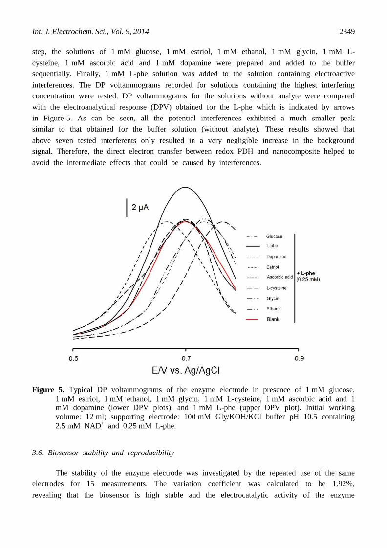
sequentially. Finally, 1 mM L-phe solution was added to the solution containing electroactive
interferences. The DP voltammograms recorded for solutions containing the highest interfering
concentration were tested. DP voltammograms for the solutions without analyte were compared
with the electroanalytical response (DPV) obtained for the L-phe which is indicated by arrows
in Figure 5. As can be seen, all the potential interferences exhibited a much smaller peak
similar to that obtained for the buffer solution (without analyte). These results showed that
above seven tested interferents only resulted in a very negligible increase in the background
signal. Therefore, the direct electron transfer between redox PDH and nanocomposite helped to
avoid the intermediate effects that could be caused by interferences.
Figure 5. Typical DP voltammograms of the enzyme electrode in presence of 1 mM glucose,
1 mM estriol, 1 mM ethanol, 1 mM glycin, 1 mM L-cysteine, 1 mM ascorbic acid and 1
mM dopamine (lower DPV plots), and 1 mM L-phe (upper DPV plot). Initial working
volume: 12 ml; supporting electrode: 100 mM Gly/KOH/KCl buffer pH 10.5 containing
2.5 mM NAD+ and 0.25 mM L-phe.
3.6. Biosensor stability and reproducibility
The stability of the enzyme electrode was investigated by the repeated use of the same
electrodes for 15 measurements. The variation coefficient was calculated to be 1.92%,
revealing that the biosensor is high stable and the electrocatalytic activity of the enzyme

Int. J. Electrochem. Sci., Vol. 9, 2014
2350
immobilized on the nanocomposite does not change upon repeated use. The peak potential for
the oxidation of NADH remained similar peak although a small decrease in the peak current
has been observed after 30 days storage in the buffer, demonstrating the stability of the
device is desirable (Figure 6). This happening, decrease in the catalytic current, after 30-days
is maybe due to the leaching or denaturing of the some redox enzyme from the electrode
surface. As can be seen in Figure 6, the GO-chitosan-PDH hybrid enzyme electrode has good
operational stability on continuous polarization at 0.7 V. Therefore, the GO-chitosan-PDH
hybrid film is high stable and does not undergo fouling by the oxidation products and
interferences.
The biodevice can be easily prepared immediately as the protocol for the fabrication of
the enzyme electrode is very simple and inexpensive. The reproducibility of the PKU detector
was investigated by using ten different electrodes for the electrocatalytic oxidation of NADH.
The peak potential for the oxidation of NADH is identical in all the ten electrodes and the
variation coefficient in the peak current was calculated to be 2.84%, showing that the results
are reproducible.
Figure 6. Control chart constructed for enzyme-functionalized electrode after 30 days in 2 mM
L-phe concentration. Initial working volume: 12 ml; supporting electrode: 100 mM
Gly/KOH/KCl buffer pH 10.5 containing 2.5 mM NAD+.
3.7. Real sample analysis
One of the main problem in biosensor applications is measuring analyte in real
samples. The clinical range of L-phe in human blood and saliva are CL-phe > 500.0 µM [34, 35,
38, 39] and CL-phe > 10.0 µM respectively for people with PKU.

Int. J. Electrochem. Sci., Vol. 9, 2014
2351
The modified biosensor was applied for determining the recoveries of different
concentrations of L-phe in human fluids (blood and saliva) by standard addition methods to
evaluate the feasibility of the PKU biosensor for possible clinical applications. Real samples was
obtained from Shariati hospital. The extract was then diluted with 0.1 M Gly/KOH/KCI buffer
containing 0.02 g NAD+ (pH 10.5). Then, the 10 diluted extraction with different L-phe spiked
concentrations of 0.5, 1.0, 3.0, and 10.0 mM (in blood) and 0.0, 10.0, 20.0, 50.0 and 100.0
µM (in saliva) were analyzed by the biosensor. The results were shown in Tables 2. The
recovery was in the range of 96.82–107.83% and the relative standard deviation (RSD%) was
in the range of 1.52–6.2%. The results showed that the developed PKU sensor can be initially
applied for monitoring PKU in biological samples (blood and saliva). Therefore, this wide linear
sensing range provides possibilities for new non-invasive biosensing protocol in which L-phe
concentrations from blood and saliva could be determined simultaneously.
Table 2. Determination and recovery results of L-phe in the human blood and saliva samples
by the biosensor based on GO-chitosan-PDH nanobiocomposite.
Samples The addition
content (mM)
The detection
content (mM)
RSD (%) Recovery (%)
Human blood
(150 µM)
0.0 0.192 mM 6.2 -
0.5 0.508 mM 2.4 98.64
1.0 1.24 mM 1.78 107.83
3.0 3.05 mM 3.43 96.82
10.0 10.18 mM 1.52 100.29
Human saliva
(3.5 µM)
0.0 4.1 µM 5.1 -
10 14.32 µM 2.4 106.07
20 23.25 µM 3.53 98.94
50 53.02 µM 2.57 99.1
100 104.12 µM 1.69 100.6
4. CONCLUSION
In this research, a biosensor based on a chitosan film amended with GO was
constructed and modified by covalent immobilization of the enzyme PDH on a functionalized
substrate for highly sensitive determination of L-phe. The results showed that chitosan-GO
nanocomposite can provide a unique microenvironment for the direct electrochemistry of PDH,
and the immobilized enzyme on the electrode possesses its native structure, function and
electrocatalytic activities. Enzyme conjugated onto the nanocomposite surface was adequate for
the great performance, high sensitivity and a long shelf life of the enzyme electrode. The
resulting GO-chitosan nanocomposite material brings new capabilities for electrochemical
sensors by combining the advantages of GO and chitosan composites. Compared with other

Int. J. Electrochem. Sci., Vol. 9, 2014
2352
types of L-phe biosensors, the preparation of the GO-chitosan/biocomposite modified-basal Au
electrode is simple, fast, and reproducible and this method can be used in designing a wide
range of new electrochemical biosensors.
ACKNOWLEDGEMENT
We would like to acknowledge Pasteur Institute of Iran for scientific and technologic support of this
work.
References
1. R. Matalon, K. Michals, Clin. Biochem, 24 (1991) 337.
2. B.K. Burton, L. Leviton, H. Vespa, H. Coon, N. Longo, B.D. Lundy, M. Johnson, A. Angelino, A.
Hamosh, D. Bilder, Mol Genet Metab, 108 (2013) 8.
3. W.B. Hanley, J.T.R. Clarke, W. Schoonheyt, Clin. Biochem, 20 (1987) 149.
4. W.B. Hanley, H. Demshar, M.A. Preston, A. Borczyk, W.E. Schoonheyt, J.T.R. Clarke, A.
Feigenbaum, Early. Hum. Dev, 47 (1997) 87.
5. T.T. Baby, S.S.J. Aravind, T. Arockiadoss, R.B. Rakhi, S. Ramaprabhu, Sensor. Actuat. B-Chem,
145 (2010) 71.
6. Y. Jiang, Q. Zhang, F. Li, L. Niu, Sensor. Actuat. B-Chem, 161 (2012) 728.
7. X. Kang, J. Wang, H. Wu, I.A. Aksay, J. Liu, Y. Lin, Biosens. Bioelectron, 25 (2009) 901.
8. J.-D. Qiu, J. Huang, R.-P. Liang, Sensor. Actuat. B-Chem, 160 (2011) 287.
9. C. Shan, H. Yang, D. Han, Q. Zhang, A. Ivaska, L. Niu, Biosens. Bioelectron, 25 (2010) 1070.
10. A. Singh, G. Sinsinbar, M. Choudhary, V. Kumar, R. Pasricha, H.N. Verma, S.P. Singh, K. Arora,
Sensor. Actuat. B-Chem, 185 (2013) 675.
11. T. Gu, Y. Zhang, F. Deng, J. Zhang, Y. Hasebe, J. Environ. Sci, 23, Supplement (2011) 66.
12. K.-J. Huang, Y.-X. Miao, L. Wang, T. Gan, M. Yu, L.-L. Wang, Process. Biochem, 47 (2012)
1171.
13. K.-J. Huang, L. Wang, J. Li, T. Gan, Y.-M. Liu, Measurement, 46 (2013) 378.
14. H.D. Jang, S.K. Kim, H. Chang, K.-M. Roh, J.-W. Choi, J. Huang, Biosens. Bioelectron, 38 (2012)
184.
15. X. Jiang, Y. Chai, R. Yuan, Y. Cao, Y. Chen, H. Wang, X. Gan, Anal. Chim. Acta, 783 (2013) 49.
16. T. Kuila, S. Bose, P. Khanra, A.K. Mishra, N.H. Kim, J.H. Lee, Biosens. Bioelectron, 26 (2011)
4637.
17. S. Liu, J. Tian, L. Wang, Y. Luo, W. Lu, X. Sun, Biosens. Bioelectron, 26 (2011) 4491.
18. C. Ruan, T. Li, Q. Niu, M. Lu, J. Lou, W. Gao, W. Sun, Electrochim. Acta, 64 (2012) 183.
19. J.-Y. Sun, K.-J. Huang, S.-F. Zhao, Y. Fan, Z.-W. Wu, Bioelectroch, 82 (2011) 125.
20. K. Wang, Q. Liu, Q.-M. Guan, J. Wu, H.-N. Li, J.-J. Yan, Biosens. Bioelectron, 26 (2011) 2252.
21. P. Wu, Q. Shao, Y. Hu, J. Jin, Y. Yin, H. Zhang, C. Cai, Electrochim. Acta, 55 (2010) 8606.
22. H. Xu, H. Dai, G. Chen, Talanta, 81 (2010) 334.
23. M. Zheng, F. Gao, Q. Wang, X. Cai, S. Jiang, L. Huang, F. Gao, Mater. Sci. Eng. C, 33 (2013)
1514.
24. Y. Zhou, H. Yin, X. Meng, Z. Xu, Y. Fu, S. Ai, Electrochim. Acta, 71 (2012) 294.
25. X. Bai, G. Chen, K.-K. Shiu, Electrochim. Acta, 89 (2013) 454.
26. L. Cui, J. Zhu, X. Meng, H. Yin, X. Pan, S. Ai, Sensor. Actuat. B-Chem, 161 (2012) 641.
27. M. Cui, B. Xu, C. Hu, H.B. Shao, L. Qu, Electrochim. Acta, 98 (2013) 48.
28. R. Devi, S. Relhan, C.S. Pundir, Sensor. Actuat. B-Chem, 150 (2009) 89.
29. Y. Zhang, Y. Liu, Z. Chu, L. Shi, W. Jin, Sensor. Actuat. B-Chem, 176 (2013) 978.

Int. J. Electrochem. Sci., Vol. 9, 2014
2353
30. Y.-Q. Zhang, Y.-J. Fan, L. Cheng, L.-L. Fan, Z.-Y. Wang, J.-P. Zhong, L.-N. Wu, X.-C. Shen, Z.-
J. Shi, Electrochim. Acta, 104 (2013) 178.
31. X.-H. Zhou, L.-H. Liu, X. Bai, H.-C. Shi, Sensor. Actuat. B-Chem, 181 (2013) 661.
32. W. Wen, W. Chen, Q.-Q. Ren, X.-Y. Hu, H.-Y. Xiong, X.-H. Zhang, S.-F. Wang, Y.-D. Zhao,
Sensor. Actuat. B-Chem, 166–167 (2012) 444.
33. G. Yang, J. Cao, L. Li, R.K. Rana, J.-J. Zhu, Carbon, 51 (2013) 124.
34. S.M. Naghib, M. Rabiee, E. Omidinia, P. Khoshkenar, Electroanal, 24 (2012) 407.
35. S.M. Naghib, M. Rabiee, E. Omidinia, P. Khoshkenar, D. Zeini, Int. J. Electrochem. Sci, 7 (2012)
120.
36. W.S. Hummers, R.E. Offeman, J. Am. Chem. Soc, 80 (1958) 1339.
37. J. Wang, Analytical Electrochemistry, Third Edition ed., Wiley-VCH, New Jersey (2006).
38. D.J. Weiss, M. Dorris, A. Loh, L. Peterson, Biosens. Bioelectron, 22 (2007) 2436.
39. R. Villalonga, A. Fujii, H. Shinohara, S. Tachibana, Y. Asano, Sensor. Actuat. B-Chem, 129 (2008)
195.
© 2014 The Authors. Published by ESG (www.electrochemsci.org). This article is an open access
article distributed under the terms and conditions of the Creative Commons Attribution license
(http://creativecommons.org/licenses/by/4.0/).


![Applied electrochemical biosensor based on covalently self ... · PDF fileAuto lab Potentiostat/Galvanostat, ... tremely corrosive and must be handled carefully]) ... Electrochemical](https://img.dokumen.tips/doc/110x75/5abe0b0e7f8b9a5d718c7cf7/applied-electrochemical-biosensor-based-on-covalently-self-lab-potentiostatgalvanostat.jpg)
![1 New Micro - and Nanotechnologies for Electrochemical ... · 2 1 New Micro- and Nanotechnologies for Electrochemical Biosensor Development Since their discovery in 1991 [5] , CNTs](https://img.dokumen.tips/doc/110x75/5f0751ff7e708231d41c66c9/1-new-micro-and-nanotechnologies-for-electrochemical-2-1-new-micro-and-nanotechnologies.jpg)