Embed Size (px)
Citation preview

Contents lists available at ScienceDirect
Biosensors and Bioelectronics
journal homepage: www.elsevier.com/locate/bios
An electrochemical biosensor based on Hairpin-DNA modified goldelectrode for detection of DNA damage by a hybrid cancer drug intercalationKatherine Lozano Untiverosa,b, Emanuella Gomes da Silvaa, Fabiane Caxico de Abreua,Edeildo Ferreira da Silva-Júniorc, João Xavier de Araújo-Juniorc, Thiago Mendoça de Aquinoa,Stephanie M. Armasb, Ricardo Olímpio de Mourad, Francisco J.B. Mendonça-Juniord,Vanessa Lima Serafimd, Karin Chumbimuni-Torresb,⁎
a Chemistry and Biotechnology Institute (IQB), Federal University of Alagoas (UFAL), Campus A.C. Simões, Tabuleiro dos Martins, Maceió, AL, 57072-970, BrazilbUniversity of Central Florida. NanoBioelectrochemistry Laboratory, Department of Chemistry, University of Central Florida, 4000 Central Florida Blvd., Orlando, FL32816, United StatescNursing and Pharmacy School (ESENFAR), Federal University of Alagoas (UFAL), Campus A.C. Simões, Tabuleiro dos Martins, Maceió, AL, 57072-970, Brazild Laboratory of Synthesis and Drug Delivery, State University of Paraíba (UEPB), Campus V, 58071-160 João Pessoa, PB, Brazil
A R T I C L E I N F O
Keywords:Stem-loop DNA (SL-DNA)Double-stranded DNA biosensorDNA damageHybrid acridine-thiophene anticancer drugGold electrode
A B S T R A C T
An efficient and new electrochemical biosensor for detection of DNA damage, induced by the interaction of thehybrid anti-cancer compound (7ESTAC01) with DNA, was studied by differential pulse voltammetry (DPV). Thebiosensor consists of a Stem-Loop DNA (SL-DNA) probe covalently attached to the gold electrode (GE) surfacethat hybridizes to a complementary DNA strand (cDNA) to form a double-stranded DNA (dsDNA). The inter-action and DNA damage induced by 7ESTAC01 was electrochemically studied based on the oxidation signals ofthe electroactive nucleic acids on the surface of the GE by DPV. As a result, the SL-DNA/GE and dsDNA/GE weretested with the reduced 7ESTAC01, showing the voltammetric signal of guanine and adenine, increase in thepresence of 7ESTAC01. Under optimum conditions, the dsDNA/GE biosensor exhibited excellent DPV responsein the presence of 7ESTAC01. The bonding interaction between 7ESTAC01 and calf thymus DNA (ctDNA) wasconfirmed by UV–Vis absorption spectroscopy, dynamic simulations (performed to investigate the DNA structureunder physiological conditions), and molecular docking. Theoretical results showed the presence of hydrogenbonding and intercalation in the minor groove of DNA, involving hydrophobic interactions.
1. Introduction
Deoxyribonucleic acid (DNA) is considered the building block forgenetic information. As such, DNA is also susceptible to chemicalmodifications via oxidation/reduction pathways, or interaction withsmall molecules (Arnold et al., 2015). Electrochemical detection of DNAinteraction with small molecules has also been applied in the design ofnovel pharmaceutical drugs (Vyskočil et al., 2010; Aydoğdu et al.,2014).
A variety of small molecules are known to interact with DNA non-covalently through (i) groove binding interactions, (ii) electrostaticinteractions, or (iii) intercalations between the stacked base pairs of thedouble-stranded DNA (Kovacic and Wakelin, 2001; Kalanur et al.,2009). Vibrational spectroscopy, fluorescence spectroscopy, surfaceplasmon resonance and nuclear magnetic resonance are just a few of thetechniques employed to investigate the binding modes, thermodynamic
properties and DNA affinity to these small molecules (Rauf et al., 2005).Unfortunately, these techniques mostly address the issues of structuralanalysis and binding mechanisms, rather than investigating DNA da-mage and its impact. Electrochemical biosensors, on the other hand,have been efficiently used to monitor the production of DNA damagevia small molecule interaction (Lucarelli et al., 2004; Vyskočil et al.,2010). An electrochemical DNA biosensor is preferred due to the highlyconducting ability provided by π-stack of nitrogenous bases, versatilityto optimize DNA immobilization on an electrode surface, and ability todetermine DNA damage induced by drug intercalation or via drug-in-duced oxidative stress (Labuda et al., 2009; Nepali et al., 2014; Arnoldet al., 2015; Huang et al., 2016).
Oxidation of electroactive nucleic acids have been used to monitorDNA lesions on modified gold, glassy carbon and mercury electrodes(Paleček et al., 1998; Li et al., 2010; Ibañez et al., 2015). Benvidi et al.employed Au-thiol chemistry to covalently bind a stem-loop (SL)-DNA
https://doi.org/10.1016/j.bios.2019.02.071Received 18 December 2018; Received in revised form 14 February 2019; Accepted 28 February 2019
⁎ Corresponding author.E-mail address: [email protected] (K. Chumbimuni-Torres).
Biosensors and Bioelectronics 133 (2019) 160–168
Available online 14 March 20190956-5663/ © 2019 Published by Elsevier B.V.
T
probe and 6-mercapto-1-hexanol (MCH) to form self-assembled mono-layer on gold surfaces (Benvidi et al., 2015). MCH played a significantrole in the overall optimized response of the DNA biosensor by avoidingnon-specific DNA adsorption and adjusting the interfacial electrontransfer on the electrode (Mills et al., 2017; McEwen et al., 2009). AStem-Loop DNA (SL-DNA) structure offers higher thermodynamic sta-bility when compared to a linear DNA structure. That stability could beexplained by the presence of the hairpin loop with a reduced negativecharge, which reduces the non-specific binding at the loop withoutcompromising the first binding on the stem (Nguyen and Wilson, 2009).
The hybrid drug, a combination of two pharmacophores, has thepotential to improve binding affinity, selectivity, and synergic activitytowards nucleic acids (Goodell et al., 2006; Cholewiński et al., 2011;Nepali et al., 2014; Harbinder et al., 2017). Recent research has alsoinvestigated the amplification of oxidative stress in relation to DNAdamage caused by sulfur, thiophene, triazole and acridine moieties(Brett et al., 2003; Pontinha et al., 2013; Sazhnikov et al., 2013; Nohet al., 2015; Deng et al., 2017). Acridine derivatives are highly inter-esting chemotherapeutic agents, which are linked to different phar-macophores in order to modify reactivity. Modifications of substituentgroups in acridine derivatives have been found to further enhance theanti-cancer drug efficacy (Putic et al., 2010; Lafayette et al., 2013).
Here, we developed a highly sensitive electrochemical biosensorbased on an SL-DNA probe that can detect DNA damage via hybriddrug, 7ESTAC01 interaction. 7ESTAC01 is composed of two anti-cancerpharmacophores, acridine, and thiophene. A complementary DNAstrand (cDNA) was introduced to hybridize the SL-DNA probe to form adouble-stranded DNA (dsDNA) biosensor. The optimization of SL-DNA/GE and dsDNA/GE modified electrode for sensitive detection of DNAdamage was assessed by DPV and cyclic voltammetry (CV).
The present work involves two sections. First, we investigated theelectrochemical oxidation of SL-DNA probe and dsDNA on the surfaceof the gold electrode induced by the presence of 7ESTAC01 to detectDNA damage by DPV. Second, the interaction of 7ESTAC01 with DNAwas analyzed via UV–Vis absorption spectroscopy, in-silico dynamicsimulations, and molecular docking.
2. Experimental section
2.1. Chemicals and reagents
All solutions were prepared with Milli-Q water using a Siemens
PURELAB Ultra system (Lowell, USA). The immobilization buffer (IB)contains 50mM Sodium Phosphate (Monobasic/Dibasic), 250mM NaClat pH 7.4. Hybridization buffer (HB) contains 50mM Tris-HCl, 25mMNaCl, 50mM MgCl2 at pH 7.4. Stock solutions of 1mM 7ESTAC01 wereprepared in HB and were evaluated in acetate solution at differentconcentrations. 1.0M Acetate buffer solution at pH 4.2 was used for theDNA oxidation in presence of the 7ESTAC01. The Tris-HCl buffer wasused for UV–Vis measurements and contains 50mM NaCl, 5 mM, Tris-HCl at pH 7.2. The pH was adjusted with either NaOH or HCl solution.The calf thymus DNA (ctDNA) was prepared from dissolution of12mgmL−1 in acetate buffer (pH 4.2; 1.0M). This stock solution waskept at 8 °C for 24 h and stirred at certain intervals to ensure homo-geneity of the final DNA solution. Trizma hydrochloride (Tris-HCl), tris(2-carboxyethyl) phosphine hydrochloride (TCEP), sodium phosphatedibasic (Na2HPO4), sodium phosphate monobasic dihydrate(NaH2PO4·2H2O), 6-mercapto-1-hexanol (MCH) and magnesiumchloride (MgCl2) were purchased from Sigma-Aldrich. Sodium chloride(NaCl), potassium chloride (KCl), sodium hydroxide (NaOH) and sul-furic acid (H2SO4) were obtained from Fisher Scientific (Pittsburgh,USA).
Gold electrodes (GEs) were purchased from CH Instruments (Austin,USA). Alumina slurry (1.0, 0.3 and 0.05 µm) was obtained from Buehler(Lake Bluff, USA). SL-DNA Probe, modified with a methylene-blue(MeB) redox marker (SL-DNA-MeB, 5´ -C6-S-S-TC GCG ACA TAC AATAGA TCG CG-MeB-3′), and complementary DNA (cDNA, 5´-CGA TCTATT GTA TGT TAA CG −3′) were obtained from BiosearchTechnologies, Inc. (Petaluma, USA) and used as received.
2.2. Synthesis of 7ESTAC01
The synthesis of 7ESTAC01 was synthesized at the Laboratory ofSynthesis and Drug Delivery (LSVM) at the State University of Paraiba,Brazil. In short, as represented in Fig. 1A, the compound was obtainedby reacting acridine-9-carboxaldehyde with 2-amino-thiophene moietypreviously obtained via the classic Gewald reaction (Gewald, 1965;Gewald et al., 1966). Subsequent purification steps and recrystallizationin ethanol yielded 7ESTAC01 as a red powder, with a melting point of155–158 °C. Comparison of physical-chemical characteristics andspectral data confirms the achievement of the hybrid.
Fig. 1. Electrochemical characterization of 7ESTAC01 A) Chemical mechanism of synthesis of 7ESTAC01: Acridine-9-carboxaldehyde with 2-aminothiophene. B)Cyclic voltammogram of 10mM 7ESTAC01 in mixture of pH 7.2, aqueous phosphate buffer and 20% DMF at a GE, with an Ag/AgCl reference electrode (RE), and thescan rate 0.1 V s−1, E= potential, V= volt, A= ampere. Cyclic voltammogram of 10mM 9-aminoacridine (B, inner graph*). (*) Red line represents the first scan.The green line represents the second scan with the potential range adjusted only to get the peak already registered on the first scan. (For interpretation of thereferences to color in this figure legend, the reader is referred to the web version of this article).
K. Lozano Untiveros, et al. Biosensors and Bioelectronics 133 (2019) 160–168
161

2.3. Preparation and immobilization of SL-DNA/GE and dsDNA/GE
GEs were used as substrate for SL-DNA probe and dsDNA im-mobilization. GEs were cleaned in a piranha solution (1:3 ratio of H2O2:H2SO4) and then polished on a micro cloth with 1.0 µm, followed by 0.3and 0.05 µm alumina slurry. The GEs were then sonicated in water andethanol to remove any residual alumina particles trapped at the surfaceof the electrode. The GE was activated in 0.5M H2SO4 via CV from+0.1 to + 1.6 V at a scan rate of 0.5 V s−1.
The SL-DNA probe was immobilized on the electrode surface via agold-thiol bond. The disulfide bonds of the SL-DNA probe were reducedwith 1mM TCEP by shaking the solution at room temperature for 1 h.The solution was then diluted with IB to yield 0.1 µM and 1.0 µM of theSL-DNA probe. 15 µL of this solution was drop casted onto the electrodeand incubated at room temperature for 30min. The electrodes wererinsed with IB and dried with nitrogen. To minimize nonspecific ad-sorption on the electrode surface, 15 µL of 2mM MCH in IB wasdropcasted on the electrode and incubated for 30min. Then, the elec-trodes were rinsed with IB and dried with nitrogen. For dsDNA/GEpreparation, SL-DNA modified GE was hybridized by dropcasting 15 µLof 50 nM cDNA in HB for 1.5 h at room temperature, which formeddsDNA at the surface of GE. Following hybridization, the GEs wererinsed using HB and then dried with nitrogen.
2.4. Optimization of 7ESTAC01 concentration and experimental timing
The protocol for analysis for the proposed biosensors consists of thefollowing steps: (i) measurement of 7ESTAC01 signal in acetate bufferpH=4.2; (ii) measurement of guanine and adenine signal obtainedfrom SL-DNA/GE and dsDNA/GE modified electrode before the inter-action with 7ESTAC01; (iii) measurements of guanine and adeninesignal after interaction between 7ESTAC01 and DNA/GE (7ESTAC01-DNA complex). The optimization of 7ESTAC01 concentration wasperformed at various final concentrations of 10 µM, 100 µM, and400 µM. Intercalation time of the compound with the SL-DNA/GE anddsDNA/GE was varied from 1 h, 2 h and 24 h.
2.5. Electrochemical measurements
Electrochemical measurements were performed with a CHI660DElectrochemical workstation (CH Instruments, USA) at room tempera-ture. A typical 3-electrode system was used where the GE served as theworking electrode, a platinum wire was used as the counter electrode(CE), and Ag/AgCl (3M KCl) was used as a reference electrode (RE).The electrochemical characterization of 7ESTAC01 was investigatedusing CV in 0.2M Phosphate buffer (pH=7.2) and 20%Dimethylformamide (DMF) in nitrogen saturated solutions (the solu-bility of the hybrid compound in DMF gave better solubility for theanalyses in protic media). CVs of 10mM 7ESTAC01 were recorded from−1.2 to + 1.0 V vs. Ag/AgCl at scan rates of 0.1 V s−1.
CV was performed to analyze the electrochemical behaviour of SL-DNA probe and dsDNA immobilized on the GE and were recorded from0.1 to 800 V s−1 scan rates. The Optimization of SL-DNA/GE anddsDNA/GE focused on the analysis of the signal of suppression (% SS)before and after the hybridization with the cDNA. The % SS was cal-culated using the equation (Eq. (1)) (Lai et al., 2013) as follows:
= ×I I I% SS ( )/ 1000 0 (1)
where I, is the current obtained upon hybridization with the cDNA andI0 is the current obtained of the immobilized SL-DNA. We analyzed %SS of 0.1 µM and 1.0 µM SL-DNA probe concentration with a fixed50 nM cDNA to obtain the dsDNA/GE biosensor.
The electro-oxidation of 7ESTAC01 and detection of DNA damage
were conducted after the interaction of 7ESTAC01 with the SL-DNA/GEand dsDNA/GE using DPV. The oxidation of 7ESTAC01 and simulta-neous determination of DNA damage was performed in acetate buffer atpH 4.2 using oxidation potentials from 0 to +1.6 V; 0.05 V amplitude;0.0167 s sample width; 0.5 s pulse period and 2 s quiet time. All theintercalation measurements of 7ESTAC01 and DNA/GE biosensors weredone with 7ESTAC01 in solution. The reduction of 7ESTAC01 wasperformed in acetate buffer at a potential range from 0.0 to −0.122 Vfor 100 s before the oxidation process.
2.6. Interaction of ctDNA with 7ESTAC01 by UV–Vis spectroscopic andmolecular docking studies
UV–Vis absorption spectroscopy was performed at a fixed 20 μM7ESTAC01 while varying ctDNA from 0 to 20 μM. The concentration ofthe stock solution of ctDNA (0.38mM per nucleotide) was determinedby UV absorption, using a molar extinction coefficient of 6600M−1
cm−1 at 260 nm. A ratio> 1.8 at A260/A280 was obtained as indicativethat DNA was sufficiently free of proteins. The intrinsic binding con-stant (Kb) of the compounds with ctDNA was calculated according toWolfe-Shimer equation (Eq. (2)) (Sirajuddin et al., 2013) as follows:
= +DNA DNAK
[ ]( )
[ ]( )
1( )a f b f b b f (2)
where [DNA] is the concentration of DNA per nucleotides, a is themolar absorption coefficient of the complex at a given DNA con-centration (Aobs. / [Compound]), f is the molar absorption coefficientof the complex in free solution, and b is the molar absorption coeffi-cient of the complex when fully bound to DNA. A plot of equation (Eq.(2)) allows the determination of the intrinsic binding constant Kb, ob-tained by the linear data fit. The value of constant was calculated as theratio between the slope and the intercept.
All molecular dynamic and Density Function Theory (DFT) calcu-lations were performed in agreement with Silva et al., (2016, 2017).The coordinates for building the molecular model were extracted fromthe X-ray crystal structure of the ctDNA dodecamer d(CGCGAATTCGCG) (PDB entry: 1BNA). Gold v.5.4 software from CambridgeCrystallographic Data Centre (CCDC) was utilized to perform all mo-lecular docking studies (Huang et al., 2013). Initially, all hydrogenswere added into the DNA structure and, then, 7ESTAC01 (ligand) wasintroduced into space. Different genetic algorithms (GA) were appliedto find the best score function for the ligand. The GoldScore, Chem-Score, Piecewise Linear Potential (ChemPLP), and Astex StatisticalPotential (ASP) functions were employed to obtain the best 10 bindingposes for the ctDNA-7ESTAC01 complex. All search coordinates weremanually introduced, as x: 1.389, y: − 1.149, and z: − 7.376 Å (Muraliet al., 2017).
After the docking calculations, the DFT calculations were performedusing quantum mechanics (QM) models from the Spartan'14 program todetermine the corrected free binding energy (ΔG) for the ctDNA-7ESTAC01 complex. The potential of intercalation of 7ESTAC01 wasinvestigated by theoretical methods. The optimized geometries of thiscompound's ability to interact with the ctDNA were taken from dockinganalysis. In addition, the coordinates of the ctDNA structure were takenfrom the crystal structure (PDB ID: 1BNA), and the ligand and watermolecules were removed. The binding energy of the DNA/ligandcomplex was calculated by applying the M06/6-31G (d) basis set. TheM06 method employed the global hybrid functional, which is the topperformer within the 6 functionals of the main group, thermochemistry,kinetics and non-covalent interactions. Moreover, frequency calcula-tions were performed to confirm the nature of the stationary point atthe same level. QM binding energies were obtained applying the
K. Lozano Untiveros, et al. Biosensors and Bioelectronics 133 (2019) 160–168
162

following formula, in which the free binding energy of Gibbs (ΔG) wascalculated as the difference between the energy of the complex (EDNA-ligands) and the sum of the ctDNA (EctDNA) and ligand (Eligand) energiesbased on the equation (Eq. (3)).
= +G E E E[ –( )]DNA ligands ctDNA ligand (3)
The final energy of the optimized structure was improved by in-cluding the single point energy from the 6-31G(d) basis set unscaledzero-point energy (ZPE) and thermal corrections (at 298.15 K and1 atm) estimated at the same level of theory, using the Spartan'14program. All these protocols were performed exactly as described bySilva-Júnior et al. (2017).
3. Results and discussion
3.1. Electrochemical characterization of 7ESTAC01
The electrochemical characterization of acridine-9-carboxaldehydewith 2-aminothiophene derivative designed as 7ESTAC01 (Fig. 1A) wasinvestigated using CV on the GE in a mixture of phosphate buffer (pH7.2) and 20% DMF in nitrogen saturated solutions. CVs were registeredin the range from −1.8 V to +1.0 V (Fig. 1B). Fig. 1B shows the re-duction of 7ESTAC01 displaying two waves. The first one exhibits aquasi-reversible reduction peak at EIc =−0.38 V vs. Ag/AgCl with apeak separation potential (ΔE=Ea-Ec) of 150mV. The second onedisplays an irreversible reduction peak at EIIc =−0.71 V. In the sameway, 9-aminoacridine, which was synthesized containing only acridine,was examined in the same potential range from −1.8 V to + 1.0 V(Fig. 1B, inner graph). The first cathodic potential for the 9-aminoa-cridine showed a single peak at EIc=−1.2 V, Fig. 1B (inner graph). It isimportant to mention that no wave was registered in the oxidationpotential range for the 9-aminoacridine. Thus, only the reduction po-tential range was further studied.
The reduction of 7ESTAC01 showed the first cathodic peak at EIc=−0.38 V, which was lower than the 9-aminoacridine alone. Thisbehaviour hinted a possible synergic activity of 7ESTAC01 due to thesignificant shift of the first cathodic peak potential. It is worth men-tioning that most of the bioactive compounds are recorded at a lessnegative potential of − 0.5 V (Bouffier et al., 2012; Dogan-Topal et al.,2014; Nepali et al., 2014; Noh et al., 2015). Thus, 7ESTAC01 representsa promising anti-cancer drug candidate; even though additionalbioactivity tests must still be performed.
3.2. Characterization and optimization of SL-DNA/GE and dsDNA/GE
SL-DNA probe contains a thermodynamically stable structure andwas used due of its capacity to detect specific site interaction (Nguyenand Wilson, 2009). Both SL-DNA and dsDNA structures were proposedand evaluated, as shown in Scheme 1. The SL-DNA probe, modifiedwith MeB redox marker, was covalently attached to the gold electrode(GE) via a thiol bond (Scheme 1). The cDNA was added to hybridize theSL-DNA probe to form a dsDNA/GE (Scheme 1B). Here, CV was used toevaluate the electrochemical characterization, and subsequently opti-mization of SL-DNA and dsDNA immobilized on the surface of the GE(see Fig. S1 in the Supplementary information). Cyclic voltammogramsof the SL-DNA probe in Fig. S1A and Fig. S1B show a high current forMeB due to the close proximity of the redox marker (MeB) to theelectrode's surface in the SL-DNA configuration (Scheme 1), since itprovides an efficient electron transfer. On the other hand, after hy-bridization with cDNA, the MeB is distant from the surface of theelectrode (Scheme 1), consequently decreasing the current of MeB (Fig.S1A and B).
The signal suppression (% SS) as a surface coverage was furtherevaluated based on equation (Eq. (1)). The % SS of the dsDNA/GE
based on 0.1 µM SL-DNA probe upon hybridization of cDNA, increasedin relation to the scan rate, and plateaued around 94% at 100 V s−1
(Fig. S1C). However, when the concentration was at 1.0 µM SL-DNAprobe, the %SS reached its maximum at 92% and 600 V s−1. This showsthat the proximity of the MeB redox marker supported efficient electrontransfer at significantly high scan rates (Fig. S1D). 1.0 µM SL-DNAprobe was used to further characterize and optimize the SL-DNA/GEand dsDNA/GE biosensors.
3.3. Electrochemical behaviour of SL-DNA/GE and dsDNA/GE byDifferential Pulse Voltammetry
The oxidative DNA damage could be induced in two ways (i) byelectro-oxidation and (ii) via oxidizing agents that interact narrowlywith DNA (Cadet and Wagner, 2013). Since DNA oxidation is the pro-cess of oxidative injury; in this experiment, the behaviour of SL-DNA/GE and dsDNA/GE was assessed only under electro-oxidation andwithout the presence of an oxidizing agent (7ESTAC01).
The electrochemical response for SL-DNA/GE and dsDNA/GE werefirst studied by DPV in acetate buffer at pH 4.2. The modified GE wasused for the electrochemical oxidation of adenine (A) and guanine (G).As shown in Fig. 2A, both types of DNA modified GE exhibited peakcurrents at 1.04 V due to the guanine bases. No adenine peaks wereobserved for either sensor, which could be explained by the stability ofthe stem-loop DNA structure through the adenine electro-oxidation(Wei et al., 2011). Otherwise, the electro-oxidation process for modifiedGE reported oxidation peaks between +0.85 V to +0.96 V for guanine(Barman and Jasimuddin, 2014). The DPV peak potentials of the gua-nosine on the ssDNA and dsDNA in acetate buffer reported by Oliveiraand Oliveira-Brett (2010) were in agreement with our values. Thoseresults showed that the electrochemical current signal of SL-DNA probeand dsDNA on the GE were mainly attributed to the electrochemicaloxidation of guanine bases, which was expected because guanine is themost readily oxidized of the DNA bases (McEwen et al., 2009). Simi-larly, the DPV peak current of guanine on SL-DNA/GE and dsDNA/GEwere further evaluated. As indicated in Fig. 2A (inset), SL-DNA/GE, anddsDNA/GE showed a reproducible guanine peak current at 7.94 and3.86 µA, respectively. The blank response of the GE does not show anypeak. All measurements were conducted in triplicates. The S.D of theguanine peak current on the SL-DNA/GE and dsDNA/GE were 0.14 µAand 0.24 µA, respectively (Fig. 2A, inner graph).
The influence of the scan number to the oxidative DNA damage wassubsequently examined as an indicator of consecutive DNA lesionsunder electro-oxidation. DNA oxidation is the process of oxidative in-jury, so it is expected to see higher oxidative damage to the DNA as thenumber of scans increases. Fig. 2B, prove this hypothesis, wherein theoxidation current of guanine increases with the number of scans, in-dicating that the electron donor was DNA itself.
Furthermore, for the dsDNA/GE, the presence of the intermediateform of 8-oxoguanine (8-oxoG) at E=+0.25 V is a reliable indicatorthat the oxidation of guanine is taking place (Fig. 2B) from the secondscan onwards. In addition, the guanine oxidation peak current increasesas the oxidative DNA damage increases for dsDNA/GE.
3.4. Optimization of the concentration of 7ESTAC01 and detection of DNAdamage
The electro-oxidation processes involved in purine DNA bases aresimilar to those affecting enzymatic oxidation. For this reason, theelectro-oxidation of the DNA immobilized on the GE was applied todetect DNA damage through the interaction with the oxidizing com-pound (7ESTAC01). The oxidative DNA damage was induced byelectro-oxidation in the presence of the given 7ESTAC01. A reductionpotential was applied to 7ESTAC01 to obtain 7ESTAC01 radicals to
K. Lozano Untiveros, et al. Biosensors and Bioelectronics 133 (2019) 160–168
163

Scheme 1. Schematic representation the fabrication the Hairpin-DNA modified Gold electrode (GE). A) Schematic of the assay procedure of the SL-DNA probe anddsDNA biosensor at GE B) SL-DNA probe and dsDNA structures. SL-DNA probe, 5´-C6-S-S-TC GCG ACA TAC AAT AGA TCG CG-MeB-3′. The number of Guanines (G).The number of adenines (A). 6-mercapto-1-hexanol (MCH). Complementary DNA (cDNA). Methylene-blue (MeB).
Fig. 2. Electrochemical behaviour of SL-DNA/GE and dsDNA/GE under electro-oxidation by DPV. (A) Comparison of DPV signals between SL-DNA probe (curve a)and dsDNA (curve b) and blank response of GE (GE/blank). Histogram graph represents the guanine peak current for SL-DNA/GE (a) and dsDNA/GE (b) modifiedelectrode (A, inner graph). (B) DPV response for dsDNA/GE from first to the fifth scan. The electro-oxidation by DPV was carried out in acetate buffer at pH 4.2 forpotentials range from 0.0 to +1.6 V; amplitude 0.05 V; sample width 0.0167 s. (For interpretation of the references to color in this figure legend, the reader isreferred to the web version of this article).
K. Lozano Untiveros, et al. Biosensors and Bioelectronics 133 (2019) 160–168
164
damage DNA. According to Abreu et al. (2002) and Dogan-Topal et al.(2014) certain anticancer drugs require the formation of short-livedradicals to interact and damage DNA.
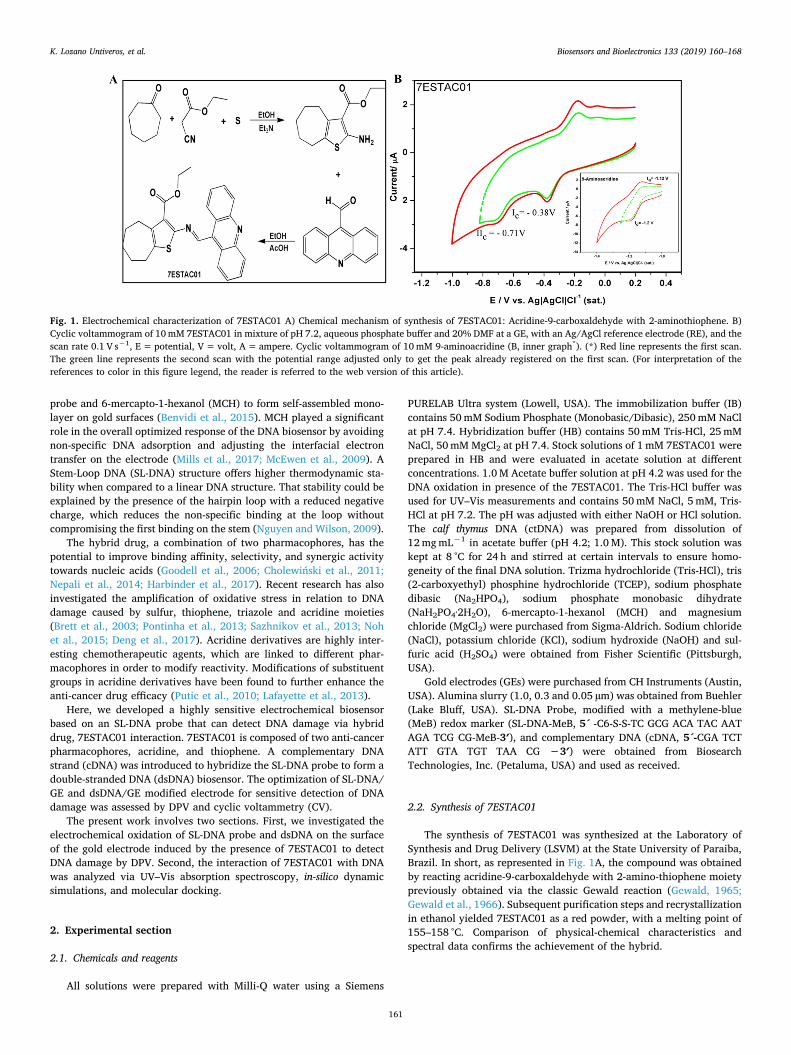
The direct determination of the oxidation of electroactive DNAbases in the presence of 7ESTAC01 were carried out by DPV from 0 to+ 1.6 V. The oxidation peak current differences between SL-DNA/GEand their corresponding SL-DNA/GE-7ESTAC01 system were furtherinvestigated in acetate buffer (pH 4.2). The reproducibility of the SL-DNA/GE was investigated in presence and absence of different con-centrations of 7ESTAC01 (Fig. 3A and B). As seen in Fig. 3A, 7ESTAC01interactions with the SL-DNA/GE were examined at different times, 1, 2and 24 h.
In general, the guanine oxidation peak current increased notoriouslyas the interaction time increased, which indicated the formation of SL-DNA/GE-7ESTAC01, increasing the electron transfer ability of the SL-DNA/GE. Another valuable data collected from Fig. 3A showed that theguanine oxidation peak current increased as the 7ESTAC01 concentra-tions increased. It is important to note that acridine, which binds to DNAby intercalation, might either donate electrons to or accept electronsfrom, the double helix, thus actively participating in electron transferreactions (Kovacic and Wakelin, 2001; Baguley et al., 2003; Nepali et al.,2014). These results support literature data of acridine and its im-portance on the electron transfer (Noh et al., 2015); showing that oxi-dation of 7ESTAC01 is facilitating the electron transfer on the SL-DNA/GE biosensor. The adenine oxidation was evaluated with the minimumrequired concentration of 7ESTAC01 (Fig. 3B). The electrochemicaloxidation of adenine followed a multiple step, six electron, six protonsoxidation (Wei et al., 2011), which implies a more demanding oxidationprocess compared to guanine. As seen in Fig. 3B, the adenine oxidationwas registered in the presence of higher concentrations of 7ESTAC01,equal to 100 µM 7ESTAC01 (Fig. 3B). Taking into account the interactiontime of 1 and 2 h under the same level of 100 µM 7ESTAC01, adeninepeak currents registered at 5.45 µA and 8.34 µA, respectively.
The reproducibility was evaluated for three different concentrationsof 7ESTAC01 with 1, 2 and 24 h to obtain the best timing conditions forthe interaction. After three successive measurements, the S.D of theguanine peak current on the SL-DNA/GE response to 10,100 and400 µM 7ESTAC01 with 1 h of interaction were 0.67 µA, 1.70 µA and1.19 µA, respectively. For the same conditions, the standard deviationsof SL-DNA/GE response to 10, 100 and 400 µM 7ESTAC01 with 2 h ofinteraction were 0.44 µA, 0.33 µA and 0.98 µA. The S.D with 24 h ofinteraction for the same concentrations showed 1.01 µA, 2.36 µA and
2.51 µA, showing an acceptable reproducibility. Nevertheless, based onbiosensor performance, the best S.D was obtained for 2 h of interaction.All further experimentation was carried out at the optimized two hoursof interaction time.
3.5. Interaction of SL-DNA/GE and dsDNA/GE with 7ESTAC01 anddetection of DNA damage
All the following experiments were carried out for different con-centrations of 7ESTAC01 with 2 h of interaction time. As depicted inFig. 4, after the addition of 7ESTAC01, the strong binding interactionbetween 7ESTAC01 with guanine and adenine base of SL-DNA/GE anddsDNA/GE occurred. Therefore, as the concentration of 7ESTAC01 in-creases, the oxidation peak current of SL-DNA probe and dsDNA in-creased (Fig. 4A and B). These results are similar to those obtained byLucarelli et al. (2002). Lucarelli and co-workers utilized screen-printedelectrodes for the detection of apolipoprotein E, where an increase ofthe electrochemical signal of the guanine base resulted from the non-specific interaction of the apolipoprotein E and the DNA immobilizedon the electrode. The increasing oxidation peak current we report hereis consistent with other electrochemical DNA/GE biosensors testing aredox-active intercalator, such as in the case of anthraquinone monosulfonic acid (AQMS) (Wong and Gooding, 2006). Electron transferfrom the DNA to AQMS intercalated into DNA duplexes reported thegrowth of the peak current signal with time. These last results supportthe electrochemical behaviour of our biosensors in the presence of7ESTAC01, which is intercalated into the double-stranded DNA.
To evaluate the level of DNA damage with the dsDNA/GE and SL-DNA/GE, we compared guanine and adenine peak current in the sameconditions. The adenine oxidation was recorded for SL-DNA/GE anddsDNA/GE, each yielding different sensitivity levels. The SL-DNA/GE-7ESTAC01 showed adenine oxidation for 100 µM (Fig. 3B) and 400 µM7ESTAC01 (Fig. 4A). Notoriously, an adenine peak for 100 and 400 µM7ESTAC01 (Fig. 4B, red histograms) with the dsDNA/GE-7ESTAC01,reached higher adenine peak current at 12.36 µA and 18.63 µA, re-spectively. On the other hand, as shown in Fig. 4 (blue histograms), theguanine oxidation was seen for dsDNA/GE and SL-DNA/GE with thepresence of the minimum concentration of 7ESTAC01. The guaninepeak current for 40 µM 7ESTAC01 with SL-DNA/GE and dsDNA/GE,reached 9.412 µA and 11.35 µA, respectively (Fig. 4A and B, innergraph). These results not only validated the strong interaction between7ESTAC01 and purine bases but also emphasized the higher sensitivity
Fig. 3. DPV peak currents responses under electro-oxidation on SL-DNA/GE in the presence of 7ESTAC01 to the detection of DNA damage. (A) Guanine peak currentin the presence of 7ESTAC01 at concentrations of (a) 10, (b) 100, (c) 400 µM for 1, 2 and 24 h of interaction. (B) Guanine (blue histogram on the left) and Adeninepeak current (red histogram, on the right) in the presence of 100 µM 7ESTAC01 (b) for 1 and 2 h of interaction. Histograms represent the guanine and adenine peakcurrent for SL-DNA/GE extrapolated from the DPV signal. Results were expressed as the average of three independent experiments. Error bars represent standarddeviations. (*) All the intercalation measurements were done with 7ESTAC01 in solution. (For interpretation of the references to color in this figure legend, the readeris referred to the web version of this article).
K. Lozano Untiveros, et al. Biosensors and Bioelectronics 133 (2019) 160–168
165

and more damage induced by employing the dsDNA/GE versus the SL-DNA/GE. The dsDNA and SL-DNA sequences exhibit 11 and 5 guanines,respectively. It is possible that 7ESTAC01-DNA intercalation can lead tobreaking hydrogen bonds and exposing guanine and adenine bases tothe surface of the GE. Therefore, it is likely that the higher oxidationpeak current exhibited by the dsDNA/GE-7ESTAC01 system is due to ahigher quantity of available guanine and adenine bases in comparisonto the SL-DNA/GE-7ESTAC01 system (Fig. 4A). In fact, two criticalparameters must be followed to improve the electron transfer effi-ciency, (i) type of DNA structure, and (ii) distance between guaninebase and electrode surface (Brett et al., 2003; McEwen et al., 2009;Ibañez et al., 2015). Similar results were observed in a single strandedDNA modified GE biosensor, wherein electron transfer efficiency wasthe highest when guanine bases were in close proximity and exposed tothe surface of the GE (Huang et al., 2016). In addition, the high chargemigration along the DNA duplex in the presence of a DNA intercalatorlike 7ESTAC01, can also account for the higher oxidation peak currentseen in the dsDNA platform (Liu and Barton, 2005; Elias et al., 2008).
The 8-oxoguanine (8-oxoG) is arguably the most important muta-genic lesion in DNA. The oxidation potentials are highly dependent onthe type of electrode and pH of the solution. With that in mind, it hasbeen reported an 8-oxoG oxidation peak at + 0.25 and +0.45 V, forneutral (Ferapontova, 2004) and acidic pH (Oliveira and Oliveira-Brett,2010), respectively. In the present study, the presence of the 8-oxoG atE=+0.5 V (Fig. 4B, black line) after the interaction with the highestconcentration of 7ESTAC01 (400 µM) with the dsDNA/GE biosensor,demonstrates a substantial DNA damage.
To understand the mechanism behind the observed increase in peakcurrent signal from purine bases in the presence of 7ESTAC01 radicals,another double-stranded ctDNA was immobilized by non-covalentbonding on a glassy carbon electrode (GCE) (Supplementary informa-tion). The double-stranded ctDNA is a natural DNA from calf thymuswidely used in studies of DNA binding anticancer compounds. Thedamage of the ctDNA in the presence of 7ESTAC01 radicals was in-vestigated in acetate buffer solution at pH 4.2 by DPV. Fig. S2 showsDPV peak potentials characteristic of the guanine and adenine bases atthe GCE (Li et al., 2010; Aydoğdu et al., 2014). In the presence of7ESTAC01 in solution, the peak current recorded a significant increaseof guanine and adenine bases at + 1.03 V and + 1.29 V, respectively,which could imply the opening of the double helix of ctDNA (Fig. S2).
Therefore, the increase in the signal, regardless of the electrode, in-dicates that the electron transfer through DNA increases in the presenceof the 7ESTAC01 (oxidizing agent). It demonstrates that the 7ESTAC01mechanism of intercalation into the DNA through electro-oxidationcaused a substantial distortion of the double-stranded ctDNA. It alsocorroborates the imminent breaking of the double helix DNA, exposingthe purine bases to the surface of the electrode as a result of the DPVcurrent increase in the presence of 7ESTAC01. Finally, the blank signalsupon oxidation of 7ESTAC01 were analyzed by DPV for both un-modified electrodes, GCE (Fig. S2) and the GE (Fig. S3) under the sameworking conditions. As recorded in Fig. S2 and Fig. S3, the blank re-sponse of the GE/GCE+7ESTAC01 does not show any peak.
3.6. Interaction of DNA with 7ESTAC01 by UV–Vis spectroscopy,molecular docking and density function theory (DFT) studies
Elucidating the binding between 7ESTAC01 and DNA provides helpin understanding drug-DNA interactions and consecutive DNA damageon the surface of the GE. The interaction of an anticancer drug7ESTAC01 with double-stranded ctDNA was studied using various ap-proaches like UV–Vis spectroscopy, Molecular Docking, and DensityFunction Theory (DFT) studies.
UV–Vis spectroscopy confirmed 7ESTAC01-DNA interaction.Importantly, as depicted in Fig. 5A, the presence of an isosbestic pointdeveloped at 297 nm in the 7ESTAC01-DNA spectra indicates inter-calation as a dominant binding mode. Moreover, UV–Vis showed abinding constant of Kb =6.57×10⁴ Lmol−1 at 260 nm using theWolfe-Shimer equation (Eq. (2)). The small molecules can interact withDNA involving a single mode of binding or mixed binding modes. Thus,the exact mode of interaction can be established merely by this tech-nique due to the presence of the isosbestic point; however, another kindof non- covalent interactions could be present.
Other types of non-covalent interactions were further studied byMolecular Docking and DFT studies. For these studies, the GoldScorewas selected as the best algorithm to predict results without large de-viations between generated poses. Based on this, the GoldScore func-tion was employed to determine different thermodynamic parametersrelated to the 7ESTAC01 compound (see Table S in the Supplementaryinformation). From this analysis, the most important type of interactioncan be identified and, consequently, it is possible to determine their
Fig. 4. Detection of the DNA damage product of the interaction between SL-DNA/GE and dsDNA/GE with 7ESTAC01 expressed by the DPV peak currents. DPV peakcurrents responses under electro-oxidation on SL-DNA/GE (A) and dsDNA/GE (B) in presence of 7ESTAC01 at concentrations of (a) 40, (b) 100 and (c) 400 µM for 2 hof interaction. Histograms represent the guanine and adenine peak current for SL-DNA/GE, and dsDNA/GE extrapolated from the DPV signal of each biosensor (A andB, inner graph). Error bars represent standard deviations (S.D). The S.D of the guanine peak current on the SL-DNA/GE for 40 and 400 µM 7ESTAC01 were 0.31 µAand 0.91 µA, respectively. The S.D of the guanine peak current on the dsDNA/GE for 40, 100 and 400 µM 7ESTAC01 were 0.44 µA, 0.52 µA and 1.32 µA, respectively.DPV signal in acetate buffer for SL-DNA/GE (*) and dsDNA/GE (+) without the presence of 7ESTAC01.
K. Lozano Untiveros, et al. Biosensors and Bioelectronics 133 (2019) 160–168
166

contribution in kcal mol−1 (Kamal et al., 2010; Arshad et al., 2017;Kundu and Chattopadhyay, 2017; Veerashekhar Goud et al., 2017).According to the quantitative results from the Molecular Docking andDFT studies (Table S), it is observed that significant interactions fromthe 7ESTAC01-DNA complex are the contributions of Van der Walls,46.35 kcal mol−1. In addition, it was observed that the 7ESTAC01compound presented a significant external H-bond value of 1.2 kcalmol−1. This happens due to 7ESTAC01's ability to form an H-bond withthe NH donor from the adenine base (DA18), at a distance of 2.836 Å(Fig. 5B). After DFT calculations from ctDNA and 7ESTAC01 (free andbinding), Gibbs free energy (ΔG) from the complex was low, suggestinga high affinity, showing its relation to the FitScore value, 63.44 (TableS). Therefore, binding of 7ESTAC01 analyzed via molecular docking(Fig. 5C), demonstrates that this compound could act via intercalationmechanism, considering that the aminothiophene and partially acridinerings are located between the DNA bases.
A careful analysis of the type of interaction between the hybrid7ESTAC01 and the DNA represents the success of producing oxidativedamage in the DNA. In this way, the interaction between DNA and7ESTAC01 characterized by UV–Vis spectroscopy, Molecular Docking,and DFT studies showed that the 7ESTAC01 act via an intercalationmechanism into the DNA. Besides, the low free energy of Gibbs (ΔG)calculated from molecular docking suggests an excellent affinity7ESTAC01-DNA.
4. Conclusions
Here, a novel electrochemical biosensor, divided into two config-urations, SL-DNA/GE and dsDNA/GE, was optimized and characterized
for detection of DNA damage caused by intercalation with a new hybridanti-cancer drug (7ESTAC01). DPV analysis of guanine and adeninebases presented high sensitivity and efficiency of the SL-DNA probemodified GE and the dsDNA. High intercalation between DNA and7ESTAC01 was determined to be a critical parameter to improve thesensitivity of the biosensor. DPV analysis of dsDNA in the presence ofhigh concentration of 7ESTAC01 led to the formation of 8-oxoguanine,which is considered a key indicator of a mutagenic lesion in DNA.Intercalation mechanism was demonstrated using molecular docking,DFT studies, and UV–Vis Spectroscopy. These studies showed ami-nothiophene and partially acridine rings located between DNA bases, aswell as an isosbestic point at 297 nm in the 7ESTAC01-DNA spectra,indicating the intercalation as a dominant binding mode.
The dsDNA/GE showed higher sensitivity in the presence of the7ESTAC01 due to a higher quantity of purine bases in comparison to theSL-DNA by itself. The high sensitivity of this novel biosensor allowsdetection of minimal DNA damage and can be further expanded tostudy DNA damage with many other drugs.
Supplementary information
Optimization of SL-DNA/GE and dsDNA/GE biosensor by CyclicVoltammetry, electrochemical ctDNA biosensor on the Glassy CarbonElectrode, Detection of the DNA damage product of the interaction ofctDNA/GCE and 7ESTAC01, DPV signal of 7ESTAC01 on the GoldElectrode, Molecular Docking and DFT studies for 7ESTAC01 and calfthymus DNA. This material is available free of charge via the Internet.
Fig. 5. Interaction of DNA with 7ESTAC01 by UV–Vis Spectroscopy, and Molecular docking. (A) UV–Visible absorption spectra of 20 µM 7ESTAC01 in presence ofdifferent concentrations of DNA (μM): (a) 0.0; (b)2; (c)10; (d)12; (e)14; (f)16; (g)18; (h)20. (B) Molecular docking of 7ESTAC01 (forward of the mage) to ctDNA(background structure) sites, represented by H-bond at DA18. (C) The binding pose of ctDNA (orange helix) acting via intercalation mechanism between theaminothiophene and acridine domains of 7ESTAC01. (For interpretation of the references to color in this figure legend, the reader is referred to the web version ofthis article).
K. Lozano Untiveros, et al. Biosensors and Bioelectronics 133 (2019) 160–168
167

CRediT authorship contribution statement
Katherine Lozano Untiveros: Conceptualization, Methodology,Validation, Investigation, Writing - original draft, Writing - review &editing. Emanuella Gomes da Silva: Methodology, Writing - review &editing. Fabiane Caxico de Abreu: Conceptualization, Validation,Investigation, Writing - review & editing, Supervision, Funding acqui-sition. Edeildo Ferreira da Silva-Júnior: Methodology. JoãoXavier de Araújo-Junior:Methodology. Thiago Mendoça de Aquino:Methodology, Writing - review & editing, Supervision, Funding acqui-sition. Stephanie M. Armas: Validation, Investigation, Writing - review& editing. Ricardo Olímpio de Moura: Methodology. FranciscoJ.B. Mendonça-Junior: Methodology, Writing - review & editing.Vanessa Lima Serafim: Methodology. Karin Chumbimuni-Torres:Conceptualization, Validation, Investigation, Writing - review &editing, Supervision, Funding acquisition.
Acknowledgements
This work is supported by Organization of American States (OAS)under the OAS-GCUB scholarship program (P.202.458.3000).Foundation for Research Support of the State Alagoas (FAPEAL), Brazil.NSF-CBET, United States, grant number #1706802 and FloridaDepartment of Health Grant #7ZK05.
Declaration of interests
None.
Appendix A. Supporting information
Supplementary data associated with this article can be found in theonline version at https://doi.org/10.1016/j.bios.2019.02.071.
References
Abreu, F.C., Goulart, M.O.F., Oliveira Brett, A.M., 2002. Biosens. Bioelectron. 17,913–919.
Arnold, A.C., Grodick, M.A., Barton, J.K., 2015. Cell Chem. Biol. 23, 183–197.Arshad, N., Perveen, F., Saeed, A., Channar, P.A., Farooqi, S.I., Larik, F.A., Ismail, H.,
Mirza, B., 2017. J. Mol. Struct. 1139, 371–380.Aydoğdu, G., Günendi, G., Zeybek, D.K., Zeybek, B., Pekyardımcı, Ş., 2014. Sens.
Actuators B Chem. 197, 211–219.Baguley, B.C., Wakelin, L.P.G., Jacintho, J.D., Kovacic, P., 2003. Curr. Med. Chem. 10,
2643–2649.Barman, K., Jasimuddin, Sk, 2014. RSC Adv. 4, 49819–49826.Benvidi, A., Dehghani Firouzabadi, A., Dehghan Tezerjani, M., Moshtaghiun, S.M.,
Mazloum-Ardakani, M., Ansarin, A., 2015. J. Electroanal. Chem. 750, 57–64.Bouffier, L., Gosse, I., Demeunynck, M., Mailley, P., 2012. Bioelectrochemistry 88,
103–109.Brett, A.M.O., da Silva, L.A., Fujii, H., Mataka, S., Thiemann, T., 2003. J. Electroanal.
Chem. 549, 91–99.Cadet, J., Wagner, J.R., 2013. Cold Spring Harb. Perspect. Biol. 5 (2), a012559.Cambridge Crystallographic Data Centre, 2015. GOLD User Guide A Component of the
GOLD Suite. 245. ⟨https://www.ccdc.cam.ac.uk⟩.Cholewiński, G., Dzierzbicka, K., Kołodziejczyk, A.M., 2011. Pharmacol. Rep. 63,
305–336.
Deng, Z., Hu, J., Liu, S., 2017. Macromol. Rapid Commun. 1600685.Dogan-Topal, B., Bozal-Palabiyik, B., Ozkan, S.A., Uslu, B., 2014. Sens. Actuators B Chem.
194, 185–194.Elias, B., Shao, F., Barton, J.K., 2008. J. Am. Chem. Soc. 130, 1152–1153.Ferapontova, E.E., 2004. Electrochim. Acta 49, 1751–1759.Gewald, K., 1965. Chem. Ber. 98, 3571–3577.Gewald, K., Schinke, E., Böttcher, H., 1966. Chem. Ber. 99, 94–100.Goodell, J.R., Madhok, A.A., Hiasa, H., Ferguson, D.M., 2006. Bioorg. Med. Chem. 14,
5467–5480.Harbinder, S., Jatinder, V.S., Manish, K.G., Ajit, K.S., Sahil, S., Kunal, N., Preet, M.S.B.,
2017. Bioorg. Med. Chem. Lett. 27, 3974–3979.Huang, S., Lu, S., Huang, C., Sheng, J., Zhang, L., Su, W., Xiao, Q., 2016. Sens. Actuators B
Chem. 224, 22–30.Huang, Z., Lin, K., You, Q., 2013. Bioorg. Med. Chem. Lett. 23, 4166–4171.Ibañez, D., Santidrian, A., Heras, A., Kalbáč, M., Colina, A., 2015. J. Phys. Chem. C 119,
8191–8198.Kalanur, S.S., Katrahalli, U., Seetharamappa, J., 2009. J. Electroanal. Chem. 636, 93–100.Kamal, A., Reddy, K.S., Khan, M.N.A., Shetti, R.V.C.R.N.C., Ramaiah, M.J., Pushpavalli,
S.N.C.V.L., Srinivas, C., Pal-Bhadra, M., Chourasia, M., Sastry, G.N., Juvekar, A.,Zingde, S., Barkume, M., 2010. Bioorg. Med. Chem. 18, 4747–4761.
Kovacic, P., Wakelin, L.P.G., 2001. Anti-Cancer Drug Des. 16, 175–184.Kundu, P., Chattopadhyay, N., 2017. Photobiol. B Biol. 173, 485–492.Labuda, J., Ovádeková, R., Galandová, J., 2009. Microchim. Acta 164, 371–377.Lafayette, E.A., Vitalino de Almeida, S.M., Pitta, M.G., Carneiro Beltrão, E.I., da Silva,
T.G., Olímpio de Moura, R., Pitta, I. a.R., de Carvalho, L.B., de Lima, M. o.C., 2013.Molecules 18, 15035–15050.
Lai, R.Y., Walker, B., Stormberg, K., Zaitouna, A.J., Yang, W., 2013. Methods 64,267–275.
Li, Q., Batchelor-McAuley, C., Compton, R.G., 2010. J. Phys. Chem. B 114, 7423–7428.Liu, T., Barton, J.K., 2005. J. Am. Chem. Soc. 127, 10160–10161. https://doi.org/10.
1021/ja053025c.Lucarelli, F., Marrazza, G., Turner, A.P., Mascini, M., 2004. Biosens. Bioelectron. 19,
515–530.Lucarelli, F., Marrazza, G., Palchetti, I., Cesaretti, S., Mascini, M., 2002. Anal. Chim. Acta
469, 93–99.McEwen, G.D., Chen, F., Zhou, A., 2009. Anal. Chim. Acta 643, 26–37.Mills, D.M., Calvo-Marzal, P., Pinzon, J.M., Armas, S., Kolpashchikov, D.M.,
Chumbimuni- Torres, K.Y., 2017. Electroanalysis 29, 873–879.Murali, S.R.S., Siddhardha, S.R.S., Babu, R.D., Venketesh, S., Basavaraju, R., Rao, N.G.,
2017. Acta Part A Mol. Biomol. Spectrosc. 180, 217–223.Nguyen, B., Wilson, W.D., 2009. J. Phys. Chem. B. 113, 14329–14335.Nepali, K., Sharma, S., Sharma, M., Bedi, P.M.S., Dhar, K.L., 2014. Eur. J. Med. Chem. 77,
422–487.Noh, J., Kwon, B., Han, E., Park, M., Yang, W., Cho, W., Yoo, W., Khang, G., Lee, D., 2015.
Nat. Commun. 6, 6907.Oliveira, S.C.B., Oliveira-Brett, A.M., 2010. Comb. Chem. High. Throughput Screen. 13,
628–640.Paleček, E., Fojta, M., Tomschik, M., Wang, J., 1998. Biosens. Bioelectron. 13, 621–628.Pontinha, A.D.R., Sparapani, S., Neidle, S., Oliveira-Brett, A.M., 2013.
Bioelectrochemistry 89, 50–56.Putic, A., Stecher, L., Prinz, H., Müller, K., 2010. Eur. J. Med. Chem. 45, 3299–3310.Rauf, S., Gooding, J.J., Akhtar, K., Ghauri, M.A., Rahman, M., Anwar, M.A., Khalid, A.M.,
2005. J. Pharm. Biomed. Anal. 37, 205–217.Sazhnikov, V.A., Khlebunov, A.A., Sazonov, S.K., Vedernikov, A.I., Safonov, A.A.,
Bagatur’yants, A.A., Kuz’mina, L.G., Howard, J.A.K., Gromov, S.P., Alfimov, M.V.,2013. J. Mol. Struct. 1053, 79–88.
Silva-Júnior, E., França, P., Quintas-Júnior, L., Mendonça-Junior, F., Scotti, L., Scotti, M.,de Aquino, T., Araújo-Júnior, J., 2017. Aided Drug Des. 13, 1–9.
Silva, M., Nascimiento, E.O., Edeildo, F.,M., 2017. Int. J. Biol. Macromol. 96, 223–233.Silva, M., Savariz, F.C., Silva, E.F., De Aquino, T.M., Sarragiotto, M.H., Santos, J.C.C.,
Figueiredo, I.M., 2016. J. Braz. Chem. Soc. 27, 1558–1568.Sirajuddin, M., Ali, S., Badshah, A., 2013. J. Photochem. Photobiol. B Biol. 124, 1–19.Veerashekhar Goud, E., Sivaramakrishna, A., Vijayakrishna, K., Brahmmananda Rao,
C.V.S., Khedkar, V.M., Jha, P.C., 2017. Inorg. Chim. Acta 461, 84–91.Vyskočil, V., Labuda, J., Barek, J., 2010. Anal. Bioanal. Chem. 397, 233–241.Wei, Y., Huang, Q.A., Li, M.G., Huang, X.J., Fang, B., Wang, L., 2011. Electrochim. Acta
56, 8571–8575.Wong, E., Gooding, J., 2006. Anal. Chem. 78, 2138–2144.
K. Lozano Untiveros, et al. Biosensors and Bioelectronics 133 (2019) 160–168
168


![Research Article Preparation of Electrochemical Biosensor ...chromatography (HPLC), capillary electrophoresis [ , ], mass spectrometry [ ], and thermospray-mass spectrometry []. Besides](https://img.dokumen.tips/doc/110x75/60d24d89e1e9ab12f6131bb0/research-article-preparation-of-electrochemical-biosensor-chromatography-hplc.jpg)

![1 New Micro - and Nanotechnologies for Electrochemical ... · 2 1 New Micro- and Nanotechnologies for Electrochemical Biosensor Development Since their discovery in 1991 [5] , CNTs](https://img.dokumen.tips/doc/110x75/5f0751ff7e708231d41c66c9/1-new-micro-and-nanotechnologies-for-electrochemical-2-1-new-micro-and-nanotechnologies.jpg)


![Applied electrochemical biosensor based on covalently self ... · PDF fileAuto lab Potentiostat/Galvanostat, ... tremely corrosive and must be handled carefully]) ... Electrochemical](https://img.dokumen.tips/doc/110x75/5abe0b0e7f8b9a5d718c7cf7/applied-electrochemical-biosensor-based-on-covalently-self-lab-potentiostatgalvanostat.jpg)