Embed Size (px)
Citation preview

JOURNAL OF BACTERIOLOGY, Jan. 1981, p. 583-595 Vol. 145, No. 10021-9193/81/010583-13$02.00/0
Elaboration of Cellulose Fibrils by Agrobacterium tumefaciensDuring Attachment to Carrot Cells
ANN G. MATTHYSSE,I* KATHRYN V. HOLMES,2 AND ROBIN H. G. GURLITZ'
Department of Botany, University of North Carolina, Chapel Hill, North Carolina 275141; and DepartmentofPathology, Uniformed Services University of the Health Sciences, Bethesda, Maryland 200142
The attachment of virulent strains ofAgrobacterium tumefaciens to plant cellsis the first step in the bacterial induction of tumors. Binding of A. tumefaciens tocarrot tissue culture cells occurred as a two-step process. The initial step was theattachment of the bacteria to the plant cell wall. Living plant cells were notrequired. Bacterial attachment to heat-killed or glutaraldehyde-fixed carrot cellsproceeded with only slightly altered kinetics and unaltered bacterial strain spec-ificity. After the bacteria bound to the carrot cell surface, scanning electronmicroscopy showed that fibrils developed, surrounded the bacteria, and anchoredthem to the plant cell surface. These fibrils were synthesized by the bacteria andnot by the plant cell since they were also made after the attachment of A.tumefaciens to dead carrot cells and since under some conditions the bacteriasynthesized fibrils in the absence of plant cells. Calcofluor staining, acid hydrol-ysis, enzymatic digestion studies, and infrared spectroscopy showed that thefibrils were composed of cellulose. The formation of these cellulose fibrils occurredduring the attachment of virulent strains of A. tumefaciens to plant cells in vitro.The fibrils anchored the bacteria to the plant cell surface and entrapped additionalbacteria. The multiplication of entrapped and attached bacteria resulted in theformation of large clusters of bacteria held close to the plant cell wall and plasmamembrane by cellulose fibrils. This high concentration of bacteria may facilitatetransfer of Ti plasmid deoxyribonucleic acid to the plant cell resulting in theformation of tumors.
Crown gall disease of dicotyledenous plants isthe only known example of a bacterially inducedtumor (15). The tumors result from the extra-cellular infection of a wound site by Agrobacte-rium tumefaciens (15). Virulent strains of A.tumefaciens harbor a large tumor-inducing (Ti)plasmid (34, 35, 39). During the course of infec-tion, the Ti plasmid is transferred from thebacterium to the plant host cell in which aportion of the Ti DNA is maintained and tran-scribed (4, 10, 17, 33). The mechanism of thistransfer is unknown.The first step in tumor formation in vivo is
the site-specific attachment ofA. tumefaciens tothe plant (13). We have demonstrated specificattachment of A. tumefaciens to tissue culturecells. The attachment is dependent on the pres-ence of the bacterial Ti plasmid (18). This man-uscript reports further studies of bacterial at-tachment to tissue culture cells and describesthe development of fibrils attaching the bacteriato the host cell wall. Evidence is presented thatthese fibrils are composed of cellulose elaboratedby the bacterium.
MATERIALS AND METHODS
Bacteria were grown, and viable cell counts weredetermined as previously described (18). Sources ofvirulent strains of A. tumefaciens A6, C58 and B6,avirulent strains NT1 and IIBNV6, and avirulentAgrobacterium radiobacter strains 6467, S1005, andTR1 were the same as previously listed (18). Esche-richia coli 23739 was obtained from the AmericanType Culture Collection. Suspension cultures of Dau-cus carota were obtained from Wendy Boss, NorthCarolina State University. They were maintained inMurashige and Skoog medium (20) with weekly trans-fers.
Attachment studies were carried out by adding asuspension of bacteria to the carrot cell cultures. Thebacteria had been well dispersed by Vortex mixing.The percentage of the bacterial inoculum attachedwas determined as previously described (18). A knownconcentration of bacteria was introduced into the car-rot cell suspension. Samples were removed at varioustimes and filtered through Miracloth (Calbiochem),which allowed the passage of bacteria but not wholeplant cells. The concentration of free bacteria wasdetermined by a viable cell count of the filtrate. Forsome experiments, the concentration of attached bac-teria was determined by a viable cell count of a ho-
583
on January 21, 2020 by guesthttp://jb.asm
.org/D
ownloaded from
584 MATTHYSSE, HOLMES, AND GURLITZ
mogenate of the carrot cells. For all experiments 1 x10⁵ to 2 x 10⁵ carrot cells per ml were used. For kineticexperiments, bacteria were added to the carrot cellsuspension to a final concentration of 1 x 10³ to 3 x10³ bacteria per ml. For microscopic observations,bacteria were added to a final concentration of 2 x 10⁶to 10 X 106 bacteria per ml.
For scanning electron microscopy, plant cells incu-bated with or without bacteria inoculated were fixedafter 0, 0.5, 1.5, 4, 8, and 19 h. Cells were fixed in 1%glutaraldehyde for 1 to 5 days and postfixed in 1%osmium tetroxide for 4 to 16 h. Cells in Flow-Thruspecimen capsules (Martin Instrument Co.) were se-quentially dehydrated in aqueous solutions of 30, 50,70, 90, 95, and 100% acetone and dried in a Polaroncritical-point drying apparatus with liquid C02. Thecells were attached to aluminum specimen holderswith Microstick (Ted Pella Co.), coated in a Laddrotary coater with about 10 nm of 60% gold-40%palladium, and examined with a JEOL JSM 35U scan-ning electron microscope with a tungsten filament at25 kV.
For some attachment studies, carrot cells werekilled by heating at 65°C for 20 min. They werecollected on a Miracloth (Calbiochem) filter andwashed twice with 10 ml of Murashige and Skoogmedium. They were then suspended in fresh Mura-shige and Skoog Medium and used for studies ofbacterial attachment. For other experiments, beforethe addition of bacteria, about 108 carrot cells werekilled by fixation with 1% glutaraldehyde for 30 min atroom temperature. To remove the glutaraldehydefrom the cells, 10 ml of fresh medium was added, andthe cells were pelleted in a tabletop centrifuge for 5min at 1,000 rpm. The cells were washed by centrifu-gation six times, suspended in fresh medium, and usedfor studies of bacterial attachment.
For scanning electron microscopy of free bacteria,living bacteria were allowed to adhere to glass coverslips coated with 1 mg of poly-L-lysine per ml for 15min. The cover slips were rinsed in water, followed by1% bovine serum albumin, and fixed in 1% glutaralde-hyde. They were then processed as described above.
For light microscopy, specimens were stained with0.025% Calcofluor white ST (American Cyanamide),pH 8, which yields fluorescent staining of cellulose,chitin, and other fl-linked polysaccharides (12).Stained preparations were examined with a Zeiss Pho-toscope by using fluorescence microscopy with exciterfilter no. 4, which transmits wavelengths of 250 to 400nm, and barrier filter no. 41, which transmits wave-lengths above 410 nm.
Cellulase purified from Trichoderma viride (29)was obtained from Ross D. Brown, Jr., Virginia Poly-technic Institute and State University. Samples ofbacterial cultures were incubated with 10 jig of cellu-lase per ml in 0.05 M citrate buffer, pH 4.9, at 45°C for24 to 72 h until digestion was complete. The enzymeis active and stable under these conditions.To characterize further the chemical composition
of the fibrils, the trifluoroacetic acid extraction pro-cedure of Romanovicz and Brown (26) was used. Sam-ples were extracted with 0.5 N trifluoroacetic acid at37°C for 3 h and centrifuged at 12,800 x g in anEppendorf microcentrifuge. The pellet was extracted
with 2 N trifluoroacetic acid at 121°C for 3 h, collectedby centrifugation, washed once with water, placed onFormvar-covered, carbon-coated copper grids, nega-tively stained with 2% phosphotungstic acid, pH 7.2,and examined in a Philips 400 transmission electronmicroscope. The washed pellet was also subjected todigestion with 6 N HCl at 100°C for 24 h or toenzymatic digestion with 10 ,ug of cellulase per ml. Thesugars released were identified by comparison withauthentic standards by using thin-layer chromatogra-phy on Baker cellulose sheets (no. 4468) and visualizedwith a spay of 1.23% p-anisidine and 0.166% phthalicacid in ethanol. The developing solvent was n-bu-tanol-pyridine-water (6:4:3) (26). In addition, the pel-let was washed once with 1 N NaOH, rinsed withwater, and characterized by infrared spectroscopy witha KBr pellet and a Beckman model 4250 spectropho-tometer. The spectrum was compared with that of a-cellulose purchased from Sigma Chemical Co.
RESULTSFormation of fibrils during attachment
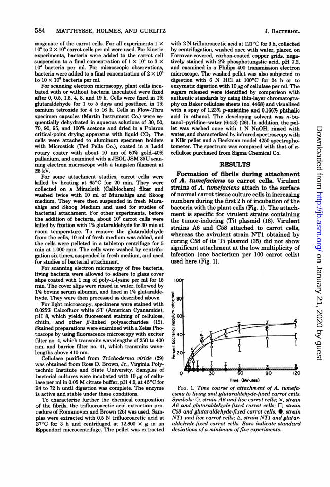
of A. tumefaciens to carrot cells. Virulentstrains of A. tumefaciens attach to the surfaceofnormal carrot tissue culture cells in increasingnumbers during the first 2 h of incubation of thebacteria with the plant cells (Fig. 1). The attach-ment is specific for virulent strains containingthe tumor-inducing (Ti) plasmid (18). Virulentstrains A6 and C58 attached to carrot cells,whereas the avirulent strain NT1 obtained bycuring C58 of its Ti plasmid (35) did not showsignificant attachment at the low multiplicity ofinfection (one bacterium per 100 carrot cells)used here (Fig. 1).
0IO
E' 60
201
0 30 A 0 90 120lime (Minues)
FIG. 1. Time course of attachment of A. tumefa-ciens to living and glutaraldehyde-fixed carrot cells.Symbols: 0, strain A6 and live carrot cells; x, strainA6 and glutaraldehyde-fixed carrot cells; E, strainC58 and glutaraldehyde-fixed carrot cells; 0, strainNT1 and live carrot cells; A, strain NT1 and glutar-aldehyde-fixed carrot cells. Bars indicate standarddeviations of a minimum offive experiments.
J. BACTERIOL.
on January 21, 2020 by guesthttp://jb.asm
.org/D
ownloaded from

AGROBACTERIUM MAKES CELLULOSE FOR ATTACHMENT 585
Scanning electron microscopy showed thatcarrot cells grown in liquid suspension cultureon a shaker were spherical and grew in clumpsof 2 to 20 cells (Fig. 2a). Dividing cells werefrequently seen. The surface of the cell wallconsisted of a flat mesh of fibrillar material (Fig.2b). The rough texture of this cell surface maybe due to surface wounding caused by the con-stant agitation of these cells in culture flasks.A. tumefaciens strain A6 was added to washed
carrot cells, and the resulting cellular interac-
tions were observed with scanning electron mi-croscopy at intervals during the subsequent 19h. By 30 min after their addition, bacteria wereattached to the surface of the carrot cells bothsingly and in small clumps (Fig. 2c and d). By 90min more bacteria were observed on the surfaceof the carrot cells, and many of these bacteriawere in well-isolated clumps (Fig. 3c). Figure 3aalso shows that bacteria could attach to a re-cently divided carrot cell. It is interesting to notethat in both control and inoculated cultures
FIG. 2. Scanning electron micrographs ofsuspension cultures ofD. carota cells before and after binding ofthe virulent strain A6 ofA. tumefaciens. (a) Normal carrot cells appeared as small clusters of spherical cells.(b) The surface of a normal carrot cell showed a finely fibrillar mesh and occasional small blebs. (c) Within30 min after the addition of A6 bacteria to the plant cell culture, bacteria were observed attached to thesurface of the carrot cells (arrowheads). (d) Higher magnification shows that bacteria may be attached to theplant cells by their ends or lateral sides and that bacteria may also attach to other bacteria (arrowheads).
VOL. 145, 1981
on January 21, 2020 by guesthttp://jb.asm
.org/D
ownloaded from
586 MATTHYSSE, HOLMES, AND GURLITZ
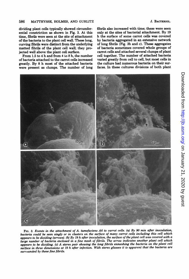
dividing plant cells typically showed circumfer-ential constriction as shown in Fig. 3. At thistime, fibrils were seen at the site of attachmentof the bacteria to the plant cell wall. These long,curving fibrils were distinct from the underlyingmatted fibrils of the plant cell wall; they pro-jected well above the plant cell surface.From 1.5 to 4 h and from 4 to 8 h, the number
of bacteria attached to the carrot cells increasedgreatly. By 8 h most of the attached bacteriawere present as clumps. The number of long
fibrils also increased with time; these were seenonly at the sites of bacterial attachment. By 19h the surface of some carrot cells was coveredby bacteria aggregated in an extensive networkof long fibrils (Fig. 3b and c). These aggregatesof bacteria sometimes covered whole groups ofcarrot cells and attached several clumps of plantcell together. The number of attached bacteriavaried greatly from cell to cell, but most cells inthe culture had numerous bacteria on their sur-faces. In these cultures divisions of both plant
FIG. 3. Events in the attachment of A. tumefaciens A6 to carrot cells. (a) By 90 min after inoculation,bacteria could be seen singly or in clusters on the surface of many carrot cells including this cell whichappears to be dividing (arrows). (b) By 19 h after inoculation, the surface of the plant cell was covered with alarge number of bacteria enclosed in a fine mesh of fibrils. The arrow indicates another plant cell whichappears to be dividing. (c) A stereo pair showing the long fibrils enmeshing the bacteria on the plant cellsurface in three dimensions at 19 h after infection. With stereo glasses it is apparent that the bacteria are
surrounded by these fine fibrils.
J. BACTERIOL.
on January 21, 2020 by guesthttp://jb.asm
.org/D
ownloaded from

AGROBACTERIUM MAKES CELLULOSE FOR ATTACHMENT
cells and bacteria were observed (Fig. 3a and b).Figure 3 shows that the bacteria were attachedto the plant cells both singly and in large clus-ters. There appeared to be no preferential ori-entation of the bacteria for attachment sincebacteria that were attached at their ends or ontheir sides were observed. A similar observationwas made earlier by using light microscopy ofliving cells (18). In the large clusters, many bac-teria were not directly attached to the plant cellbut rather were entrapped in a three-dimen-sional network of long fibrils attached to otherbacteria which were directly bound to the sur-face of the plant cell.Synthesis of fibrils by A. tumefaciens. To
determine whether the plant cell or the bacte-rium synthesized the fibrils, the interaction ofbacteria with killed carrot cells was examined.Virulent strains of A. tumefaciens attached tocarrot cells which had been killed by treatmentwith 1% glutaraldehyde or by high temperature.The kinetics of attachment to killed plant cellswere only slightly different from the kinetics ofattachment to living carrot cells (Fig. 1 and 4).The bacterial strain specificity of attachmentwas the same as that with live plant cells. Viru-lent A. tumefaciens strains A6 and C58 showedattachment to killed cells, whereas the avirulentstrain NT1 did not (Fig. 1 and 4). Scanningelectron microscopy showed large clumps of bac-teria attached to glutaraldehyde- or heat-killedcarrot cells after 19 h of incubation (Fig. 5a tod). In some cases, mats of bacteria coveredgroups of carrot cells (Fig. 5b). Numerous longfibrils were attached to the bacteria (Fig. 5a andd). The fibrils often seemed to arise from theside of the bacterium and frequently appearedtwisted together into wide filaments. Althoughthe glutaraldehyde-killed cells were not morpho-logically distinguishable from cells fixed withglutaraldehyde after bacterial attachment, thesurface of heat-killed cells was irregular. Sincethe long fibrils were made during the attachmentof live bacteria to dead carrot cells, it is probablethat the bacteria rather than the plant cellssynthesize these fibrils.
Characteristics of bacterial growth, ag-gregation, and fibril formation. Although A.tumefaciens was usually grown in LB medium(19), the bacteria also grew on other media. Infresh cultures of Agrobacterium growing expo-nentially in LB medium with vigorous aeration,very little fibrillar material was made, and thebacteria appeared to be well dispersed (Fig. 6b).When the cultures became stationary, some bac-terial aggregation was visible to the unaided eye,and small amounts of fibrillar material could beseen in the light microscope after Calcofluorstaining.
C. 60
0
406.,
20
230 60 90 120Time (Minutes)
FIG. 4. Time course of attachment of A. tumefa-ciens to living or heat-killed carrot cells. Symbols:0, strain A6 and live carrot cells; x, strain A6 andheat-killed carrot cells; *, strain NT1 and live carrotcells; A, strain NT1 and heat-killed carrot cells. Barsindicate standard deviations of a minimum of fiveexperiments.
Carrot cells grew as small clusters of 4 to 20cells in Murashige and Skoog medium. Theyshowed no tendency to form larger aggregates(Fig. 6a). The addition of virulent strains of A.tumefaciens to carrot cells resulted in the for-mation of massive aggregates of bacteria andplant cells after 19 h of incubation (Fig. 6c).Agrobacterium grew in Murashige and Skoogmedium in the presence of carrot cells, but thebacteria did not grow in the absence of the plantcells. However, the addition of 0.02% Soytone(soluble soybean extract [Difco Laboratories])to this medium allowed good bacterial growth.The bacteria were highly aggregated, and largeamounts of fibrillar material were made by bothvirulent and avirulent agrobacteria. Strains ofAgrobacterium in which we observed fibril for-mation and clumping include the virulent A.tumefaciens strains A6, B6, and C58, the aviru-lent A. tumefaciens strains NT1 and IIBNV6,and the avirulent A. radiobacter strains 6467,S1005, and TR1. Since the avirulent NT1 andthe A. radiobacter strains lack the Ti plasmid(7, 35, 39) found in the virulent strains and haveno known plasmid in common, the genes in-volved in the synthesis of fibrils are presumablylocated on the bacterial chromosome.A. tumefaciens strains A6 and NT1 grown in
Murashige and Skoog medium synthesized fila-ments, aggregated, and attached to Whatmanno. 1 filter paper (Fig. 7a and b). By scanningelectron microscopy, strain A6 was seen at-
587VOL. 145, 1981
on January 21, 2020 by guesthttp://jb.asm
.org/D
ownloaded from

588 MATTHYSSE, HOLMES, AND GURLITZ
. f ,.
1''
k-,1i;'W48'-5.
0~~~~~~~~
FIG. 5. Attachment of A. tumefaciens to killed plant cells. (a) Bacteria attached to glutaraldehyde-fixedplant cells, and numerous fine fibrils were elaborated during this process. (b) The surface of this cluster ofheat-killed plant cells was nearly covered by a mass of adherent bacteria. (c) At a higher magnification, theindividual bacterial cells that were attached to the heat-killed plant cells can be distinguished. (d) At highmagnification, a three-dimensional view of bacteria attached to glutaraldehyde-fixed plant cells shows thatthe numerous long, slender fibrils originated on the sides of bacterial cells (arrow). The fibrils frequentlybecame entwined together to form large fibrils.
tached to the filter paper apparently by anchor-ing fibrils which connected to the sides of thebacteria (Fig. 7b). Fibrils were observed toanastomose and to wind around each other. At-tachment to filter paper required much longertimes, about 24 to 48 h, than attachment tocarrot cells.
Aggregation of the A6 strain ofA. tumefaciensgrown in Murashige and Skoog medium with0.02% Soytone was also observed with bacteria
attached to poly-L-lysine-coated glass coverslips. Bacterial aggregation on the cover slipswas visible to the unaided eye. In the scanningelectron microscope, these aggregates ofbacteriacould be seen to be held together by extensivenetworks of fibrils (Fig. 7c). Thus, under certainconditions, A. tumefaciens can form fibrils in theabsence of the host plant.The bacteria also possess at least a limited
capacity to digest pectin, a component of the
J. BACTERIOL.
on January 21, 2020 by guesthttp://jb.asm
.org/D
ownloaded from

AGROBACTERIUM MAKES CELLULOSE FOR ATTACHMENT
FIG. 6. Cultures ofA. tumefaciens and D. carota. (a) Cultures ofD. carota were well dispersed and did notshow aggregation. (b) Cultures of A. tumefaciens were also well dispersed and showed minimal clumpingwhen grown in LB medium. (c) A mixture of D. carota cells with A. tumefaciens A6 showed enormousaggregates ofplant and bacterial cells after 19 h. (d) A. tumefaciens grown in the presence ofSoytone (solublesoybean extract) exhibited massive clumping.
host cell wall. A. tumefaciens A6, C58, and NT1grew slowly on minimal medium (16) with pectinrather than glucose as the sole carbon source.The pectin was not contaminated with a suffi-cient amount of glucose or other carbon sourceto support the growth of E. coli 23739, which didnot grow on medium with pectin as the solecarbon source, but did grow on medium withglucose as the carbon source.Composition of bacterial fibrils. When A.
tumefaciens was grown under conditions whichpromoted fibril synthesis in the absence of plantcells, long, thin strands of material whichfluoresced after Calcofluor staining were ob-served in the light microscope (Fig. 8a). Cellu-lose, chitin, and some fl-1,3-glucans show fluores-cent staining with Calcofluor (12). When thesebacterial cultures were treated with purifiedTrichoderma cellulase, no Calcofluor stainingwas observed. The bacteria themselves showedan intrinsic fluorescence which was not Calco-fluor-dependent and which was not affected by
cellulase. Thin-layer chromatography of theproducts of enzymatic digestion showed onlycellobiose and glucose at 24 h and only glucoseat 72 h. The sole product of hydrolysis with 6 NHCl at 1000C as determined by thin-layer chro-matography was glucose. Treatment of the bac-terial cultures with proteinase K (100 ,ug/ml, pH7.4, at 370C for 1 to 4 h) did not affect theCalcofluor fluorescent strands and left fibrilswhich were visible with negative staining in thetransmission electron microscope. To determinewhether these strands were indeed cellulose, thetrifluoroacetic acid extraction procedure of Ro-manovicz and Brown was used (26). Treatmentwith 2 N trifluoracetic acid at 1210C for 3 h leftvisible fibrils which showed fluorescent stainingwith Calcofluor. These fibrils were also visibleafter negative staining in the transmission elec-tron microscope (Fig. 8b). An infrared spectrumof the purified fibrillar material was comparedwith the spectrum of cellulose (Sigma) preparedin a similar manner (Fig. 9). The two spectra
589VOL. 145, 1981
on January 21, 2020 by guesthttp://jb.asm
.org/D
ownloaded from

590 MATTHYSSE, HOLMES, AND GURLITZ
FIG. 7. Elaboration of fibrils by A. tumefaciens in the absence ofplant cells. (a) Bacteria grown in thepresence offilter paper developed numerous long fibrils and became aggregated. The arrowhead indicates aportion of the filter paper. (b) At higher magnification, several bacteria growing on the surface of the filterpaper can be seen. Long fibrils originate from the sides of the bacteria. (c) Bacteria grown in the presence ofsoluble plant extract also developed long fibrils. These fibrils formed bridges between adjacent bacteria andlead to massive clumping of the bacteria in the culture.
were almost identical. Thus, it appears that thefibrils synthesized by Agrobacterium are com-posed of cellulose.
DISCUSSIONAttachment of A. tumefaciens to
wounded plants and tissue culture cells.The initial step in tumor formation by A. tume-faciens is a site-specific attachment of the bac-terium to the host plant (13). A similar site-specific attachment has been observed betweenthe bacterium and plant tissue culture celLs (18).In the whole plant, the bacterial infection andresulting tumor formation generally occurs in awound site. In suspension cultures of carrots, thesurface of the cells appeared slightly damaged,and occasional cells were broken by the mechan-ical agitation. This may provide equivalent stim-uli and surfaces to those generated by a wound.Both in the wounded plant (36) and in tissueculture (18) bacterial lipopolysaccharide in-hibited the interaction between the bacteriumand the plant cell suggesting that the bacterialreceptor on the plant cell surface may recognizethe lipopolysaccharide in the bacterial outer en-velope.
AvirulentA. tumefaciens strain NT1 inhibitedtumor formation in plants apparently by block-ing attachment sites for virulent A. tumefaciens
(14). NT1 did not show significant attachmentto tissue culture cells at the low multiplicity ofinfection used in kinetic studies (Fig. 1). How-ever, data not shown here indicate that at thehigher multiplicity of infection used for micro-scopic studies, NT1 showed some attachment tothe carrot cells. NT1 was also capable of fibrilformation although at a lower rate that thevirulent strain A6.
Specific attachment ofA. tumefaciens to plantcells has been observed in several in vitro sys-tems including carrot (18), tobacco (18, 32), andDatura (22) tissue culture cells and carrot (38)and potato disks (8). Scanning electron micros-copy of A. tumefaciens attached to habituatedtobacco cells suggested that the bacteria gener-ally attached end on to the host cell surface (32),whereas our observations suggest that the bac-teria could attach in both polar and lateral ori-entations to the carrot cell surface.The attachment of A. tumefaciens to Datura
tissue culture cells has been examined withtransmission electron microscopy of sectionedmaterial (22). The bacteria were tightly boundto the surface of the plant cell wall and weresurrounded by a network of fine fibrils. Some ofthe fibrils connected loosely bound bacteria tothe host cell wall (22). These fibrils, previouslyobserved in the transmission electron micro-
J. BACTERIOL.
on January 21, 2020 by guesthttp://jb.asm
.org/D
ownloaded from

AGROBACTERIUM MAKES CELLULOSE FOR ATTACHMENT
L.OpmbFIG. 8. Cellulose fibrils elaborated by A. tumefaciens. (a) Calcofluor staining of a bacterial culture grown
in the presence of soytone. Calcofluor stains cellulose and other f8-linked polysaccharides (12). In otherexperiments (not shown) in which the bacteria were treated with cellulase before Calcofluor staining nofluorescent stain was observed. (b) Bacterial cultures were extracted with trifluoroacetic acid at 121°C for 3h. The fibrils which remained were cellulose since they were still positive for fluorescent staining withCalcofluor and appeared as fine filaments in negatively stained preparations.
scope, are probably identical to the fibrils de-scribed here since their size, location, and con-figuration appear similar.The presence of large aggregates of bacteria
attached to the carrot cell surface noted previ-ously (18) has also been observed with tobacco(32) and Datura (22) tissue culture cells. Thedivision of attached bacteria which has beenobserved with time-lapse microcinematography(R. H. G. Gurlitz and A. G. Matthysse, manu-script in preparation) may be responsible, inpart, for the growth of clusters of attached bac-teria. However, bacterial growth has a lag timeof 2 to 3 h after the addition of the bacteria tocarrot cell cultures, and thereafter the bacterialdoubling time is about 2 h. Thus, one bacteriumwhich attached to a carrot cell within 1 h ofincubation would have been unlikely to havedivided at 90 min, but could have given rise to128 bacteria after 19 h. By 30 min bacterialclumps were visible by scanning electron mi-croscopy (Fig. 2c); these clumps increased in sizewith longer incubation times. Therefore, theclusters of bacteria seen on the plant cell surfaceprobably arose both from the division of at-tached bacteria and from the tendency ofincom-ing bacteria to attach near previously attachedbacteria (A. G. Matthysse, S. Matthysse, and R.H. G. Gurlitz, manuscript in preparation).Numerous dividing plant cells were seen by
scanning electron microscopy. These cells typi-cally showed a circumferential constriction (Fig.3a) and retained their spherical shape after di-vision. This method of cytokinesis appearedslightly different from the usual higher plant cellplate formation in which the original cell wall isunaltered in shape.Role of fibrils in bacterial infections of
plants. In contrast to many animal pathogensin which the ability to attach to the host isassociated with bacterial virulence, infection ofplants by many species of bacterial pathogens isfollowed by the attachment of avirulent strainsto the host cell wall; in general, virulent strainsdo not attach. Pathogens for which this has beenobserved include Pseudomonas putida (31),Pseudomonas phaseolicola (25, 30), Pseudom-onas pisi (9, 24), Pseudomonas solanacearum(28), and Xanthomonas malvacearum (2, 3).During attachment of avirulent strains to thehost cell wall, the bacteria were covered withfibrils and granular material. The compositionof these fibrils is unknown. In some cases apellicle also covered the attached bacteria. At-tachment was usually followed by a hypersensi-tive response, including changes in membranepermeability and localized necrosis. Saprophyticbacteria such as E. coli and Bacillus subtilisalso attached to the host cell wall but theirattachment was not followed by a hypersensitive
591VOL. 145, 1981
on January 21, 2020 by guesthttp://jb.asm
.org/D
ownloaded from

592 MATTHYSSE, HOLMES, AND GURLITZ
I
4000 3500 3000 2500 2000 1800 1600 1400 1200 1000 800 600WAVENUMUER (CM 1)
FIG. 9. Infrared spectra of a purified preparation of bacterial fibrils and cellulose.
response (28). In the only one of these cases inwhich the source of the attachment fibrils hasbeen examined, it appeared that the plant, andnot the bacterium, was responsible for fibril syn-thesis. Heat-killed P. solanacearum were at-tached by fibrils to living tobacco mesophyllcells (28). As was the case for virulent strains ofA. tumefaciens, bacterial lipopolysaccharide ap-pears to be involved in the attachment of avir-ulent strains of P. solanacearum to potato cells(37).The various species of rhizobia, unlike agro-
bacteria to which they are closely related (11),have a limited host range (5). Both specific andnonspecific interactions were involved in theinitial attachment of Rhizobium cells to roothairs (5). Some species of Rhizobium have beenshown to synthesize cellulose fibrils (5, 21) whichcould have been involved in the subsequenttighter binding of the bacteria to the root sur-face. Bacterial lipopolysaccharide appeared tobe involved in the initial attachment of Rhizo-bium cells to the plant root hair (5, 27).Thus, although the interaction of A. tumefa-
ciens and other plant pathogens with the host
cell surface was similar in some respects, inter-esting differences were observed. Avirulentstrains of most phytopathogens attached to theplant cell, and virulent strains remained free. Incontrast, virulent strains of A. tumefaciens at-tached to the plant cell, and avirulent strainsremained free. P. solanacearum attached to theplant cell by fibrils synthesized by the host,whereas A. tumefaciens was attached to theplant cell by fibrils synthesized by the bacte-rium. These differences may, in part, reflect thedifference between an invasive pathogen whichdestroys host tissue and a noninvasive pathogenwhose food supply depends on materials (pre-sumably the opines) excreted by living planttumor cells (23).Bacterial cellulose synthesis. The compo-
sition of the fibrils involved in the attachmentof other phytopathogens to the host is unknown.However, we have shown that in the case ofAgrobacterium cells, the attachment fibrils arecomposed of cellulose elaborated by the bacte-rium. Cellulose synthesis accompanied by bac-terial aggregation has previously been observedin several genera of gram-negative bacteria iso-
J. BACTERIOL.
on January 21, 2020 by guesthttp://jb.asm
.org/D
ownloaded from

AGROBACTERIUM MAKES CELLULOSE FOR ATTACHMENT 593
lated from sewage (6). On the basis of biochem-ical tests, some of these bacteria were identifiedas agrobacteria. Rhizobium trifolii, which has a50% chromosomal DNA homology with A. tu-mefaciens (11), has also been shown to synthe-size cellulose (21). As with Agrobacterium spp.the ability to synthesize cellulose was not cor-related with infectivity. However, the cellulosefibrils were apparently involved in the adherenceof the bacteria to root hair surfaces (21).
All of the strains of A. tumefaciens and A.radiobacter that we examined synthesized cel-lulose, although they differed in the quantityproduced. Since these strains do not contain anyknown plasmids in common, the genes involvedmay be presumed to be chromosomal.The ultrastructure of bacterial cellulose syn-
thesis has been examined in Acetobacter xy-linum. Cellulose microfibrils are synthesized atsites on the side of the bacterium; the microfi-brils that formed aggregate into a ribbon ofcellulose (1). Cellulose fibrils also appeared toarise from the side ofAgrobacterium cells; how-ever, the fibrils did not aggregate to form aribbon.A model for the attachment of A. tume-
faciens to the plant host cell. The first stepin attachment of A. tumefaciens to the planthost cell (Fig. 10.1) appears to be the attachmentof a bacterium (probably a motile, flagellatedbacterium) to a receptor on the surface of theplant cell. Bacterial lipopolysaccharide appearsto be a recognition factor in this step both in thewounded plant (36) and in tissue culture (18).After attachment, the bacterium synthesizes cel-lulose fibrils (Fig. 10.2). Substances releasedfrom wounded plant tissues or from damagedcells in tissue culture may act as Soytone doesin synthetic medium to stimulate the synthesisof these cellulose fibrils. The newly developedfibrils anchor the bacteria to the surface of theplant cell (Fig. 10.3). Because the fibrils aremade of cellulose, which is also a component ofthe plant cell wall, the plant host cell does notreadily digest the fibrils to detach the bacteria.As the fibrils become longer they may entrapother bacteria and attach them indirectly to thesurface of the plant cell (Fig. 10.4). Fibrils mayalso hold both daughter cells of a dividing bac-terium to the surface of the plant cell. Once thebacteria are firmly attached, both directly byinteraction with receptor sites and indirectly bycellulose fibrils to the outer surface of the plantcell wall, they probably need to make contactwith the host cell plasma membrane to transferbacterial Ti plasmid DNA to the host cell. Howthis occurs is unknown. However, we haveshown that Agrobacterium cells can digest pec-
FIG. 10. A model for the attachment ofA. tumefa-ciens to carrot cells. A bacterium attaches to a specificreceptor site (R) on the surface of the plant cell (step1) and is induced to elaborate cellulose fibrils (step2). The fibrils anchor the bacterium to the surface ofthe plant cell wall (CW) (step 3) and entrap addi-tional bacteria (step 4). These entrapped bacteriamay themselves begin to synthesize fibrils (step 6).This facilitated attachment and multiplication of at-tached bacteria result in the development of a bacte-rial colony attached to the plant cell wall via recep-tors and cellulose fibrils. The attached bacteria se-crete enzymes which digest theplant cell wall (step 5)and may aid in establishing contact of the bacteriawith the plant cell plasma membrane (PM) to facili-tate transfer of the Ti plasmid from the bacteria tothe plant cell.
tin. Possibly other plant cell wall componentscan be digested as well. This may aid the bac-terium in approaching the host plasma mem-brane (Fig. 10.5). Bacteria which are entrappedin fibrils begin to synthesize fibrils themselves(Fig. 10.6). Steps 2 through 6 may then be re-peated to cause the formation of a large clusterof bacteria attached to the plant cell surface byfibrils. At the same time, attached and en-trapped bacteria may divide, also increasing thesize of the bacterial clump. Unoccupied receptorsites may still be present on the plant cell sur-face, and additional individual bacteria may at-tach to them, beginning the sequence again.Thus, at late times, some cells will have largeclumps of bacteria representing early initial at-tachment followed by amplification. Other cellsor other areas on the same cell may have indi-vidual bacteria or small bacterial clusters result-ing from relatively late initial attachment events,with little time available for bacterial entrap-ment or multiplication.The initial step in the pathogenic interaction
of A. tumefaciens and the plant cell is the at-tachment of the bacterium to the host cell sur-face. The studies reported here indicate that thisinitial interaction is not a simple one-step proc-ess. Instead, there exists a complicated series of
VOL. 145, 1981
on January 21, 2020 by guesthttp://jb.asm
.org/D
ownloaded from

594 MATTHYSSE, HOLMES, AND GURLITZ
events, including bacterial attachment to hostreceptors, bacterial synthesis of cellulose fibrils,the digestion of a portion of the host cell wail bybacterial enzymes, and amplification ofthe num-ber ofattached bacteria by entrapment and mul-tiplication.
ACKNOWLEDGMENTSWe thank Ross D. Brown, Jr., for the gift of purified
cellulase and Joy A. Sawyer for assistance with the infraredspectra. We thank R. Malcolm Brown, Jr., Alan White, andCandace Haigler for assistance with fluorescent staining ofcellulose and for helpful discussions of bacterial cellulosesynthesis. We are grateful for the excellent technical assistanceof Gustave Boesch.
This research was supported by Public Health Serviceresearch grant CA 18604 from the National Cancer Institute.
LITERATURE CiMD1. Brown, R. M., Jr., J. H. M. Willison, and C. L Rich-
ardson. 1976. Cellulose biosynthesis in Acetobacterxylinum: visualization of the site of synthesis and directmeasurement of the in vivo process. Proc. Natl. Acad.Sci. U.S.A. 73:45654569.
2. Cason, E. T., Jr., P. E. Richardson, L. A. Brinkerhoff,and R. K. Gholson. 1977. Histopathology of immuneand susceptible cotton cultivars inoculated with Xan-thomonas malvacearum. Phytopathology 67:196-198.
3. Cason, E. T., Jr., P. E. Richardson, M. K. Essenberg,L. A. Brinkerhoff, W. M. Johnson, and R. J. Ven-ere. 1978. Ultrastructural cell wall alterations in ih-mune cotton leaves inoculated with Xanthomonas mal-vacearum. Phytopathology 68:1015-1021.
4. Chilton, M. D., M. H. Drummond, D. J. Merlo, D.Sciaky, A. L Montoya, M. P. Gordon, and E. W.Nester. 1977. Stable incorporation of plasmid DNAinto higher plant cells: the molecular basis of crown galltumorigenesis. Cell 3:263-271.
5. Dazzo, F. B. 1980. Adsorption of microorganisms to rootsand other plant surfaces, p. 253-316. In G. Bitton andK. C. Marshall (ed.), Adsorption of microorganisms tosurfaces. John Wiley & Sons, New York, N.Y.
6. Dienema, M. H., and L. P. T. M. Zevenhuizen. 1971.Formation of cellulose fibrils by gram-negative bacteriaand their role in bacterial flocculation. Arch. Mikrobiol78:42-57.
7. Genetello, C., N. Van Larebeke, M. Holsters, A.DePicker, M. Van Montagu, and J. Schell. 1977. Tiplasmids of Agrobacterium as conjugative plasmids.Nature (London) 265:561-563.
8. Glogowski, W., and A. G. Galsky. 1978. Agrobacteriumtumefaciens site attachment as a necessary prerequisitefor crown gall tumor formation on potato discs. PlantPhysiol. 61:1031-1033.
9. Goodman, R. N., P.-Y. Huang, and J. A. White. 1976.Ultrastructural evidence for immobilization of an in-compatible bacterium, Pseudomonas pisi, in tobaccoleaf tissue. Phytopathology 66:754-764.
10. Gurley, W. B., J. D. Kemp, M. J. Albert, D. W. Sutton,and J. Callis. 1979. Transcription of Ti plasmid-de-rived sequences in three octopine-type crown gall tumorlines. Proc. Natl. Acad. Sci. U.S.A. 76:2828-2832.
11. Heberlein, G. T., J. DeLey, and R. Tijtgat. 1967. De-oxyribonucleic acid homology and taxonomy of Agro-bacterium, Rhizobium, and Chromobacterium. J. Bac-teriol. 94:116-124.
12. Hughes, J., and M. E. McCully. 1975. The use of anoptical brightener in the study of plant structure. StainTechnol. 50:319-329.
13. Lippincott, B. B., and J. A. Lippincott. 1969. Bacterial
J. BACTERIOL.
attachment to a specific wound site as an essential stagein tumor initiation by Agrobacterium tumefaciens. J.Bacteriol. 97:620-628.
14. Lippincott, B. B., M. H. Whatley, and J. A. Lippin-cott. 1977. Tumor induction byAgrobacterium involvesattachment of the bacterium to a site on the host plantcell wall. Plant Physiol. 59:388-390.
15. Lippincott, J. A., and B. B. Lippincott. 1975. The genusAgrobacterium and plant tumorigenesis. Annu. Rev.Microbiol. 29:377-406.
16. Matthysse, A. G. 1977. Variation in plasmid DNA se-quences present in crown gall tumour lines. J. Gen.Microbiol. 102:427-420.
17. Matthysse, A. G., and A. J. Stump. 1976. The presenceof Agrobacterium tumefaciens plasmid DNA in crowngall tumor cells. J. Gen. Microbiol. 95:9-16.
18. Matthysse, A. G., P. M. Wyman, and K. V. Holmes.1978. Plasmid-dependent attachment ofAgrobacteriumtumefaciens to plant tissue culture cells. Infect. Immun.22:516-522.
19. Miller, J. H. 1972. Experiments in molecular genetics, p.433. Cold Spring Harbor Laboratory, Cold Spring Har-bor, N.Y.
20. Murashige, T., and F. Skoog. 1962. A revised mediumfor rapid growth and bioasays with tobacco tissuecultures. Physiol. Plant. 15:473-497.
21. Napoli, C., F. Dazzo, and D. Hubbell. 1975. Productionof cellulose microfibrils by Rhizobium. Appl. Microbiol.30:123-131.
22. Ohyama, K., L E. Pekher, A. Schaefer, and L C.Fowke. 1979. In vitro binding of Agrobacterium tu-mefaciens to plant cells from suspension culture. PlantPhysiol. 63:382-387.
23. Petit, A., S. Delhaye, J. Temp6, and G. Morel. 1970.Recherches sur les guanidines des tissus de crown gall.Mise en evidence d'une relation biochimique specifiqueentre lea souches d'Agrobacterium tumefaciens et lestumeurs qu'elles induisent. Physiol. V6g. 8:205-213.
24. Politis, D. J., and R. N. Goodman. 1978. Localized cellwall appositions: incompatibility response of tobaccocells to Pseudomonaspisi. Phytopathology 68:309-316.
25. Roebuck, P., R Sexton, and J. W. Mansfield. 1978.Ultrastructural observations on the development of thehypersensitive reaction in leaves of Phaseolus vulgariscv. Red Mexican inoculated with Pseudomonas phas-eolicola (race 1). Physiol. Plant. Path. 12:151-157.
26. Romanovicz, D. K., and RK M. Brown, Jr. 1976. Bio-genesis and structure of Golgi-derived cellulosic scalesin Pleurochrysis. II. Scale composition and supramo-lecular structure. Appl. Polym. Symp. 28:587-610.
27. Sequeira, L. 1978. Lectins and their role in host pathogenspecificity. Annu. Rev. Phytopathol. 16:453-481.
28. Sequeira, L, G. Gaard, and G. A. DeZoeten. 1977.Interaction of bacteria and host cell walls: Its relationto mechanisms of induced resistance. Physiol. PlantPath. 10:43-50.
29. Shoemaker, S. P., and R. D. Brown, Jr. 1978. Charac-terization of endo-1,4-,6-D-glucanases purified fromTrichoderma viride. Biochim. Biophys. Acta 523:147-161.
30. Sigee, D. C., and H. A. S. Epton. 1976. Ultrastructuralchanges in resistant and susceptible varieties of Phas-eolus vulgaris following artificial inoculation with Pseu-domonasphaseolicola. Physiol. Plant Path. 9:1-8.
31. Sing, V. O., and M. N. Schroth. 1977. Bacteria-plant cellsurface interactions: Active immobilization of sapro-phytic bacteria in plant leaves. Science 197:759-761.
32. Smith, V. A., and J. Hindley. 1978. Effect of agrocin 84on attachment of Agrobacterium tumefaciens to cul-tured tobacco cells. Nature (London) 276:498-500.
33. Thomashow, M. F., R. Nutter, A. L. Montoya, M. P.Gordon, and E. W. Nester. 1980. Integration and
on January 21, 2020 by guesthttp://jb.asm
.org/D
ownloaded from

AGROBACTERIUM MAKES CELLULOSE FOR ATTACHMENT 595
organization of Ti plasmid sequences in crown galltumors. Cell 19:729-739.
34. Van Larebeke, N., G. Engler, M. Holsters, S. Van denElsacker, I. Zaenen, R. A. Schilperoort, and J.Schell. 1974. Large plasmid in Agrobacterium tumefa-ciens essential for crown gall-inducing ability. Nature(London) 253:169-170.
35. Watson, B., T. C. Currier, M. P. Gordon, M. D. Chil-ton, and E. W. Nester. 1975. Plasmid required forvirulence of Agrobacterium tumefaciens. J. Bacteriol.123:255-264.
36. Whatley, M. H., J. S. Bodwin, B. B. Lippincott, andJ. A. Lippincott. 1976. Role for Agrobacterium cellenvelope lipopolysaccharide in infection site attach-
ment. Infect. Immun. 13:1080-1083.37. Whatley, M. H., N. Hunter, M. A. Cantrell, C. Hen-
drick, K. Keegstra, and L. Sequeira. 1980. Lipo-polysaccharide composition of the wilt pathogen, Pseu-domonas solanacearum: correlation with the hypersen-sitive response in tobacco. Plant Physiol. 65:557-559.
38. Yajko, D. M., and G. D. Hegeman. 1971. Tumor induc-tion by Agrobacterium tumefaciens: specific transfer ofbacterial deoxyribonucleic acid to plant tissue. J. Bac-teriol. 108:973-979.
39. Zaenen, L, N. Van Larebeke, H. Teuchy, M. VanMontagu, and J. Schell. 1974. Supercoiled circularDNA in crown-gall inducing Agrobacterium strains. J.Mol. Biol. 86:109-127.
VOL. 145, 1981
on January 21, 2020 by guesthttp://jb.asm
.org/D
ownloaded from