Embed Size (px)
Citation preview
Proc. Natl. Acad. Sci. USAVol. 93, pp. 5888-5893, June 1996Plant Biology
Enhanced green fluorescence by the expression of an Aequoreavictoria green fluorescent protein mutant in mono- anddicotyledonous plant cells
(plants/protoplasts/mutagenesis/reporter gene/blue fluorescent protein)
CHRISTOPH REICHEL*, JAIDEEP MATHUR, PETER ECKESt, KERSTIN LANGENKEMPER, CSABA KONCZ, JEFF SCHELL,BERND REISS, AND CHRISTOPH MAAStMax-Planck-Institut fur Zichtungsforschung, Abteilung Genetische Grundlagen der Pflanzenzfichtung, Carl-von-Linne Weg 10, D-50829 Koln, Germany; andtHoechst-Schering AgrEvo GmbH, Forschung Biochemie H872N, Postfach 800320, D-65926 Frankfurt, Germany
Contributed by Jeff Schell, January 4, 1996
ABSTRACT The expression of the jellyfish green fluores-cent protein (GFP) in plants was analyzed by transientexpression in protoplasts from Nicotiana tabacum, Arabidopsisthaliana, Hordeum vulgare, and Zea mays. Expression of GFPwas only observed with a mutated cDNA, from which arecently described cryptic splice site had been removed.However, detectable levels of green fluorescence were onlyemitted from a small number of protoplasts. Therefore, othermutations in the GFP cDNA leading to single-amino acidexchanges in the chromophore region, which had been previ-ously studied in Escherichia coli, were tested in order toimprove the sensitivity of this marker protein. Of the muta-tions tested so far, the exchange of GFP amino acid tyrosine66 to histidine (Y66H) led to detection of blue fluorescence inplant protoplasts, while the exchange of amino acid serine 65to cysteine (S65C) and threonine (S65T) increased the inten-sity of green fluorescence drastically, thereby significantlyraising the detection level for GFP. For GFP S65C, thedetectable number of green fluorescing tobacco (BY-2) pro-toplasts was raised up to 19-fold, while the fluorimetriclydetermined fluorescence was raised by at least 2 orders ofmagnitude.
A powerful tool for the rapid analysis of promoters is the useof marker genes for which expression can be easily monitoredby autoradiography (NPT II, CAT), light emission (LUC,GUS), or color production (GUS). Commonly used reportergenes are CAT, NPT II, GUS, and LUC, of which GUS andLUC are of special interest since their assays do not involve anyradioactivity (1). However, none of these reporter genes allowsconvenient, noninvasive in vivo detection of the respectiveenzyme in intact plant cells. An attractive alternative turnedout to be the green fluorescent protein (GFP) from thejellyfishAequorea victoria. Use of this marker protein has beendescribed for Escherichia coli, Caenorhabditis elegans, Drosoph-ila melanogaster, yeast, and HeLa cells (2-5). Detection of GFPis noninvasive and nondestructive, which is a clear advantageover formerly used reporter genes such as f-glucuronidase orfirefly luciferase (6, 7). Illumination of GFP with long-waveUV light (395 nm) or blue light (475 nm) leads to bright greenfluorescence (510 nm) without any need for additional sub-strates, since chromophore formation and light emission areintrinsic properties of this marker protein (8).
Expression and detection of wild-type GFP in maize andsweet orange (Citrus sinensis) protoplasts using constructsdriven by a heat-shock promoter or by a constitutive promoter(9-11), using a potato virus X expression system in Nicotianaclevelandii and Nicotiana benthamiana plants (12, 13), or using
The publication costs of this article were defrayed in part by page chargepayment. This article must therefore be hereby marked "advertisement" inaccordance with 18 U.S.C. §1734 solely to indicate this fact.
tobacco mosaic virus in N. benthamiana and tobacco proto-plasts (14, 15), has been described recently. However, in ourhands expression of a wild-type GFP cDNA driven by a CaMV35S promoter was neither detectable in transgenic tobaccoplants nor in transient expression studies in protoplasts fromArabidopsis thaliana, Nicotiana tabacum, and Hordeum vulgare.Recently it has been described that the A. victoria wild-typeGFP mRNA can be aberrantly processed in plant cells, due tothe recognition of internal cryptic splice sites leading toinefficient expression of the GFP. Mutation of a cryptic splicesite improves expression of GFP in cells from transgenic A.thaliana plants significantly (J. Haseloff, K. Siemering, D.Prasher, and S. Hodge, personal communication; ref. 16).Moreover, mutagenesis of the GFP cDNA in Escherichia colihas led to changes in the fluorescence properties of this newmarker protein. A number of GFP amino acid exchangemutants have been isolated. They exhibit modifications in theirexcitation and emission spectra (17-21). Of these reportedmutations, at least three are of great interest for expression inplant cells. GFP mutation Y66H (19) was shown to have ashifted emission peak leading to blue fluorescence. Analysis ofa second set of GFP mutations, S65C and S65T (20, 21),revealed increased excitation and emission values, which mightsignificantly improve brightness of green fluorescence also inplant cells.
In order to evaluate marker gene expression in plants, plantprotoplasts provide a powerful tool. Transient gene expressionstudies in protoplasts from a variety of plant species and organshave been used widely as a rapid and powerful method tomonitor gene expression and to analyze expression levels ofdifferent marker gene constructs (22-24). The investigation ofseveral GFP derivatives by transient gene expression in pro-toplasts from widely used plant species would thus providevaluable information about the potential of mutations in theGFP cDNA to improve the brightness of green fluorescenceand to alter the GFP emission spectrum.Here we report transient expression studies of GFP genes,
driven by the CaMV 35S promoter, in protoplasts of variousmono- and dicotyledonous plant species. The cDNA of theseGFP constructs was mutated to abolish aberrant splicing inplant cells. Additionally, this cDNA was modified to introducesingle amino acid changes into the GFP chromophore region,leading to significantly improved brightness of green fluores-
Abbreviations: GFP, green fluorescent protein; CaMV, cauliflowermosaic virus; CAT, chloramphenicol acetyltransferase; GUS, 3-gluc-uronidase; LUC, firefly luciferase; NPT II, neomycin phosphotrans-ferase II.*To whom reprint requests should be addressed. e-mail:[email protected].
tPresent address: Hoechst-Schering AgrEvo GmbH, Forschung Bio-chemie H872N, Postfach 800320, D-65926 Frankfurt, Germany.
5888
Proc. Natl. Acad. Sci. USA 93 (1996) 5889
cence (S65C and S65T) or to the emission of blue fluorescence(Y66H) when expressed in plant cells.
MATERIALS AND METHODS
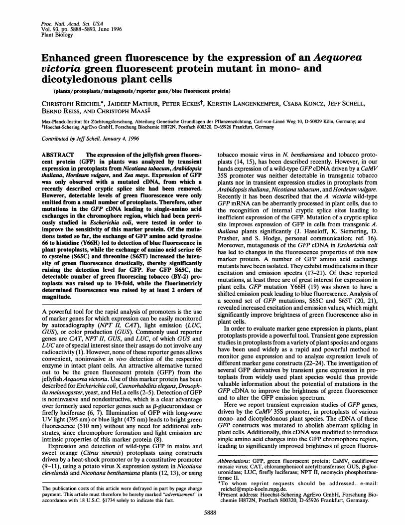
Cloning Strategies. All plasmids used in this study are basedon a commercially available GFP cDNA (pGFP-1; Clontech)carrying a NcoI restriction site at the translation start codonand a modified 3' end leading to the addition of four aminoacids. By site-directed mutagenesis, an internal NcoI restric-tion site was destroyed (oligoS435: CCTGTTCCTTGGC-CAACAC), and the wild-type stop codon was restored alongwith the introduction of BamHI, BglII, XbaI, and AvrII re-striction sites 3' of the new stop codon (oligo5618: CTATA-CAAATAAGGATCCAGATCTAGAATCCTAGGC). Plas-mid pCRGFP was constructed by inserting the modified GFPcDNA as a NcoI/BamHI fragment into the plant expressionvector pRTL2 GUS/NIa[A]Bam (25), thereby removing theGus/NIa cassette and placing the modified GFP cDNA be-tween the CaMV 35S promoter with a duplicated transcrip-tional enhancer, a tobacco etch virus translational enhancerand the CaMV 35S polyadenylation signal (Fig. 1). PlasmidpCKGFP 10 was constructed by exchanging an internal NdeI/AccI restriction fragment with the corresponding fragmentfrom plasmid pBIN 35S-mGFP4, thus supplying the mutationsfor the removal of an internal cryptic splice site found in thewild-type GFP cDNA (J. Haseloff, K. Siemering, D. Prasherand S. Hodge; personal communication) (Fig. 1). PlasmidspCKGFP S65C, pCKGFP S65T, and pCKGFP Y66H werecreated by site-directed mutagenesis of plasmid pCKGFP 10introducing single amino acid exchanges S65C (oligo5637:CTACrTTCTGTTATGGTGTACAATGC), S65T (oligo5858:CTACTTTCACTTATGGTGTACAATGC), and Y66H(oligo5652: CTACTTTCTCTCATGGTGTACAATGC).Plasmid pCKGFP S65Cmono contains the modified GFPcDNA from pCKGFP S65C driven by the CaMV35S promoterand harboring the Shrunken-1 exon 1/intron 1 sequencesshown to significantly enhance reporter gene activity in mono-cot cells (22). During the course of our experiments, DNAsequence analysis of pGFP-1 (Clontech) directly from the
1161T
wt-cDNA
pCR GFP
pCK GFP 10
.deI .-cci
t - -
pCK GFP S65C/T
p35^ TL *I161T wtTAA
.* *p.A.
... I.l..
N 4p35S TL * " -i* * pA
T161I735S TL 1611
VI.pCK GFP S65C [ II *
mono p35S Sh- ex it S65Cfp3-5S TL *
pCK GFP Y66H
T161IL. n4
Y66H
FIG. 1. Cloning strategies for GFP plant expression vectors. Thewild-type GFP cDNA (pGFP-1) was modified and introduced into a
plant expression vector to give plasmid pCRGFP. Insertion of aNdeI-AccI restriction fragment, containing mutations in order todelete a cryptic splice site, led to plasmid pCKGFP 10. By site-directedmutagenesis of pCKGFP 10, chromophore amino acids 65, (Ser ->
Cys) and (Ser -> Thr), and 66 (Tyr His) were exchanged, leadingto plasmids pCKGFP S65C, pCKGFP S65T and pCKGFP Y66H.pCKGFP S65Cmono was constructed by insertion of GFP codingregion from pCKGFP S65C into a monocot expression vector. TL,translational enhancer; wt, wild-type; *, point mutations.
originally supplied plasmid DNA, revealed a previously unre-ported point mutation in the GFP cDNA of this plasmid,leading to amino acid exchange I161T. The wild-type sequence(26) was restored by site-directed mutagenesis of the plasmidspCKGFP S65T, pCKGFP S65C(mono), and pCKGFP Y66H(oligo5653: GAATGGAATCAAAGTCAACTTCAA).
Protoplast Transfection Protocols. Preparation and PEG-mediated DNA uptake with A. thaliana mesophyll protoplastswas performed as described in Mathur et al. (27). Transfectionof tobacco and barley protoplasts followed protocols fromNegrutiu et al. (28) and Maas et al. (23), respectively. Elec-troporation of mesophyll protoplasts from etiolated maizeseedlings isolated essentially according to Sheen (29) wasperformed with a Dialog Electroporator II (Dusseldorf, Ger-many). Electroporation conditions were 500 V/cm and 200 ms.Each sample contained 3 x 10⁵ protoplasts and 40 jig ofplasmid DNA in 0.3 ml of 0.8 M mannitol and 20 mM KCI.After electroporation, the protoplasts were cultivated in 1 mlof 0.8 M mannitol and 10 mM Mes (pH 5.7) for 20 h at roomtemperature in the dark prior to microscopical analysis. Pro-toplasts from other species were analyzed by fluorescencemicroscopy 48 h after transfection.
Microscopic Analysis. Microscopic studies were performedusing an Aristoplan fluorescence microscope (Leitz, Germa-ny). For fluorescence studies, filter blocks A (UV light exciterBP 340-380 nm; beamsplitter RKP 400 nm; emitter LP 430nm) and 13 (blue light exciter BP 450-490 nm; beamsplitterRKP 510 nm; emitter LP 520 nm) were used (Leitz, Germany).For the elimination of chlorophyll autofluorescence in tobaccoSR1 mesophyll protoplasts, the filter set 41014 (exciter HQ450/50; beamsplitter Q 480 LP; emitter HQ 510/50) was used(Chroma Technology). Experiments were documented usingKodak Ektachrome 320T and Kodak Ektachrome PantherP1600x films.
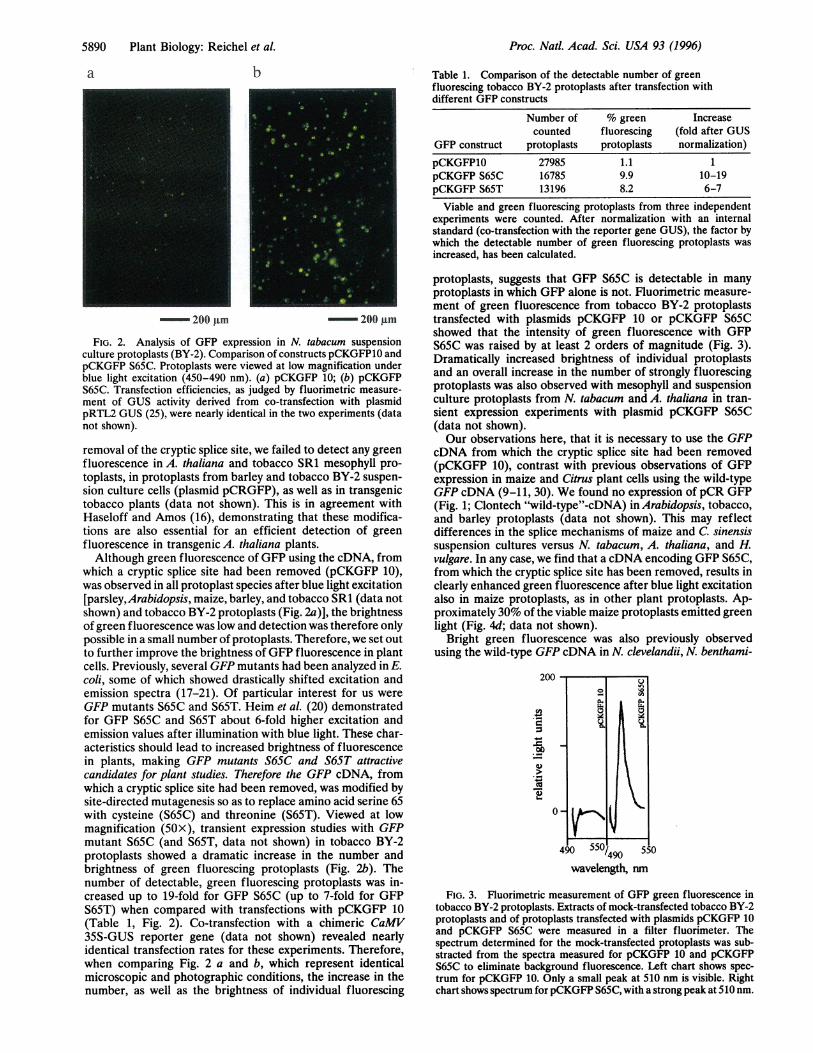
Fluorimetry. For the fluorimetric measurement of GFPgreen fluorescence, tobacco BY-2 protoplasts were trans-fected with plasmids pCKGFP 10 and pCKGFP S65C. Viableand dead protoplasts were separated 24 h after transfection bycentrifugation (3,300 x g for 20 min) through 33% Percol.Viable protoplasts were collected at gradient top and dis-rupted by six passages through an 18-gauge needle. Extractswere diluted 10-fold in 50 mM sodium phosphate buffer (pH7), and an equivalent of approximately 50,000 protoplasts wasmeasured in a filter fluorimeter (LS-2B, Perkin-Elmer) usingexcitation filter 139168 (480 nm) for blue light excitation. Theemission spectra were scanned between 495 and 540 nm, andthe emission spectrum determined for a mock-transfectedtobacco BY-2 sample was substracted from the spectra deter-mined for transfections with plasmids pCKGFP 10 and pCK-GFP S65C.
RESULTS AND DISCUSSIONSeveral plasmids containing the GFP cDNA driven by theCaMV35S promoter were tested in transient gene expressionstudies in protoplasts derived from suspension culture cells aswell as leaf mesophyll. Protoplasts were transfected usingPEG-mediated gene transfer or electroporation (maize), andGFP expression was monitored 20-48 h after transfection bythe detection of green fluorescence using fluorescence mi-croscopy.
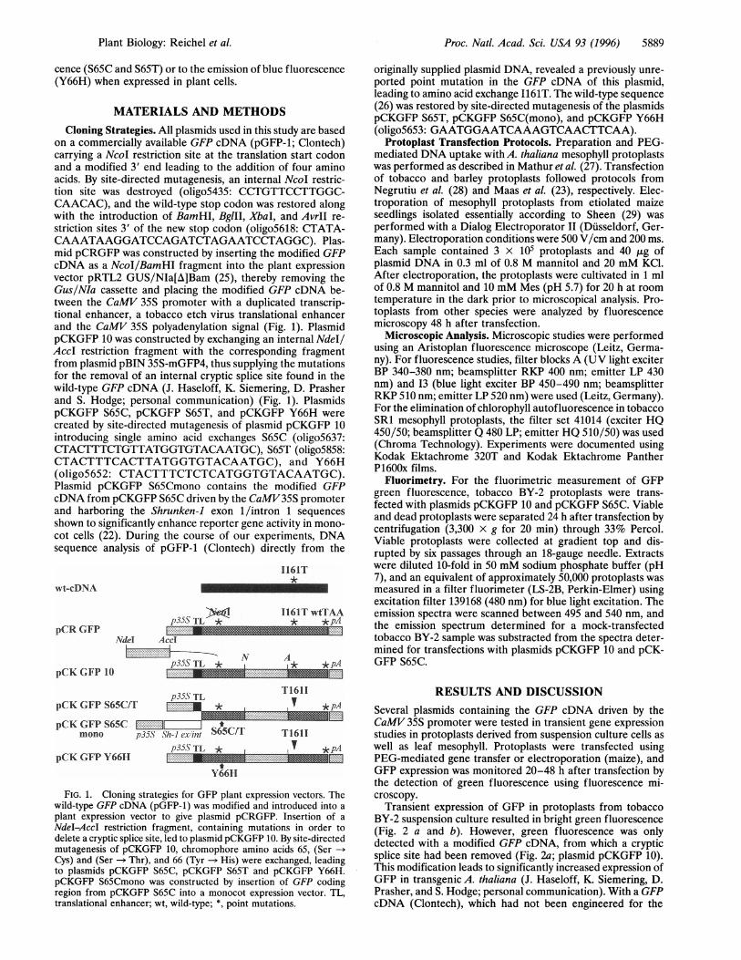
Transient expression of GFP in protoplasts from tobaccoBY-2 suspension culture resulted in bright green fluorescence(Fig. 2 a and b). However, green fluorescence was onlydetected with a modified GFP cDNA, from which a crypticsplice site had been removed (Fig. 2a; plasmid pCKGFP 10).This modification leads to significantly increased expression ofGFP in transgenic A. thaliana (J. Haseloff, K. Siemering, D.Prasher, and S. Hodge; personal communication). With a GFPcDNA (Clontech), which had not been engineered for the
Plant Biology: Reichel et al.
5890 Plant Biology: Reichel et al.
a b
200 gm -^ 200 gmFIG. 2. Analysis of GFP expression in N. tabacum suspension
culture protoplasts (BY-2). Comparison of constructs pCKGFP10 andpCKGFP S65C. Protoplasts were viewed at low magnification underblue light excitation (450-490 nm). (a) pCKGFP 10; (b) pCKGFPS65C. Transfection efficiencies, as judged by fluorimetric measure-ment of GUS activity derived from co-transfection with plasmidpRTL2 GUS (25), were nearly identical in the two experiments (datanot shown).removal of the cryptic splice site, we failed to detect any greenfluorescence in A. thaliana and tobacco SR1 mesophyll pro-toplasts, in protoplasts from barley and tobacco BY-2 suspen-sion culture cells (plasmid pCRGFP), as well as in transgenictobacco plants (data not shown). This is in agreement withHaseloff and Amos (16), demonstrating that these modifica-tions are also essential for an efficient detection of greenfluorescence in transgenic A. thaliana plants.Although green fluorescence of GFP using the cDNA, from
which a cryptic splice site had been removed (pCKGFP 10),was observed in all protoplast species after blue light excitation[parsley, Arabidopsis, maize, barley, and tobacco SR1 (data notshown) and tobacco BY-2 protoplasts (Fig. 2a)], the brightnessof green fluorescence was low and detection was therefore onlypossible in a small number of protoplasts. Therefore, we set outto further improve the brightness of GFP fluorescence in plantcells. Previously, several GFP mutants had been analyzed in E.coli, some of which showed drastically shifted excitation andemission spectra (17-21). Of particular interest for us wereGFP mutants S65C and S65T. Heim et al. (20) demonstratedfor GFP S65C and S65T about 6-fold higher excitation andemission values after illumination with blue light. These char-acteristics should lead to increased brightness of fluorescencein plants, making GFP mutants S65C and S65T attractivecandidates for plant studies. Therefore the GFP cDNA, fromwhich a cryptic splice site had been removed, was modified bysite-directed mutagenesis so as to replace amino acid serine 65with cysteine (S65C) and threonine (S65T). Viewed at lowmagnification (50x), transient expression studies with GFPmutant S65C (and S65T, data not shown) in tobacco BY-2protoplasts showed a dramatic increase in the number andbrightness of green fluorescing protoplasts (Fig. 2b). Thenumber of detectable, green fluorescing protoplasts was in-creased up to 19-fold for GFP S65C (up to 7-fold for GFPS65T) when compared with transfections with pCKGFP 10(Table 1, Fig. 2). Co-transfection with a chimeric CaMV35S-GUS reporter gene (data not shown) revealed nearlyidentical transfection rates for these experiments. Therefore,when comparing Fig. 2 a and b, which represent identicalmicroscopic and photographic conditions, the increase in thenumber, as well as the brightness of individual fluorescing
Table 1. Comparison of the detectable number of greenfluorescing tobacco BY-2 protoplasts after transfection withdifferent GFP constructs
Number of % green Increasecounted fluorescing (fold after GUS
GFP construct protoplasts protoplasts normalization)pCKGFP10 27985 1.1 1pCKGFP S65C 16785 9.9 10-19pCKGFP S65T 13196 8.2 6-7
Viable and green fluorescing protoplasts from three independentexperiments were counted. After normalization with an internalstandard (co-transfection with the reporter gene GUS), the factor bywhich the detectable number of green fluorescing protoplasts wasincreased, has been calculated.
protoplasts, suggests that GFP S65C is detectable in manyprotoplasts in which GFP alone is not. Fluorimetric measure-ment of green fluorescence from tobacco BY-2 protoplaststransfected with plasmids pCKGFP 10 or pCKGFP S65Cshowed that the intensity of green fluorescence with GFPS65C was raised by at least 2 orders of magnitude (Fig. 3).Dramatically increased brightness of individual protoplastsand an overall increase in the number of strongly fluorescingprotoplasts was also observed with mesophyll and suspensionculture protoplasts from N. tabacum and A. thaliana in tran-sient expression experiments with plasmid pCKGFP S65C(data not shown).Our observations here, that it is necessary to use the GFP
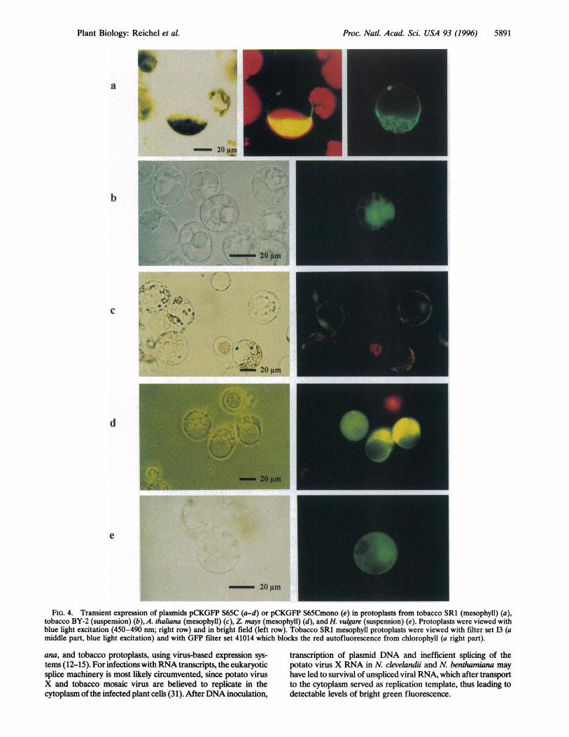
cDNA from which the cryptic splice site had been removed(pCKGFP 10), contrast with previous observations of GFPexpression in maize and Citrus plant cells using the wild-typeGFP cDNA (9-11, 30). We found no expression of pCR GFP(Fig. 1; Clontech "wild-type"-cDNA) in Arabidopsis, tobacco,and barley protoplasts (data not shown). This may reflectdifferences in the splice mechanisms of maize and C. sinensissuspension cultures versus N. tabacum, A. thaliana, and H.vulgare. In any case, we find that a cDNA encoding GFP S65C,from which the cryptic splice site has been removed, results inclearly enhanced green fluorescence after blue light excitationalso in maize protoplasts, as in other plant protoplasts. Ap-proximately 30% of the viable maize protoplasts emitted greenlight (Fig. 4d; data not shown).
Bright green fluorescence was also previously observedusing the wild-type GFP cDNA in N. clevelandii, N. benthami-
200 1
0'
wavelength, nm
FIG. 3. Fluorimetric measurement of GFP green fluorescence intobacco BY-2 protoplasts. Extracts of mock-transfected tobacco BY-2protoplasts and of protoplasts transfected with plasmids pCKGFP 10and pCKGFP S65C were measured in a filter fluorimeter. Thespectrum determined for the mock-transfected protoplasts was sub-stracted from the spectra measured for pCKGFP 10 and pCKGFPS65C to eliminate background fluorescence. Left chart shows spec-trum for pCKGFP 10. Only a small peak at 510 nm is visible. Rightchart shows spectrum forpCKGFP S65C, with a strong peak at 510 nm.
r 1
Proc. Natl. Acad. Sci. USA 93 (1996)
Proc. Natl. Acad. Sci. USA 93 (1996) 5891
Ia
20 un
...... ~'._
I.r-
I ..'t - _
·, ~ .,
,l
t
C 4
0l,..
A1 *1
'- 20 gmorr0lm
d
e
-_ 20gm
FIG. 4. Transient expression of plasmids pCKGFP S65C (a-d) or pCKGFP S65Cmono (e) in protoplasts from tobacco SR1 (mesophyll) (a),tobacco BY-2 (suspension) (b),A. thaliana (mesophyll) (c), Z. mays (mesophyll) (d), and H. vulgare (suspension) (e). Protoplasts were viewed withblue light excitation (450-490 nm; right row) and in bright field (left row). Tobacco SR1 mesophyll protoplasts were viewed with filter set I3 (amiddle part, blue light excitation) and with GFP filter set 41014 which blocks the red autofluorescence from chlorophyll (a right part).
ana, and tobacco protoplasts, using virus-based expression sys-tems (12-15). For infections withRNA transcripts, the eukaryoticsplice machinery is most likely circumvented, since potato virusX and tobacco mosaic virus are believed to replicate in thecytoplasm of the infected plant cells (31). AfterDNA inoculation,
transcription of plasmid DNA and inefficient splicing of thepotato virus X RNA in N. clevelandii and N. benthamiana mayhave led to survival of unspliced viral RNA, which after transportto the cytoplasm served as replication template, thus leading todetectable levels of bright green fluorescence.
S
-, ~., r;. -.
,'b s. ,m^
**f ~.** ..
''^:
I
Plant Biology: Reichel et aL.
.t^t7
.4..ti,
5892 Plant Biology: Reichel et al.
In subsequent experiments we used the GFP cDNA exclu-sively, from which the cryptic splice site had been removed,combined with mutation S65C, either under control of theCaMV 35S promoter for expression in dicotyledonous cells, oroptimized for expression in monocot cells by combination withthe Shrunken-1 intron 1 and exon 1 (22). The excitationmaximum (479 nm) of this particular mutant lies within thelimits of the used fluorescein isothiocyanate filter set (exciterBP 450-490 nm), allowing an optimal excitation of GFP S65C,while the range of this filter set is already suboptimal for theexcitation maximum of GFP S65T (488 nm). This also mayexplain why GFP mutant S65T in our hands leads to a slightlylower increase (6-7-fold) in the number of detectable greenfluorescing protoplasts, when compared with GFP S65C (up to19-fold; Table 1).
In protoplasts from tobacco SR1 and BY-2 (Fig. 4 a and b),A. thaliana (Fig. 4c), Zea mays (Fig. 4d), and H. vulgare (Fig.4e), bright green fluorescence was observed in transient ex-pression experiments with the respective plasmids designed formonocotyledonous (pCKGFP S65Cmono; Fig. 1) or dicotyle-donous plant cells (pCKGFP S65C; Fig. 1). Green fluores-cence of GFP in tobacco SR1 mesophyll protoplasts wasinitially somewhat obscured by the strong red autofluores-cence of chlorophyll after illumination with blue light. Nev-ertheless, in tobacco mesophyll protoplasts expressing highlevels of GFP or with chloroplasts out of focus or concentratedin one half of the cell, detection of bright green fluorescencewas clearly possible (Fig. 4a middle panel). To reduce prob-lems due to autofluorescence of chloroplasts, we subsequentlyused filtersets, specially designed for GFP detection, that blockred light without affecting green fluorescence derived fromGFP expression. In the tobacco mesophyll protoplast, thestrong background of red chlorophyll fluorescence (Fig. 4amiddle panel) was clearly reduced when using filter set 41014(Fig. 4a right panel).
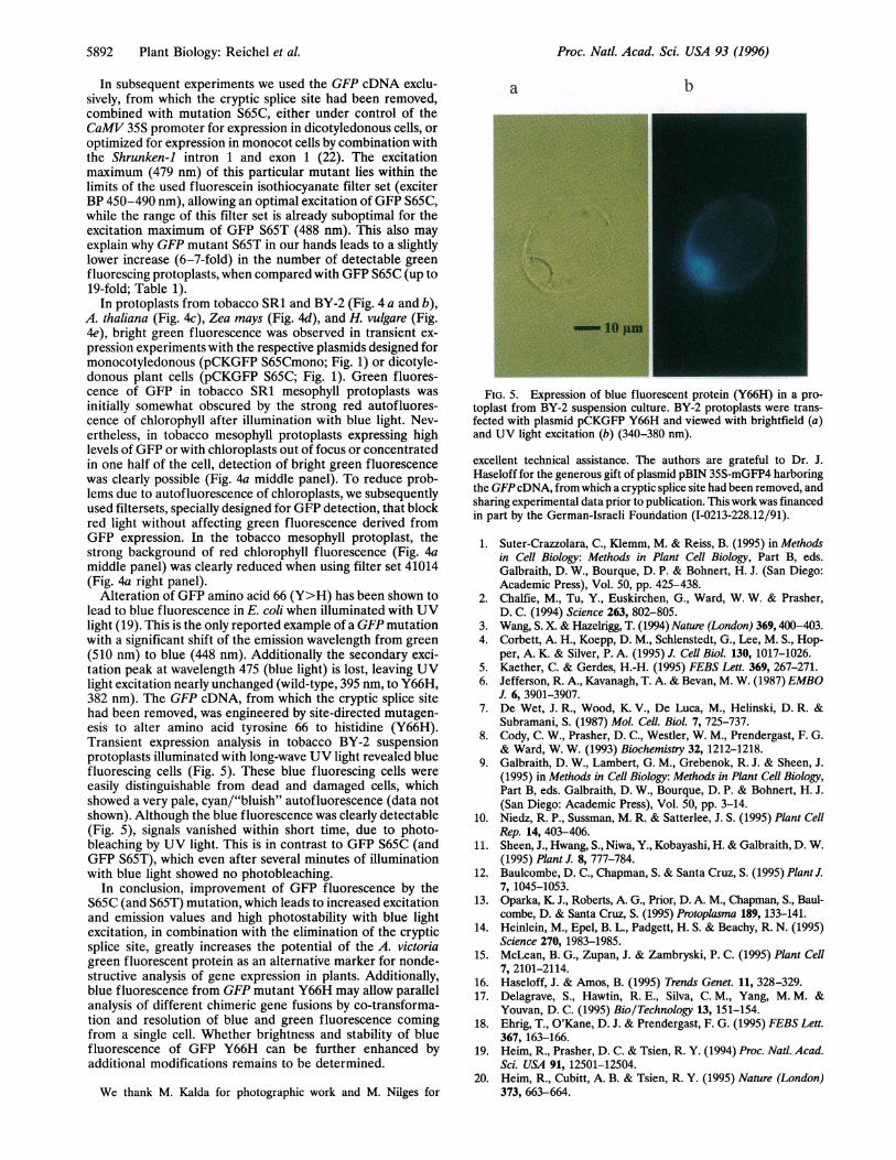
Alteration of GFP amino acid 66 (Y>H) has been shown tolead to blue fluorescence in E. coli when illuminated with UVlight (19). This is the only reported example of a GFP mutationwith a significant shift of the emission wavelength from green(510 nm) to blue (448 nm). Additionally the secondary exci-tation peak at wavelength 475 (blue light) is lost, leaving UVlight excitation nearly unchanged (wild-type, 395 nm, to Y66H,382 nm). The GFP cDNA, from which the cryptic splice sitehad been removed, was engineered by site-directed mutagen-esis to alter amino acid tyrosine 66 to histidine (Y66H).Transient expression analysis in tobacco BY-2 suspensionprotoplasts illuminated with long-wave UV light revealed bluefluorescing cells (Fig. 5). These blue fluorescing cells wereeasily distinguishable from dead and damaged cells, whichshowed a very pale, cyan/"bluish" autofluorescence (data notshown). Although the blue fluorescence was clearly detectable(Fig. 5), signals vanished within short time, due to photo-bleaching by UV light. This is in contrast to GFP S65C (andGFP S65T), which even after several minutes of illuminationwith blue light showed no photobleaching.
In conclusion, improvement of GFP fluorescence by theS65C (and S65T) mutation, which leads to increased excitationand emission values and high photostability with blue lightexcitation, in combination with the elimination of the crypticsplice site, greatly increases the potential of the A. victoriagreen fluorescent protein as an alternative marker for nonde-structive analysis of gene expression in plants. Additionally,blue fluorescence from GFP mutant Y66H may allow parallelanalysis of different chimeric gene fusions by co-transforma-tion and resolution of blue and green fluorescence comingfrom a single cell. Whether brightness and stability of bluefluorescence of GFP Y66H can be further enhanced byadditional modifications remains to be determined.
We thank M. Kalda for photographic work and M. Nilges for
a b
FIG. 5. Expression of blue fluorescent protein (Y66H) in a pro-toplast from BY-2 suspension culture. BY-2 protoplasts were trans-fected with plasmid pCKGFP Y66H and viewed with brightfield (a)and UV light excitation (b) (340-380 nm).excellent technical assistance. The authors are grateful to Dr. J.Haseloff for the generous gift of plasmid pBIN 35S-mGFP4 harboringthe GFP cDNA, from which a cryptic splice site had been removed, andsharing experimental data prior to publication. This workwas financedin part by the German-Israeli Foundation (I-0213-228.12/91).1. Suter-Crazzolara, C., Klemm, M. & Reiss, B. (1995) in Methods
in Cell Biology: Methods in Plant Cell Biology, Part B, eds.Galbraith, D. W., Bourque, D. P. & Bohnert, H. J. (San Diego:Academic Press), Vol. 50, pp. 425-438.
2. Chalfie, M., Tu, Y., Euskirchen, G., Ward, W. W. & Prasher,D. C. (1994) Science 263, 802-805.
3. Wang, S. X & Hazelrigg, T. (1994) Nature (London) 369,400-403.4. Corbett, A. H., Koepp, D. M., Schlenstedt, G., Lee, M. S., Hop-
per, A. K. & Silver, P. A. (1995) J. Cell Biol. 130, 1017-1026.5. Kaether, C. & Gerdes, H.-H. (1995) FEBS Lett. 369, 267-271.6. Jefferson, R. A., Kavanagh, T. A. & Bevan, M. W. (1987) EMBO
J. 6, 3901-3907.7. De Wet, J. R., Wood, K. V., De Luca, M., Helinski, D. R. &
Subramani, S. (1987) Mol. Cell. Biol. 7, 725-737.8. Cody, C. W., Prasher, D. C., Westler, W. M., Prendergast, F. G.
& Ward, W. W. (1993) Biochemistry 32, 1212-1218.9. Galbraith, D. W., Lambert, G. M., Grebenok, R. J. & Sheen, J.
(1995) in Methods in Cell Biology: Methods in Plant Cell Biology,Part B, eds. Galbraith, D. W., Bourque, D. P. & Bohnert, H. J.(San Diego: Academic Press), Vol. 50, pp. 3-14.
10. Niedz, R. P., Sussman, M. R. & Satterlee, J. S. (1995) Plant CellRep. 14, 403-406.
11. Sheen, J., Hwang, S., Niwa, Y., Kobayashi, H. & Galbraith, D. W.(1995) Plant J. 8, 777-784.
12. Baulcombe, D. C., Chapman, S. & Santa Cruz, S. (1995) Plant J.7, 1045-1053.
13. Oparka, K. J., Roberts, A. G., Prior, D. A M., Chapman, S., Baul-combe, D. & Santa Cruz, S. (1995) Protoplasma 189,133-141.
14. Heinlein, M., Epel, B. L., Padgett, H. S. & Beachy, R. N. (1995)Science 270, 1983-1985.
15. McLean, B. G., Zupan, J. & Zambryski, P. C. (1995) Plant Cell7, 2101-2114.
16. Haseloff, J. & Amos, B. (1995) Trends Genet. 11, 328-329.17. Delagrave, S., Hawtin, R. E., Silva, C. M., Yang, M. M. &
Youvan, D. C. (1995) Bio/Technology 13, 151-154.18. Ehrig, T., O'Kane, D. J. & Prendergast, F. G. (1995) FEBS Lett.
367, 163-166.19. Heim, R., Prasher, D. C. & Tsien, R. Y. (1994) Proc. Natl. Acad.
Sci. USA 91, 12501-12504.20. Heim, R., Cubitt, A. B. & Tsien, R. Y. (1995) Nature (London)
373, 663-664.
Proc. Natl. Acad. Sci. USA 93 (1996)

Plant Biology: Reichel et al.
21. Cubitt, A. B., Heim, R., Adams, S. R., Boyd, A. E., Gross, L. A.& Tsien, R. Y. (1995) Trends Biochem. Sci. 20, 448-455.
22. Maas, C., Laufs, J., Grant, S., Korfhage, C. & Werr, W. (1991)Plant Mol. Biol. 16, 199-207.
23. Maas, C., Reichel, C., Schell, J. & SteinbiB, H.-H. (1995) inMethods in Cell Biology: Methods in Plant CellBiology, Part B, eds.Galbraith, D. W., Bourque, D. P. & Bohnert, H. J. (San Diego:Academic Press), Vol. 50, pp. 383-399.
24. Fischer, R. & Hain, R. (1995) in Methods in Cell Biology: Methodsin Plant Cell Biology, Part B, eds. Galbraith, D. W., Bourque,D. P. & Bohnert, H. J. (San Diego: Academic Press), Vol. 50, pp.401-410.
Proc. Natl. Acad. Sci. USA 93 (1996) 5893
25. Carrington, J. C., Freed, D. D. & Leinicke, A. J. (1991) Plant Cell3, 953-962.
26. Prasher, D. C., Eckenrode, V. K., Ward, W. W., Prendergast,F. G. & Cormier, M. J. (1992) Gene (Amst.) 111, 229-233.
27. Mathur, J., Koncz, C. & Szabados, L. (1995) Plant Cell Rep. 14,221-226.
28. Negrutiu, I., Shillito, R., Potrykus, I., Biasini, G. & Sala, F. (1987)Plant Mol. Biol. 8, 363-373.
29. Sheen, J. (1990) Plant Cell 2, 1027-1038.30. Hu, W. & Cheng, C.-L (1995) FEBS Lett. 369, 331-334.31. Matthews, R. E. F. (1991) Plant Vrology. 3rd Ed., (San Diego:
Academic Press).