Embed Size (px)
Citation preview
Accepted Manuscript
Efficacy of Endoscopic Dilation of gastroduodenal Crohn’s disease strictures: ASystematic Review and Meta-analysis of Individual Patient Data
Dominik Bettenworth, Marcus M. Mücke, Rocio Lopez, Amandeep Singh, WeimingZhu, Feilong Guo, Toshiyuki Matsui, Theodore W. James, Hans Herfarth, MartinGoetz, Ren Mao, Satya Kurada, Jochen Hampe, Katja Matthes, John GásdalKarstensen, Piero V. Valli, Marjolijn Duijvestein, Geert D’Haens, Vipul Jairath, TianBai Qiu, Nik Sheng Ding, Gerhard Rogler, Florian Rieder
PII: S1542-3565(18)31325-9DOI: https://doi.org/10.1016/j.cgh.2018.11.048Reference: YJCGH 56220
To appear in: Clinical Gastroenterology and HepatologyAccepted Date: 24 November 2018
Please cite this article as: Bettenworth D, Mücke MM, Lopez R, Singh A, Zhu W, Guo F, Matsui T,James TW, Herfarth H, Goetz M, Mao R, Kurada S, Hampe J, Matthes K, Karstensen JG, Valli PV,Duijvestein M, D’Haens G, Jairath V, Qiu TB, Ding NS, Rogler G, Rieder F, Efficacy of EndoscopicDilation of gastroduodenal Crohn’s disease strictures: A Systematic Review and Meta-analysis ofIndividual Patient Data, Clinical Gastroenterology and Hepatology (2018), doi: https://doi.org/10.1016/j.cgh.2018.11.048.
This is a PDF file of an unedited manuscript that has been accepted for publication. As a service toour customers we are providing this early version of the manuscript. The manuscript will undergocopyediting, typesetting, and review of the resulting proof before it is published in its final form. Pleasenote that during the production process errors may be discovered which could affect the content, and alllegal disclaimers that apply to the journal pertain.

MANUSCRIP
T
ACCEPTED
ACCEPTED MANUSCRIPT
1
Efficacy of Endoscopic Dilation of gastroduodenal Crohn’s disease strictures: A
Systematic Review and Meta-analysis of Individual Patient Data
Dominik Bettenworth1, Marcus M. Mücke2, Rocio Lopez3 Amandeep Singh4, Weiming Zhu5,
Feilong Guo5, Toshiyuki Matsui6, Theodore W. James7, Hans Herfarth7, Martin Goetz8, Ren
Mao9,10, Satya Kurada10,11, Jochen Hampe12, Katja Matthes12, John Gásdal Karstensen13,14,
Piero V. Valli15, Marjolijn Duijvestein16, Geert D’Haens16, Vipul Jairath17,18, Tian Bai Qiu19,
Nik Sheng Ding19, Gerhard Rogler15, Florian Rieder10,11
1Department of Medicine B, University Hospital Münster, Münster, Germany 2Department of Internal Medicine 1, University Hospital Frankfurt, Frankfurt a.M., Germany 3Department of Quantitative Health Sciences, Lerner Research Institute, Cleveland Clinic
Foundation, Cleveland, Ohio, USA 4Department of Gastroenterology and Hepatology, Center for Human Nutrition, Cleveland
Clinic Foundation, Cleveland, Ohio, USA 5Department of General Surgery, Jinling Hospital, School of Medicine, Nanjing University,
Nanjing, People’s Republic of China 6Department of Gastroenterology, Fukuoka University Chikushi Hospital, Chikushino, Japan 7Division of Gastroenterology and Hepatology, University of North Carolina at Chapel Hill,
Chapel Hill, North Carolina, USA 8First Department of Internal Medicine I, University Hospital Tübingen, Tübingen, Germany 9Department of Gastroenterology, First Affiliated Hospital of Sun Yat-sen University, China 10Department of Inflammation and Immunity, Lerner Research Institute, Cleveland Clinic
Foundation, Cleveland, Ohio, USA
11Department of Gastroenterology, Hepatology & Nutrition, Digestive Diseases and Surgery
Institute, Cleveland Clinic Foundation, Cleveland, Ohio, USA
12Medical Department 1, University Hospital Dresden, Technical University Dresden,
Dresden, Germany 13Gastro Unit, Division of Endoscopy, Copenhagen University Hospital Herlev, Denmark 14Gastro Unit, Division of Surgery, Copenhagen University Hospital Hvidovre, Denmark 15Department of Gastroenterology and Hepatology, University Hospital Zurich, Zurich,
Switzerland 16Academic Medical Center, Amsterdam, The Netherlands

MANUSCRIP
T
ACCEPTED
ACCEPTED MANUSCRIPT
2
17Department of Medicine, University of Western Ontario, London, ON, Canada; 18Department of Epidemiology and Biostatistics, University of Western Ontario, London, ON,
Canada; 19Department of Gastroenterology, St. Vincent, Melbourne, Australia
Short title: Upper GI Crohn’s disease strictures
Address for correspondence: Dr. Dominik Bettenworth, Department of Medicine B,
Gastroenterology and Hepatology, University Hospital Münster, Albert-Schweitzer-Campus
1, D-48149 Münster, Germany; Phone +49-251-83-47661, fax +49-251-83-47570
E-mail: [email protected]
Specific author contributions: DB and FR planed and conducted the study and drafted the
manuscript. MMM, RL, AS, WZ, FG, TM, TWJ, HH, MG, RM, SK, JH, KM, JGK, PV, MD,
GD, VJ, NSD; TBQ and GR collected and interpreted data. All authors have contributed to
drafting the manuscript and approved the final version of the manuscript.
Grant support: This work was supported by grants from the National Institutes of Health
[T32DK083251, P30DK097948 Pilot, K08DK110415] to F.R.
Conflicts of interest and potential competing interests: None
Abbreviations: EBD, endoscopic balloon dilatation; GI, gastrointestinal; Psi, pounds per
square inch; SP, strictureplasty; TNF, tumor necrosis factor; CD, Crohn’s disease; TTS,
through-the-scope.
Writing assistance: None
ABSTRACT

MANUSCRIP
T
ACCEPTED
ACCEPTED MANUSCRIPT
3
Background & Aims: Little is know about the effects of endoscopic balloon dilation (EBD)
for strictures of the upper gastrointestinal (UGI) tract in patients with Crohn’s disease (CD).
We performed a pooled analysis of the efficacy and safety of EBD for UGI CD-associated
strictures.
Methods: We searched the EMBASE, Medline and Cochrane library, as well as
bibliographies of relevant articles, for cohort studies of adults with CD and strictures of the
stomach, or duodenum (up to the ligament of Treitz) who underwent EBD through December
2016. We obtained data from 7 international referral centers on 94 patients who underwent
141 EBD. We performed a patient-level meta-analysis of data from published and
unpublished cohort studies to determine mechanical and clinical success. We performed time
to event analysis to assess symptom recurrence and need for re-dilation or surgery. The
patients analyzed had strictures of the duodenum (n=107), stomach (n=30), or spanning both
(n=4).
Results: The rate of technical success for EBD was 100%, with 87% short term clinical
efficacy; major complications arose from 2.9% of all procedures. During median follow up of
23.1 months, 70.5% patients had recurrence of symptoms, 59.6% required re-dilation, and
30.8% required surgical intervention. Patients whose disease was located in the small bowel
had a higher risk for symptom recurrence (hazard ratio [HR], 2.1; P=.003). Asian race (HR,
2.8; P<.001) and location of disease in the small bowel (HR, 1.9; P=.004) increased the need
for re-dilation. Prestenotic dilation was a risk factor for earlier need for surgery (HR, 1.9;
P=.001).
Conclusions: In a meta-analysis, we found EBD for CD-associated strictures of the UGI to be
a effective alternative to surgery, with a high rate of short-term technical and clinical success,
moderate long-term efficacy, and an acceptable rate of complications.
KEY WORDS : Therapy; fibrosis; stenosis; IBD; endoscopy.
MANUSCRIP
T
ACCEPTED
ACCEPTED MANUSCRIPT
4
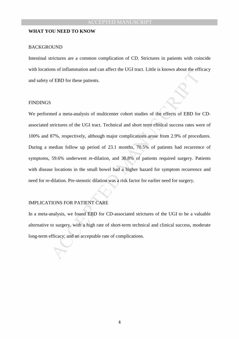
WHAT YOU NEED TO KNOW
BACKGROUND
Intestinal strictures are a common complication of CD. Strictures in patients with coincide
with locations of inflammation and can affect the UGI tract. Little is known about the efficacy
and safety of EBD for these patients.
FINDINGS
We performed a meta-analysis of multicenter cohort studies of the effects of EBD for CD-
associated strictures of the UGI tract. Technical and short term clinical success rates were of
100% and 87%, respectively, although major complications arose from 2.9% of procedures.
During a median follow up period of 23.1 months, 70.5% of patients had recurrence of
symptoms, 59.6% underwent re-dilation, and 30.8% of patients required surgery. Patients
with disease locations in the small bowel had a higher hazard for symptom recurrence and
need for re-dilation. Pre-stenotic dilation was a risk factor for earlier need for surgery.
IMPLICATIONS FOR PATIENT CARE
In a meta-analysis, we found EBD for CD-associated strictures of the UGI to be a valuable
alternative to surgery, with a high rate of short-term technical and clinical success, moderate
long-term efficacy, and an acceptable rate of complications.

MANUSCRIP
T
ACCEPTED
ACCEPTED MANUSCRIPT
5
INTRODUCTION
Crohn’s disease (CD) may affect the entire gastrointestinal (GI) tract (1). As a consequence of
transmural inflammation and a relapsing and remitting disease course, clinically apparent
fibrostenosis of the intestine occurs in 20% of patients within 20 years after initial diagnosis
(2). Despite recent advances in the medical treatment of CD, prevention and treatment of
stricturing CD remains a large unmet need (3). Due to the absence of specific anti-fibrotic
therapies (4), CD patients with intestinal obstruction are commonly treated by surgical
intervention such as strictureplasty or bowel resection (5), both of which can be associated
with significant complications (6, 7).
While CD may affect all parts of the GI tract, involvement of the stomach and duodenum is
rarely reported. More specifically, the incidence of CD-associated strictures of the upper GI
tract is below 4% (8). Beside the occurrence of strictures, fistulae development in the upper
GI tract has been reported as well (9, 10). Historically, in the pre-steroid era, surgery was the
solely available treatment modality for obstructive duodenal CD, but was often accompanied
by a complicated postoperative course with postoperative abscesses (11, 12). Subsequently,
with the advent of corticosteroids and immunosuppressive drugs, some case reports are
available that illustrate cases with a – at least temporary - successfully medical treatment (8,
9, 11). Therefore, stricturing as well as fistulizing complications of the upper GI tract may
generate challenging clinical scenarios for both, affected CD patients as well as health care
providers alike. The very limited body of published evidence for this treatment scenario is
further aggravating this clinical dilemma.
Endoscopic balloon dilation (EBD) has emerged as an alternative to surgery and has been
proven effective for the treatment of fibrotic CD-associated strictures of the ileocecum. EBD

MANUSCRIP
T
ACCEPTED
ACCEPTED MANUSCRIPT
6
has therefore been implemented in current CD treatment algorithms and guidelines (13-15).
EBD for CD-associated strictures of the upper GI tract is rarely reported, with the largest
cohort comprising 35 patients from a single center (16). Therefore, the aim of this pooled
analysis of international multicenter cohort studies, combining published cohorts with a
multicenter investigation for a patient level meta-analysis, was to evaluate safety and efficacy
of EBD in upper GI Crohn’s disease associated strictures, in order to provide evidence-based
guidance for this clinical situation.

MANUSCRIP
T
ACCEPTED
ACCEPTED MANUSCRIPT
7
METHODS AND MATERIAL
Literature search and data
Detailed information regarding the literature search and data, as well as a PRISMA diagram
(Supplemental Figure 1) can be found in the supplement. Corresponding authors of papers
that fulfilled the inclusion and exclusion criteria were contacted via e-mail to obtain single
patient level data of the subjects included in the studies. These data represent part one of the
pooled individual patient analysis.
In addition, 7 high volume inflammatory bowel disease (IBD) endoscopy centers were
contacted and provided clinical data of cumulative 24 patients. These data represent part two
of the pooled individual patient analysis. Of note, comparable criteria were used to assess the
performance of dilation procedures as well as treatment outcomes in relation to the already
published studies and as defined below.
Data collection
Used definitions for the explored of included studies (16-19) were reported in the
supplemental text and supplemental table 1, Tables 2 & 3 in detail.
Statistical analysis
Detailed information about the statistical analysis can be found in the supplement.

MANUSCRIP
T
ACCEPTED
ACCEPTED MANUSCRIPT
8
RESULTS
Pooled individual patient analysis
In total, we included 94 patients from 11 different tertiary referral centers for IBD (located in
the United States, Australia, China, Japan, Denmark, Germany, Netherlands and Switzerland).
The number of total dilation procedures was 141. 70 patients undergoing 112 dilations have
been published in multiple individual smaller series prior (16-21) and 24 patients undergoing
29 dilations were not previously published and added through our multicenter collaboration
(Table 1). The demographics of this population are depicted in Table 2. The median age at
diagnosis of CD was 24 ± 10.6 years while the median age at time of stricture diagnosis was
33.7 ± 14.3 years. 75.9% of strictures were located in the duodenum, 21.3% of patients
developed strictures in the stomach and 2.8% of patients presented with strictures that
spanned from the stomach to the duodenum (Table 2). Most strictures were de novo strictures
(89.1%; as opposed to anastomotic strictures) at sites without prior surgery. The median
length of stricture was 3 cm [25th percentile 2, 75th percentile 10] with 85.4% of strictures
being less than 5 cm long. All strictures were dilated by through-the-scope (TTS) systems and
70.1% of the investigators applied graded dilation. The median maximum balloon diameter
used was 15 mm [14.0, 18.0] and the vast majority of investigators employed a balloon of 5.5
[5.5, 5.5] cm length. The median time of balloon inflation was 2 min [2, 3] and a pressure of
20 psi was applied (Table 3). Technical success rate was 100% and EBD lead to short term
clinical efficacy in 87% of all patients. Of note, technical success did not result in clinical
improvement of obstructive symptoms in all patients
Major complications (defined as perforation, bleeding or dilation-related need for surgery),
occurred in 2.9% per procedure and in 4% per patient, considering the possibility of multiple
dilations over time in the same study subject. During a median follow-up period of 23.1

MANUSCRIP
T
ACCEPTED
ACCEPTED MANUSCRIPT
9
months, 70.5% of patients reported symptomatic recurrence, 59.6% required another dilation
and 30.8% of all patients had to undergo surgical intervention (Table 2, 3 and Fig. 1 A-C).
Table 1 presents descriptive statistics for patients from each center as well as the random
effects analysis and measures of variability. The overall rates for symptom, dilation and
surgery free survival at 6, 12 and 24 months are depicted in Table 4.
To assess potential bias, we included both published and unpublished studies (Suppl. Tables
2a, 3a, 4a and 5a) and performed a sensitivity analysis separately after excluding unpublished
studies (Suppl. Tables 2b, 3b, 4b and 5b). These results are presented and discussed in the
supplement to this article and the below data refers to the complete cohort only.
Factors associated with short-term dilation outcome
Given the achievement of technical success in all included subjects, no analysis for factors
associated with technical success could be performed. We hence evaluated factors associated
with short term clinical efficacy of dilation procedures. Patients with active disease in the
ileocecum had a lower likelihood of relief of symptoms in the adjusted and unadjusted
analysis (OR 0.27, confidence interval (CI)95%: 0.058-1.2; p=0.087 and OR 0.27, CI95%:
0.06 - 1.2; p=0.083). No other factor was linked to clinical efficacy (Supplemental Table 2a).
Factors associated with symptom recurrence
We next assessed factors associated with symptom recurrence. In univariate analysis, disease
location in the jejunum/proximal ileum (Hazard ratio (HR) 1.8; CI95%: 1.09-2.9; p=0.022)
was associated with an increased hazard for symptom recurrence. In contrast, a stricture
length of less than 5 cm (HR 0.41; CI95%: 0.24-0.7; p=0.001) was negatively associated with
time to symptom recurrence. While disease location in the jejunum/ileum was linked to

MANUSCRIP
T
ACCEPTED
ACCEPTED MANUSCRIPT
10
symptom recurrence in the multivariate analysis (HR 2.1; CI95%: 1.3-3.5; p=0.003), age at
diagnosis was associated with a diminished risk (HR 0.85; CI95%: 0.72-1.0; p=0.054). Uni-
and multivariate analysis did not detect any other factors that were linked to an increased risk
of symptom recurrence (Supplemental Table 3a).
Factors associated with need for re-dilation
Regarding the need for re-dilation, univariate as well as multivariate analysis showed that
Asian race (HR 2.8; CI95%: 1.7-4.5; p<0.001 and HR 2.8; CI95%: 1.8-4.5; p<0.001) and
disease location in the jejunum/proximal ileum (HR 1.7; CI95%: 1.1-2.6; p=0.015 and HR
1.9; CI95%: 1.2-2.9; p=0.004) were associated with a significantly increased risk of re-
dilation (Supplemental Table 4a). None of the other investigated factors, including smoking,
stricture location, type of stricture (de novo vs. anastomotic), prestenotic dilation, graded
dilation or maximum caliber of dilation or anti-TNF treatment were linked to the need for
additional endoscopic therapy in the univariate or multivariate analysis (Supplemental Table
4a).
Factors associated with the need for surgery
Prestenotic dilation was linked to an increased risk for surgery in the univariate as well as
multivariate analysis (HR2.0; CI95%: 1.4-2.7, p<0.001 and HR 1.9; CI95%: 1.3-2.7; p=0.001;
Supplemental Table 5a). In addition, in the univariate analysis, Asian race and younger age at
time of dilation were found to be associated with an increased risk for surgery (HR 1.7; CI95:
1.2-2.3; p=0.003 and HR 0.92; CI95%: 0.87-0.98; p=0.007), while increased BMI and a
positive family history of CD were associated with a reduced risk of need for surgery, but
could not be confirmed by the multivariate analysis (HR 0.91; CI95%: 0.87-0.96; p<0.001

MANUSCRIP
T
ACCEPTED
ACCEPTED MANUSCRIPT
11
and HR 0.71; CI95: 0.51-0.99; p=0.046). None of the other investigated factors, including
smoking, disease location, type of stricture, graded dilation or maximum caliber of dilation,
was linked to need for earlier surgery (Supplemental Table 5a).
Complications after EBD
Complications associated with dilations occurred in 5 patients. This included 2 perforations
and 3 bleeding events. No patient had to undergo dilation-associated surgery. Due to the low
number of complications in the cohort, no meaningful analysis for risk factors could be
performed. The patients that experienced a perforation were treated by their first dilation
procedure, the stricture length was below 5 cm and both patients did not receive steroid
treatment at the time of intervention.
Efficacy of serial dilations of upper GI strictures
Finally, we analyzed the efficacy of repeat dilations of the same stricture. Data on a second
dilation were available for 22 episodes and a third dilation for 9 episodes. With regard to the
technical success, all second and third dilations were successful. While a symptomatic relief
of symptoms could be achieved in 86.2% of patients through the first dilation, clinical success
was found in 85% of patients being treated by a second dilation and 88.9% of patients
requiring a third dilation. The need for re-dilation was documented in 61.6% of patients
following the first dilation and in 54.5% and 66.7% of patients being treated by a second and
third dilation (all p>0.05). Interestingly, time to re-dilation did not differ significantly
between first, second and third dilation procedure (p>0.05; Supplemental Table 6).

MANUSCRIP
T
ACCEPTED
ACCEPTED MANUSCRIPT
12
DISCUSSION
EBD is frequently used for the treatment of CD-associated strictures throughout the
gastrointestinal tract. A pooled analysis of 3213 dilation procedures supports the efficacy and
safety of this approach (14). Most studies, however, do not provide a separate analysis on the
use of EBD for CD-associated strictures of the upper GI tract. More specifically, the two
largest available studies on this indication only include 24 and 35 patients, respectively (16,
19), precluding a thorough analysis of short and long term efficacy, safety and predictors of
success.
Our pooled analysis of international multi-center cohort studies comprises individual datasets
of 94 CD patients who underwent EBD for upper GI strictures. While 70 of the 94 analyzed
cases were published prior, we were able to include 24 additional cases in the analysis. We
found rates for technical and clinical success of 100% and 87%, respectively, while major
complications occurred in 2.9% per procedure. During a median follow up period of 23.1
months, the pooled individual patient analysis revealed that 70.5% of patients experienced
symptomatic recurrence, 59.6% underwent re-dilation and 30.8% of patients required surgery.
Patients with small bowel disease location had a higher hazard for symptom recurrence and
need for re-dilation. Pre-stenotic dilation was found to be risk factors for earlier need for
surgery.
The largest pooled analysis of EBD for ileocecal strictures reported technical and clinical
success rates of 89.1% and 80.8% (14). Our study shows that dilation therapy for CD-
associated strictures of the upper GI tract is comparably successful in the short term. The very
high technical success rate observed in our study may result from the fact that we defined this
parameter differently from prior studies by the ability to dilate the stricture after starting the

MANUSCRIP
T
ACCEPTED
ACCEPTED MANUSCRIPT
13
procedure (but not by the ability to traverse the stricture after dilation). During a follow up
period of 23 months, long-term outcome parameters for dilation of CD-associated strictures of
the upper GI tract such as symptoms recurrence, need for re-dilation and surgery were not
significantly different as compared to pooled data for CD-associated strictures of the
ileocecum during a follow up of 24 months (70.5 vs. 75.9%; 59.6 vs. 73.5% and 30.8 vs.
42.9%) (14), which came as a surprise, given the common belief of clinicians that upper GI
CD-associated stricture dilation is less durable. Finally, the complications rates per dilation
procedure for ileocecal and upper GI strictures were comparable (2.8% vs. 2.9%) (14). These
results are also in line with the findings from other meta-analyses on EBD for CD-associated
strictures (22, 23). We were not able to assess if fluoroscopy impacted the complications rates
of EBD as the vast majority of investigators reported to use fluoroscopy at the discretion of
the operator and did not record its performance systematically. Taken together, our study
provides evidence that endoscopic dilation of CD-associated strictures of the upper GI tract
by EBD is effective and safe, with rates comparable to dilation therapy for CD-associated
strictures of the ileocecum.
Identification of predictive factors for the long-term success of EBD of the upper GI tract
would allow risk stratification of patients benefitting from a procedure. Patients with disease
location in jejunum/proximal ileum showed a higher rate of symptom recurrence and need for
re-dilation. The patients with pre-stenotic dilation were at a higher risk for surgery. Pre-
stenotic dilation is a feature that is not observed in every patient with stricturing CD but is
regarded as a sign of advanced, longer standing strictures with a higher fibrotic component,
even though evidence supporting this notion is limited. In our cohort, patients’ symptoms
were less likely to improve when pre-stenotic dilation was present and the time to surgery was
shorter. This is in concordance with the recently published CREOLE study, which evaluated
the efficacy of anti-TNF treatment for symptomatic small-bowel strictures (3). Patients with

MANUSCRIP
T
ACCEPTED
ACCEPTED MANUSCRIPT
14
pre-stenotic dilation were at an increased risk for surgery. As jejunal/proximal ileal disease
was found to be negatively associated with symptom recurrence and need for re-dilation, it
can be hypothesized that CD patients with disease manifestation at different locations along
the GI tract may suffer from a more severe phenotype or symptoms classified as obstructive
originating from other regions of the GI tract. This assumption is supported by data from
population-based studies indicating that CD patients with ileal involvement at time of
diagnosis are at an increased risk for a more aggressive disease phenotype (2). Interestingly,
race showed an effect on dilation efficacy. Aside from the fact that Asians may have a higher
risk for stricture recurrence it may be explained by different strategies for the utilization of
endoscopy and management strategy in this region.
Interestingly, active smoking was not found to be a risk factor for worse outcome of dilation
therapy in our study. There is a body of evidence indicating the smoking aggravates the
course of CD (24, 25), increases the risk of postoperative recurrence (26, 27) and smoking
cessation decreases the risk for CD-related surgery (28). One studies reports an increased risk
for stricture recurrence after endoscopic dilation, however, the included patients exclusively
suffered from ileocecal strictures (29).
Given the fact that stricture development follows the location of inflammation, it is logical
that an early appropriate medical treatment of inflammatory CD of the upper GI tract may be
the best approach to prevent the occurrence of CD-associated strictures. Indeed, a study by
Decker and colleagues evaluating 20 patients with esophageal CD from the Mayo Clinic
(Rochester) in the pre-anti-TNF era found modest treatment effects of steroid or
immunomodulatory therapy (clinical response rates of 67% and 55%)(30), and three out of 20
patients finally required esophagectomy. In contrast, the findings from a more recent study
from the same institution analyzing 24 cases of esophageal CD demonstrated that early
aggressive therapy including different anti-TNF agents, systemic and topical steroid therapy

MANUSCRIP
T
ACCEPTED
ACCEPTED MANUSCRIPT
15
resulted in the complete resolution of clinical symptoms in 83% of patients and prevented
surgical interventions in all patients (31). Future prospective studies will need to show, if
early biologic therapy can prevent the occurrence of strictures or their need for redilation or
surgery.
Our study has several limitations. Individual patients’ datasets were retrieved from 11
different centers and variations in the care patterns and approaches of the endoscopists may
influence the results. Additionally and as delineated in the method section, definitions
regarding parameters such as technical success and clinical efficacy differ between various
studies. Key features, however, such as the use of TTS balloons in all cases, maximum caliber
of dilation as well as the duration and pressure of balloon inflation were quite similar among
different centers. Furthermore, we were not able to exclude publication or reporting bias so
that patients in whose dilation therapy could not be technically performed, may be
underrepresented in available publications. The retrospective non-controlled observational
nature of the study did not allow randomization based on risk factors or other criteria. Finally,
the timepoint of evaluation of clinical efficacy was not standardized across studies. However,
clinical efficacy was measured closely to dilation in all cases as symptom relief occurs almost
immediately post procedure. According to the Cochrane risk of bias tool, our study carries all
potential inherent biases of cohort studies with retrospective data collection. In addition,
reporting bias may apply as our study is a pooled analysis of already published studies.
Centers with poor outcomes or high complication rates may not publish their cases. However,
the largest published study included in this investigation by Singh et al. includes all dilations
performed at this tertiary center. (16) Finally, since we do have incomplete data for some
variables and outcomes attrition bias may apply: However, of the 39 items assessed for
patients characteristics, stricture characteristics and outcome, 29 items were available in 85%

MANUSCRIP
T
ACCEPTED
ACCEPTED MANUSCRIPT
16
of included patients or more. In particular, short- and long-term outcome parameters were
available for the vast majority of patients.
While our study adds important information to the literature, from the clinical point of view,
our study cannot fully answer the question, which patients are treated best by EBD and which
by surgical intervention. This clinical dilemma would require a head-to-head trial of the two
modalities. The main value of this investigation lies in providing practicing providers with
robust data for informed decision-making in patients with upper GI CD.
Taken together, the results of this largest multicenter evaluation of EBD for CD-associated
strictures of the upper GI tract, demonstrate high rates of short-term technical and clinical
success. Given the moderate long-term efficacy and acceptable complication rate, EBD is a
valuable treatment option in patients with stricturing CD of the upper GI tract when
contraindications such as abscess, fistula, phlegmon, dysplasia, or malignancy have been
excluded.

MANUSCRIP
T
ACCEPTED
ACCEPTED MANUSCRIPT
17
FIGURE LEGENDS
Figure 1: Risk for symptomatic recurrence, re-dilation and stricture surgery in patients
with endoscopic balloon dilation for primary CD-associated strictures in the upper GI
tract over time. A) Evaluating the future course following endoscopic dilation therapy
indicates that symptom recurrence occurred in 58.2% and 70.5% of patients within 6 and 12
months respectively. B) Assessing the risk for re-dilation reveals that 46.7% and 58.3% of
patients require another dilation within 6 and 12 months after initial EBD while C) 23.6% and
32.5% needed surgery within 6 and 12 months post dilation.

MANUSCRIP
T
ACCEPTED
ACCEPTED MANUSCRIPT
18
REFERENCES
1 Torres J, Mehandru S, Colombel JF, et al. Crohn's disease. Lancet 2017;389:1741-
55.
2 Thia KT, Sandborn WJ, Harmsen WS, et al. Risk factors associated with
progression to intestinal complications of Crohn's disease in a population-based cohort.
Gastroenterology 2010;139:1147-55.
3 Bouhnik Y, Carbonnel F, Laharie D, et al. Efficacy of adalimumab in patients with
Crohn's disease and symptomatic small bowel stricture: a multicentre, prospective,
observational cohort (CREOLE) study. Gut 2018;67:53-60.
4 Bettenworth D, Rieder F. Medical therapy of stricturing Crohn's disease: what the
gut can learn from other organs - a systematic review. Fibrogenesis Tissue Repair
2014;7:5.
5 Rutgeerts P, Geboes K, Vantrappen G, et al. Predictability of the postoperative
course of Crohn's disease. Gastroenterology 1990;99:956-63.
6 Yamamoto T, Fazio VW, Tekkis PP. Safety and efficacy of strictureplasty for
Crohn's disease: a systematic review and meta-analysis. Diseases of the colon and
rectum 2007;50:1968-86.
7 Diederen K, de Ridder L, van Rheenen P, et al. Complications and Disease
Recurrence After Primary Ileocecal Resection in Pediatric Crohn's Disease: A
Multicenter Cohort Analysis. Inflammatory bowel diseases 2017;23:272-82.
8 Nugent FW, Roy MA. Duodenal Crohn's disease: an analysis of 89 cases. The
American journal of gastroenterology 1989;84:249-54.

MANUSCRIP
T
ACCEPTED
ACCEPTED MANUSCRIPT
19
9 McCabe RP, Jr., Chow CJ, Rothenberger DA. Duodenal-pleural fistula in Crohn's
disease: successful long-term medical management. Inflammatory bowel diseases
2013;19:E38-9.
10 Steel A, Dyer NH, Matthews HR. Cervical Crohn's disease with oesophago-
pulmonary fistula. Postgraduate medical journal 1988;64:706-9.
11 Yamamoto T, Allan RN, Keighley MR. An audit of gastroduodenal Crohn disease:
clinicopathologic features and management. Scandinavian journal of gastroenterology
1999;34:1019-24.
12 Yamamoto T, Bain IM, Connolly AB, et al. Gastroduodenal fistulas in Crohn's
disease: clinical features and management. Diseases of the colon and rectum
1998;41:1287-92.
13 Rieder F, Latella G, Magro F, et al. European Crohn's and Colitis Organisation
Topical Review on Prediction, Diagnosis and Management of Fibrostenosing Crohn's
Disease. Journal of Crohn's & colitis 2016;10:873-85.
14 Bettenworth D, Gustavsson A, Atreja A, et al. A Pooled Analysis of Efficacy, Safety,
and Long-term Outcome of Endoscopic Balloon Dilation Therapy for Patients with
Stricturing Crohn's Disease. Inflammatory bowel diseases 2017;23:133-42.
15 Gionchetti P, Dignass A, Danese S, et al. 3rd European Evidence-based Consensus
on the Diagnosis and Management of Crohn's Disease 2016: Part 2: Surgical
Management and Special Situations. Journal of Crohn's & colitis 2017;11:135-49.
16 Singh A, Agrawal N, Kurada S, et al. Efficacy, Safety, and Long-term Outcome of
Serial Endoscopic Balloon Dilation for Upper Gastrointestinal Crohn's Disease-
associated Strictures-A Cohort Study. Journal of Crohn's & colitis 2017;11:1044-51.
17 Karstensen JG, Hendel J, Vilmann P. Endoscopic balloon dilatation for Crohn's
strictures of the gastrointestinal tract is feasible. Dan Med J 2012;59:A4471.

MANUSCRIP
T
ACCEPTED
ACCEPTED MANUSCRIPT
20
18 Matsui T, Hatakeyama S, Ikeda K, et al. Long-term outcome of endoscopic balloon
dilation in obstructive gastroduodenal Crohn's disease. Endoscopy 1997;29:640-5.
19 Guo F, Huang Y, Zhu W, et al. Efficacy and Safety of Endoscopic Balloon Dilation
for Upper Gastrointestinal Strictures of Crohn's Disease. Dig Dis Sci 2016;61:2977-85.
20 Hirai F, Beppu T, Sou S, et al. Endoscopic balloon dilatation using double-balloon
endoscopy is a useful and safe treatment for small intestinal strictures in Crohn's
disease. Dig Endosc 2010;22:200-4.
21 Hirai F, Beppu T, Takatsu N, et al. Long-term outcome of endoscopic balloon
dilation for small bowel strictures in patients with Crohn's disease. Dig Endosc
2014;26:545-51.
22 Navaneethan U, Lourdusamy V, Njei B, et al. Endoscopic balloon dilation in the
management of strictures in Crohn's disease: a systematic review and meta-analysis of
non-randomized trials. Surgical endoscopy 2016;30:5434-43.
23 Morar PS, Faiz O, Warusavitarne J, et al. Systematic review with meta-analysis:
endoscopic balloon dilatation for Crohn's disease strictures. Alimentary pharmacology &
therapeutics 2015;42:1137-48.
24 Bernstein CN, Rawsthorne P, Cheang M, et al. A population-based case control
study of potential risk factors for IBD. The American journal of gastroenterology
2006;101:993-1002.
25 Lakatos PL, Vegh Z, Lovasz BD, et al. Is current smoking still an important
environmental factor in inflammatory bowel diseases? Results from a population-based
incident cohort. Inflammatory bowel diseases 2013;19:1010-7.
26 Sutherland LR, Ramcharan S, Bryant H, et al. Effect of cigarette smoking on
recurrence of Crohn's disease. Gastroenterology 1990;98:1123-8.

MANUSCRIP
T
ACCEPTED
ACCEPTED MANUSCRIPT
21
27 De Cruz P, Kamm MA, Hamilton AL, et al. Crohn's disease management after
intestinal resection: a randomised trial. Lancet 2015;385:1406-17.
28 Lawrance IC, Murray K, Batman B, et al. Crohn's disease and smoking: is it ever
too late to quit? Journal of Crohn's & colitis 2013;7:e665-71.
29 Gustavsson A, Magnuson A, Blomberg B, et al. Smoking is a risk factor for
recurrence of intestinal stricture after endoscopic dilation in Crohn's disease.
Alimentary pharmacology & therapeutics 2013;37:430-7.
30 Decker GA, Loftus EV, Jr., Pasha TM, et al. Crohn's disease of the esophagus:
clinical features and outcomes. Inflammatory bowel diseases 2001;7:113-9.
31 De Felice KM, Katzka DA, Raffals LE. Crohn's Disease of the Esophagus: Clinical
Features and Treatment Outcomes in the Biologic Era. Inflammatory bowel diseases
2015;21:2106-13.

MANUSCRIP
T
ACCEPTED
ACCEPTED MANUSCRIPT
Table 1. Pooled Per Study Analysis
Study Rieder1 Zhu1 Matsui1 Bettenworth2 Herfarth2 Hampe2 Gotz2 Ding2 Karstensen1 Duijvestein2 Rogler2 All Studies
Published Studies
Unpublished Studies
No. Patients 34 23 10 4 7 4 4 1 3 2 2 94 70 24
No. Dilations (included in study)
76 23 10 7 7 4 4 3 3 2 2 141 112 29
Stricture Location (non-exclusive) Stomach Strictures % 26.3 13 50 0 28.6 50 0 0 0 50 50 24.1 (16.2, 32.0) 25.0 (12.1, 37.9) 20.7 (0.00, 42.1)
Duodenum Strictures % 73.7 87 90 100 71.4 50 100 100 100 50 50 78.7 (70.4, 87.1) 78.6 (64.2, 93.0) 79.3 (57.9, 100.0)
Strictures ≤ 5 cm % 93.8 66.7 0 100 100 100 100 100 100 100 100 85.4 (62.3, 100.0) 81.3 (28.8, 100.0) 100
TTS Balloon Dilation, % 100 100 100 100 100 100 100 100 100 100 100 100 100 100
Maximal Caliber of Dilation (mm) 15 15.7 16.8 15.6 16.1 13.5 14.5 12 13.3 18 20 15.3 (14.7, 15.8) 15.3 (14.3, 16.2) 15.2 (13.7, 16.7)
Steroid Injection % 3.9 0 0 0 28.6 0 0 0 0 0 0 3.6 (0.20, 6.9) 2.7 (0.00, 6.5) 7.1 (0.00, 22.8)
Technical Success % 100 100 100 100 100 100 100 100 100 100 100 100 100 100 Short term Clinical Efficacy %
86.1 63.6 100 100 100 100 50 100 100 100 100 87.0 (80.1, 93.9) 85.4 (74.4, 96.5) 92.6 (74.1, 100.0)
Major Complications Per Patient %
7.1 4.3 0 0 0 0 0 0 0 0 0 4.0 (0.59, 7.3) 5.1 (0.34, 9.9) 0
Major Complications Per Procedure %
4.2 4.3 0 0 0 0 0 0 0 0 0 2.9 (1.1, 4.7) 3.7 (1.9, 5.5) 0
Cutting Techniques used %
0 0 10 0 0 0 0 0 0 0 0 0.71 (0.00, 2.5) 0.89 (0.00, 4.7) 0
Stent Used % 0 0 0 0 0 0 0 0 33.3 0 0 0.71 (0.00, 2.6) 0.89 (0.00, 4.9) 0
Re-dilation During Follow-up %
50.7 78.3 100 42.9 71.4 25 25 100 100 50 0 59.6 (43.7, 75.4) 62.6 (28.3, 96.9) 48.3 (22.7, 73.8)
Symptomatic Recurrence During Follow-up %
71.2 57.1 100 83.3 71.4 0 0 50 100 50 100 70.5 (60.1, 80.9) 75.0 (57.0, 93.0) 56.5 (23.9, 89.1)
Surgery During Follow-up %
32.3 34.8 30 25 28.6 25 50 100 0 0 0 30.8 (25.7, 35.8) 31.3 (24.4, 38.3) 29.2 (13.9, 44.5)
1: Published study; 2: Unpublished study
Overall estimates are obtained using survey methodology with study as a clustering effect.
Overall estimates: pooled estimate (95% CI).

MANUSCRIP
T
ACCEPTED
ACCEPTED MANUSCRIPT
Table 2. Patient Characteristics
Total
(N=94)
Factor n Statistics
Female gender 94 56(59.6)
Race/Ethnicity 92
. Caucasian 56(60.9)
. African-American 2(2.2)
. Asian 33(35.9)
. Other 1(1.1)
BMI 75 21.0[17.3, 24.3]
Family history of CD 82 4(4.9)
Smoking 84
. Never 63(75.0)
. Current 11(13.1)
. Former 10(11.9)
Age at diagnosis (years) 92 24.0±10.6
Age at time of stricture diagnosis (years) 94 33.7±14.3
Age at time of 1st dilation (years) 89 34.3±14.6
Disease location
Upper GI 93 93(100.0)
Jejunum/proximal ileum 93 26(28.0)
Ileocecal 92 54(58.7)
Colon 93 41(44.1)
Rectum 93 27(29.0)
Any EIM 86 26(30.2)
Num. upper GI strictures 94
. 1 74(78.7)
. 2 18(19.1)
. 3 2(2.1)
Patient outcomes
Total follow-up (months) 88 23.1[9.8,47.5]
Dilations during FU 73
. 1 25(34.2)
. 2 13(17.8)
. 3 14(19.2)
. 4 9(12.3)
. 5+ 12(16.4)
Stricture surgery 91 28(30.8)
Months to surgery 28 3.6[1.7,7.3] Statistics presented as Mean ± SD, Median [P25, P75] or N (column %).

MANUSCRIP
T
ACCEPTED
ACCEPTED MANUSCRIPT
Table 3. Stricture & Dilation Characteristics
Total
(N=141)
Factor n Statistics
Stricture
Stricture location 141
. Stomach 30(21.3)
. Stomach & Duodenum 4(2.8)
. Duodenum 107(75.9)
Type of Stricture 64
. Post-surgical/anastomotic 7(10.9)
. De novo 57(89.1)
Length of stricture (cm) 39 3.0[2.0,10.0]
Length of stricture 103
. > 5 cm 15(14.6)
. ≤ 5 cm 88(85.4)
Pre-stenotic dilation 136 63(46.3)
PPI at the time of dilation 137 97(70.8)
Anti-TNF at time of dilation 138 30(21.7)
No therapy 138 8(5.8)
Dilation
Graded dilation 137 96(70.1)
Abnormal mucosa at time of dilation 139 96(69.1)
Maximum caliber of dilation (mm) 134 15.0[14.0,18.0]
Length of balloon (cm) 46 5.5[5.5,5.5]
Time of balloon inflation 23 2.0[2.0,3.0]
Pressure of dilation (psi) 14 20.0[20.0,87.0]
Steroid injection 140 5(3.6)
Cutting techniques used 140 1(0.71)
Stent placement 140 1(0.71)
Outcomes of dilation
Technical success 140 140(100.0)
Passage of scope after dilation 132 122(92.4)
Relief of symptoms after dilation (clinical efficacy)
123 107(87.0)
Major complications per procedure 136 4(2.9)
Re-dilation 136 81(59.6)
Months to re-dilation 81 2.0[1.2,7.5]
If clinical efficacy, symptom recurrence 95 67(70.5)
Months to symptom recurrence 67 2.0[1.00,5.1]
Medications between first and second dilation 141 31(22.0)
Duration of medical therapy between first and second dilation (weeks)
24 6.5[4.5,20.2]
Statistics presented as Median [P25, P75] or N (column %).

MANUSCRIP
T
ACCEPTED
ACCEPTED MANUSCRIPT
Table 4 - Post-dilation event rates Post-dilation follow-up (months) Surgery Re-dilation
Symptom Recurrence
6 23.6 (13.4, 32.6) 46.7 (37.2, 54.7) 58.2 (44.4, 68.6) 12 32.5 (20.5, 42.8) 58.3 (48.0, 66.5) 70.5 (57.9, 79.3) 24 44.3 (28.7, 56.5) 67.1 (56.5, 75.2) 78.3 (64.9, 86.6)
Values presented as cumulative rate (95% CI)

MANUSCRIP
T
ACCEPTED
ACCEPTED MANUSCRIPT

MANUSCRIP
T
ACCEPTED
ACCEPTED MANUSCRIPTSUPPLEMENT
Statistical analysis
Individual patient data meta-analysis was performed using a one-step approach where data
from all studies were modeled simultaneously. Summary data was obtained using survey
methodology with study as a clustering effect. In addition, regression accounts for
correlations between subjects within the same study as well as multiple dilations for same
patients. Complete-case analysis was performed. Short term Clinical Efficacy. Dilation-level
data was used to assess factors associated with clinical efficacy using generalized linear
mixed models with a logit link for binary data; random effects for center, study and subject
were used to account for correlation between multiple dilations performed on the same patient
and between patients seen at the same center. Only unadjusted analysis was performed as < 20
dilations did not achieve clinical efficacy. Re-Dilation and Recurrence of Symptoms. Dilation-
level data was used to assess factors associated with recurrence of symptoms and need of re-
dilation. Some dilations did not have follow-up information on either re-dilation or symptom
recurrence and were excluded from this part of the analysis. To assess re-dilation, follow-up
time was defined as months from current dilation to time of re-dilation; subjects were
censored at the time of last follow-up visit if they had no re-dilations. Symptom recurrence
was only assessed in subjects with clinical efficacy and follow-up time was defined as months
from current dilation to time of symptom recurrence; subjects were censored at the time of re-
dilation, surgery or last follow-up visit if they had no recurrence. Cox marginal model
regression analysis was performed and standard errors and p-values are based on a robust
(sandwich) variance estimator that accounts for patients having multiple dilations and study
clustered data. Factors that were seen in 5 or more patients and those that were reported for
most dilations were considered for inclusion in the multivariable model and a stepwise

MANUSCRIP
T
ACCEPTED
ACCEPTED MANUSCRIPTvariable selection method was used to choose the final model. Surgery. Patient-level data was
used to assess factors associated with need of surgery. There were 4 patients that had no
information regarding surgery and were excluded from this part of the analysis. Follow-up
time was defined as months from first dilation to time of surgery; patients were censored at
the time of last follow-up visit if they did not have surgery. Unadjusted and multivariable Cox
marginal model regression analysis was performed to assess factors associated with surgery;
standard errors and p-values are based on a robust (sandwich) variance estimator that
accounts for patient clustering by study. Factors that were seen in 5 or more patients and those
that were reported for most dilations were considered for inclusion in the multivariable model
and a stepwise variable selection method was used to choose the final model. A P < 0.05 was
considered statistically significant. All analyses were performed using SAS (version 9.4; The
SAS Institute, Cary, NC) or R (meta-package, version 3.3.2; The R Institute for Statistical
Computing, Vienna, Austria).
Literature search and data
We performed a formal systematic review with a comprehensive literature search to identify
all relevant citations in Embase, Medline (service of the US National Library of Medicine and
the National Institutes of Health) and the Cochrane library for the following key words:
(‘Crohn’s disease (CD)‘ OR ‘Crohn’s’ AND (‘stricture’ OR ‘endoscopic dilatation’ OR
’endoscopic dilation’ OR ’balloon dilation’ OR ’balloon dilatation’)). A recursive search of
bibliographies of relevant articles was also performed. The search included cohort studies
since inception until December 2016 and only included full-text articles in English language.
Eligible studies enrolled adult patients (> 18 years) with a confirmed diagnosis of CD,
strictures of the stomach or duodenum (up to the ligament of Treitz) associated with CD that
were dilated using through the scope endoscopic balloon dilation. Exclusion criteria were
unclear diagnosis or use of dilation methods other than through the scope balloons. We

MANUSCRIP
T
ACCEPTED
ACCEPTED MANUSCRIPTdecided to exclude patients with esophageal CD as the exact etiology of esophageal strictures
in these patients can often not be elucidated. This is particularly true for the distinction
between reflux related strictures and CD-associated strictures.
Two reviewers (D. B., M. M. M.) independently screened citations and abstracts. The full-text
publications of potentially eligible studies were reviewed in duplicate by two pairs of
researchers (D. B., M. M. M.). Disagreements regarding inclusion or extraction were resolved
through discussion, or arbitration was performed by F. R.
In addition, 7 high volume inflammatory bowel disease (IBD) endoscopy centers were
contacted and asked to contribute adult patients (> 18 years) with a confirmed diagnosis of
CD, strictures of the stomach or duodenum (up to the ligament of Treitz) associated with CD
that were dilated using through the scope endoscopic balloon dilation. Clinical data of
cumulative 24 patients was transferred into an anonymized, secured database. Data checks
were performed. If discrepancies were detected they were resolved with the respective
investigators. Ethical approval for this data collection was obtained by each local center and
data were provided in a de-identified fashion. Non-responding corresponding investigators
were re-contacted up to two times. 4 out of 8 contacted authors provided their complete
datasets of cumulative 70 CD patients, whereas 4 authors did not respond to our query.
Ethical approval of this pooled analysis was not needed, because only published data were
provided in a de-identified fashion. Missing individual level data was handled as described
under Statistical analysis.
Data collection (A) Technical success was mainly defined as the ability to dilate the stricture after starting the
procedure. The definitions for technical success as mentioned in the individual publications

MANUSCRIP
T
ACCEPTED
ACCEPTED MANUSCRIPTcan be found in Supplemental Table 1; (B) Definition for short term clinical efficacy were
improvement or relief of symptoms of obstruction. The definitions for clinical efficacy as
mentioned in the individual publications can be found in Supplemental Table 1; (C) Long-
term success was defined as the absence of recurrent symptoms, re-dilation-free interval and
intervention-free period with no need for surgery after first dilation; (D) Major complications
were defined as perforation, bleeding or dilation-related surgery; (E) Need for surgery was
defined as surgery at the site of the dilated stricture only. This did not include patients that
had surgery in other areas of their intestine. For the additionally collected unpublished
patients the following definitions were used. (A) Technical success was defined as the ability
to dilate the stricture after starting the procedure; (B) Definition for short term clinical
efficacy included improvement or relief of symptoms of obstruction; (C) Long-term success
was defined as the absence of recurrent symptoms, redilation-free interval and intervention-
free period with no need for surgery after first dilation; (D) Major complications were defined
as perforation, bleeding or dilation-related surgery; (E) Need for surgery was defined as
surgery at the site of the dilated stricture only. Only symptomatic strictures with no
concomitant fistula, abscess, dysplasia or malignancy were included in the analysis.
For the individual per-patient analysis a protocol was developed and items regarding
demographics, disease phenotype, medications and dilation procedures were collected for all
included subjects. A detailed list depicting all assessed variables is shown in Tables 2 & 3.
Since the organizers of this article did not have access to the individual patient charts we used
the descriptors provided by the investigators and no patient was re-classified.

MANUSCRIP
T
ACCEPTED
ACCEPTED MANUSCRIPTSupplemental Table 1: Definitions for Technical success and short term clinical efficacy in the individual studies
Study Definition for technical success Reference
1 Ability to pass the scope beyond stricture after dilation (12)
2 Passage of the endoscope through the stricture without resistance immediately after the dilation performed safely (15)
3 No definition provided (14)
4 Dilatation of initially non-traversable strictures to a balloon diameter of 15 mm had been reached (13)
Study Definition for short term clinical efficacy Reference
1 Relief of obstructive symptoms (12)
2 Return to normal diet (15)
3 Symptomatic relief (without postprandial fullness) (14)
4 Remission of obstructive symptoms (13)

MANUSCRIP
T
ACCEPTED
ACCEPTED MANUSCRIPT
Supplemental Table 2a - Analysis of Factors Associated with short term Clinical Efficacy: Generalized Linear Mixed Models (All Studies)
Factor
Unadjusted Analysis Adjusted Analysis
OR (95% CI) p-value OR (95% CI) p-value
Female vs. Male 1.8 (0.46, 7.3) 0.38 --- ---
Asian vs. Caucasian 0.52 (0.03, 8.8) 0.64 --- ---
BMI (1 kg/m2 increment) 1.2 (0.94, 1.4) 0.16 --- ---
Family history of CD 0.56 (0.03, 9.6) 0.68 --- ---
Smoking (past or present) 5.2 (0.48, 56.5) 0.17 --- ---
Age at diagnosis (5 year increment) 0.86 (0.64, 1.2) 0.33 --- ---
Age at time of stricture diagnosis (5 year increment) 1.03 (0.80, 1.3) 0.82 --- ---
Age at time of dilation (5 year increment) 1.1 (0.84, 1.5) 0.40 --- ---
Disease in jejunum/proximal ileum 1.08 (0.23, 5.0) 0.92 --- ---
Disease in ileocecum 0.27 (0.06, 1.2) 0.083 0.27 (0.058, 1.2) 0.087 Disease in colon 2.1 (0.48, 9.5) 0.31 --- ---
Disease in rectum 4.5 (0.81, 24.6) 0.083 --- ---
EIM 0.70 (0.12, 4.2) 0.69 --- ---
Stomach stricture 1.08 (0.24, 4.9) 0.92 --- ---
Duodenum stricture 1.05 (0.23, 4.9) 0.95 --- ---
De novo vs. post-surgical/anastomotic stricture 1.4 (0.03, 72.1) 0.82 --- ---
≤ 5 cm vs. > 5 cm in length 0.31 (0.015, 6.2) 0.44 --- ---
Pre-stenotic dilation 0.30 (0.08, 1.2) 0.084 0.31 (0.079, 1.3) 0.099
PPI at the time of dilation 1.03 (0.22, 4.9) 0.97 --- ---
Anti-TNF at time of dilation 3.6 (0.35, 36.5) 0.28 --- ---
Graded dilation 1.9 (0.38, 9.7) 0.43 --- ---
Abnormal mucosa at time of dilation 0.77 (0.19, 3.1) 0.71 --- ---
Maximum caliber of dilation (1 mm increment) 1.2 (0.97, 1.6) 0.092 --- ---
Steroid Injection 0.40 (0.03, 6.2) 0.50 --- --- OR: odds ratio; CI: confidence interval; EIM: extraintestinal manifestation. ---: Factor not included in multivariable model

MANUSCRIP
T
ACCEPTED
ACCEPTED MANUSCRIPT
Supplemental Table 2b - Analysis of Factors Associated with short term Clinical Efficacy: Generalized Linear Mixed Models (Published Studies*)
Factor
Unadjusted Analysis Adjusted Analysis
OR (95% CI) p-value OR (95% CI) p-value
Female vs. Male 2.6 (0.52, 12.5) 0.24 --- ---
Asian vs. Caucasian 0.73 (0.01, 52.6) 0.88 --- ---
BMI (1 kg/m2 increment) 1.1 (0.93, 1.4) 0.19 --- ---
Family history of CD 0.62 (0.04, 10.8) 0.74 --- ---
Smoking (past or present) 5.5 (0.49, 62.3) 0.16 --- ---
Age at diagnosis (5 year increment) 0.98 (0.68, 1.4) 0.90 --- ---
Age at time of stricture diagnosis (5 year increment) 1.1 (0.83, 1.5) 0.45 --- ---
Age at time of dilation (5 year increment) 1.2 (0.85, 1.6) 0.35 --- ---
Disease in jejunum/proximal ileum 1.8 (0.28, 11.7) 0.52 --- ---
Disease in ileocecum 0.35 (0.07, 1.8) 0.20 0.32 (0.059, 1.8) 0.20 Disease in colon 2.1 (0.43, 10.6) 0.35 --- ---
Disease in rectum 5.2 (0.86, 31.0) 0.071 --- ---
EIM 0.94 (0.15, 5.7) 0.94 --- ---
Stomach stricture 1.00 (0.20, 5.1) 0.99 --- ---
Duodenum stricture 1.2 (0.23, 6.5) 0.80 --- ---
De novo vs. post-surgical/anastomotic stricture 1.5 (0.012, 150.2) 0.87 --- ---
≤ 5 cm vs. > 5 cm in length 0.25 (0.012, 5.1) 0.37 --- ---
Pre-stenotic dilation 0.25 (0.05, 1.2) 0.087 0.25 (0.050, 1.2) 0.084
PPI at the time of dilation 1.3 (0.25, 6.6) 0.76 --- ---
Anti-TNF at time of dilation 2.7 (0.23, 31.3) 0.41 --- ---
Graded dilation 1.3 (0.16, 10.5) 0.81 --- ---
Abnormal mucosa at time of dilation 1.01 (0.22, 4.6) 0.98 --- ---
Maximum caliber of dilation (1 mm increment) 1.3 (1.00, 1.8) 0.050 --- ---
Steroid Injection 0.24 (0.01, 5.5) 0.36 --- --- *Published studies include: Rieder, Zhu, Matsui and Karstensen. OR: odds ratio; CI: confidence interval; EIM: extraintestinal manifestation. ---: Factor not included in multivariable model

MANUSCRIP
T
ACCEPTED
ACCEPTED MANUSCRIPT
Supplemental table 3a - Analysis of Factors Associated with Recurrence of Symptoms After Clinical Efficacy: Cox Marginal Models (All Studies)
Factor
Unadjusted Analysis Multivariable Analysis
HR (95% CI) p-value HR (95% CI) p-value
Female vs. Male 1.2 (0.70, 2.1) 0.50 --- ---
Asian vs. Caucasian 1.1 (0.65, 2.0) 0.68 --- --- BMI (1 kg/m2 increment) 0.97 (0.93, 1.02) 0.29 --- --- Family history of CD 0.99 (0.19, 5.2) 0.99 --- ---
Smoking (past or present) 1.08 (0.62, 1.9) 0.80 --- ---
Age at diagnosis (5 year increment) 0.89 (0.75, 1.04) 0.15 0.85 (0.72, 1.00) 0.054
Age at time of stricture diagnosis (5 year increment) 1.02 (0.93, 1.1) 0.65 --- ---
Age at time of dilation (5 year increment) 1.02 (0.93, 1.1) 0.61 --- ---
Disease in jejunum/proximal ileum 1.8 (1.09, 2.9) 0.022 2.1 (1.3, 3.5) 0.003 Disease in Ileocecal 1.4 (0.83, 2.4) 0.20 1.6 (0.96, 2.6) 0.073
Disease in Colon 1.05 (0.63, 1.7) 0.85 --- ---
Disease in Rectum 1.2 (0.70, 2.0) 0.50 --- ---
EIM 1.6 (0.88, 2.8) 0.12 --- ---
Stomach stricture 1.6 (0.85, 2.9) 0.15 --- ---
Duodenum stricture 0.74 (0.38, 1.4) 0.37 --- ---
De Novo vs. post-surgical/anastomotic stricture 0.79 (0.29, 2.1) 0.64 --- ---
≤ 5 cm vs. > 5 cm in length 0.41 (0.24, 0.70) 0.001 --- --- Pre-stenotic dilation 1.2 (0.68, 2.0) 0.59 --- --- PPI at the time of dilation 1.08 (0.58, 2.0) 0.81 --- ---
Anti-TNF at time of dilation 1.2 (0.68, 2.0) 0.56 --- --- Graded dilation 1.01 (0.55, 1.8) 0.98 --- ---
Abnormal mucosa at time of dilation 1.1 (0.65, 2.0) 0.65 --- ---
Maximum caliber of dilation (1 mm increment) 0.96 (0.86, 1.06) 0.41 --- ---
Steroid Injection 0.62 (0.06, 6.7) 0.69 --- --- HR: Hazard ratio; CI: confidence interval ---: Factor not included in multivariable model

MANUSCRIP
T
ACCEPTED
ACCEPTED MANUSCRIPT
Supplemental table 3b - Analysis of Factors Associated with Recurrence of Symptoms After Clinical Efficacy: Cox Marginal Models (Published Studies*)
Factor
Unadjusted Analysis Multivariable Analysis
HR (95% CI) p-value HR (95% CI) p-value
Female vs. Male 1.09 (0.60, 2.0) 0.78 --- ---
Asian vs. Caucasian 0.83 (0.47, 1.5) 0.53 --- --- BMI (1 kg/m2 increment) 0.97 (0.91, 1.03) 0.27 --- --- Family history of CD 0.90 (0.16, 5.0) 0.90 --- ---
Smoking (past or present) 1.05 (0.57, 1.9) 0.89 --- ---
Age at diagnosis (5 year increment) 0.92 (0.80, 1.07) 0.27 0.90 (0.77, 1.04) 0.16
Age at time of stricture diagnosis (5 year increment) 1.01 (0.91, 1.1) 0.86 --- ---
Age at time of dilation (5 year increment) 1.01 (0.91, 1.1) 0.91 --- ---
Disease in jejunum/proximal ileum 1.5 (0.92, 2.6) 0.10 1.7 (1.02, 2.9) 0.042 Disease in Ileocecal 1.2 (0.65, 2.1) 0.60 --- ---
Disease in Colon 0.73 (0.41, 1.3) 0.28 --- ---
Disease in Rectum 0.89 (0.50, 1.6) 0.71 --- ---
EIM 1.2 (0.67, 2.2) 0.50 --- ---
Stomach stricture 1.2 (0.62, 2.5) 0.53 --- ---
Duodenum stricture 0.97 (0.45, 2.1) 0.93 --- ---
De Novo vs. post-surgical/anastomotic stricture 0.23 (0.09, 0.58) 0.002 --- ---
≤ 5 cm vs. > 5 cm in length 0.48 (0.26, 0.87) 0.015 --- --- Pre-stenotic dilation 0.64 (0.34, 1.2) 0.18 0.65 (0.33, 1.3) 0.22 PPI at the time of dilation 0.71 (0.30, 1.6) 0.42 --- ---
Anti-TNF at time of dilation 0.95 (0.48, 1.9) 0.88 --- --- Graded dilation 1.5 (0.74, 3.1) 0.25 --- ---
Abnormal mucosa at time of dilation 1.2 (0.67, 2.2) 0.52 --- ---
Maximum caliber of dilation (1 mm increment) 0.94 (0.83, 1.07) 0.33 --- --- *Published studies include: Rieder, Zhu, Matsui and Karstensen. HR: Hazard ratio; CI: confidence interval ---: Factor not included in multivariable model

MANUSCRIP
T
ACCEPTED
ACCEPTED MANUSCRIPT
Supplemental Table 4a - Analysis of Factors Associated with Stricture Re-Dilation: Cox Marginal Models (All Models4
Factor
Unadjusted Analysis Multivariable Analysis
HR (95% CI) p-value HR (95% CI) p-value
Female vs. Male 1.3 (0.82, 2.0) 0.28 --- ---
Asian vs. Caucasian 2.8 (1.7, 4.5) <0.001 2.8 (1.8, 4.5) <0.001 BMI (1 kg/m2 increment) 0.97 (0.92, 1.02) 0.24 --- --- Family history of CD 0.37 (0.09, 1.5) 0.17 --- ---
Smoking (past or present) 0.91 (0.54, 1.5) 0.73 --- ---
Age at diagnosis (5 year increment) 0.97 (0.87, 1.09) 0.63 --- ---
Age at time of stricture diagnosis (5 year increment) 0.92 (0.81, 1.04) 0.16 --- ---
Age at time of dilation (5 year increment) 0.92 (0.82, 1.04) 0.18 --- ---
Disease in jejunum/proximal ileum 1.7 (1.1, 2.6) 0.015 1.9 (1.2, 2.9) 0.004 Disease in Ileocecal 0.85 (0.55, 1.3) 0.48 --- ---
Disease in Colon 1.1 (0.74, 1.7) 0.56 --- ---
Disease in Rectum 0.85 (0.55, 1.3) 0.46 --- ---
EIM 0.74 (0.44, 1.2) 0.25 --- ---
Stomach stricture 1.2 (0.77, 2.0) 0.40 --- ---
Duodenum stricture 0.91 (0.55, 1.5) 0.73 --- ---
De Novo vs. post-surgical/anastomotic stricture 1.00 (0.58, 1.7) 0.99 --- ---
≤ 5 cm vs. > 5 cm in length 0.49 (0.22, 1.07) 0.075 --- --- Pre-stenotic dilation 1.4 (0.90, 2.2) 0.13 --- --- PPI at the time of dilation 1.5 (0.86, 2.6) 0.16 --- ---
Anti-TNF at time of dilation 0.81 (0.49, 1.3) 0.42 --- --- Graded dilation 0.80 (0.52, 1.2) 0.31 --- ---
Abnormal mucosa at time of dilation 1.6 (1.00, 2.6) 0.051 --- ---
Maximum caliber of dilation (1 mm increment) 1.03 (0.95, 1.1) 0.44 --- ---
Steroid Injection 0.35 (0.09, 1.4) 0.13 --- --- HR: Hazard ratio; CI: confidence interval ---: Factor not included in multivariable model

MANUSCRIP
T
ACCEPTED
ACCEPTED MANUSCRIPT
Supplemental Table 4b - Analysis of Factors Associated with Stricture Re-Dilation: Cox Marginal Models (Published Studies*)
Factor
Unadjusted Analysis Multivariable Analysis
HR (95% CI) p-value HR (95% CI) p-value
Female vs. Male 1.2 (0.75, 2.0) 0.44 --- ---
Asian vs. Caucasian 2.7 (1.7, 4.3) <0.001 3.6 (2.2, 6.1) <0.001 BMI (1 kg/m2 increment) 0.95 (0.90, 1.01) 0.13 --- --- Family history of CD 0.37 (0.09, 1.5) 0.17 --- ---
Smoking (past or present) 1.10 (0.65, 1.8) 0.73 --- ---
Age at diagnosis (5 year increment) 1.00 (0.90, 1.1) 0.99 --- ---
Age at time of stricture diagnosis (5 year increment) 0.94 (0.83, 1.06) 0.29 --- ---
Age at time of dilation (5 year increment) 0.93 (0.82, 1.05) 0.26 --- ---
Disease in jejunum/proximal ileum 1.5 (0.98, 2.4) 0.060 1.5 (0.99, 2.3) 0.056
Disease in Ileocecal 0.81 (0.51, 1.3) 0.37 --- ---
Disease in Colon 0.96 (0.61, 1.5) 0.86 --- ---
Disease in Rectum 0.74 (0.46, 1.2) 0.20 --- ---
EIM 0.62 (0.37, 1.05) 0.075 --- ---
Stomach stricture 1.2 (0.75, 2.0) 0.43 --- ---
Duodenum stricture 0.93 (0.54, 1.6) 0.78 --- ---
De Novo vs. post-surgical/anastomotic stricture 1.2 (0.81, 1.8) 0.35 --- ---
≤ 5 cm vs. > 5 cm in length 0.50 (0.23, 1.08) 0.078 --- --- Pre-stenotic dilation 1.08 (0.70, 1.7) 0.74 --- --- PPI at the time of dilation 1.2 (0.58, 2.5) 0.62 --- ---
Anti-TNF at time of dilation 0.51 (0.31, 0.82) 0.006 --- --- Graded dilation 0.82 (0.51, 1.3) 0.39 1.9 (1.2, 3.0) 0.011 Abnormal mucosa at time of dilation 1.9 (1.04, 3.3) 0.037 2.1 (1.09, 3.9) 0.025 Maximum caliber of dilation (1 mm increment) 1.05 (0.97, 1.1) 0.24 --- ---
Steroid Injection 0.22 (0.05, 0.94) 0.041 --- --- *Published studies include: Rieder, Zhu, Matsui and Karstensen. HR: Hazard ratio; CI: confidence interval ---: Factor not included in multivariable model

MANUSCRIP
T
ACCEPTED
ACCEPTED MANUSCRIPT
Supplemental Table 5a - Analysis of Factors Associated with Stricture Surgery: Cox Marginal Models (All Studies)
Factor
Unadjusted Analysis Multivariable Analysis
HR (95% CI) p-value HR (95% CI) p-value
Female vs. Male 0.81 (0.31, 2.1) 0.67 --- ---
Asian vs. Caucasian 1.7 (1.2, 2.3) 0.003 0.96 (0.90, 1.02) 0.15 BMI (1 kg/m2 increment) 0.91 (0.87, 0.96) <0.001 --- --- Family history of CD 0.71 (0.51, 0.99) 0.046 --- ---
Smoking (past or present) 0.89 (0.39, 2.1) 0.79 --- ---
Age at diagnosis (5 year increment) 1.03 (0.89, 1.2) 0.68 --- ---
Age at time of stricture diagnosis (5 year increment) 0.95 (0.88, 1.02) 0.18 --- ---
Age at time of dilation (5 year increment) 0.92 (0.87, 0.98) 0.007 --- ---
Disease in jejunum/proximal ileum 1.4 (0.72, 2.7) 0.32 --- ---
Disease in Ileocecal 1.6 (0.90, 2.9) 0.11 --- ---
Disease in Colon 0.99 (0.63, 1.5) 0.96 --- ---
Disease in Rectum 0.73 (0.29, 1.8) 0.51 --- ---
EIM 0.85 (0.51, 1.4) 0.55 --- ---
Stomach stricture 1.2 (0.60, 2.4) 0.61 --- ---
Duodenum stricture 0.78 (0.39, 1.6) 0.49 --- ---
De Novo vs. post-surgical/anastomotic stricture 1.00 (0.55, 1.8) 0.99 --- ---
≤ 5 cm vs. > 5 cm in length 0.79 (0.46, 1.3) 0.38 --- --- Pre-stenotic dilation 2.0 (1.4, 2.7) <0.001 1.9 (1.3, 2.7) 0.001 PPI at the time of dilation 0.94 (0.48, 1.9) 0.86 --- ---
Anti-TNF at time of dilation 1.6 (0.72, 3.7) 0.24 --- --- Graded dilation 0.88 (0.50, 1.5) 0.65 --- ---
Abnormal mucosa at time of dilation 1.8 (0.67, 4.8) 0.25 --- ---
Maximum caliber of dilation (1 mm increment) 0.90 (0.79, 1.03) 0.12 --- ---
Steroid Injection 1.8 (0.12, 25.7) 0.68 --- --- HR: Hazard ratio; CI: confidence interval ---: Factor not included in multivariable model

MANUSCRIP
T
ACCEPTED
ACCEPTED MANUSCRIPT
Supplemental Table 5b - Analysis of Factors Associated with Stricture Surgery: Cox Marginal Models (Published Studies*)
Factor
Unadjusted Analysis Multivariable Analysis
HR (95% CI) p-value HR (95% CI) p-value
Female vs. Male 0.54 (0.24, 1.2) 0.15 --- ---
Asian vs. Caucasian 1.8 (1.4, 2.3) <0.001 3.0 (2.3, 3.9) <0.001 BMI (1 kg/m2 increment) 0.90 (0.88, 0.92) <0.001 --- --- Family history of CD 0.69 (0.49, 0.96) 0.029 --- ---
Smoking (past or present) 0.74 (0.29, 1.8) 0.51 --- ---
Age at diagnosis (5 year increment) 0.94 (0.85, 1.05) 0.28 --- ---
Age at time of stricture diagnosis (5 year increment) 0.94 (0.90, 0.97) <0.001 --- ---
Age at time of dilation (5 year increment) 0.93 (0.89, 0.96) <0.001 --- ---
Disease in jejunum/proximal ileum 0.90 (0.43, 1.9) 0.77 --- ---
Disease in Ileocecal 1.9 (1.7, 2.1) <0.001 --- ---
Disease in Colon 0.90 (0.67, 1.2) 0.48 --- ---
Disease in Rectum 0.73 (0.25, 2.1) 0.56 --- ---
EIM 0.63 (0.35, 1.1) 0.11 --- ---
Stomach stricture 1.4 (0.65, 2.8) 0.42 --- ---
Duodenum stricture 0.67 (0.33, 1.4) 0.28 --- ---
De Novo vs. post-surgical/anastomotic stricture 0.44 (0.02, 10.8) 0.62 --- ---
≤ 5 cm vs. > 5 cm in length 0.73 (0.43, 1.2) 0.24 --- --- Pre-stenotic dilation 1.8 (1.08, 2.9) 0.023 --- --- PPI at the time of dilation 0.74 (0.31, 1.8) 0.50 --- ---
Anti-TNF at time of dilation 2.0 (1.3, 3.2) 0.003 3.5 (2.6, 4.8) <0.001 Graded dilation 0.87 (0.44, 1.7) 0.68 --- ---
Abnormal mucosa at time of dilation 1.6 (0.63, 3.8) 0.33 --- ---
Maximum caliber of dilation (1 mm increment) 0.89 (0.76, 1.04) 0.14 --- ---
Steroid Injection 21.5 (10.3, 44.8) <0.001 47.5 (24.8, 91.1) <0.001
*Published studies include: Rieder, Zhu, Matsui and Karstensen. HR: Hazard ratio; CI: confidence interval ---: Factor not included in multivariable model

MANUSCRIP
T
ACCEPTED
ACCEPTED MANUSCRIPT
Supplemental Table 6 - Outcomes by Dilation Number
1st Dilation
(N=103) 2nd Dilation
(N=22) 3rd Dilation
(N=9)
p-value Factor n Summary n Summary n Summary
Technical success 102 102(100.0) 22 22(100.0) 9 9(100.0) ---
Clinical success 87 75(86.2) 20 17(85.0) 9 8(88.9) 0.96c
Re-dilation 99 61(61.6) 22 12(54.5) 9 6(66.7) 0.77c
Months to re-dilation 61 2.0[1.2,6.0] 12 1.6[1.02,7.0] 6 9.1[5.8,16.8] 0.23b Values presented as median [P25, P75] or N (column %). This is restricted to the 1st three dilations.

MANUSCRIP
T
ACCEPTED
ACCEPTED MANUSCRIPT
From: Moher D, Liberati A, Tetzlaff J, Altman DG, The PRISMA Group (2009). Preferred Reporting Items for Systematic Reviews and Meta-Analyses: The PRISMA Statement. PLoS Med 6(7): e1000097. doi:10.1371/journal.pmed1000097
Supplemental Figure 1: PRISMA Flow Diagram
Records identified through
electronic database searching
(n = 2424)
Scr
ee
nin
g
Incl
ud
ed
E
lig
ibil
ity
Id
en
tifi
cati
on
Additional records identified
through other sources (n = 0)
Records after duplicates removed
(n = 1496)
Records screened
(n = 1496)
Records excluded
(n = 1467) due to
- stricture location not in the upper
GI tract
- no endoscopic dilation therapy
- non-english language
- review or guideline articles
Full-text articles assessed
for eligibility
(n = 29)
Full-text articles excluded due to
combined analysis of CD associated
strictures in different parts of the GI
tract and no primary data available
(n = 25)
Studies included in
qualitative synthesis
(n = 8)
Studies included in
quantitative synthesis
(meta-analysis)
(n = 4)
Articles excluded due non-response
of contacted authors
(n=4)
Studies included from IBD tertiary
referral centers (n=7)
Studies included in
quantitative synthesis
(meta-analysis)
(n = 11)

MANUSCRIP
T
ACCEPTED
ACCEPTED MANUSCRIPT