Embed Size (px)
Citation preview

EFFICACY OF CRUDE EXTRACTS FROM ALLIUM SATIVUM,
CALLISTEMON CITRINUS AND MORINGA STENOPETALA OF
KENYA AGAINST LEISHMANIA MAJOR
GEOFFREY KARIUKI KINUTHIA (M. Phil)
I 84/15304/05
A Thesis Submitted in Fulfillment for the Requirements of the Award of
Doctor of Philosophy Degree in Medical Parasitology of Kenyatta University
NOVEMBER, 2013

ii
DECLARATION
This thesis is my original work and has not been presented for a degree in any other
University or any other award.
Signature: ___________________________________Date____________________
Geoffrey Kariuki Kinuthia
Department of Pathology, Kenyatta University
Supervisors
We confirm that the work reported in this thesis was carried out by the candidate under
our supervision.
Signature _________________________________Date ______________________
Prof. Ephantus W. Kabiru
Department of Pathology, Kenyatta University
Signature __________________________________Date _____________________
Prof. Nicholas K. Gikonyo
Department of Pharmacy and Complementary/Alternative Medicine, Kenyatta University
Signature___________________________________ Date ____________________
Dr. Christopher O. Anjili
Centre for Biotechnology Research & Development, Kenya Medical Research Institute-
Nairobi

iii
DEDICATION
In loving memory of my late dad and mum, Mr. Charles Kariuki Njuguna (1915 – 2012)
and Mrs. Racheal Wanjiku Kariuki (1926 – 2008)

iv
ACKNOWLEDGEMENTS
Special thanks to Prof. Ephantus Kabiru, Prof. Nicholas Gikonyo and Dr. Chris Anjili for
graciously providing good supervision, critical review and commitment to shaping the
PhD project. I appreciate Dr. Kimani Gachuhi, the director of the Center of Biotechnology
and Research Development (CBRD) at Kenya Medical Research Institute (KEMRI),
Nairobi for accepting to host this project in the Leishmania laboratory. I am grateful to
National Council of Science and Technology (NCST) in Kenya for awarding me the
permit to carry out the research. I appreciate the contributions of Mr. David Muchina of
Njoro and Mr. Simeon Mathenge from University of Nairobi Herbarium for collecting and
identifying the study plants. I appreciate the support accorded to me by the researchers and
technologists at CBRD and the Center of Traditional Medicine & Drug Research
(CTMDR) in KEMRI especially Dr. Lunah Kamau, Mr. Johnny Ingonga, Mr. Lukas
Ogutu and Ms. Elizabeth Kigondu, for their professional contribution in designing the
experimental protocols and preparation of the extracts from the herbal materials. To my
colleagues in Daystar University and Ms. Afolayan Funmilayo of Ibadan University, I
appreciate your encouragements and concern. Thanks a lot Prof. Peter Ngure, Dr. Martha
Kiarie and Dr. Jon Masso for keeping track of my studies and also Dr Patrick Kanyi of
United States International University who availed SPSS Version 17.0 for data analysis
and gave advice on organization of the table of content. To my wife Jane Wanjiku and our
children Ted Kiki, Cyndi Ciku and Duncan Minja, thank you for prayers and being there
for me when the journey was rough. Finally, I am grateful to the Graduate School at
Kenyatta University and the thesis defense panel for facilitating the completion of this
program. This work formed part of the requirements for the degree of PhD of Kenyatta
University, Kenya.

v
TABLE OF CONTENTS PAGE NO. Declaration ……………………………………………………………………………….. ii
Dedication ………………………………………………………………………………...iii
Acknowledgements …………………………………………………………………….... iv
Table of Contents ………………………………………………………………………….v
List of Tables ……………………………………………………………………………...xi
List of Figures ………………………………………………………………………….....xv
List of Appendices …………………………………………………………………….....xx
List of Abbreviations and Acronyms ……………………………………………………xxi
List of Operational Terms ………………………………………………………………xxii
Abstract ………………………………………………………………………………...xxiii
CHAPTER ONE: INTRODUCTION……………………………………………...……...1
1.1 Background Information ................................................................................. 1
1.2 The Statement of the Problem ........................................................................ 3
1.3 Justification ..................................................................................................... 4
1.4 Research Questions......................................................................................... 6
1.5 Hypotheses...................................................................................................... 6
1.6 Objectives ....................................................................................................... 7
1.6.1 General Objective ............................................................................................ 7
1.6.2 Specific Objectives .......................................................................................... 7
1.7 Significance of the study ................................................................................ 8
1.8 Scope of the study........................................................................................... 8
1.9 Limitations of the study .................................................................................. 9

vi
1.10 Assumptions of the study ............................................................................... 9
1.11 Conceptual Frame work ................................................................................ 10
CHAPTER TWO: LITERATURE REVIEW……………………………………............13
2.1 Background of Leishmaniases ...................................................................... 13
2.2 Transmission and lifecycle of Leishmania ................................................... 13
2.3 Clinical manifestions and pathology of leishmaniases ................................. 15
2.4 Forms of leishmaniases in Kenya ................................................................. 15
2.5 Diagnosis of leishmaniases ........................................................................... 16
2.6 Standard treatment of leishmaniases ............................................................ 17
2.7 Combination of standard drugs and/or extracts as a treatment for
leishmaniases ................................................................................................ 17
2.8 Selected medicinal plant extracts with antileishmanial potential ................. 20
2.8.1 Overview ....................................................................................................... 20
2.8.2 Allium sativum: Family Amaryllidaceae ....................................................... 20
2.8.3 Moringa stenopetala: Family Moringaceae .................................................. 23
2.8.4 Callistemon citrinus (Curtis) Skeels: Family Myrtaceae .............................. 28
2.8.5 Determining cytotoxicity of the potential herbal drugs ................................ 31
CHAPTER THREE: MATERIALS AND METHODS …………………………….........34
3.1 Plant materials .............................................................................................. 34
3.2 Plant extraction ............................................................................................. 34
3.2.1 Aqueous extraction ........................................................................................ 34
3.2.2 Methanolic extraction .................................................................................... 35
3.3 Leishmania parasites..................................................................................... 35
3.4 Experimental animals ................................................................................... 35

vii
3.5 Preparation of the stock solutions of test extracts ........................................ 36
3.6 Evaluation of minimum inhibitory concentration (MIC) ............................. 36
3.7 Promastigote proliferation measurement by colorimetric MTT assay ......... 37
3.8 Determination of the blend effect ................................................................. 38
3.9 In vitro anti-amastigote assay ....................................................................... 38
3.10 Determination of nitric oxide production ..................................................... 40
3.11 Cytotoxicity assay using vero cells .............................................................. 41
3.12 Infection and treatment of BALB/c mice with L. major .............................. 42
3.13 Estimation of parasite burden in mice spleens ............................................. 43
3.14 Experimental design for the in vitro assays .................................................. 44
3.14.1 Evaluation of MIC ......................................................................................... 44
3.14.2 Tests for the viability and cytotoxicity (IC50) of the promastigotes ............. 44
3.14.3 The design for anti - amastigotes assay ......................................................... 45
3.15 Experimental design for the in vivo assays ................................................... 46
3.16 Disposal of Animals ..................................................................................... 47
3.17 Data Analysis ................................................................................................ 47
CHAPTER FOUR: RESULTS……………………………………………………………48
4.1 Plant extracts yields ...................................................................................... 48
4.2 In vitro bioassays .......................................................................................... 48
4.2.1 The survival of L. major promastigotes after exposure to extracts and
controls .......................................................................................................... 49
4.2.2 Minimum inhibitory concentrations (MIC), IC50 values and viabilities of
L. major promastigotes .................................................................................. 53
4.2.3 Interaction of the extracts after combining (Blend effect) ........................... 60
viii
4.2.4 Anti-amastigote assay [Macrophage assay] .................................................. 65
4.2.5 In vitro nitric oxide assay .............................................................................. 74
4.2.6 Cytotoxicity assay using vero cells ............................................................... 80
4.3 In vivo bioassays ........................................................................................... 85
4.3.1 Treatment of L. major infected BALB/c mice .............................................. 85
4.3.1.1 Effects of single aqueous extracts on BALB/c mice lesion sizes
and body weights...……………………………….......................................86
4.3.1.2 Effects of individual methanolic extracts on BALB/c mice lesion
sizes and body weights…………………………………………………......91
4.3.1.3 Effects of blends of two different aqueous extracts on BALB/c
mice lesion sizes and body weights ……………………………………….97
4.3.1.4 Effects of combining two different methanolic extracts lesion
sizes and body weights ……………………………………………….......102
4.3.1.5 Effects of combining three different aqueous extracts on lesion
sizes and body weights ………………………………………………......107
4.3.1.6 Effects of blends of three methanolic extracts on lesion sizes
and body weights ………………………………………………………..111
4.3.2 Estimation of number of Leishmania parasites in the infected BALB/c
mice splenocytes…………………………………………………………..112
4.4 Culturing of tissues obtained from infected and treated BALB/c mice ..... 122
CHAPTER FIVE:DISCUSSION, CONCLUSIONS AND RECOMMENDATIONS…126
5.1 Discussion ................................................................................................... 126
5.1.1 Introduction ................................................................................................. 126

ix
5.1.2 The in vitro efficacy of test single extracts in inhibting L. major
promastigotes .............................................................................................. 126
5.1.3 The effect of combining different test extracts (blends) on in vitro survival
of L. major ................................................................................................... 130
5.1.4 Cytotoxicity of the test extracts against L. major promastigotes and vero
cells ............................................................................................................. 131
5.1.5 Effects of the test extracts on infection rates and multiplication of L. major
jor amastigotes in BALB/c mice macrophages ........................................... 133
5.1.6 In vitro stimulation of Nitric Oxide (NO) production by the test extracts in
peritoneal macrophages ............................................................................... 137
5.1.7 In vivo efficacy of test extracts against L. major using BALB/c mice........ 140
5.1.7.1 Effects of single aqueous and methanolic test extracts on lesion sizes and
body weights in BALB/c mice………………………................................141
5.1.7.2 Effects of blends of aqueous or methanolic test extracts on
lesion sizes and body weights in BALB/c mice………………………..…143
5.1.7.3 Effects of test extracts on parasite burden in BALB/c mice
spleens …………………………………………………………………....145
5.1.7.4 Effects of the route of test extract administration during treatment of the
infected BALB/c mice ……………………………………………...…....149
5.1.7.5 Establishing clearance of L. major amastigotes in treated BALB/c mice
………………………………………………….........................................149
5.2 Conclusions ................................................................................................ 151
5.2.1 In vitro activity of extracts against promastigotes and amstigotes .. ......... 151
5.2.2 In vitro activity of the extracts against promastigotes and vero cells. ........ 152

x
5.2.3 Interaction of individual extracts in combiicacy nation (blend effect) ...... 152
5.2.4 Efficacy of the extracts on L. major induced cutaneous lesions ................. 152
5.2.5 L. major amastigotes burden in the spleens of treated mice ....................... 153
5.2.6 Summary .................................................................................................... 153
5.3 Recommendations ...................................................................................... 154
5.4 Suggestions for further research ................................................................. 155
REFERENCES…………………………………………………………………………..156
APPENDICES…………………………………………………………………………..168
Appendix I: Aproval of Research Proposal .................................................................... 168
Appendix II: Research Authorization .............................................................................. 169
Appendix III: Research authorization letter by National Council for Science and
Technology (NCST) ................................................................................... 170
Appendix IV: Research Permit by NCST ....................................................................... 171

xi
LIST OF TABLES
PAGE NO. Table 3.1: Design for the in vivo protocol for testing extracts using BALB/c mice…46
Table 4.1: The percentage yields of the plant extracts obtained from the study
plants………………………………………………………………………48
Table 4.2: Survival of the L. major promastigotes in varying concentrations of the
plant extracts as observed under a light microscope……………………...50
Table 4.3: Survival of L. major promastigotes in varying concentrations of double
combination of the plant extracts………………………………………….51
Table 4.4: Survival of L. major promastigotes in different concentrations of triple
combination of the plant extracts………………………………………….52
Table 4.5: The MIC and IC50 levels for test extracts and viability (%) of L. major
promastigotes following in vitro treatment with the extracts……………..54
Table 4.6: The IC50 levels for blends of test extracts and viability of the L. major
promastigotes after treatment with the blends at MIC based
concentrations..............................................................................................56
Table 4.7: Viability (%) of L. major promastigotes following in vitro treatment with
blends of the plant extracts………………………………………………..57
Table 4.8: The SFIC values that indicate the interaction of aqueous extracts of M.
stenopetala (A) with C. citrinus (B) and A. sativum (C)………………….61
Table 4.9: The SFIC values that indicate the interraction of aqueous extracts of C.
citrinus (B) with A. sativum C)…………………………………………...62
Table 4.10: The SFIC values to indicate interaction of methanolic extract of A. sativum
(F) with C. citrinus (G) and M. stenopetala (H)…………………………..63

xii
Table 4.11: Showing SFIC values to indicate interaction of methanolic extract of C.
citrinus (G) with M. stenopetala (H)……………………………………...64
Table 4.12: The in vitro infection rates (IR) and multiplication indices (MI) of L. major
amastigotes per 100 peritoneal macrophages following treatment with test
extracts at concentrations that ranged between 125 to 15.60µg/ml……….66
Table 4.13: The in vitro infection rates (IR) and multiplication indices (MI) of L. major
amastigotes per 100 BALB/c mice peritoneal macrophages following
treatment with blends of test extracts in ratios 1:1 and 2:2:1…………….71
Table 4.14: Absorbance (OD) units ± SD of the standard nitrite (positive control),
RPMI-1640 (negative control) and the aqueous extracts from the study
plants………………………………………………………………………75
Table 4.15: Nitric oxide production by the BALB/c mice peritoneal macrophages after
being stimulated with blends of aqueous test extracts at fixed ratios at
concentrations of 125µg/ml……………………………………………….77
Table 4.16: The absorbances (OD) units ± SD of the standard nitrite, RPMI-1640 and
the test methanolic extracts……………………………………………….78
Table 4.17: Nitric oxide (NO) production by the Balb/c mice macrophages after
stimulation with combined methanolic plant extracts…………………….80
Table 4.18: The IC50 (µg/ml) of the test extracts and their effects on vero cells
expressed as cell viability (%)…………………………………………….82
Table 4.19: Viability (%) of vero cells after in vitro exposure to specific concentrations
(µg/ml) of single plant extracts against L. major promastigotes………….83

xiii
Table 4.20: The mean lesion size (mm) and body weights (g) in BALB/c mice infected
with L. major and treated with single aqueous test extracts and Leishmania
drugs administered orally or intra-peritoneal (ip) over a 5 weeks (wks)
period……………………………………………………………………...90
Table 4.21: The average lesion size (mm) and body weights (g) in BALB/c mice
infected with L. major and treated with single methanolic extracts and
Leishmania drugs administered orally or intra-peritoneally (ip) over 5
weeks (wks) period………………………………………………………..96
Table 4.22: The average lesion size (mm) and body weights (g) in BALB/c mice
infected with L. major and treated with combination of two aqueous
extracts and control drugs administered orally or intra-peritoneal over 5
weeks (wks) period………………………………………………………..99
Table 4.23: Summary of paired sample t test analysis to compare aqueous blends (1:1
ratio) of the test extracts on the mean lesion sizes in treated BALB/c
mice……………………………………………………………………...101
Table 4.24: Summary of paired sample t test analysis comparing methanolic blends
constituted in a 1:1 ratio………………………………………................103
Table 4.25: The average lesion size (mm) and body weights (g) in BALB/c mice
infected with L. major and treated with blends of two methanolic test
extracts and Leishmania drugs administered orally or intra-peritoneal (ip)
over 5 weeks (wks) period……………………………………….............105

xiv
Table 4.26: The average lesion size (mm) and body weights (g) in BALB/c mice
infected with L. major and treated with blends of three aqueous or
methanolic test extracts and control drugs administered orally and intra-
peritoneally (ip) interchangeably over a period of 5 weeks……………..109
Table 4.27: Specific blends of three different extracts that reduced significantly the
lesion sizes as indicated by Tukey’s multiple comparison tests…………110
Table 4.28: Average spleen index ± SE, LDU ± SE and total LDU±SE for infected
BALB/c mice that were treatment with single aqueous and methanolic
extracts, pentostam, liposomal amphotericin B and PBS
controls…………………………………………………………………..115
Table 4.29: Shows the average spleen index, LDU and total LDU for groups of L.
major infected BALB/c mice that were treated with blends of both test
aqueous and methanolic extracts………………………………………...118
Table 4.30: The cultures of tissues from L. major infected BALB/c mice that were
previously under treatment………………………………………………123
Table 4.31: Cultures of spleen tissues from L. major infected BALB/c mice that had
been treated with different blends of extracts……………………………125

xv
LIST OF FIGURES
PAGE NO. Figure 1.1 Conceptual framework flow………………………………………………12
Figure 2.1: Summary of the lifecycle of Leishmania parasite ………………………..14
Figure 2.2: Bulbs of Allium sativum (a) and the cloves retrieved from the bulbs (b)... 21
Figure 2.3: Conversion of alliin to allicin by the enzyme allinase, and of allicin into
various sulphur containing compounds ………………………………… 22
Figure 2.4: (a) Young Moringa stenopetala tree (b) Mature leaves on a branch of a
Moringa stenopetala tree........................................................................... 24
Figure 2.5: Structural formulae of selected active compounds from Moringa plant… 27
Figure 2.6: Callistemon citrinus ornamental tree and its red flower inflorescence…. 28
Figure 2.7: Structural formulae for essentials oils found in C. citrinus ……………... 30
Figure 3.1: The 24 wells plate design for evaluation of MIC ……………………….44
Figure 3.2: The 96 well plate design for the cytotoxicity assay of the plant extracts
(drugs) ……………………………………………………………………45
Figure 4.1: Viability of L. major promastigotes when exposed to combinations of two
aqueous extracts at different ratios in vitro. MICs of extracts were used to
constitute the blends at different fixed ratios............................................. 58
Figure 4.2: Viability of promastigotes after treatment with blends of methanolic
extracts at different ratios in vitro. MICs were used to constitute the blends
…………………………………………………………………………….58
Figure 4.3: Viability of L. major promastigotes after treatment with serially diluted
Leishmania drugs, pentostam and liposomal amphotericin B…………….59

xvi
Figure 4.4: Showing a BALB/c mice peritoneal macrophage having engulfed L. major
amastigotes at 5 days post infection in RPMI-1640 medium
culture……..................................................................................................65
Figure 4.5: The in vitro activities of the test extracts and controls against L. major
amastigotes in BALB/c mice peritoneal macrophages……………………69
Figure 4.6: In vitro activities for blends of aqueous and methanolic extracts ( 1:1,
125µg/ml) indicated by infection rates of L. major amastigotes in treated
BALB/c mice peritoneal macrophages………............................................72
Figure 4.7: In vitro activities of combined methanolic extracts and control drugs as
represented by MIs of L. major amastigotes in treated BALB/c mice
peritoneal macrophages…………………………………………………...73
Figure 4.8: Nitric Oxide (µM) produced by macrophages after exposure to different
concentrations of single aqueous test extracts ……………………...........76
Figure 4.9: Nitric oxide (µM) from BALB/c mice peritoneal macrophages treated
with different concentrations of methanolic extracts……………………79
Figure 4.10: The viability of vero cells in varying concentration of aqueous extracts,
pentostam and amphotericin B…………………………………………...84
Figure 4.11: The viability of vero cells in varying concentration of methanolic test
extracts, pentostam and amphotericin B………………………………….84
Figure 4.12: (a) Swollen foot pad of a BALB/c mouse after infection with L. major
promastigotes compared to non- infected contra-lateral right footpad;
(b) Advanced lesion on infected left foot pad of a BALB/c mouse……...85
Figure 4.13: The foot pad swelling after oral treatment infected BALB/c mice with
aqueous extracts…………………………………………...........................87
xvii
Figure 4.14: Foot pad swelling after intra-peritoneal treatment of L. major infected
BALB/c mice with aqueous extracts and control drugs………………….89
Figure 4.15: Footpad swelling after oral treatment of infected BALB/c mice with test
methanolic extracts ………………………………………………………92
Figure 4.16: Shows the congregating and less mobility behavior of the BALB/c mice
(ears not marked) with their eyes closed and hairs becoming rough
immediately after an ip treatment with methanolic C. citrinus extracts….93
Figure 4.17 (a): Foot pad swelling after intra peritoneal (ip) treatment of L. major
infected BALB/c mice with methanolic extracts on the progression of
footpad lesions……………………………………………………………94
Figure 4.17 (b): Foot pad swelling after intra peritoneal (ip) treatment of L. major
infected BALB/c mice with methanolic M. stenopetala (H)……………...95
Figure 4.18: Foot pad swelling after oral treatment of L. major-infected BALB/c mice
with aqueous blends (1:1) of M. stenopetala and C. citrinus (AB); C.
citrinus and Allium sativum (BC); M. stenopetala and A. sativum (AC);
Pentostam; Liposomal amphotericin B, phosphate buffered saline
administered orally or intraperitoneally…………………………………..98
Figure 4.19: Foot pad swelling post ip-treatment of infected BALB/c mice with aqueous
blends (1:1) of M. stenopetala and C. citrinus (AB); C. citrinus and Allium
sativum (BC); M. stenopetala and A. sativum (AC); Pentostam; Liposomal
amphotericin B, phosphate buffered saline administered orally or
intraperitoneally………………………………………………………….102

xviii
Figure 4.20: Foot pad swelling after oral treatment of BALB/c mice with methanolic
blends (ratio 1:1) of A. sativum and C. citrinus (FG); A. sativum and M.
stenopetala (FH); C. citrinus and M. stenopetala (GH); pentostam,
liposomal amphotericin B and phosphate buffered saline ………………104
Figure 4.21: Foot pad swelling after ip treatment of BALB/c mice with methanolic
blends (ratio 1:1) of A. sativum and C. citrinus (FG); A. sativum and M.
stenopetala (FH); C. citrinus and M. stenopetala (GH); Pentostam,
Liposomal amphotericin B, and phosphate buffered saline……………..106
Figure 4.22: Foot pads after oral/ip treatment of BALB/c mice with blends of three
aqueous extracts (2:2:1) of M. stenopetala, C. citrinus and A. sativum
(ABC); C. citrinus, A. sativum and M. stenopetala (BCA); M. stenopetala,
A. sativum and C. citrinus (ACB); pentostam, amphotericin B and oral
phosphate buffered saline………………………………………………..108
Figure 4.23: Foot pad swelling after ip/oral treatment of infected BALB/c mice with
methanolic extracts blends (2:2:1) of A. sativum, C. citrinus and M.
stenopetala (FGH); A. sativum, M. stenopetala and C. citrinus (FHG); M.
stenopetala, C. citrinus and A. sativum (HGF); Pentostam; Liposomal
amphotericin B; phosphate buffered saline……………………………...112
Figure 4.24 Giemsa-stained impression spleen smears obtained from infected BALB/c mice. The slides shown are those for extracts B, FG, AC, and
ACB ……………………………………………………………………..113

xix
Figure 4.25: (a) Amastigotes (Am) among splenocytes (Sp cell) in smear of infected
BALB/c mouse post-treatment with oral PBS for five weeks. (b)
Splenocytes (Sp cell) with no amastigotes in spleen smear of mouse post-
treatment with aqueous C. citrinus extracts (B) ….……………………..114
Figure 4.26: Parasite burden in spleens of nfected BALB/c mice treated with test
extracts, pentostam, liposomal amphotericin B, and PBS……………….117
Figure 4.27: Parasite burden in spleens of infected BALB/c mice following treatment
with blends of aqueous extracts……………………………………….....120
Figure 4.28: Parasite burden in spleens of infected BALB/c mice following treatment
with blends of methanolic extracts ..…….................................................121
xx
LIST OF APPENDICES
APPENDICES…………………………………………………………………………...168
Appendix I: Aproval of Research Proposal .................................................................... 168
Appendix II: Research Authorization .............................................................................. 169
Appendix III: Research authorization letter by National Council for Science and
Technology (NCST) .................................................................................. 170
Appendix IV: Research Permit by NCST ..................................................................... 171

xxi
DEFINITION OF OPERATIONAL TERMS
Allicin: It is a sulphur containing compound (allyl 2-propene thiosulfinate
or diallyl thiosulfinate) which produces the oduor in fresh cut garlic.
Allyl-isothiocyanate: It is an organo-sulphur compound which is colorless oil with
characteristic pungent taste. It is slightly soluble in water but
soluble in many organic compounds.
Amastigote: Round, non-flagellated Leishmania developmental stage found in
the macrophages of a mammalian host.
Anti-leishmanial: Substance that inhibits Leishmania parasites or shows activity
against the Leishmania parasites.
Combination therapy: Most often it refers to the simultaneous administration of two or
more medications to treat a single disease.
Gluconic acid: It is an organic acid which forms gluconates salt. Some drugs are
injected in the form of gluconates.
Glucosinolates: A family of natural products present in Moringa plants
(Moringaceae) which contain sulfur and nitrogen and are derived
from glucose and an amino acid.
In-bred BALB/c: A strain of mice, which are genetically susceptible to leishmaniases.
Leishmania: A protozoan parasite (Kinetoplastida: Trypanosomatidae) that may
cause leishmaniasis in human.
Niazimicin: An organo-sulphur compound that has been isolated from moringa
plant (M. oleifera).
Promastigote: Elongated and flagellated developmental form of Leishmania that
survives in the gut of a sand fly (vector) or in culture.

xxii
LIST OF ABBREVIATIONS AND ACRONYMS
ACUC: Animal care and use committee.
AIDS: Aquired immunodeficiency syndrome.
ANOVA: Analysis of variance.
AVL: American visceral leishmaniasis.
CL: Cutaneous leishmaniasis.
DMSO: Dimethyl sulfoxide
ELISA: Enzyme linked immunosorbent assay
FBS: Fetal bovine serum
HIV: Human immunodeficiency virus.
HPLC: High pressure liquid chromatography.
IR: Infection rate.
LD units: Leishman-donovan units.
MCL: Mucocutaneous leishmaniasis.
MEM: Minimum essential medium.
MI: Multiplication index.
MIC: Minimum inhibitory concentration
MTT: 3-(4,5-Dimethylthiazol-2-yl)-2,5-diphenyltetrazolium bromide
NNN: Novy-McNeal-Nicole diphasic parasite nutrient growth medium.
PBL: Peripheral blood lymphocyte.
PBS: Phosphate buffered saline.
RPMI 1640: Roswell Park Memorial Institute liquid medium used for supporting many
types of cell cultures
VL: Visceral leishmaniasis.

xxiii
ABSTRACT Cutaneous leishmaniasis (CL) is endemic in more than 80 countries worldwide and it causes skin ulcers and disfigurement. Leishmania major causes CL in Kenya and its drugs are expensive, toxic and require prolonged use. Multidrug combination therapy prevents drug resistance and reduces toxicity. Herbal extracts can be safe and cheaper. This study investigated in vitro and in vivo efficacy of single and blends of crude aqueous and methanolic extracts from Moringa stenopetala, Callistemon citrinus, and Allium sativum against L. major. Controls were contemporary Leishmania drugs pentostam and liposomal amphotericin B, and phosphate buffered saline. Dry ground test materials were soaked in H2O at 70oC for 1½ hours, filtered, and freeze dried. Similarly, ground test materials were soaked in 500 ml of analytical grade methanol for 72 hours at room temperature, filtered and concentrated using rotary evaporator. T-test and ANOVA were used for data analysis. P-value of < 0.05 was considered significant. The minimum inhibitory concentrations (MICs) of aqueous extracts of M. stenopetala (A), C. citrinus (B), and A. sativum (C) ranged from 3 to 5mg/ml while IC50 from 297.79 to 575.75µg/ml against L. major promastigotes as compared to MICs of 12.50 and 6.25µg/ml and IC50 of 0.26 and 0.82µg/ml for pentostam and liposomal amphotericin B respectively. MICs of methanolic extracts of M. stenopetala (H), C. citrinus (G), and A. sativum (F) ranged from 1 to 5mg/ml with IC50 of 572.69 to 1752.92µg/ml against L. major promastigotes. The extracts’s cytotoxicity against vero cells ranged from 467.11 to 2105.93µg/ml as compared to 60.95µg/ml and 108.58µg/ml for pentostam and liposomal amphotericin B respectively. Methanolic extracts of C. citrinus and aqueous A. sativum extracts stimulated production of 20µM nitric oxide in BALB/c mice peritoneal macrophages signifying their immuno-modulatory role. Blends of M. stenopetala & C. citrinus (AB), M. stenopetala & A. sativum (AC) and C. citrinus & A. sativum (BC) at concentrations based on MICs of individual extracts, were active at ratios 1:1, 1:9 and 1:1 with promastigotes’ viabilities of 33.82%, 17.41% and 60.74 % respectively. The ratios and promastigotes viabilities for methanolic blends were, FG (1:1; 31.32%), FH (1:9; 34.59%) and GH (9:1; 7.44%). The IC50 for any blends of two extracts ranged from 174µg/ml to 1314µg/ml against L. major promastigotes. There was strong synergistic (1:9) and additive (1:1 and 2:8) interactions for the blend AC. Blend BC interacted additively at ratio 1:1. Blend AC at 125µg/ml had infection rate (IR) of 71% and multiplication index (MI) of 48.20% for L. major amastigotes in vitro, and compared well to pentostam at 12.50µg/ml with IR of 67% and MI of 47.51%. Methanolic blends of three different extracts were more efficacious with MIs of 33.48 to 38.24%. Oral aqueous and methanolic A. sativum extracts (A and F) reduced the foot pad lesion sizes significantly (P < 0.05) in infected BALB/c mice. Oral blend BC reduced the footpad lesion size significantly (P < 0.05) like Leishmania control drugs. Oral blends BC and AC reduced spleen amastigotes in mice by 48.33% and 60.94% corresponding to total LDUs of 6.35±0.66 and 4.80±0.95 respectively. Oral/ip blend HGF (2:2:1) had amastigotes inhibition rate of 63.95% compared to 66.40% and 60.62% for pentostam and liposomal amphotericin B respectively. In conclusion, aqueous and methanolic crude extracts of C. citrinus, A. sativum and M. stenopetala were less toxic but active against L. major in vitro and in vivo and their blends that had additive or synergistic interaction lowered L. major survival. This study recommends that Kenyatta University in collaboration with relevant stakeholders to consider developing natural products based on these results for the management of CL in poverty stricken leishmaniases endemic areas of Kenya.

1
CHAPTER ONE: INTRODUCTION
1.1 Background Information
The Leishmaniases are sandfly-transmitted protozoal tropical diseases prevalent in more
than 80 countries in the world. Approximately 12 million people in 88 countries are
infected with leishmaniases and 2 million new cases occur each year (Anez et al., 1999).
Leishmaniases are infectious diseases caused by parasites of the genus Leishmania. The
pathogenic Leishmania species produce three strikingly different diseases in man namely,
Kala-azar or visceral leishmaniasis (VL), cutaneous leishmaniasis (CL) and muco-
cutaneous leishmaniasis (MCL) (Molyneux and Ashford, 1983). Cutaneous leishmaniasis
(CL) is the most common form of Leishmania infections with an estimated incidence
range of 0.7 to 1.2 million cases each year (Alvar et al., 2012). CL often causes pain,
unsightly long lasting lesion, stigmatization, discrimination and psychological emotional
problems (Kassi et al., 2008). CL is caused by several species of Leishmania, which
include L. major, L. tropica and L. ethiopica in Africa, Mediterranean basin, Asia Minor,
Middle East, Afghanistan, and India (Mebrahtu et al., 1989). Leishmania major is
prevalent in many regions of the world including Baringo County in Kenya (Gicheru et al.,
2001).
The standard drugs for leishmaniases are pentavalent antimonials, mainly sodium
stibogluconate (Pentostam) and meglumine antimoniate (Glucantime) (Berman & Lee,
1984). These drugs are expensive and are known to exert severe toxic effects in treated
individuals (Haidaris & Bonventre, 1983). Leishmania strains that are resistant to
Pentavalent antimonials have been detected and furthermore the compounds have low
activity against CL infections and they are administered intravenously which calls for

2
hospitalization (Grogyl et al., 1992). Pentavalent antimonials have been associated with
prolonged healing time of 3 to 4 months and serious side effects which include
pancreatitis, liver enzymes abnormalities, and cardiac arrhythmia (Almeida et al., 2005).
Amphotericin B and Pentamidine are alternative leishmaniasis drugs whch are of greater
toxicity; however, liposomal amphotericin B is less toxic, more effective but expensive.
Over 60% of patients with VL in Bihar State, India do not respond to treatment with
pentavalent antimonials (Croft et al., 2006a). Quite often, relapse of the disease after an
initial chemotherapy treatment occurs (Gamboa-Leon et al., 2006) and it has also been
reported that therapeutic failure in about 5% of patients with CL due to L. braziliensis
occurs (Koff & Rosen, 1994). Therefore more effective and less toxic inexpensive
therapeutic drugs are urgently needed.
In recent years, there has been growing interest in alternative natural products for the
treatment of the leishmaniases (Dutta et al., 2007). Extracts from Allium sativum Linn
(Family Amaryllidaceae) bulbs commonly known as garlic have been shown to be
effective against L. major in BALB/c mice when applied topically or orally (Wabwoba et
al., 2010). The active ingredient in garlic has been shown to be a polysulphur compound,
diallyl disulphide. So far, extensive research has been carried out on Moringa oleifera Lam
(Moringaceae) as compared to Moringa stenopetala (Baker F) Cufodontis. Glucosinolate
isothiocyanates from M. oleifera leaves and roots have been shown to elicit in vitro
inhibitory effects against L. donovani Laveran & Mesnil (Mekonnen et al., 1999). The
anti-parasite compounds from M. oleifera are thought to be; 4(4’-O-α-
rhamnopyranosyloxyl) benzyl isothiocyanate, 4-(α-L-rhamnopyranosyloxyl) benzyl
isothiocyanate, and 4-(α-L-rhamnopyranosyloxyl) benzyl glucosinolate among others

3
(Mekonnen & Drager, 2003; Fahey, 2005). M. stenopetala leaf extracts have previously
shown in vitro activity against trypanomastigotes, family trypanosomatidae (Mekonnen et
al., 1999) and significant anti-bacterial effects (Pal et al., 1995). There are chances that the
M. stenopetala extract may inhibit Leishmania protozoa which also belong to the family
trypanosomatidae. The activity of M. stenopetala extracts has been associated with
glucosinolate isothiocyanates compounds present in the plant. Carbon tetrachloride
extracts of the bottle brush tree, Callistemon citrinus leaves have previously shown
significant antimicrobial activity (Islam et al., 2010). The C. citrinus flowers are more
potent inhibitors of bacterial growth (Cock, 2008a). The active derivatives of C. citrinus
species are the essential oils which include 1,8-cineole and α-pinene (Oyedeji et al., 2009),
in addition to C-methyl flavonoids, triterpenoids and phloroglucinol (Islam et al., 2010).
The listed bioactive compounds from A. sativum, M. stenopetala and C. citrinus species
are all different, and therefore this study aimed at preparing several blends from the three
plants to treat cutaneous leishmaniasis caused by L. major using the BALB/c mice models.
The additive and /or synergistic effects of the blended extracts were tested against the
Leishmania parasite in vitro and in vivo. The blends of the extracts were expected to show
a higher antileishmanial activity when compared to the individual plant extracts.
1.2 The Statement of the Problem
The leishmaniases affect about 12 million people in the world and it is one of the most
common infectious diseases in the Middle East, South America and Sub-Saharan Africa
including Kenya. CL is the most common form of the Leishmania infections caused by L.
major in the old world and it often causes pain, unsightly long lasting lesion,
stigmatization, discrimination and psychological emotional problems (Kassi et al., 2008).
An estimated incidence range of 0.7 to 1.2 million cases of CL occur each year worldwide

4
(Alvar et al., 2012). With advent of the human immunodeficiency virus (HIV) pandemic,
the leishmaniases are rapidly becoming opportunistic infections in HIV and AIDS patients
(WHO, 1994).
The standard drugs used to treat leishmaniases are costly, highly toxic, and require a
protracted administration in patients (Martinez & Marr, 1992). Drugs like pentavalent
antimonials are known to accumulate in the body tissues causing health complications
including cardiac arrhythmia, pancreatitis and liver problems. Drug resistances by
Leishmania parasites and therapeutic failure have been reported in leishmaniasis patients
leading to delayed healing, high costs and patients frustrations.
Leishmniases is neglected tropical infectious disease that is common in poverty stricken
areas. Many patients therefore seek for herbal therapies which are cheaper and readily
available. However, most of the herbs have not been evaluated scientifically. Therefore
these challenges associated with leishmaniases treatment and the high leishmaniases
incidence validates an intensive search for alternative cheaper natural therapies especially
in developing countries.
1.3 Justification
There is need to identify local alternative less expensive and less toxic drugs or herbal
therapy for leishmaniases treatment. Allium sativum extracts which are locally available,
possess antiprotozoal aromatic polysulphur compounds that include allicin, alliin and
diallyl disulphide (Pamploma-Roger, 2000). The extracts from M. stenopetala which
possess sulphur containing glucosinolate isothiocyanates compounds, have been shown to
have anti-trypanosomal and Leishmania inhibitory activities (Mekonen et al., 1999). Anti

5
leishmanial efficacy of M. stenopetala would be advantageous because the plant grows in
Baringo County in Kenya, which is a leishmaniasis endemic area. Leaf extracts from
Callistemon species have been associated with significant antimicrobial activitiy (Cock,
2008a) and in Jamaica, it is locally used to treat gastro-enteritis, diarrhoea and skin
infections (Cowan, 1999). According to Cock, (2012), Callistemon flowers were used as
food while the leaves were used to cure respiratory tract infections by the Australian
Aborigines. Methanolic and aqueous extracts of Callistemon viminalis have been reported
to be effective against bacterial that causes intestinal illnesses (Delahaye et al., 2009). In
addition, little research has been done to test Callistemon plant species extracts on
Leishmania parasites.
The active compounds of A. sativum and M. stenopetala contain sulphur. Many synthetic
drugs that have sulphur components have been used as anti-protozoa agents. For instance,
the drugs paromomycin ointment (El-safi et al., 1990) and liposomal paromomycin sulfate
(PM) (Jafaari et al., 2009) that are used to treat cutaneous leishmaniases in susceptible
BALB/c mice contain sulphur.
In this study, methanolic and aqueous extracts obtained from A. sativum, M. stenopetala,
and C. citrinus plant materials were tested singly or in combinations against L. major.
Previous studies have also shown that alcohols are reliable and consistent solvents for
extractions (Ahmad et al., 1998). Combining different drugs to treat infectious agents has
several advantages that include lowering the treatment failure rate, lowering case-fatality
ratios, slowing development of resistance and saving money. Using a combination of
herbal drugs is a common practice in Kenya and it has existed in many cultural systems

6
for centuries (Gathirwa et al., 2008). The rationale of combining the plant extracts in the
present study was to test for their antileishmanial activity and to determine whether the
combinations (blends) could have synergistic and /or additive effects when used against L.
major in vitro and in vivo.
1.4 Research Questions
a) What are the in vitro inhibition activity levels of crude aqueous and
methanolic extracts of A. sativum, C. citrinus and M. stenopetala against L.
major promastigotes and amastigotes?
b) How are the in vitro toxicity levels of aqueous and methanolic crude
extract’s against L. major promastigotes and vero cells?
c) What kinds of in vitro interactions occur when two different aqueous or
methanolic crude extracts are blended at fixed ratios?
d) What effect do the crude extracts of aqueous and methanolic extracts from
A. sativum, C. citrinus and M. stenopetala have on L. major induced
cutaneous lesions in BALB/c mice?
e) What are the parasite burdens in the spleens of BALB/c mice after
treatment with aqueous or methanolic extracts or the leishmaniases drugs?
1.5 Hypotheses
(a) The aqueous and methanolic crude extracts from A. sativum, M. stenopetala
and C. citrinus are not efficacious against L. major promastigotes and
amastigotes in vitro as compared to pentostam and liposomal amphotericin
B leishmaniases drugs.

7
(b) The aqueous and methanolic crude extract fom A. sativum, M. stenopetala
and C. citrinus are not toxic in vitro against L. major promastigotes and
vero cells.
(c) The individual aqueous and methanolic crude extracts when in a
combination (blend) at fixed ratios interact antagonistically.
(d) The crude extracts do not inhibit L. major amstigotes in vivo compared to
the standard leishmaniasis drugs.
1.6 Objectives
1.6.1 General Objective
The general objective of this study was to determine the efficacies of combined crude
extracts obtained from selected plants of Kenya against Leishmania major.
1.6.2 Specific Objectives
a) To determine the in vitro inhibition activity of A. sativum, C. citrinus and M.
stenopetala aqueous and methanolic extracts against L. major promastigotes and
amastigotes;
b) To establish the in vitro toxicity levels of aqueous and methanolic extracts against
L. major promastigotes and vero cells;
c) To establish the in vitro blend effect of any two different aqueous or methanolic
extracts when combined at fixed ratios;
d) To determine the inhibition activity of A. sativum, C. citrinus and M. stenopetala
aqueous and methanolic extracts against L. major induced cutaneous lesions in
BALB/c mice;
e) To determine the parasite burden in the spleens of BALB/c mice following
treatment with aqueous and methanolic extracts or the standard Leishmania drugs.

8
1.7 Significance of the study
The findings highlighted the importance of plant extracts as therapeutic agents in the
control of leishmaniases. The M. stenopetala is a nutrient rich edible drought resistant
plant that grows in the leishmaniases endemic semi arid parts of Kenya including Baringo
County. Residents of such areas in Kenya show low economic status and therefore any
commercial medical use of the M. stenopetala plant would empower them economically,
and provide a cheap and easily accessible leishmaniases drugs. Similarly A. sativum is
common in both rural and urban areas in Kenya and it is locally cultivated in many parts of
Kenya. A. sativum has been associated with many medical properties and therefore the
current study would verify its medicinal properties. C. citrinus are common ornamental
exotic trees in many urban areas of Kenya and currently many researchers have focused on
it for its antimicrobial properties globally. However, few studies on the medicinal
properties of C. citrinus species have been carried out here in Kenya.
1.8 Scope of the study
The current study was an experimental laboratory based research which involved testing
the efficacy of aqueous and methanolic crude extracts obtained from selected plants
against L. major in vitro and in vivo. In different studies, aqueous and methanolic plant
extracts have been shown to be very effective, reliable and consistent (Ahmad et al.,
1998). The crude extracts were obtained from flowers of C. citrinus trees, leaves of M.
stenopetala and the bulbs of A. sativum. The plants chosen for the study were locally
available. The crude extracts were tested for their efficacy, individually or in combinations
of two or three different extracts (blends).

9
1.9 Limitations of the study
The in vivo assays involved infecting BALB/c mice with L. major promastigotes in order
for them to develop cutaneous lesions followed by treatment of mice with single or
combined crude extracts. The in vivo assays were limited to using combined crude extracts
in equal doses (1:1 ratio) for any two different crude extracts and using combined crude
extracts of three different extracts in 2:2:1 ratio. The control assays involved treating a
group of infected mice with leishmaniases drugs that included pentostam and liposomal
amphotericin B as the positive controls and also treating groups of infected mice with
sterile phosphate buffered saline (PBS) as the negative controls. At least five mice were
used per treatment since inbred BALB/c mice were expensive costing 12 US Dollars per
mouse from the International livestock Research Institute (ILRI). The parasites chosen for
this study were L. major because they are known to cause cutaneous lesions in BALB/c
mice that are highly susceptible to L. major (Scott et al., 1996). This minimized the time
frame for the whole study.
1.10 Assumptions of the study
Interpretation of the data obtained from the in vivo assay using BALB/c mice could have
been influenced by factors like, the experimental design, control experiments set up, and
ancillary variables including body size, age, sex, weight and strain of the mice. Therefore
in addition to observing and recording the primary interest in the study, all other variables,
which could have affected the analysis and interpretation of the data, were considered,
observed and recorded in order to have meaningful and reliable results. The results could
have been influenced by experimental practice procedures adopted in the study, in
particular, the handling and care of the BALB/c mice during the study period. Increasing

10
the number of BALB/c mice used per treatment could have minimized experimental bias.
However, this was limited by the high prices of these pure inbred mice.
The in vitro evaluation of the efficacy of crude extracts and control drugs involved
counting of giemsa stained intracellular L. major amastigotes, in peritoneal macrophages.
This conventional procedure may have introduced some possible counting errors.
Similary, at the end of the experiment, all the mice were sacrificed and the relative number
of parasites in their spleens was estimated by calculating the Leishman-Donovani Unit
(LDU) and total Leishman-Donovani Unit (total LDU). This may have led to errors arising
from staining and counting procedures. Other assumptions made included: - the active
compounds in the extracts were either soluble in water or methanol; the extraction protocol
did not lower the efficacy of active ingredients; that all BALB/c mice responded uniformly
after being infected with L. major. These aspects relating to extracts and mice were
assumed not to have implications on the data collected and conclusions made from it.
1.11 Conceptual Frame work
Some of the approaches used in rational drug design involve generating structural
modifications in an initial molecule called the lead compound, which could be of natural
or synthetic origin, for obtaining a derivative series (Santos et al., 2008). Besides rational
drug design, natural product research shows promise in finding new lead structures
(Polonio & Efferth, 2008).
According to Croft et al. (2006b), the biological tests required in the process of
discovering new drugs against leishmaniases includes (a) the in vitro assay against
extracellular stage of the protozoan (promastigotes) and against different species of

11
Leishmania using as host cells the murine peritoneal macrophages or axenic cultures of the
amastigotes and (b) the in vivo tests that allow determination of drug absorption (route of
administration) and distribution (site of infection and toxicity levels). For validation of the
in vitro results, in vivo studies are essential and will also determine whether the test
compound enters the clinical trials (Polonio & Efferth, 2008).
Cytotoxicity studies were based on the concept that dead cells are unable to metabolize
various tetrazolium salts. This made it possible to use MTT (3-(4,5-Dimethylthiazol-2-yl)-
2,5-diphenyltetrazolium bromide) calorimetric assay to measure cell survival. Live cells
possess mitochondrial enzymes that reduced MTT to insoluble purple formazan dye. By
adding a solubilization solution, dimethyl sulfoxide (DMSO), the formazan dissolve into a
coloured solution. The absorbances of the coloured solution, measured at 500 to 600 nm
wavelengths by a microtiter plate reader, can be used to determine the viability of
promastigotes and toxicity (IC50) of the test extracts. IC50 represent the concentration of a
drug or an extract that is required for 50% in vitro inhibition of cells or promastigotes in
this case. The aim was to keep the test extract or drug concentration above the IC50 but
below the level that would cause serious side effects.
Infected macrophages elicit a killing mechanism that involves production of nitric oxide
(NO). Nitrite (NO2-) is one of the products released when the breakdown of NO occurs in
the infected macrophages. Griess reaction test for nitrites can indirectly quantify the NO
produced by the peritoneal macrophages that have been exposed to the test extracts. This
would establish the immunological role of the test extracts. Figure 1.1 gives a summary of
the framework flow.

12
Figure 1.1: Conceptual framework flow (Adapted from Polonio & Efferth, 2008)
Rational drug design (Inventive process based on facts
about the biological target)
Medicinal natural products (Ethnomedicine, Culture, Literature)
Lead compound/Initial molecule (Natural or Synthetic)
Synthetic drug
Biological tests (3 steps)
Step 1: in vitro tests (Parasite viability; Toxicity; Immunological role)
Step 2: in vivo tests (Efficacy of test extracts on cutaneous lesions
and spleen amastigotes burden when administered orally or intra-peritoneally into
BALB/c mice)
Step 3: Clinical trials
(Identify & Prepare)
(Crude extracts and fractions)
(Validate)
(Structure modifications)
(Identify)

13
CHAPTER TWO: LITERATURE REVIEW
2.1 Overview of Leishmaniases
The leishmaniases are infectious diseases caused by parasites of the genus Leishmania
(Kinetoplastida: Trypanosomatidae). The genus comprises of many pathogenic species,
which cause a variety of geographically widespread clinical syndromes in man and
animals (Uliana et al., 1994). Leishmaniases are the most neglected tropical diseases with
a major impact among the poorest. Leishmaniases are transmitted by sandflies of the
genus Phlebotomus in Africa. Leshmania species are obligate intracellular parasites in the
vertebrate host, although they live extracellularly as slender elongated flagellated forms in
the sand fly vector (Kutish and Janovy, 1981). Leishmania multiply in cells of the
mononuclear phagocytic system, which includes blood monocytes, and macrophages in
the skin, liver, spleen and lymphoid tissues (Ridley, 1987). The Leishmania forms found
in the sandflies and in vitro cultures are called promastigotes while the forms that have a
round shape with no exterior flagella in vertebrate host’s macrophages are called
amastigotes.
2.2 Transmission and life cycle of Leishmania
Leishmaniases are transmitted through bites of the hematophagus phlebotomine sand flies
(Diptera: Psychodidae) endemic in many tropical areas of Africa, Asia, and Europe and
also through Lutzomyia sand flies in South America, and the Carribean region (Santos et
al., 2008). In Kenya, visceral leishmaniasis (VL) which is caused by Leishmania donovani
is transmitted by Phlebotomus martini in the endemic sites which include Baringo District.
Phlebotomus orientalis is a confirmed vector for VL in Sudan and it is also suspected to be
a probable vector for VL in northern parts of Kenya (Ngumbi et al., 2010). Cutaneous

14
leishmaniasis (CL) caused by L. major is transmitted by P. dubosqui (Beach et al., 1984)
while CL caused by L. tropica and L. aethiopica is transmitted by P. guggisbergi. The
transmission of Leishmania parasites comprises of sandfly and human stages as
summarized below (Figure 2.1). Leishmania parasites live a dual-form lifecycle (digenetic
life cycle), as either a flagellated promastigote or an amastigote form (Freitas-Junior et al.,
2012). The sand fly bites an infected human being and ingests macrophages infected with
amastigotes which then transforms into promastigotes in the gut of the sand fly. The
infected sand fly injects the promastigotes into the skin of man when sucking blood. The
injected promastigotes are then phagocytized by macrophages in which they transform into
amastigotes that go ahead to multiply inside the macrophages, in various tissues including
the skin, liver, spleen and lymphoid organs.
Figure 2.1: Summary of the life cycle of Leishmania parasite.
(http://rpmedia.ask.com/ts?u=/wikipedia/commons/8/83/Leishmania_LifeCycle.gif)

15
2.3 Clinical manifestions and pathology of leishmaniases
The leishmaniases are designated as neglected tropical diseases which tend to thrive
among the poverty stricken areas of the world. Leishmaniases comprise of three main
forms namely, cutaneous leishmaniasis (CL), visceral leishmaniasis (VL) and muco-
cutaneous leishmaniasis (MCL). The typical manifestation of CL is a single ulceration
localized predominantly in the areas exposed to the sand fly bites. After healing, CL often
leaves a permanent and disfiguring scar which can stigmatize the victim. VL (Kala-azar) is
often severe and frequently fatal and currently it affects approximately 2 million
individuals or more and about 1/10 of the world’s population is at risk (Gamboa-Leon et
al., 2006). The main clinical features of VL caused by L. donovani include fever,
anaemia, cachexia, hypergammaglobulinemia, hepato-splenomegaly and progressive
suppressive cellular immune response. MCL results to serious infection of the mucus
membranes and/or disfigurement of the patient (Soulsby, 1982) and it occurs in areas that
are endemic for VL. The Sudanese MCL is mainly caused by L. donovani (El-Hassan &
Zijlstra, 2001). Post Kala-azar dermal leishmaniasis (PKDL) is a complication of VL
mainly seen in Sudan and India and it follows the treatment of VL (Zijlstra et al., 2003).
2.4 Forms of leishmaniases in Kenya
In sub-Saharan Africa, L. donovani, responsible for VL, occurs in the southern Sudan, the
borders of Ethiopia, Somalia and northern Kenya (Perkins et al., 1988). In Kenya, both CL
and VL are endemic (Tonui, 2006). VL cases in Kenya have been reported in Baringo
District and Turkana District that neighbour South Sudan, Machakos District, Pokot
District and North Eastern Districts of Isiolo, Wajir and Mandera. Half of the reported VL
patients are between age 5 and 14 years of age and 66% of them are males (Ngure et al.,

16
2009a). Leishmania major, which causes cutaneous leishmaniasis, is prevalent in many
regions of the world including sub-Saharan Savanna areas of Africa including Sudan and
Kenya (Githure et al., 1995). In Kenya, CL caused by L. major is common in Baringo
District while CL caused by L. aethiopica is endemic in Mount Elgon area (Ngure et al.,
2009a). In Kenya, CL cases caused by L. tropica are endemic in central Rift Valley
Districts of Naivasha and Laikipia.
2.5 Diagnosis of leishmaniases
Laboratory diagnosis involves demonstration of parasites in materials obtained from the
patient. Such materials include peripheral blood, skin lesions aspirates, spleen aspirates,
bone marrow aspirates and lymph node aspirates (Jayaram Panicker, 2007). The aspirates
are stained with Leishman or Giemsa stains and observed under the microscope for the
presence of amastigotes inside the patient’s macrophages. The aspirates can also be
cultured in NNN medium with an overlay of Schneider’s insect medium and heat
inactivated foetal bovine serum (FBS), antibiotics (Streptomycin, gentamycin, penicillin)
and 5- fluorocytocine as an antifungal (Kimber et al.,1981). The amastigotes are expected
to grow into flagellated promastigotes. The aspirates can also be inoculated into BALB/c
mice that are genetically susceptible to Leishnmania. The mice become infected with time.
Antibodies against Leishmania or Leishmania antigens can be demonstrated using
immunodiagnostic procedures like enzyme linked immunosorbent assay (ELISA),
complement fixation tests, immunofluorescence assay (IFA), polymerase chain reaction
(PCR) assay and others. Leishmanin skin test (Montenegro’s skin test) in which 0.1ml of
killed Leishmania antigens are injected intradermally can be used and if an inflammatory

17
response (induration) of greater than 5 mm in diameter develops after 72 hours at the
inoculation site, it is considered as positive (Jayaram Panicker, 2007).
2.6 Standard treatment of leishmaniases
According to Nilforoushzadeh et al. (2007), there is no effective and safe treatment for
leishmaniases. Pentavalent antimonials have been used for long despite their side effects
and the need for daily parenteral injection for 20 – 30 days (Almeida et al., 2005). The
most commonly used organic compounds of antimony are sodium antimony gluconate
(SAG) and meglumine antimoniate (MA) (Nilforoushzadeh et al., 2007). Other alternate
drugs includes amphotericin B and pentamidine which are highly toxic and expensive
(Soto-mancipe et al., 1993). Since victims of leishmaniases are generally poor, lengthy
treatment that may require hospitalization and close monitoring using expensive drugs is
far beyond the means of poor families (Kalczinski et al., 2008). Systemic administration of
pentavalent antimonials has remained the treatment of choice of CL, however, in the
recent years; topical formulations of paromomycin have improved the treatment of CL
(Chakravarty & Sundar, 2010). Numerous treatment failures following administration of
pentavalent antimonials in leishmaniases patients have been reported and according to
Chakravarty & Sundar (2010), the standard pentavalent antimonials are being threatened
by development of resistance. The growing resistance of the parasites to anti-leishmanial
drugs suggests that the currently used monotherapy should be revised.
2.7 Combination of standard drugs and/or extracts as a treatment for leishmaniases
Drug combination therapy has been used successfully for malaria, tuberculosis and leprosy
(Chakravarty & Sundar, 2010). Drug combination therapy has several advantages
including; it increases the activity through use of compounds that have synergistic or

18
additive activity and this prevents the emergence of drug resistance. In addition, a lower
dose of the drug combination may be required hence minimizing the level of toxicity and
costs (Chakravarty & Sundar, 2010). Combination therapy may have several
disadvantages which may include antagonistic effects, increased risk of drug interactions,
increased toxicity and increased costs (Tahany et al., 2010). Drug combinations to treat
leishmaniasis have demonstrated positive results and according to Sundar et al. (2011),
such combinations may provide a short term solution to delay or prevent the emergence of
resistance, increasing efficacy and shortening the course of treatment. A combination
therapy of miltefosine and amphotericin B or paromomycin is very efficient and could be
used to treat antimony-resistant VL infections in India (Freitas-Junior et al., 2012). In
Sudan, paromomycin in combination with sodium stibogluconate (Pentostam) was
reported to be more efficacious than the sodium stibogluconate alone (Melaku et al.,
2007). Similarly, Ghazanfari and others (2000) have demonstrated that garlic extract in
combination with glucantime was much effective in decreasing lesion size caused by L.
major than either garlic or glucantime alone. Barragan et al. (2010), also reported a case of
HIV - infected patient who had several relapses of visceral leishmaniasis after treatment
with standard therapies, however, the patient responded to a combined therapy of
antimonials/paromomycin drugs followed by itraconazole/miltefosine drugs.
Use of herbal drugs as combinations has also been in practice for centuries in several
cultural systems to treat various infectious diseases, for instance, the traditional Chinese
medical knowledge and practice emphasized on combining Artemisia annua in
combination with other plants in the treatment of fevers (Gathirwa et al., 2008). According
to Yousefi et al. (2009), combination of Alkana tincturia and Peganum harmala crude

19
extracts in a ratio of 1:1 (10 µg:10 µg) had a better in vitro effect at low dose against L.
major as compared to the effect of separated extracts. Combination therapy however may
not always work, for instance, glutamine and topical honey has been shown to decrease the
rate of healing of skin lesions caused by Leishmania parasites (Nilforoushzadeh et al.,
2007).
According to Rasoanaivo et al. (2011), there is evidence for several different types of
positive interactions between different components of medicinal plants used to treat
malaria. Drug synergy occurs when drugs interact in ways that enhance or magnify one or
more effects, or side effects, of those drugs. In synergy, the effect of the combination is
greater than the sum of the individual drugs effects. The additive effect is the term used
when two or more drugs are taken at the same time and the action of one plus the action of
the other results in an action as if just one drug had been given. Antagonism is action in
which two drugs given together will have an opposite effect on the body. Side effects refer
to unintended effects developing after the drug administration in the body. The
combination of garlic extracts and the standard drug omeprazole (proton pump-inhibitor)
have been shown to have synergistic effect against Helicobacter pylori (Cellini et al.,
1996). Essential oils from Chenopodium ambrosioides L (Mexican tea) show a synergic
activity after incubation in conjuction with pentamidine against L. amazonensis
promastigotes (Monzote et al., 2007). However interactions of the combinations of
meglumine antimoniate and the essential oils or amphotericin B and the essential oils gave
indifferent effects (Monzote et al., 2007).

20
2.8 Selected medicinal plant extracts with antileishmanial potential
2.8.1 Overview
Of the 2.5 million higher plant species on earth, more than 80, 000 species are reported to
have at least some medicinal value and about 5,000 species have specific therapeutic value
(Joy et al., 2001). The herbal products or the natural products are increasingly becoming
important because they symbolize safety in contrast to the synthetics (Joy et al., 2001).
Crude extracts of medicinal plants may be used as medicaments. Alternatively, the active
components in the herbal extracts can be identified and separated for use in drug industry.
The efficacy dosage, safety and active principles of most of the herbal preparations are not
known (Kigondu et al., 2009). Most studies directed towards the detection of plant extracts
that have leishmanicidal activity, have been done using the promastigotes form of the
parasite because it is easy to maintain under in vitro conditions. Reliable leishmanicidal
activity must be complemented with use of intracellular amastigotes in macrophages
(Chan-Bacab & Pena-Rodriguez, 2001). Anti-leishmanial activity of several herbal
extracts has been investigated in experimental mice. Methanol extracts of Aloe nyeriensis,
Albizia coriaria and Acacia tortilis have shown reasonable anti-leishmanicidal activity
(Kigondu et al., 2009). Hexane extracts of Warbugia ugandanensis similarly have shown
anti-leishmanial activity against L. major promastigotes that is comparable to that of the
leishmaniasis standard drugs in particular Pentostam (Sodium stibogluconate) and
Amphotericin B (Ngure et al., 2009b).
2.8.2 Allium sativum: Family Amaryllidaceae
A. sativum (garlic) is a perennial plant that originally came from central Asia, and it’s now
cultivated throughout the world. A. sativum is a member of the Alliaceae family and it has

21
been recognized as an antimicrobial agent (Singh & Singh, 2008). However, family
Alliceae has since been replaced by family Amaryllidaceae according to the new
classification system of APG III (Angiosperm Phylogeny Group III) in 2009 (Salgado et
al., 2011). A. sativum has been used as food, spice and medicine for thousands of years
dating back to ancient times in many different parts of the world (Singh & Singh, 2008;
Islam et al., 2011). Medicinal properties of A. sativum are multiple and they range from
antimicrobial, hypolipidemic, antithrombotic to antitumour activities (Augusti, 1996). The
most important part of the garlic plant for medicinal purposes is the compound bulb which
is made up of 4 to 20 cloves, each clove weighing about one gram (Figure 2.2).
(a) (b)
Figure 2.2: Bulbs of Allium sativum (a) and the cloves retrieved from the bulbs (b).
Medicinal properties of A. sativum have been attributed to organosulfur compounds
present in the bulbs (Islam et al., 2011). The main sulfur containing compound that occurs
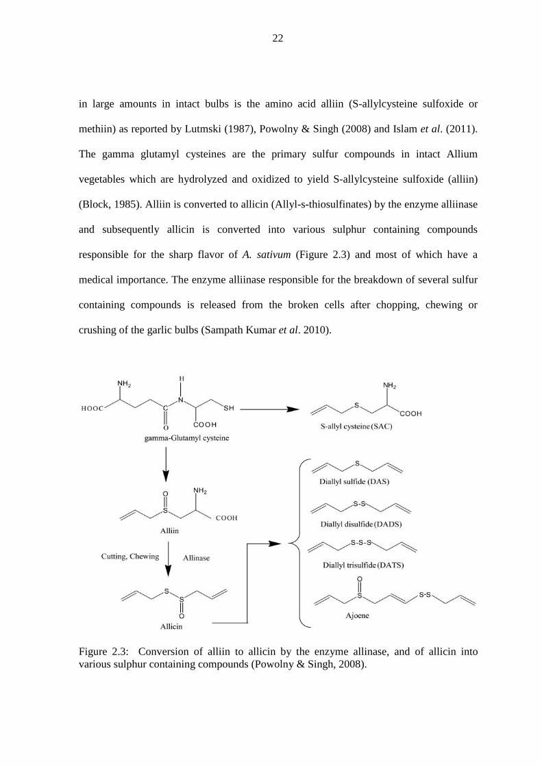
22
in large amounts in intact bulbs is the amino acid alliin (S-allylcysteine sulfoxide or
methiin) as reported by Lutmski (1987), Powolny & Singh (2008) and Islam et al. (2011).
The gamma glutamyl cysteines are the primary sulfur compounds in intact Allium
vegetables which are hydrolyzed and oxidized to yield S-allylcysteine sulfoxide (alliin)
(Block, 1985). Alliin is converted to allicin (Allyl-s-thiosulfinates) by the enzyme alliinase
and subsequently allicin is converted into various sulphur containing compounds
responsible for the sharp flavor of A. sativum (Figure 2.3) and most of which have a
medical importance. The enzyme alliinase responsible for the breakdown of several sulfur
containing compounds is released from the broken cells after chopping, chewing or
crushing of the garlic bulbs (Sampath Kumar et al. 2010).
Figure 2.3: Conversion of alliin to allicin by the enzyme allinase, and of allicin into various sulphur containing compounds (Powolny & Singh, 2008).

23
When A. sativum extracts are injected intra-peritoneally into Leishmania infected BALB/c
mice, the engulfment and destruction of amastigotes by the peritoneal macrophages is
enhanced (Ghazanfari et al., 2006). Garlic extracts stimulate production of interferon
gamma (IFN-γ) and nitric oxide (NO) both of which enhances the killing of Leishmania
parasites in experimental BALB/c mice (Gamboa-Leon et al., 2007). Garlic extracts are
commonly used in Europe and Asia for medicinal benefits in healing wounds (Bolton et
al., 1982). Today, garlic is also used to prevent heart diseases, cancer, hypertension, high
cholesterol levels as well as boosting the immune system. A. sativum is rich in antioxidants
which destroy or neutralize the free radicals in the body. Ethanolic extracts of A. sativum
have been shown to have an in vitro antioxidant activity which has been attributed to the
flavonoid and steroids fractions in the extracts (Narendhirakannan & Rajeswari, 2010).
The organo sulphur compounds in garlic have been reported to increase the synthesis of
glutathione that directly protects the cells from damage by free radicals as well as
destroying cancer cells in vitro (Islam et al., 2011). Recently, aqueous extracts of A.
sativum have been shown to destroy cancer cells in vitro (Islam et al., 2011). Earlier
studies have attributed the anti cancer potential of A. sativum to steroidal saponins and
organic selenium compounds present in intact garlic cloves (Arnault & Auger, 2006).
Garlic is considered to have low toxicity in humans.
2.8.3 Moringa stenopetala: Family Moringaceae
Moringa stenopetala Cufodontis is a smooth barked, deciduous flowering plant widely
distributed in southern parts of Ethiopia and northern Kenya (Mekonnen et al., 1999) and
on the Lake Baringo islands in Kenya. M. stenopetala is popularly known as the African
moringa tree or the cabbage tree and it belongs to the monogeneric family Moringaceae. In

24
southern Ethiopia, the leaves and fruits of M. stenopetala, locally known as aleko or
shiferaw are eaten as vegetables and they are rich in proteins, calcium, phosphorous, iron
as well as vitamins A and C. In Kenya, the residents of lake Baringo islands use the M.
stenopetala which is locally known as loresienjo, lorsenjo, or larsanjo as a medicinal plant
instead of a vegetable (Figure 2.4). The leaves, roots, fruits and the barks of the M.
stenopetala have been used in Ethiopia to treat stomach problems, malaria, hypertension,
diabetes, asthma as well as expelling retained placenta (Mekonnen et al., 1999).
(a) (b)
Figure 2.4: (a) Young Moringa stenopetala tree (b) Mature leaves on a branch of a
Moringa stenopetala tree. The picture was taken at the slopes of Lake Baringo islands,
Kenya by author.

25
On the other hand, Moringa oleifera Lam is a closely related species, widely distributed in
Asian sub continent and popularly known as ‘the miracle tree’. M oleifera is well
documented for its benefits and among the thirteen species of the genus Moringa (Ray et
al., 2006), M oleifera (local names: horseradish tree, drumstick, kelor, marango, mlonge,
mzunze, mboga chungu, benzolive tree, moonga, mulangay, saijhan, sajna or ben oil tree)
is an outstanding source of nutrients in underdeveloped nations and in Niger, M. oleifera is
the most preferred leafy vegetables (Bellostas et al., 2010). In Kenya, cultivation of M.
oleifera is rapidly picking up in such areas as Taita, Voi, and Kibwezi among others.
Other than being a food product, M. oleifera preparations have been reported to have a
variety of medicinal properties which include antibiotic, hypotensive, antitrypanosomal,
hypoglyceamic, anti-inflammatory, hypocholesterolemic, antispasmodic, and antiulcer in
addition to being used in water purification by flocculation, sedimentation and antibiosis (
Fahey, 2005).
Both M. stenopetala and M. oleifera have been demonstrated to have antimicrobial activity
(Eilert et al., 1980). The leaves and root extracts of M. stenopetala have shown in vitro
activity against trypomastigotes (Mekonnen et al., 1999). Since trypomastigotes are in the
same family of trypanosomatidae with Leishmania parasites, the expectation from the
current study was that the M. stenopetala extracts were likely to show activity against
Leishmania. In support of this, crude ethanolic extracts of the leaves and roots of the M.
stenopetala have been reported to have an adverse effect on Leishmania promastigotes
(TWAS, 2008).

26
The active components of M. stenopetala are mainly glucosinolates (Mekonnen & Drager,
2003). Glucosinolates are a class of organic compounds that contain sulphur and nitrogen
derived from glucose and amino acid. Glucosinolates are hydrolysed by an enzyme
myrosinase (found in plants and bowel microflora) to form isothiocyanates (Shapiro et al.,
2001). The active compounds of M. oleifera include glucosinolates, 4(α-L-
rhamnopyranosyloxy)-benzyl isothiocyanate, niazimicin, gluconic acid, allyl
isothiocyanate among others (Figure 2.5). Isothiocyanates also known as mustard oils have
been shown to be strong chemoprotective agents against the development and proliferation
of cancers, and they have also been shown to protect animals against chemically induced
cancer. Fahey (2005) reports a study carried out by Bharali and others, who examined a
significant skin tumour reduction in laboratory mice following their ingestion of M.
oleifera seed pods. Glucosinolate content vary in different parts of Moringa plants. Young
leaves and seeds have higher glucosinolate content, with M. oleifera being richer as
compared to M. stenopetala (Bellostas et al., 2010). Glucosinolates along with their
cognate isothiocyanates have been associated with positive metabolic effects in humans
which include anticarcinogenic, antioxidants and detoxificants (Polat, 2010). Gluconic
acids (dextronic acids) which arise from the oxidation of glucose naturally occur in fruits
and honey. Concentrated gluconic acid shows antiseptic property (Ramanchandran et al.,
2006).

27
[1] Glucosinolates:
[2] Allyl isothiocyanate (CH2CHCH2NCS):
[3]. 4(α-L-rhamnopyranosyloxy)-benzyl isothiocyanate:
[4]. Gluconic Acid (C6H12O7):
Figure 2.5: Structural formulae of selected active compounds from Moringa plant. [http://en.wikipedia.org/wiki ]

28
2.8.4 Callistemon citrinus (Curtis) Skeels; Family Myrtaceae
Callistemon citrinus is an ornamental evergreen tree in Kenya, whose origin is Australia
and it is commonly known as bottle brush tree. It is characterized by bright red flower
spikes and woody capsules that look like bead bracelets on the bark as described by
Seyydnejad et al. (2010) (Figure 2.6). It has been demonstrated that plants of the genus
Callistemon possess significant activities as antistaphylococcal, nematocidal, larvicidal,
pupicidal, antioxidants and antithrombotic (Ali et al., 2010) as well as antifungal (Dongmo
et al., 2010). Previously, C. citrinus leaf extracts have been reported as having effective
antibacterial activity (Cock, 2008a). The genus Callistemon is known in folk medicine as
anticough, antibronchitis, insecticidal agent (Abdelhady & Aly, 2012), and as a treatment
of gastro-enteritis, diarrhea and skin infections.
Figure 2.6: Callistemon citrinus ornamental tree and its red flower inflorescence.
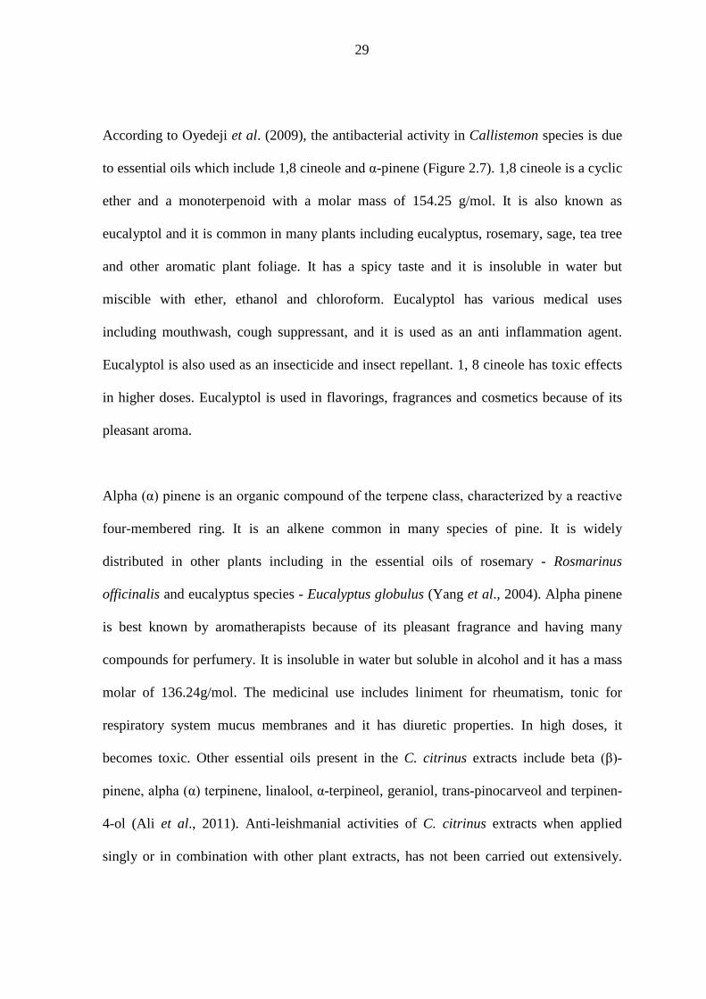
29
According to Oyedeji et al. (2009), the antibacterial activity in Callistemon species is due
to essential oils which include 1,8 cineole and α-pinene (Figure 2.7). 1,8 cineole is a cyclic
ether and a monoterpenoid with a molar mass of 154.25 g/mol. It is also known as
eucalyptol and it is common in many plants including eucalyptus, rosemary, sage, tea tree
and other aromatic plant foliage. It has a spicy taste and it is insoluble in water but
miscible with ether, ethanol and chloroform. Eucalyptol has various medical uses
including mouthwash, cough suppressant, and it is used as an anti inflammation agent.
Eucalyptol is also used as an insecticide and insect repellant. 1, 8 cineole has toxic effects
in higher doses. Eucalyptol is used in flavorings, fragrances and cosmetics because of its
pleasant aroma.
Alpha (α) pinene is an organic compound of the terpene class, characterized by a reactive
four-membered ring. It is an alkene common in many species of pine. It is widely
distributed in other plants including in the essential oils of rosemary - Rosmarinus
officinalis and eucalyptus species - Eucalyptus globulus (Yang et al., 2004). Alpha pinene
is best known by aromatherapists because of its pleasant fragrance and having many
compounds for perfumery. It is insoluble in water but soluble in alcohol and it has a mass
molar of 136.24g/mol. The medicinal use includes liniment for rheumatism, tonic for
respiratory system mucus membranes and it has diuretic properties. In high doses, it
becomes toxic. Other essential oils present in the C. citrinus extracts include beta (β)-
pinene, alpha (α) terpinene, linalool, α-terpineol, geraniol, trans-pinocarveol and terpinen-
4-ol (Ali et al., 2011). Anti-leishmanial activities of C. citrinus extracts when applied
singly or in combination with other plant extracts, has not been carried out extensively.

30
Majority of the researchers have tested C. citrinus extracts against bacteria and seemingly
it is now evident that the extracts have powerful broadspectrum antimicrobial activities.
[1] 1,8 Cineole (C10H18O) ;
[2] α-Pinene (C10H16):
Figure 2.7: Structural formulae for the essential oils found in C. citrinus.
[http://en.wikipedia.org/wiki/File: Eucalyptol and Alpha-Pinene]

31
2.8.5 Determining cytotoxicity of the potential herbal drugs
Cytotoxicity is the quality of being toxic to cells. When cells are treated with cytotoxic
compounds, a variety of changes in the cells may occur. Such changes include lose of
membrane integrity, rapid cell lysis, a decrease in cell viability, and controlled cell death
(apoptosis). In the current study, vero cells were used for cytotoxicity tests. Vero cells are
a lineage of cells originally isolated from kidney epithelial cells that were extracted from
an African green monkey, Cercopithecus aethiops. The vero lineage was developed in
1962 by Yasumura and Kawakita at the Chiba University, Japan. Vero cells are also used
for screening toxins, growing viruses and as hosts for eukaryotic parasites, among other
uses.
Cytotoxicity can be measured using various assays which include identifying the cytotoxic
compounds, assessing the cell membrane integrity and quantifying the occurrence of
specific metabolic reactions in the cells. MTT (3-(4,5-Dimethylthiazol-2-yl)-2,5-
diphenyltetrazolium bromide) assay is a colorimetric procedure for measuring the activity
of enzymes that reduce MTT to formazan dye which is purple in colour. A solubilization
solution, for instance, Dimethyl sulfoxide (DMSO) is added to dissolve the insoluble
purple formazan into a colored solution whose absorbance is quantified by a
spectrophotometric measurement at 540-nm wavelength using a microplate reader
(Gamboa-Leon et al., 2007), in order to estimate the activity of reductase enzymes. Dead
cells are unable to metabolize MTT and other tetrazolium salts. Therefore MTT assay is a
direct measure of cell survival or viability (live) of the cells. According to Khademvatan et
al. (2011a), the mitochondrial enzymes in viable cells (living) convert MTT dye

32
(tetrazolium) into formazan and therefore relative numbers of live cells can be determined
by using optical absorbance of treated and untreated samples and blank wells in the
formula: Viable Cells (%) = (AT – AB)/(AC – AB) × 100, where AC is the absorbance of
the untreated samples, AT is the absorbance of treated samples and AB is the absorbance of
the blank.
The absorbance values obtained from MTT assay are used to calculate the concentration of
the potential drugs that provides 50% inhibition; this is commonly referred to as half
maximal inhibitory concentration (IC50). IC50 is a measure of the effectiveness of the drug
or compound being tested. The drug concentration preferred should be above the IC50 but
below the level that will cause serious side effects. The zone between these two lines is
called the ‘therapeutic window’, which represent the range of drug concentration where
it’s doing more good than harm. Higher IC50 values imply that high concentration of the
drug is required to inhibit 50% of the target.
Researchers from various disciplines are now directing their efforts towards discovering
the effects of A. sativum on human health. Interest in A. sativum has emanated from the
search for a drug that has a broad spectrum therapeutic effect with minimal toxicity (Singh
& Singh, 2008). Garlic has been listed as Generally Recognized As Safe (GRAS) by the
US Food and Drug Administration (FDA). In vitro cytotoxicity studies on methanolic
extracts of A. sativum against L. major and L. donovani promastigotes have reported that it
has low toxicity (Wabwoba et al., 2010). However, apart from the valuable food and
medicinal properties, some studies have reported that A. sativum may act toxically when
overdosed. Such toxicity affects weight growth, biologic parameters and histological

33
structures (Fehri et al., 1991). Other common complains associated with A. sativum in
humans include ‘garlic breath’ and a strong body odor, allergic reactions, alteration of
platelet functions, and reduction of the efficacy of antiAIDS drug Saquinavir among
others (Borrelli et al., 2007).
The aqueous extracts of M. stenopetala leaves have been reported not to affect cell
viability or to alter glutathione (GSH) and lactate dehydrogenase (LDH) levels in HEPG 2
cells (human liver carcinoma cell line) in vitro, suggesting that they are non toxic
(Mekonnen et al.,2005). Further studies have shown that M. stenopetala aqueous crude
leaf extracts are tolerable in mice with no mortality and behavioural changes. This justifies
why the leaves can be a safe source of food. M. stenopetala leaves, flowers and fruits are
used as vegetables in southern Ethiopia.
Crude extracts of C. citrinus have been reported to be a 100% lethal (acute toxicity) at a
concentration of 1000 mg/kg but safe at a concentration of 250 mg/kg in laboratory rabbits
(Ali et al., 2011). Similarly, Nazreen et al. (2011) administered C. lanceolatus (synonym
C. citrinus) ethanol extracts at a dose of 150 mg/kg and 350 mg/kg into streptozotocin
induced diabetic rats resulting to a significant hypoglycaemic effect. Ethanolic extracts of
C. lanceolatus at an oral dose of 200 mg/kg in albino Wistar rats have been demonstrated
to have potent cardioprotection against doxorubicin induced cardiotoxicity (Firoz et al.,
2011). The albino rat mortality was found following Callistemon extracts at a dose of
3,000 mg/kg, and this confirmed that Callistemon extracts at high dose are toxic.

34
CHAPTER THREE: MATERIALS AND METHODS
3.1 Plant materials
Young leaves of M. stenopetala were collected from the slopes of one of the islands in
Lake Baringo, Baringo County, Kenya. The C. citrinus flowers inflorescences were
harvested from randomly selected homesteads in Nakuru County, Kenya. Bulbs of A.
sativum were purchased from Nakumat Prestige Store in Nairobi, Kenya. The plants were
positively identified at University of Nairobi, Department of Botany herbarium, Chiromo
campus. The young leaves of M. stenopetala, individual flowers of C. citrinus, and thin
slices of A. sativum cloves were then dried at room temperature until they became brittle
and attained a constant weight, at the Kenya Medical Research Institute (KEMRI)
Leishmania laboratory, Nairobi. The dried plant materials were packaged separately and
labeled appropriately.
3.2 Plant extraction
The dried plant materials were transferred to the Center of Traditional Medicine & Drug
Research (CTMDR) at KEMRI, where they were separately ground using an electric mill
(Christy & Norris Ltd., Chelmsford, England) into powder followed by extraction using
water and analytical grade methanol.
3.2.1 Aqueous extraction
The aqueous extracts were prepared as described by Delahaye et al. (2009). Briefly, 100g
of the dried ground plant material in 600 ml of distilled water was placed in a water bath at
70oC for 1.5 hours. The mixture was filtered using Whatman No 1 filter papers and then
the filtrate was freeze dried and weighed. The aqueous extracts of M. stenopetala, C.
35
citrinus, and A. sativum were coded as extracts A, B and C respectively. The extracts were
then stored at 4oC until required for the bioassays.
3.2.2 Methanolic extraction
The methanolic extracts were prepared as described by Mekonnen (1999) and Cock
(2012). Briefly, 100g of ground plant material was soaked in 500 ml of analytical grade
methanol for 72 hours at room temperature with gentle shaking, followed by filtering
using Whatman No 1 filter papers and concentrating using a rotary evaporator to obtain
dry methanolic extracts. The methanolic extracts of A. sativum, C. citrinus, and M.
stenopetala, were coded as extracts F, G, and H respectively. The dry extracts were
weighed and stored at 4oC until required for the bioassays.
3.3 Leishmania parasites
The Leishmania major strain (IDUB/KE/94=NLB-144) was acquired from Institute of
Primate Research (IPR), Kenya where it had been cryopreserved in liquid nitrogen. The
parasites were grown to stationary phase at 25oC in Schneider’s Drosophila medium
supplemented with 20% heat inactivated fetal bovine serum (FBS)-HYCLONE® USA),
100 U/ml penicillin and 500μg/ml streptomycin (Hendricks & Wright, 1979), and
250μg/ml 5-fluorocytosine arabinoside (Kimber et al., 1981). The stationary-phase
metacyclic stage promastigotes were then harvested by centrifugation at 1500 g for 15
minutes at 4oC. The metacyclic promastigotes were then used for the in vitro and in vivo
assays.
3.4 Experimental animals
Inbred BALB/c mice were obtained from International Livestock Research Institute
(ILRI), Kenya. They were housed at the KEMRI animal house at 23oC to 25oC and were

36
fed on standard commercial diet that was in form of mice pencils and given tap water ad
libitum. The mice were handled in accordance with the regulations that have been set by
Animal Care and Use Committee (ACUC) at KEMRI. Eight week old male BALB/c mice
were used for both in vitro and in vivo assays.
3.5 Preparation of the stock solutions of test extracts
Stock solutions of the crude extracts were made in Schneider’s Drosophila culture media
for anti-leishmanial assays and filtered through 0.22μm filter flasks in a laminar flow hood
(Biological Safety Cabinet). The stock solutions were stored at 4oC and retrieved when
being used for in vitro and in vivo bioassays.
3.6 Evaluation of minimum inhibitory concentration (MIC)
The MICs were determined as described by Wabwoba et al. (2010). Briefly, the L. major
metacyclic promastigotes at concentration of 1×10⁶ promastigotes per ml of the culture
medium were treated with several concentrations of the individual aqueous test plant
extracts (A, B, and C) that ranged between 5mg/ml and 0.5mg/ml. Similarly, the
promastigotes were treated with combined test aqueous extracts in fixed ratios that ranged
from 9:1 to 1:9 at MIC based concentrations for combinations M. stenopetala & C.
citrinus (AB), M. stenopetala & A. sativum (AC), C. citrinus & A. sativum (BC). Blend of
three different extracts in fixed ratios of 2:2:1 (ABC, ACB, and BCA) that were based on
MICs of the individual extracts were also tested for their efficacy in inhibiting the
promastigotes in vitro. These test procedures were repeated for methanolic individual test
extracts (F, G, and H) and combined methanolic extracts (FG, GH, FH, FGH, FHG, and
GHF). The lowest concentration of the individual test plant extracts or in combinations in
which no live promastigotes were observed was taken to be the MIC.

37
3.7 Promastigote proliferation measurement by colorimetric MTT assay
MTT (3-(4, 5-methylthiazol-2-yl)-2, 5-diphenyltetrazolium bromide) colorimetric assay
was used to measure the reduction of MTT dye (tetrazolium) into formazan by
mitochondrial enzymes (Khademvatan et al., 2011a) in viable promastigotes. Metacyclic
promastigotes at a concentration of 1x10⁶ promastigotes per ml of the culture medium
were grown for 48 hours in a 24 well microtiter plates at 25oC and treated with various
concentrations (5mg/ml - 0.5mg/ml) of M. stenopetala (A), C. citrinus (B) and A. sativum
(C) aqueous extracts. Aliquots of the promastigotes were transferred into 96 well
microtiter plates and incubated further at 27oC for 24 hours after which 200μl of the
highest concentrations of the individual test extracts were added before a serial dilution
was carried out. For the combined extracts (AB, BC, and AC), seven fixed ratios (9:1 to
1:9) that were based on MICs of the individual extracts, were used. For combined extracts
(ABC, BCA, and ACB) six fixed ratios ranging from 2:2:2 to 2:2:0 were used. The control
wells contained L. major promastigotes in culture medium alone. The blank wells had
culture medium alone.
The plates were then incubated further at 27oC for 48 hours. Ten microliters of MTT
reagent was added into each well and incubated further for 4 hours. The medium and MTT
were aspirated off the wells. Then in each well, 100μl of Dimethyl sulfoxide (DMSO) was
added and the plates shaken for 5 minutes. Absorbances were read at 562 nm using a
micro titer reader. The absorbances readings were used to generate the 50% inhibitory
concentration (IC50) values for the different plant extracts using the Chemosen software.
The viabilities of the promastigotes were determined as described by Mosmann (1983) in
the formula shown below for each concentration or ratio for the combined extracts.

38
Average absorbance in duplicate drug wells – average blank wells Viability (%) = ____________________________________________________ × 100 Average absorbance in control wells
This MTT assay was repeated for the single methanolic extracts (F, G, and H) and
combined methanolic extracts (FG, FH, GH, FGH, FHG and GHF) at the same fixed
ratios.
3.8 Determination of the blend effect
The calculated IC50 values for the extracts at fixed ratios were used to determine the
interaction of plant extracts in the blends (blend effect). Corresponding IC50 values were
determined for each extract alone and in combination (Sixthmith et al., 1984). The degree
of synergy was evaluated according to Berenbaum (1978). Sum of fractional inhibition
concentration (SFIC) abbreviated as K was calculated using the formula: Ac/Ae + Bc/Be =
K. Where, Ac and Bc are equally effective concentrations (IC50) when used in
combinations (blends), and Ae and Be are the equally effective concentrations when used
alone. In this system, SFIC < 1 denotes synergism, 1 ≤ SFIC < 2 denotes additive
interaction, while SFIC ≥ 2 denotes antagonism (Gupta et al., 2002). The SFIC values
were calculated for the blends of aqueous extracts in ratios that ranged from 9:1 to 1:9.
This was also repeated for the blends of methanolic extracts.
3.9 In vitro anti-amastigote assay
The anti-amastigote assay was carried out as described by Delorenzi et al. (2001). The
peritoneal macrophages were obtained from 4 clean BALB/c mice. The mice were
anaesthetized using 100μl pentobarbitone sodium (Sagatal®). The body surface of the
mouse was disinfected with 70% ethanol after which it was torn dorso-ventrally to expose

39
the peritoneum. Ten milliliters of sterile cold PBS was injected into the peritoneum. After
injection, the peritoneum was gently massaged for 2 minutes to dislodge and release
macrophages into the PBS. The peritoneal macrophages were then harvested by
withdrawing the PBS. The PBS containing the macrophages was washed through
centrifugation at 2,000 g for 10 minutes and the pellet obtained was re-suspended in RPMI
culture medium. The macrophages were adsorbed in 24-well plates for 4 hours at 37oC in
5% CO2. Non-adherent cells were washed with cold sterile PBS and the adherent
macrophages were incubated overnight in RPMI culture medium. Adherent macrophages
were then infected with L. major promastigotes and were further incubated at 37oC in 5%
CO2 for 4 hours after which they were washed with sterile PBS to remove the free
promastigotes, that were not engulfed by the macrophages. This was followed by
incubation of the preparation for 24 hours in RPMI 1640 culture medium. The infected
macrophages were then treated with individual aqueous extracts of M. stenopetala (A), C.
citrinus (B), and A. sativum (C) or combinations M. stenopetala & C. citrinus (AB), C.
citrinus & A. staivum (BC) and M. stenopetala & A. sativum (AC) at MIC based
concentrations in fixed ratios of 1:1. For combined extracts (ABC, BCA, and ACB) an
MIC based fixed ratio of 2:2:1 was used to treat the infected macrophages. Pentostam and
liposomal amphotericin B were used as positive control drugs to compare the parasite
inhibition with that by the plant extracts. The medium and test extracts or drug were
replenished daily for 3 days. After 5 days, the macrophages were washed with sterile PBS
at 37oC, fixed in methanol and stained with 10% Giemsa. The number of amastigotes was
determined by counting at least 100 macrophages in duplicate cultures, and the count was
expressed as infection rate (IR) and multiplication index (MI) as described by Berman &
Lee (1984) in the two formulae below.

40
IR (%) = Number of infected macrophages per 100 macrophages.
Number of amastigotes in experimental culture/100 macrophages MI (%) = ____________________________________________________× 100.
Number of amastigotes in control culture/100 macrophages
This anti-amastigote assay was repeated for the single methanolic extracts (F, G, and H),
for combination of two methanolic extracts (FG, FH, and GH) in ratio 1:1 and for
combination of three methanolic extracts (FGH, FHG and GHF) in a fixed ratio of 2:2:1.
3.10 Determination of nitric oxide production
Measurement of nitric oxide (NO) production was carried out as described by Gamboa-
Leon et al. (2007). BALB/c peritoneal macrophages at a concentration of 1 × 10⁵ cells per
culture medium were placed in each well in 96-well microtiter plates and allowed to
adhere at 37oC in 5% CO2 humidified atmosphere. Two hours later, the peritoneal
macrophages were incubated further in RPMI-1640 with 10% FBS for 48 hours in
presence of aqueous or methanolic test individual plant extracts (extracts A, B, C, F, G,
and H) or the controls. At least 100 µl of macrophage culture supernants were collected
and frozen until when they were required for NO measurement. NO was measured using
the Greiss reaction test for nitrites (NO2-) as described by Hollzmuller et al., 2002. NO2
-
is one of the products released when the breakdown of NO occurs in the macrophages. NO
in the collected supernatants was therefore estimated by quantifying the NO2- content. A
nitrite standard reference curve was first prepared by dispensing 50 µl of RPMI 1640 with
10% FBS into wells in rows B – H of the first 3 columns in a 96 well plate. A hundred
microliters of 100µM sodium nitrite solution was added to the remaining 3 wells in row A

41
of a 96- well micro titer plate, and immediately a six serial two fold dilutions (50 µl/well)
was performed in triplicate columns 1, 2 and 3 down the plate to generate a curve that
corresponded to the concentrations 100, 50, 25, 12.5, 6.25, 3.125 and 1.563 µM. Secondly,
50 µl of the experimental sample supernatant from the wells that had macrophages treated
with test extracts, was added into the wells in duplicate at a concentration of 100µg/ml,
starting from column 4 of the micro titer plate followed by serial dilution by a factor of
two. Fifty microliters of Greiss reagent A (1% sulphanilamide in 5% phosphoric acid) was
dispensed to all the experimental samples and into the wells containing sodium nitrite
solution. Following an incubation of 5 minutes at room temperature, 50 µl of Greiss
reagent B (0.1% N- [1-naphthyl] ethylenediamine dihydrochloride (NED) in water was
dispensed into all the wells and incubated for a further 5 minutes at room temperature
before measuring the absorbance of a purple/magenta azo compound at 520-550 nm in a
micro titer plate reader. The average absorbance values for the standard nitrite and each of
the single plant extract sample were calculated and plotted. The NO production elicited by
the combined plant extracts was read from the standard curve plotted, by reading the NO
concentration that corresponded to the absorbance values of the blends.
3.11 Cytotoxicity assay using vero cells
The assay was used to test the cytotoxicity of the individual extracts against vero cells.
The assay was carried out as described by Wabwoba et al. (2010). Vero cells were grown
in minimum essential medium (MEM) supplemented with 10% FBS, penicillin (100
IU/ml) and streptomycin (100µg/ml) in 25ml cell culture flasks incubated at 37oC in a
humidified 5% CO2 atmosphere for 24 hours. The vero cells were harvested by
trypsinization, and pooled in 50 ml centrifuge tubes from where a 100μl of the cell
suspension were put into 2 wells of rows A-H in a 96-well flat bottomed microtiter plate at

42
a concentration of 1 × 10⁶ cells per ml of the culture medium per well and incubated at
37oC in 5% Co2 in order to attach. The MEM was gently aspirated off and 150 μl of the
highest concentration (1000µg/ml) of the test extracts (extracts A, B, C, F, G, and H) was
added and serially diluted by a factor of two up to a concentration of 15.63µg/ml. The
microtitre plates containinig the vero cells and test extracts were further incubated at 37oC
for 48 hours in a humidified 5% CO2 atmosphere. The controls wells comprised of vero
cells and medium while the blank wells had medium alone. 10μl of MTT reagent was
added into each plate well and incubated further for 2 to 4 hours until a purple precipitate
(formazan) was visible under the microscope. The media together with MTT reagent were
gently aspirated off, after which 100 μl of DMSO was added, and vigorously shaken for 5
minutes in order to dissolve formazan. The absorbance (optical density) was measured for
each well plate using a micro-titer plate reader at wavelength of 570 nm. Cell viability was
calculated at each concentration using the Mosmann (1983) formula. The IC50 values of
the extracts were determined automatically using the Chemosen software program.
3.12 Infection and treatment of BALB/c mice with L. major
Each experimental group comprised of at least 5 mice. The procedure of infecting the mice
was as described by Wabwoba et al. (2010). Briefly, the thickness of hind footpads of the
mice were measured using a reading vernier caliper prior to infection. The left hind
footpads of the mice were subcutaneously inoculated with 1×10⁶ stationary phase infective
metacyclic promastigotes of L. major in 40μl sterile PBS. Lesions development and
progression was monitored weekly by using a vernier caliper to measure the thickness of
the infected left hind foot pad and comparing it with that of non infected right hind foot
pad (Nolan & Farrel, 1987). Treatment with plant extracts or control drugs commenced
one month post infection. Treatment was done orally using a cannula and intraperitoneally

43
using fine 1ml 30 gauge Insulin needles (BD Micro-Fine Plus®, USA) at a dose of 20
mg/kg daily. The positive control groups were treated with Pentostam and Liposomal
Amphotericin B which were administered intra peritoneally at the standard dose of 20
mg/kg per day. Selection of the doses (20mg/kg/day) for the test plant extracts was
adopted from earlier studies (Nok et al., 1996; Meckonnen et al., 1999; Seyydnejad et al.,
2010). Determination of lesions progression, as the mice were being treated with the plant
extracts and standard leishmaniasis drugs, was done as described by Nolan & Farrel, 1987.
3.13 Estimation of parasite burden in mice spleens
At the end of the experiment, all the mice were sacrificed using 100μl pentobarbitone
sodium (Sagatal®). At necropsy, the spleens were weighed and their impression smears
made as described by Chulay & Bryceson (1983). Portions of the spleens and the livers
were also cultured in Schneider’s Drosophila medium to verify the presence and viability
of amastigotes observed. The impression smears were fixed in methanol and stained with
Giemsa. The slides were examined under a microscope to enumerate the number of
amastigotes per 1000 nucleated cells. The relative and total numbers of parasites in spleen
were estimated by calculating the Spleen index (%), Leishman-Donovani Unit (LDU) and
Total Leishman-Donovani Unit (Total LDU) as described by Bradley & Kirkley (1977).
Spleen index was determined by dividing the spleen weight by whole body weight
multiplied by 100. LDU was the number of amastigote per 1000 nucleated splenocytes
while the total LDU was calculated by multiplying LDU × spleen weight (g) × (2 × 10⁵) (
Bradley & Kirkley, 1977).

44
3.14 Experimental design for the in vitro assays
3.14.1 Evaluation of MIC
The Leishmania promastigotes (1×10⁶ per ml) were cultured at 27oC in 24 wells plates in
culture medium containing the individual test extracts or the blends of the test plant
extracts. The plate design that was used is shown in Figure 3.1. The extracts (A, B, C)
were tested in duplicates.
5000 4000 3000 2000 1000 500µg/ml A A B
B
Figure 3.1: The 24 wells plate design for evaluation of MIC Key:
The concentration (µl/ml) of the test extracts decreased from Well 1 to Well 6 in the order; 5, 4, 3, 2, 1, 0.5 mg/ml [1 mg = 1000 µg]; Efficacy of extract A against promastigotes in Schneider’s Insect medium Efficacy of extract B against promastigotes in Schneider’s Insect medium
3.14.2 Tests for the viability and cytotoxicity (IC 50) of the promastigotes
The promastigotes were incubated at 27oC for 24 hours at different concentrations of the
plant extracts and MTT in 96 wells plate. Absorbance in the wells was measured at 562
nm using microtitre plate reader. The design of the 96 wells plate is as illustrated in Figure
3.2. The plate design was used for the single and combined plant extracts. Fixed ratios for
blends of two extracts ranged from 9:1 to 1:9 while the ratio for blends ranged from 2:2:2
to 2:2:0.
Well-1 Well-6

45
1 2 3 4 5 6 7 8 9 10 11 12 A
B
C
D
E
F
G
H
Figure 3.2: The 96 well plate design for the cytotoxicity assay of the plant extracts (drugs).
Key: The direction of extracts’ dilution (concentration decreased upwards from H to B in the order; 5, 4, 3, 2, 1, 0.5 mg/ml (1 mg = 1000 µg). Concentrations of control drugs were in the order of 100, 50, 25, 12.5, 6.25, 3.125, 3.125 µg/ml. Wells with L. major promastigotes in Schneider’s insect medium (SIM) and MTT
Wells with promastigotes, SIM, MTT and plant extracts or with promastigotes, SIM, MTT and standard drug, where concentration decreased from H to B. The blank wells had the MTT and Schneider’s insect medium (SIM) only.
3.14.3 The design for anti - amastigotes assay
The infected macrophages were treated with the plant extracts. Pentostam and liposomal
amphotericin B were used as positive control drugs. The design of the 96 wells plate was
similar to that shown in Figure 3.2 above.

46
3.15 Experimental design for the in vivo assays
All the BALB/c mice were infected with L. major metacyclic promastigotes
subcutaneously on their left hind foot pads and then they were treated with the aqueous
and methanolic plant extracts, PBS, pentostam and liposomal amphotericin B for thirty
days. The treatment commenced 4 weeks post infection when the foot pad lesions were
observable and distinct. The plant extracts were used as single or in combination of two or
three extracts in ratios 1:1 and 2:2:1 respectvely. Table 3.1, shows the experimental
protocol, for the single aqueous and methanolic extracts as well as the controls. This
design was repeated for the different blends of two and three different aqueous or
methanolic extracts represented as AB, BC, AC, ABC, BCA, ACB for the aqueous
extracts and FG, FH, GH, FGH, FHG, and HGF for the methanolic extracts.
Table 3.1: Design for the in vivo protocol for testing extracts using BALB/c mice. _____________________________________________________________________ Plant extracts Mice and controls Code Route Cage No. Mice markings _____________________________________________________________________ Aqueous extracts M. stenopetala (leaves) A oral A 5 Ears marked, M. stenopetala (leaves) A ipb 5 Ears unmarked C. citrinus (Flowers) B oral B 4 Ears marked C. citrinus (Flowers) B ip 5 Ears unmarked A. sativum (bulbs) C oral C 6 Ears marked A. sativum (bulbs) C ip 5 Ears unmarked Methanolic extracts M. stenopetala (leaves) H oral H 5 Ears marked, M. stenopetala (leaves) H ip 5 Ears unmarked C. citrinus (Flowers) G oral G 5 Ears marked C. citrinus (Flowers) G ip 5 Ears unmarked A. sativum (bulbs) F oral F 5 Ears marked A. sativum (bulbs) F ip 5 Ears unmarked Controls: Pentostam Pento ip D 5 Ears unmarked Liposomal amphotericin B Lip ampB ip 5 Ears marked Sterile PBS PBS ip 3 Ears unmarked Sterile PBS PBS oral 3 Ears marked _____________________________________________________________________

47
3.16 Disposal of Animals
All the mice were sacrificed and disposed off in accordance with the regulations that have
been set by Kenya Medical Research Institute (KEMRI)’s Animal Care and Use
Committee (ACUC).
3.17 Data Analysis
Data was analyzed using SPSS version 17.0 for windows at 5% level of significance.
Comparison of lesion sizes in infected BALB/c mice that were treated with different
extracts and control drugs was done using standard errors and standard deviations. Foot
pad lesions were also compared using single and two factors ANOVA (F test) to establish
the significance of the differences between and within multiple treatment groups or
controls. Other variables compared were MICs and viabilities (%) of promastigotes treated
with extracts and drugs; viabilities (%) of amastigotes and vero cells exposed to different
treatments in vitro; infection rates (IRs) and multiplication indices (MIs) of amastigotes in
peritoneal macrophages that were under treatments; nitric oxide production in peritoneal
macrophages that were under treatments with different extracts and finally to compare
parasite load in the spleens of infected BALB/c mice that had been treated with different
plant extracts and control drugs. When homogeneity test of variance (Levene’s test) was
significant, robust tests of equality of means that included Brown-forsythe and Welch tests
were carried out as alternative versions of the F-statistics. Multiple comparisons of the
individual treatments were done using Tukey and Games-Howell post hoc tests. Tukey
post hoc test assumes that the population variances and sample sizes are equal. Games-
Howell does not assume equal population variances nor sample sizes. Paired samples t test
was used for comparing variables in any pair of treatment groups, including the foot pad
lesion sizes or body weights in two groups of mice treated with different plant extracts.

48
CHAPTER FOUR: RESULTS
4.1 Plant extracts yields
The herbal materials were collected between August and September 2010, identified, and
air dried under shade until they attained a constant weight. They were then ground using a
laboratory mill into a powder. Aqeous and methanolic extractions for the powdered plant
materials were carried out as described earlier in section 3.1.4. The aqueous extraction
gave higher yields compared to the methanolic extracts for A. sativum and C. citrinus
(Table 4.1). A. sativum and C. citrinus aqueous yields were highest at 18.59 % and 17.58
% respectively, while A. sativum gave the lowest yield of methanolic extracts at 5.69%.
The yield for methanolic extracts of M. stenopetala and C. citrinus were 9.22% and 9.24%
respectively. The mean percentage yield for aqueous extracts was 14.19% while that of the
methanolic extracts was 8.05%. Though the aqueous yields were generally higher (Table
4.1) than that of the methanolic extracts, the difference was not significant (P = 0.207).
Table 4.1: The percentage yields of the plant extracts obtained from the study plants. _________________________________________________________________________ Plant species Part used Type of Code Initial Yield Yield Extraction wt (g) (g) (%) _________________________________________________________________________ A. sativum Bulbs Aqueous A 50 9.293 18.59 Bulbs Methanolic F 100 5.698 5.69 C. citrinus Flowers Aqueous B 50 8.790 17.58 Flowers Methanolic G 100 9.240 9.24 M. stenopetala Leaves Aqueous C 50 3.200 6.40 Leaves Methanolic H 100 9.217 9.22 _________________________________________________________________________ 4.2 In vitro bioassays
These assays were meant to determine the effects of the plant extracts on the L. major
promastigotes by establishing their survival and viability upon exposure. Secondly it was
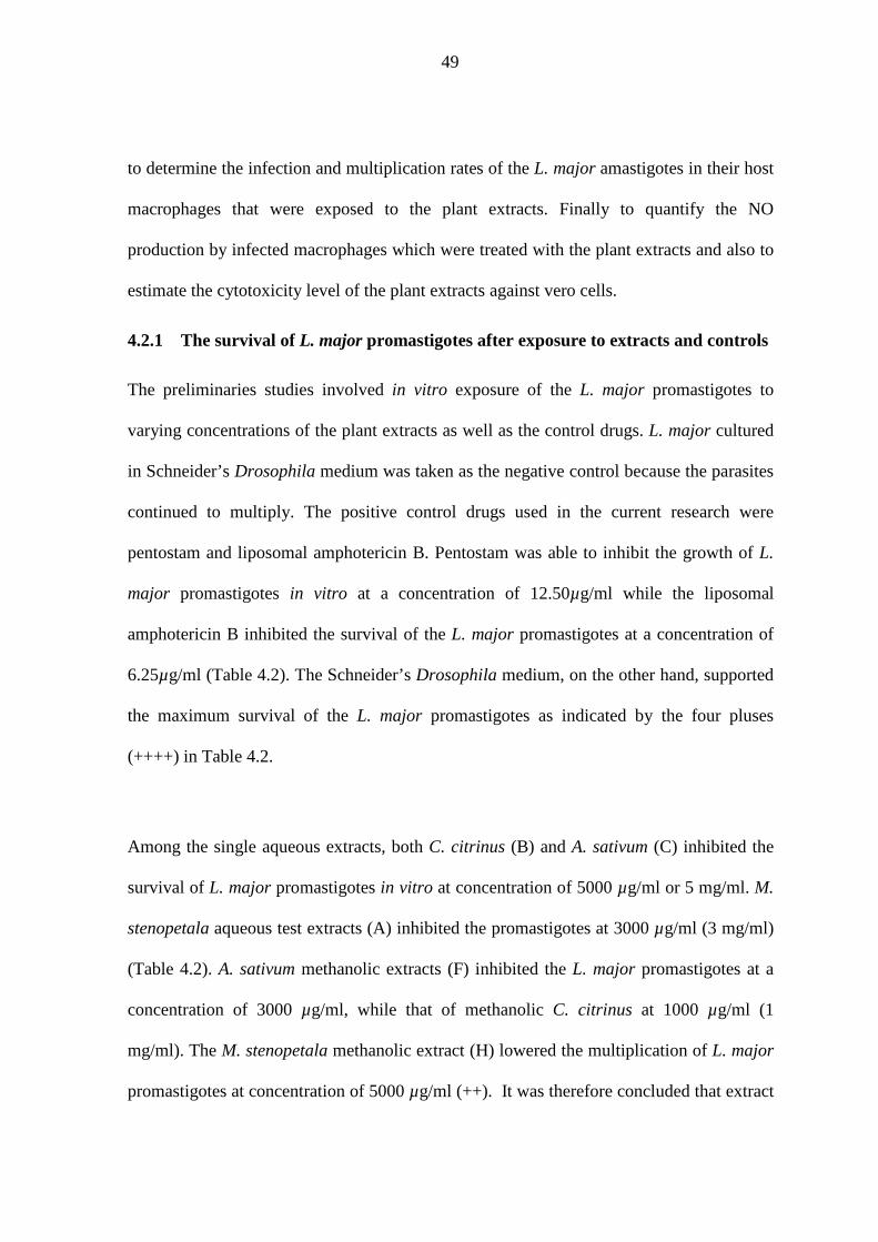
49
to determine the infection and multiplication rates of the L. major amastigotes in their host
macrophages that were exposed to the plant extracts. Finally to quantify the NO
production by infected macrophages which were treated with the plant extracts and also to
estimate the cytotoxicity level of the plant extracts against vero cells.
4.2.1 The survival of L. major promastigotes after exposure to extracts and controls
The preliminaries studies involved in vitro exposure of the L. major promastigotes to
varying concentrations of the plant extracts as well as the control drugs. L. major cultured
in Schneider’s Drosophila medium was taken as the negative control because the parasites
continued to multiply. The positive control drugs used in the current research were
pentostam and liposomal amphotericin B. Pentostam was able to inhibit the growth of L.
major promastigotes in vitro at a concentration of 12.50µg/ml while the liposomal
amphotericin B inhibited the survival of the L. major promastigotes at a concentration of
6.25µg/ml (Table 4.2). The Schneider’s Drosophila medium, on the other hand, supported
the maximum survival of the L. major promastigotes as indicated by the four pluses
(++++) in Table 4.2.
Among the single aqueous extracts, both C. citrinus (B) and A. sativum (C) inhibited the
survival of L. major promastigotes in vitro at concentration of 5000 µg/ml or 5 mg/ml. M.
stenopetala aqueous test extracts (A) inhibited the promastigotes at 3000 µg/ml (3 mg/ml)
(Table 4.2). A. sativum methanolic extracts (F) inhibited the L. major promastigotes at a
concentration of 3000 µg/ml, while that of methanolic C. citrinus at 1000 µg/ml (1
mg/ml). The M. stenopetala methanolic extract (H) lowered the multiplication of L. major
promastigotes at concentration of 5000 µg/ml (++). It was therefore concluded that extract

50
H probably would inhibit the L. major promastigotes at a concentration of > 5 mg/ml. The
concentrations of the crude extracts that were effective against L. major promastigotes in
vitro were relatively high (> 1 mg/ml) as compared to those of the Leishmania standard
drugs, pentostam and liposomal amphotericin B which inhibited the promastigotes at
0.0125 mg/ml and 0.00625 mg/ml respectively. Apart from M. stenopetala methanolic
crude extracts, the efficacy of methanolic extracts was better than their respective aqueous
counter parts (Table 4.2).
Table 4.2: Survival of the L. major promastigotes in varying concentrations of the plant extracts as observed under a light microscope. _________________________________________________________________________ Test extracts Type of Code Concentrations of the extracts (µg/ml) & Controls Extraction 5000 4000 3000 2000 1000 500 _________________________________________________________________________ Test extracts M. stenopetala Aqueous A -a - - ++ ++++ ++++ C. citrinus Aqueous B - + ++ +++ ++++ ++++ A. sativum Aqueous C - + ++ +++ ++++ ++++ A. sativum Methanolic F - - - + +++ ++++ C. citrinus Methanolic G - - - - - + M. stenopetala Methanolic H ++ +++ ++++ ++++ ++++ ++++ Positive Controls: Concentrations of the standard drugs (µg/ml) 100 50 25 12.5 6.25 3.125 Pentostam Pento - - - - + ++ Liposomal Amphotericin B Amph B - - - - - + Negative control Schneider’s Insect Medium SIM ++++b ++++ ++++ ++++ ++++ ++++ ________________________________________________________________________ -a represented absence of detectable and live promastigotes while ++++b represented maximum survival (density) of the L. major promastigotes.
A blend of M. stenopetala and C. citrinus aqueous extracts in a ratio of 1:1 (AB) inhibited
the L. major promastigotes in vitro 100% while ratios 9:1, 8:2 and 6:4 decreased the
promastigotes to minimal levels (+) and ratios 4:6, 2:8 and 1:9 supported the survival of

51
promastigotes to moderate (++) levels (Table 4.3). A blend of M. stenopetala and A.
sativum (AC) was efficacious in inhibiting L. major promastigotes at ratios 9:1 (+), 8:2
(++) and 1:1 (++). A blend of C. citrinus and A. sativum (BC) had moderate and uniform
inhibition of the L. major promastigotes at all the ratios between 9:1 and 1:9 (Table 4.3).
Blends of any two methanolic extracts in the ratio of 1:1 (extracts FG, HF and GH) had a
moderate efficacy in inhibiting L. major promastigotes in vitro. It was expected that the in
vitro survival of L. major promastigotes in medium (Table 4.3) would directly correspond
to their viabilities (%) that were determined from the microtitre plate readings. This was
the case for combination FG whose L. major survival at 1:1 was minimal (+) and it
corresponded to a viability of 31.32%. Pentostam, liposomal amphotericin B and
combination of M. stenopetala and C. citrinus (AB) had a similar trend.
Table 4.3: Survival of the L. major promastigotes in varying concentrations of double combination of the plant extracts. _________________________________________________________________________ Blends & Ratio of the two extracts based on mica Controls Test extracts & drugs 9:1 8:2 6:4 1:1 4:6 2:8 1:9 _________________________________________________________________________ Aqueous A:B M. stenopetala : C. citrinus + + + - ++ ++ ++ A:C M. stenopetala : A. sativum + ++ +++ ++ ++++ ++++ ++++ B:C C. citrinus : A. sativum ++ ++ ++ ++ ++ ++ +++ Methanolic F:G A. sativum : C. citrinus ++ ++ ++ + ++ +++ +++ H:F M. stenopetala : A. sativum ++ ++ +++ ++ ++++ ++++ ++++ G:H C. citrinus : M. stenopetala ++ +++ ++ + ++++ ++++ ++++ Positive controls: Concentrations of the standard drugs (µg/ml) 100 50 25 12.5 6.25 3.125 Pentostam - - - - + ++ Liposomal Amphotericin B - - - - - + Negative control Schneider’s Insect Medium ++++ ++++ ++++ ++++ ++++ ++++ _________________________________________________________________________ mica = minimum inhibitory concentration which indicated the level at which the extract was inhibiting the promastigotes.

52
Blends of three aqueous extracts at different ratios, allowed minimal (+) to moderate (++)
survival of L. major promastigotes (Table 4.4). The blends of M. stenopetala, C. citrinus,
and A. sativum (ABC) in a ratio of 2:2:1/8, and a blend of C. citrinus, A. sativum and M.
stenopetala (BCA) in the ratio of 2:2:2 showed a 100% inhibition of L. major
promastigotes survival. The efficacy of the blends of three aqueous extracts in ratios
ranging from 2:2:2 to 2:2:1/2 compared closely with that of standard Leishmania drugs
pentostam and liposomal amphotericin B at concentrations of 6.25µg/ml and 3.125µg/ml
respectively (Table 4.4). Similarly, majority of blends of three methanolic extracts in
majority of the ratios ranging from 2:2:2 to 2:2:0 had efficacy that compared closely with
that of pentostam at a concentration of 3.125µg/ml. Blend GHF at ratios 2:2:1/8 and 2:2:0,
allowed high survival (+++) of L. major promastigotes (Table 4.4).
Table 4.4: Survival of the L. major promastigotes in different concentrations of triple combination of the plant extracts.
_________________________________________________________________________ Blends & Ratio of the concentrations Controls Extracts & drugs 2:2:2a 2:2:1 2:2:1/2 2:2:1/4 2:2:1/8 2:2:0 _________________________________________________________________________ Aqueous A:B:C Mor:Calliste:Allium + + + ++ - ++ A:C:B Mor:Allium:Calliste + + + + ++ + B:C:A Calliste:Allium:Mor - + + + + + Methanolic F:G:H Allium:Calliste:Mor + + + ++ ++ ++ F:H:G Mor:Allium:Calliste + ++ + ++ ++ ++ G:H:F Calliste:Mor:Allium ++ ++ ++ ++ +++ +++ Positive controls Concentrations of the standard drugs (µg/ml) 100 50 25 12.5 6.25 3.125 Pentostam - - - - + ++ Liposomal Amphotericin B - - - - - + Negative control Schneider’s Insect Medium ++++ ++++ ++++ ++++ ++++ ++++ ________________________________________________________________________ a In each combination, the first two extracts had equal concentration but the third extract was serially diluted by a factor of 2 up to 0. The initial concentrations used to constitute the extracts combination ratios were based on the minimum inhibitory concentrations (MIC) for the single extracts. Mor = M. stenopetala; Calliste = C. citrinus; Allium = A. sativum.

53
4.2.2 Minimum inhibitory concentrations (MIC), IC50 values and % viabilities of L.
major promastigotes
The MICs of the individual aqueous extracts, M. stenopetala (extract A), C. citrinus
(extract B) and A. sativum (extract C) against L. major promastigotes were 3mg/ml,
5mg/ml and 5mg/ml respectively. The MICs of the individual methanolic extracts of C.
citrinus (G), A. sativum (F) and M. stenopetala (H) were 1mg/ml, 3mg/ml and 5mg/ml
respectively. In comparison, the MICs of pentostam and liposomal amphotericin B against
L. major promastigotes were 12.50µg/ml and 6.25µg/ml respectively and the difference
was significant (t test, P < 0.05). The Schneider’s Drosophila medium supported the
survival of the L. major promastigotes to the maximum (Table 4.5).
The IC50 values determined indicated the effectiveness of the test extracts or the control
drugs in inhibting the promastigotes by 50%. Conversion of the IC50 values into pIC50
scale (-log10IC50) gave the measure of the effectiveness on a scale of 10. Pentostam and
liposomal amphotericin B had IC50 values of 0.26 µg/ml and 0.82 µg/ml respectively,
which implied that very little quantities of these drugs were needed to inhibit L. major
promastigotes in vitro (Table 4.5). The single aqueous extracts of M. stenopetala, C.
citrinus, and A. sativum had IC50 values that ranged from 297µg/ml to 575µg/ml.
Similarly, the methanolic extracts that included extract F, G and H had IC50 values that
ranged between 572µg/ml to 1752µg/ml. On a scale of 10, the most effective drug or
extract was indicated by a high -log10IC50 value (Table 4.5).
For extracts that shared the same MICs, higher IC50 value tended to correspond to
increased in vitro viability (%) of L. major promastigotes. For instance, aqueous A.

54
sativum extract had an MIC of 3mg/ml, an IC50 value of 299.79µg/ml and viability of
52.55% while methanolic A. sativum extract had an MIC of 3mg/ml, an IC50 value of
863.12µg/ml and viability of 67.80%. Methanolic M. stenopetala extract had the highest
IC50 of 1752.92µg/ml which corresponded to a viability of 80.84% (Table 4.5). Generally,
all the aqueous and methanolic extracts obtained from A.sativum, C. citrinus and M.
stenopetala plant materials showed a dose-dependent in vitro cytotoxic effect on L. major
promastigotes whereby an increase in the dose of the extracts led to higher inhibition of
the parasites. Based on this, methanolic C. citrinus extracts seemed to be more efficacious
in vitro since it knocked out the L. major promastigotes at a lower MIC level (1mg/ml)
when compared to all the other extracts whose effective MIC level was ≥ 3 mg/ml.
Table 4.5: The MIC and IC50 levels for test extracts and viability (%) of L. major promastigotes following in vitro treatment with the extracts. ______________________________________________________________________ Test extracts MIC IC50 -log10IC50 & controls Code (mg/ml) (µg/ml) (pIC50 scale) Viability (%)
______________________________________________________________________ Aqueousa M. stenopetala A 3 299.79 -2.48 52.55 C. citrinus B 5 297.75 -2.47 75.74 A. sativum C 5 575.75 -2.76 60.57
Methanolica A. sativum F 3 863.12 -2.94 67.80 C. citrinus G 1 572.69 -2.76 69.58 M. stenopetala H 5 1752.92 -3.24 80.84 Positive controlsb Pentostam 0.0125 0.26 0.59 18.41 Liposomal amphotericin B 0.00625 0.82 0.09 12.22 Negative control Schneider’s Medium - - - 81.65 ______________________________________________________________________ a = the concentration of the extracts ranged between 5mg/ml to 0.5mg/ml; b = the initial concentration was 100 µg/ml (0.1mg/ml) serially diluted by a factor of 2.
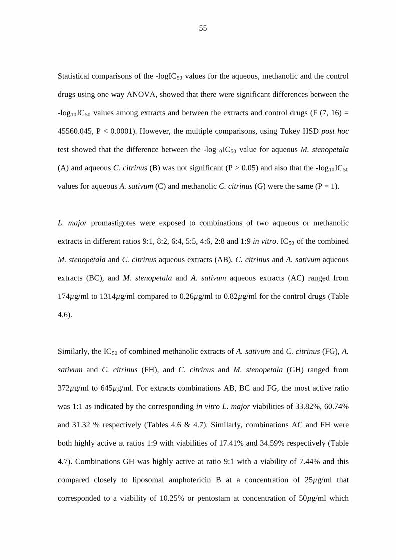
55
Statistical comparisons of the -logIC50 values for the aqueous, methanolic and the control
drugs using one way ANOVA, showed that there were significant differences between the
-log10IC50 values among extracts and between the extracts and control drugs (F (7, 16) =
45560.045, P < 0.0001). However, the multiple comparisons, using Tukey HSD post hoc
test showed that the difference between the -log10IC50 value for aqueous M. stenopetala
(A) and aqueous C. citrinus (B) was not significant (P > 0.05) and also that the -log10IC50
values for aqueous A. sativum (C) and methanolic C. citrinus (G) were the same (P = 1).
L. major promastigotes were exposed to combinations of two aqueous or methanolic
extracts in different ratios 9:1, 8:2, 6:4, 5:5, 4:6, 2:8 and 1:9 in vitro. IC50 of the combined
M. stenopetala and C. citrinus aqueous extracts (AB), C. citrinus and A. sativum aqueous
extracts (BC), and M. stenopetala and A. sativum aqueous extracts (AC) ranged from
174µg/ml to 1314µg/ml compared to 0.26µg/ml to 0.82µg/ml for the control drugs (Table
4.6).
Similarly, the IC50 of combined methanolic extracts of A. sativum and C. citrinus (FG), A.
sativum and C. citrinus (FH), and C. citrinus and M. stenopetala (GH) ranged from
372µg/ml to 645µg/ml. For extracts combinations AB, BC and FG, the most active ratio
was 1:1 as indicated by the corresponding in vitro L. major viabilities of 33.82%, 60.74%
and 31.32 % respectively (Tables 4.6 & 4.7). Similarly, combinations AC and FH were
both highly active at ratios 1:9 with viabilities of 17.41% and 34.59% respectively (Table
4.7). Combinations GH was highly active at ratio 9:1 with a viability of 7.44% and this
compared closely to liposomal amphotericin B at a concentration of 25µg/ml that
corresponded to a viability of 10.25% or pentostam at concentration of 50µg/ml which

56
corresponded to a viability of 10.15%. The difference between the viabilities (%) of L.
major promastigotes when treated with pentostam and when treated with liposomal
amphotericin B was significant (P < 0.05) in all the concentrations except at 25µg/ml.
The mean viability of L. major promastigotes upon exposure to the most active
combinations ratios, tended to compare closely with the viability of liposomal
amphotericin B at a concentration of ≤ 3.125µg/ml (Table 4.7).
Table 4.6: The IC50 levels for blends of test extracts and viability of the L. major promastigotes after treatment with the blends at MIC based concentrations. _____________________________________________________________________ Blends & Most active controls Test extracts & drugs ratio IC50 (µg/ml) Viability (%) _________________________________________________________________________ Aqueous A: B M. stenopetala : C. citrinus 1:1 1.314 × 10³ 33.82a (58.91b) A: C M. stenopetala : A. sativum 1:9 1.740 × 10² 17.41 (97.45) B: C C. citrinus : A. sativum 1:1 3.785 × 10² 60.74 (93.13) Methanolic F: G A. sativum : C. citrinus 1:1 5.608 × 10² 31.32 (56.92) F: H M. stenopetala : A. sativum 1:9 3.721 × 10² 34.59 (71.03) G: H C. citrinus : M. stenopetala 9:1 6.455 × 10² 7.44 (90.30) Positive controlsc Pento Pentostam - 8.0 ×10⁻² 18.41 Lip amph B Liposomal amphotericin B - 9.1× 10⁻² 12.22 Negative control SIM Schneider’s Medium - - 81.65 _________________________________________________________________________ a Represent the viability (%) at the most active ratio of the combination as indicated. b The number in brackets represent the overall viability (%) for all the ratios (1:9 to 9:1). c The initial concentration was 100 µg/ml which was diluted by a factor of 2.
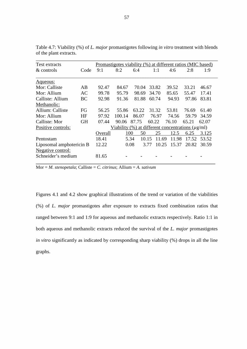
57
Table 4.7: Viability (%) of L. major promastigotes following in vitro treatment with blends of the plant extracts. _________________________________________________________________________ Test extracts Promastigotes viability (%) at different ratios (MIC based) & controls Code 9:1 8:2 6:4 1:1 4:6 2:8 1:9 _________________________________________________________________________ Aqueous: Mor: Calliste AB 92.47 84.67 70.04 33.82 39.52 33.21 46.67 Mor: Allium AC 99.78 95.79 98.69 34.70 85.65 55.47 17.41 Calliste: Allium BC 92.98 91.36 81.88 60.74 94.93 97.86 83.81 Methanolic: Allium: Calliste FG 56.25 55.86 63.22 31.32 53.81 76.69 61.40 Mor: Allium HF 97.92 100.14 86.07 76.97 74.56 59.79 34.59 Calliste: Mor GH 07.44 90.06 87.75 60.22 76.10 65.21 62.07 Positive controls: Viability (%) at different concentrations (µg/ml) Overall 100 50 25 12.5 6.25 3.125 Pentostam 18.41 5.34 10.15 11.69 11.98 17.52 53.52 Liposomal amphotericin B 12.22 0.08 3.77 10.25 15.37 20.82 30.59 Negative control: Schneider’s medium 81.65 - - - - - - ________________________________________________________________________ Mor = M. stenopetala; Calliste = C. citrinus; Allium = A. sativum
Figures 4.1 and 4.2 show graphical illustrations of the trend or variation of the viabilities
(%) of L. major promastigotes after exposure to extracts fixed combination ratios that
ranged between 9:1 and 1:9 for aqueous and methanolic extracts respectively. Ratio 1:1 in
both aqueous and methanolic extracts reduced the survival of the L. major promastigotes
in vitro significantly as indicated by corresponding sharp viability (%) drops in all the line
graphs.

58
Figure 4.1: Viability of L. major promastigotes when exposed to combinations of two aqueous extracts at different ratios in vitro. MICs of extracts were used to constitute the blends at different fixed ratios.
Figure 4.2: Viability of L. major promastigotes when exposed to combinations of two methanolic extracts at different ratios in vitro. MICs were used to constitute the blends.

59
When L. major promastigotes were exposed to the standard drugs, pentostam and
liposomal amphotericin B in vitro, the viabilities were statistically compared using one
way Anova. The difference between the viabilities (%) of L. major in these two drugs at
concentrations of 3.125, 6.25, 12.50, 50.00, 100.00 µg/ml was significant (P < 0.05) but
not at concentration 25 µg/ml, where the difference was not significant (P > 0.05).
Liposomal amphotericin B had a higher potency in lowering the survival of L major
promastogotes in vitro than pentostam, for example at concentrations of 3.125µg/ml for
liposomal amphotericin B and about 5.0 µg/ml for pentostam, L. major promastigotes
viability was 30 % (Figure 4.3).
Figure 4.3: Viability of L. major promastigotes after treatment with serially diluted Leishmania drugs, pentostam (Pento) and liposomal Amphotericin B (Amph B).

60
4.2.3 Interaction of the extracts after combining (Blend effect)
The interaction of the extracts in each blend was evaluated by calculating the sum of
fractional inhibition concentration (SFIC). The SFIC values for aqueous and methanolic
blends of extracts at fixed ratios ranged from 9:1 to 1:9 as shown in Tables 4.8 to 4.11.
Aqueous blend of M. stenopetala and C. citrinus extracts (AB) showed a marked additive
interaction at ratios 8:2, 6:4, 4:6, and 1:9. Synergistic interaction occurred at ratios 5:5, 2:8
and weak antagonistic interaction at ratio 9:1 for blend AB (Table 4.8). As the amount of
C. citrinus decreased in blend AB, the interaction changed from antagonistic to additive
and vice versa. Strong antagonistic interactions in 4 out of 7 ratios were noted for the M.
stenopetala and A. sativum aqueous blends (AC), however, additive interaction at ratios
5:5 and 2:8 and strong synergism at ratio 1:9 were also observed for blend AC (Table 4.8).
Blend of C. citrinus and A. sativum aqueous extracts (blend BC) showed strong
antagonistic interaction in all the ratios except ratio 5:5 which had moderate additive
interaction (Table 4.9). Combined methanolic extracts of A. sativum and C. citrinus (blend
FG) exhibited additive interactions in most of the ratios except ratio 2:8 which exhibited
weak antagonistic interactions (Table 4.10).
For the blend of methanolic extracts of A. sativum and M. stenopetala (FH), it was noted
that, as the content of garlic increased, the interaction tended to exhibit strong antagonism
but as the garlic content decreased, the interaction tended to become additive except for
ratio 4:6 (Table 4.10). Interaction between C. citrinus and M. stenopetala methanolic
extracts (blend GH) was antagonistic in majority of the ratios (8:2, 6:4, 4:6, 2:8, 1:9 ) but
synergistic in the remaining ratios 9:1 and 5:5 (Table 4.11).

61
Table 4.8: Showing the SFIC values to indicate the interaction of aqueous extracts of M. stenopetala (A) with C. citrinus (B) and A. sativum (C). ____________________________________________________________________________________________________________ Ratio (A: B and A: C)
______________________________________________________________________________
Test extract 90: 10 80: 20 60: 40 50: 50 40: 60 20: 80 10: 90 ____________________________________________________________________________________________________________ C. citrinus (B) 2.201c 1.854b 1.602b 0.976a 1.075b 0.966a 1.198b A. sativum (C) 3.176c 3.857c 3.798c 1.180b 2.993c 1.606b 0.686a ____________________________________________________________________________________________________________ The figures in the table are SFIC values, a = synergistic (SFIC < 1); b = additive (1 < SFIC < 2); c = antagonistic (SFIC ≥ 2). The mean SFIC values were derived from average absorbance and viability values.

62
Table 4.9: Showing SFIC values to indicate the interraction of aqueous extracts of C. citrinus (B) with A. sativum (C). ____________________________________________________________________________________________________________ Ratio (B: C)
______________________________________________________________________________
Test extract 90: 10 80: 20 60: 40 50: 50 40: 60 20: 80 10: 90 ____________________________________________________________________________________________________________ A. sativum (C) 3.039c 2.995c 2.730c 1.444b 3.094c 3.175c 2.783c ____________________________________________________________________________________________________________ The figures in the table are SFIC values, a = synergistic (SFIC < 1); b = additive (1 < SFIC < 2); c = antagonistic (SFIC ≥ 2). The mean SFIC values were derived from average absorbance and viability values.

63
Table 4.10: Showing SFIC values to indicate interaction of methanolic extract of A. sativum (F) with C. citrinus (G) and M. stenopetala (H). ____________________________________________________________________________________________________________ Ratio (F: G and F: H)
______________________________________________________________________________
Test extract 90: 10 80: 20 60: 40 50: 50 40: 60 20: 80 10: 90 ____________________________________________________________________________________________________________ C. citrinus (G) 1.670b 1.663b 1.818b 1.143b 1.619b 2.103c 1.780b M. stenopetala (H) 3.683c 3.966c 3.216c 1.948b 2.853c 1.612b 1.120b ____________________________________________________________________________________________________________ The figures in the table are SFIC values, a = synergistic (SFIC < 1); b = additive (1 < SFIC < 2); c = antagonistic (SFIC ≥ 2). The mean SFIC values were derived from average absorbance and viability values.

64
Table 4.11: Showing SFIC values to indicate interaction of methanolic extract of C. citrinus (G) with M. stenopetala (H). ____________________________________________________________________________________________________________ Ratio (G: H)
______________________________________________________________________________
Test extract 90: 10 80: 20 60: 40 50: 50 40: 60 20: 80 10: 90 ____________________________________________________________________________________________________________ M. stenopetala (H) 0.101a 2.995c 2.930c 0.782a 2.368c 2.293c 2.148c ____________________________________________________________________________________________________________ The figures in the table are SFIC values, a = synergistic (SFIC < 1); b = additive (1 < SFIC < 2); c = antagonistic (SFIC ≥ 2). The mean SFIC values were derived from average absorbance and viability values.

65
4.2.4 Anti-amastigote assay [Macrophage assay]
RPMI 1640 medium which had no drug in-cooperated supported the growth of L. major
amastigotes in peritoneal macrophages (Figure 4.4) more effectively and this was
indicated by a high infection rate (IR) of 84.67± 2.96 % (Table 4.12). In contrary, the
leishmaniasis drugs, liposomal amphotericin B and pentostam inhibited the in vitro
survival of L. major amastigotes in BALB/c mice peritoneal macrophages more
effectively and this corresponded to low IRs of 6% and 14% respectively at a
concentration of 50µg/ml (Table 4.12).
Figure 4.4: Showing a BALB/c mice peritoneal macrophage having engulfed L. major amastigotes at 5 days post infection in RPMI-1640 medium culture. Am = amastigote, Nuc = Nucleus, and Cyt = Cytoplasm.
Nuc
Cyt
Am

66
Table 4.12: The in vitro infection rates (IR) and multiplication indices (MI) of L. major amastigotes per 100 peritoneal macrophages following treatment with test extracts at concentrations that ranged between 125 to 15.60µg/ml. Test extracts Conc (µg/ml) IR (%) Amastigotes MI (%) & controls per 100 cells Aqueous A 125.00 58 112 25.34 62.50 65 161 36.43 31.25 70 201 45.48 15.63 84 320 72.40 B 125.00 75 293 66.28 62.50 80 309 69.91 31.25 86 363 82.13 15.63 85 350 79.19 C 125.00 51 163 36.88
62.50 61 213 48.19 31.25 80 221 50.00 15.63 89 306 69.23 Methanolic F 125.00 46 193 43.67
62.50 49 201 45.48 31.25 51 216 48.87 15.63 70 230 52.04 G 125.00 81 271 61.31 62.50 84 293 66.29 31.25 86 310 70.14 15.63 89 320 72.40 H 125.00 70 203 45.93 62.50 75 210 47.51 31.25 83 316 71.49 15.63 87 353 79.86 Controls RPMIa 1st 89 451 n/a 2nd 79 403 n/a 3rd 86 472 n/a
Average 84.67±2.96 442 100.00 Pentostam 50.00 14 51 11.54 25.00 38 123 27.83 12.50 67 210 47.51 6.25 81 307 69.46 Lip ampho B 50.00 6 38 8.60 25.00 10 49 10.86 12.50 19 54 12.22 6.25 23 62 14.03 RPMIa: Negative control in which the amastigotes multiplied maximally. A and H = M. stenopetala; B and G = C. citrinus; C and F = A. sativum.

67
At a concentration of 125µg/ml, the aqueous extracts of M. stenopetala (A), C. citrinus
(B), and A. sativum (C) had infection rates of 58%, 75%, and 51% respectively. Similarly,
the infection rates of methanolic extracts of M. stenopetala (H), C. citrinus (G), and A.
sativum (F) were 70%, 81%, and 46%. Therefore the activity of extracts A, C and F at a
concentration of 125µg/ml in L. major amastigotes was relatively higher and it compared
closely with that of pentostam drug which was 67% at a concentration of 12.5 µg/ml
(Figure 4.5).
The methanolic extracts of A. sativum (F) inhibited the survival of L. major amastigotes
bettter than the aqueous extracts of A. sativum (C) in all the concentrations studied (Figure
4.5), however the difference between their IRs was not significant (paired t test: P =
0.053). High concentrations of the test plant extracts and the control drugs resulted to low
IRs and MIs of L. major amstigotes. Their efficacies were dose-dependent. The difference
between the IRs of test extracts and the control drugs were statistically significant (P <
0.05).
When the MIs of amastigotes in peritoneal macrophages that were treated with 125µg/ml
of test aqueous extracts (A, B, and C) were compared with those treated with 50µg/ml of
the liposomal amphotericin B or pentostam, using one way ANOVA, there was a
statistically significant difference (P = 0.001). A Tukey post hoc test revealed that the MI
of aqueous C. citrinus (B) was significantly different from those of pentostam (P = 0.046)
and liposomal amphotericin B (P = 0.0001). Similarly, MI for A. sativum (C) was
significantly different from that of liposomal amphotericin B (P = 0.022). Further
comparisons showed that the MI of aqueous extracts of M. stenopetala at 125 µg/ml were

68
not significantly different from those of pentostam (P = 0.984) and liposomal amphotericin
B (P = 0.062) at concentrations of 50µg/ml. The MI of liposomal amphotericin B at
50µg/ml was 8.60% while that of pentostam was 11.54% and the difference did not reach
a significance level (P = 0.155).
When the IRs for the methanolic extracts of A. sativum (F), C. citrinus (G), and M.
stenopetala H) were compared with those of liposomal amphotericin B using Tukey’s post
hoc test, the difference in each case was statistically significant with P values of 0.016,
0.0001 and 0.0001 respectively. However, it was only the IR for extract G that was
statistically significant (P = 0.036) when compared to that of the drug pentostam. The
difference between the IR for the drugs pentostam and liposomal amphotericin B was
significant (P = 0.033). Comparisons of the MIs for the extracts F, G and H with those of
liposomal amphotericin B, followed a similar trend, where by the Tukey’s post hoc tests
indicated significant difference (P < 0.05) for each comparison. None of the methanolic
extract had an MI that was statistically different from that of pentostam (P > 0.05). The
MIs of pentostam and liposomal amphotericin B were not statistically different (P = 0.081)
at concentration of 50 µg/ml.
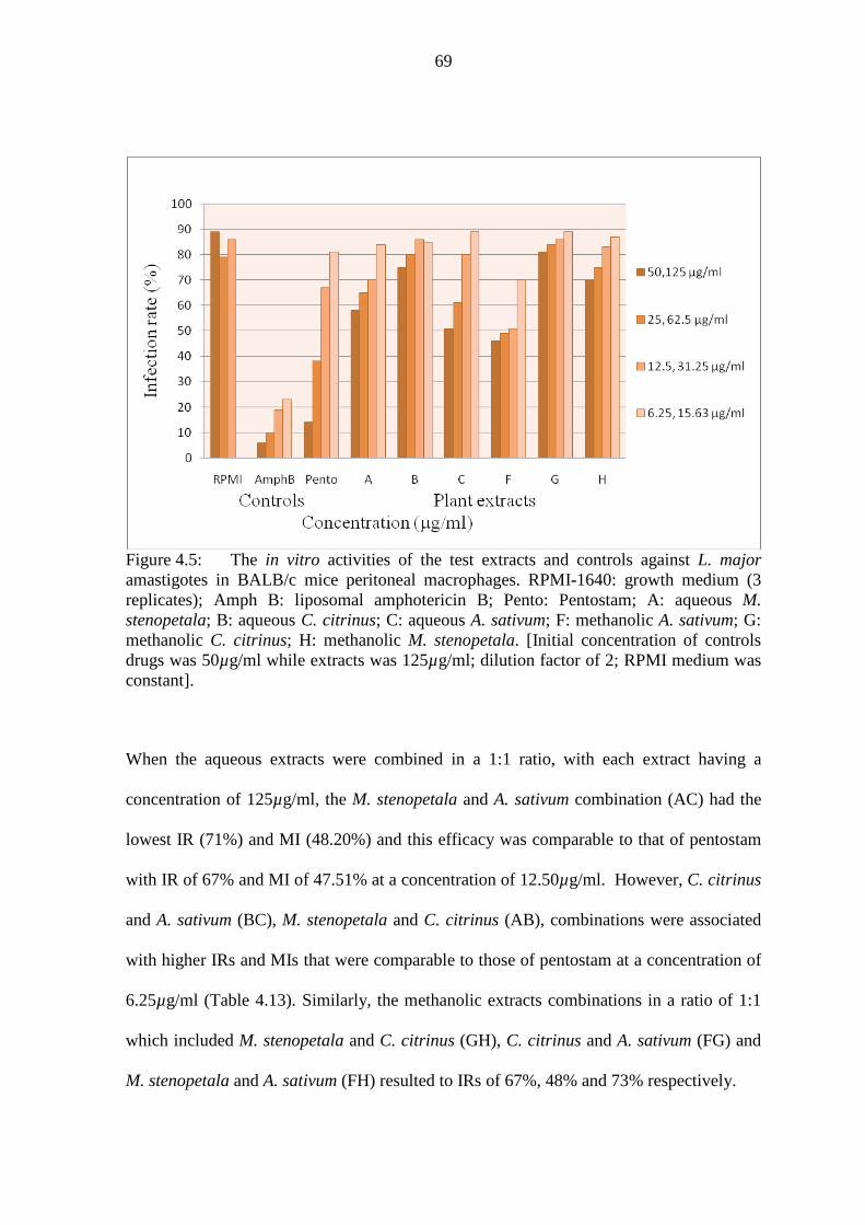
69
Figure 4.5: The in vitro activities of the test extracts and controls against L. major amastigotes in BALB/c mice peritoneal macrophages. RPMI-1640: growth medium (3 replicates); Amph B: liposomal amphotericin B; Pento: Pentostam; A: aqueous M. stenopetala; B: aqueous C. citrinus; C: aqueous A. sativum; F: methanolic A. sativum; G: methanolic C. citrinus; H: methanolic M. stenopetala. [Initial concentration of controls drugs was 50µg/ml while extracts was 125µg/ml; dilution factor of 2; RPMI medium was constant].
When the aqueous extracts were combined in a 1:1 ratio, with each extract having a
concentration of 125µg/ml, the M. stenopetala and A. sativum combination (AC) had the
lowest IR (71%) and MI (48.20%) and this efficacy was comparable to that of pentostam
with IR of 67% and MI of 47.51% at a concentration of 12.50µg/ml. However, C. citrinus
and A. sativum (BC), M. stenopetala and C. citrinus (AB), combinations were associated
with higher IRs and MIs that were comparable to those of pentostam at a concentration of
6.25µg/ml (Table 4.13). Similarly, the methanolic extracts combinations in a ratio of 1:1
which included M. stenopetala and C. citrinus (GH), C. citrinus and A. sativum (FG) and
M. stenopetala and A. sativum (FH) resulted to IRs of 67%, 48% and 73% respectively.

70
The efficacy of combined methanolic extracts in a ratio of 1:1 at a concentration of
125µg/ml, in inhibiting the infectivity and multiplication of L. major amastigotes in
BALB/c mice peritoneal macrophages in vitro was higher than their corresponding
combined aqueous extracts (Figure 4.6). The C. citrinus and A. sativum (FG) combination
performed the best with MI of 23%.
Combinations AB, AC and BC at concentrations of 125µg/ml (1:1 ratios) were associated
with IRs and MIs that were significantly close (P > 0.05) to those of pentostam at
concentrations ranging from 6.25 to 12.5µg/ml. The IRs and MIs associated with
combined extracts AB, AC, and BC were significantly different (P < 0.05) from those of
FG, FH and GH at the same concentrations (Table 4.13).
Similarly, the combinations of the methanolic extracts FG, FH and GH in the same ratio
(1:1) and same concentration (125µg/ml) resulted to infection rates and multiplication
indices that differed significantly from each other and from the control drugs at a
concentration of 12.50 µg/ml (Student’s t test, P < 0.05). This was also confirmed using
one way ANOVA that showed significant differences (P = 0.0001). Tukey’s post hoc test
indicated significant differences (P < 0.05) for all the pair wise comparisons made for the
combined methanolic extracts (FG, FH, and GH) and the control drugs (pentostam and
liposomal amphotericin B). Combination GH and pentostam at a concentration of
12.50µg/ml had the same IR of 67% (P = 1.000), however, their MIs differed significantly
(P < 0.05).

71
Table 4.13: The in vitro infection rates (IR) and multiplication indices (MI) of L. major amastigotes per 100 BALB/c mice peritoneal macrophages following treatment with blends of test extracts in ratios 1:1 and 2:2:1. Blends IR (%) Amastigotes MI (%) & controls Test extracts per 100 cells Blends (1:1), 125µg/ml: 125µg/ml Aqueous AB Mor: Calliste 86 320 72.40 BC Calliste: Allium 81 291 65.84 AC Mor: Allium 71 213 48.20 Methanolic FG Allium: Calliste 48 106 23.98 FH Allium: Mor 73 193 43.67 GH Calliste: Mor 67 176 39.82 Blends (2:2:1) 125µg/ml: 125µg/ml: 62.5µg/ml Aqueous ABC Mor:Calliste:Allium 70 184 41.63 ACB Mor:Allium:Calliste 73 181 40.95 BCA Calliste:Allium:Mor 69 172 38.91 Methanolic FHG Allium:Mor:Calliste 50 169 38.24 HGF Mor:Calliste:Allium 41 161 36.43 FGH Allium:Calliste:Mor 43 148 33.48 Positive controls (µg/ml) Pentostam 50.00 14 51 11.54 25.00 38 123 27.83 12.50 67 210 47.51 6.25 81 307 69.46 Amphotericin 50.00 6 38 8.60 25.00 10 49 10.86 12.50 19 54 12.22 6.25 23 62 14.03 Negative control RPMIa 1st test 89 451 n/a (Medium) 2nd test 79 403 n/a 3rd test 86 472 n/a
Average 84.67± 2.96 442 100.00 RPMIa: Negative control in which the amastigotes multiplied maximally. The average multiplication index for RPMI medium was therefore taken as 100%. Mor = Moringa stenopetala;

72
Calliste = C. citrinus; Allium = A. sativum.
Figure 4.6: In vitro activities for blends of aqueous and methanolic plant extracts (1:1 ratio at concentration of 125µg/ml) as represented by infection rates of L. major amastigotes in treated BALB/c mice peritoneal macrophages.
Blends of three aqueous plant extracts had higher MIs as compared to their corresponding
methanolic extracts. For example, aqueous blend ABC (M. stenopetala, C. citrinus and A.
sativum at concentrations of 125µg/ml in 2:2:1 ratio) had MI of 41.63% while methanolic
blend HGF (M. stenopetala, C. citrinus and A. sativum at concentrations of 125µg/ml in
2:2:1 ratio) had MI of 36.43%. This trend was similar for the remaining aqueous and
methanolic blends. Therefore blends of three methanolic extracts in the ratio of 2:2:1 were
more effective in inhibiting in vitro multiplication of L. major amastigotes in BALB/c
mice peritoneal macrophages. The efficacy of these combined methanolic extracts was
higher than than those of RPMI medium but closely comparable to that of the pentostam at
a concentration of 12.5µg/ml with MI of 47.51% (Figure 4.7).

73
Figure 4.7: In vitro activities of combined methanolic extracts (125 µg/ml) and control drugs (12.5 µg/ml) as represented by MIs of L. major amastigotes in treated BALB/c mice peritoneal macrophages.
Two-way ANOVA statistical test was used to compare both IRs and MIs of L. major
amastigotes in BALB/c mice peritoneal macrophages cultured in vitro and treated with
aqueous and methanolic combinations of three extracts or control drugs. The IRs and the
MIs of the triple combinations and those of the control drugs differed significantly (F =
2291.59; P = 0.0001). Multiple comparisons using Tukey’s post hoc test indicated that the
differences of IRs and MIs among the aqueous combinations (ABC, ACB and BCA) were
not significant (P > 0.05). However, the IRs and MIs of all the blends of three aqueous
extracts were significantly different from those of liposomal amphotericin B (P < 0.05).
Only combination BCA (C. citrinus, A. sativum and M. stenopetala was significantly
different from pentostam (P = 0.398). Therefore, combinations ABC and ACB were

74
associated with IRs and MIs that were significantly close to pentostam drug at a
concentration of 12.5µg/ml (P > 0.05).
Similarly, the methanolic blends of three extracts FHG, HGF and FGH at a concentration
125 µg/ml in a ratio of 2:2:1 showed that their in vitro activities against the L. major
amastigotes differed significantly among themselves and from those of pentostam and
liposomal amphotericin B drugs (P < 0.05). However, multiple statistical comparison
using Tukey’s HSD post hoc test showed that methanolic blend of HGF (M. stenopetala,
C. citrinus and A. sativum) and FGH (A. sativum, M. stenopetala and C. citrinus ) differed
non-significantly (P = 0.976) as illustrated in Figure 4.7. It was therefore observed that the
methanolic combinations of three (FHG, HGF and FGH) compared closely with efficacy
of pentostam drug when applied at a concentration of 12.5µg/ml.
In vitro inhibition rates of amastigotes in macrophages by aqueous combinations of AB,
BC, and AC were -1.6%, 4%, and 16% respectively while inhibition rates by aqueous
triple combinations ABC, BCA, and ACB were 17%, 19% and 14%. Similarly,
amastigotes inhibition rates for methanolic combinations FG, FH, and GH were 43%,
14%, and 21% respectively while the inhibition rates for tipple methanolic combinations
FGH, FHG, and GHF were 49%, 41% and 52% respectively.
4.2.5 In vitro nitric oxide assay
Nitric oxide (NO) plays a key role as a leishmanicidal effector molecule in host
macrophages (Gamboa-leon et al., 2007). Therefore the effect of the single aqueous and
methanolic plant extracts in NO production was evaluated in vitro. Since the breakdown of

75
NO occurs in the macrophages to release nitrite (NO2-) as one of the products, NO
produced was estimated by quantifying the NO2- content. The representative nitrite
standard reference curve in RPMI medium was prepared as described in the Griess reagent
system instructions manual (Promega® USA). In summary, BALB/c mice peritoneal
macrophages were incubated in RPMI-1640 medium for 48 hours in the presence or in
absence of the plant extracts. The optical densities (OD) readings of the majority aqueous
extracts were all less than 0.10 (Table 4.14) at concentrations that ranged between 3.125 to
100 µg/ml and this implied that, negligible amount of NO was produced and this could not
be estimated from the standard nitrite curve. RPMI-1640 medium produced similar
negligible levels of NO. However, the aqueous extract of A. sativum (extract C) triggered
production of 20µM of nitric oxide at a concentration of 100µg/ml (Figure 4.8) since its
optical density was 0.162 (Table 4.14). The NO production triggered by aqueous A.
sativum extracts was significantly higher (F (4, 10) = 223744.34, P = 0.0001) than that of
the other aqueous extracts at a concentration of 100µg/ml and that of RPMI medium.
Table 4.14: Absorbance (OD) units ± SD of the standard nitrite (positive control), RPMI-1640 (negative control) and the aqueous extracts from the study plants. _________________________________________________________________________ Conc (µM) Standard Nitrite RPMI M. stenopetala C. citrinus A. sativum (+ve Control) (-ve Control) (A) (B) (C) _________________________________________________________________________ 0 0.054± 0.011 0.021 0.051 0.032 0.046
20 0.153± 0.013 0.030 0.053 0.035 0.048
40 0.178±0.001 0.038 0.061 0.036 0.054
60 0.331± 0.000 0.048 0.051 0.040 0.053
80 0.599± 0.004 0.055 0.059 0.042 0.087
100 0.983± 0.010 0.079 0.066 0.045 0.162 _________________________________________________________________________

76
Figure 4.8: Nitric Oxide (µM) produced by BALB/c mice macrophages after exposure to different concentrations of single aqueous test extracts (100µg/ml serially diluted to 3.125µg/ml by a factor of 2).
In order to determine the amount of NO triggered by the combined aqueous extracts, their
optical densities (absorbances) were determined using a micro-titre reader. All the
absorbances for all the combined aqueous extracts ranged between 0.041 and 0.081 (Table
4.15). These optical densities corresponded to ≤ 20 µM of NO in the standard nitrite curve
for the blended extracts of M. stenopetala & C. citrinus (blend AB), C. citrinus & A.
sativum (blend BC) and M. stenopetala, C. citrinus & A. sativum (blend ABC).

77
Table 4.15: Nitric oxide production by the BALB/c mice peritoneal macrophages after being stimulated with blends of aqueous test extracts at fixed ratios at concentrations of 125µg/ml. _________________________________________________________________________ Combinations Code Ratio OD units NO
(µM). _________________________________________________________________________ M. stenopetala & C. citrinus AB 1:1 0.081 < 20
C. citrinus & A. sativum BC 1:1 0.057 < 20
M. stenopetala & A. sativum AC 1:1 0.041 -a
M. stenopetala, C. citrinus & A. sativum ABC 2:2:1 0.066 < 20
M. stenopetala, A. sativum & C. citrinus ACB 2:2:1 0.045 -
A. sativum, C. citrinus & M. stenopetala BCA 2:2:1 0.042 - _________________________________________________________________________ -a = the concentration of the NO released by the stimulated macrophages was below the detectable limit in the standard reference curve.
Methanolic C. citrinus extracts (G) stimulated production of 20µM of NO at a
concentration of 6.25µg/ml from infected BALB/c mice macrophages. The remaining
methanolic extracts of A. sativum (F) and M. stenopetala (H) stimulated production of low
levels of less than 10 µM of NO from BALB/c mice macrophages. Extracts F and H had
an efficacy of NO stimulation that compared closely with that of RPMI-1640. The OD
readings for the drugs F, G and H were statistically compared with OD readings for the
RPMI-1640 and those of the standard nitrite to establish the significance difference using
one way ANOVA. At a concentration of 6.250 µg/ml, the difference of the extracts’ OD
readings and those of standard nitrite and RPMI were significant (F (4, 10) = 16.680, P =
0.0001). Tukey HSD Post hoc test indicated that the differences between the OD reading
for extract G and the OD readings for standard nitrite, RPMI-1640, extracts H and F were
siginificant (P < 0.05). Similarly, at a concentration of 100 µg/ml, the differences between

78
the OD readings for the methanolic extracts and those of standard nitrite and RPMI-1640
were significant (F (4, 10) = 290.579, P = 0.0001), except for methanolic extracts of M.
stenopetala (H) and A. sativum (F) whose OD readings were not significantly different (P
> 0.05) as indicated in Table 4.16. Methanolic extracts of C. citrinus stimulated BALB/c
mice peritoneal macrophages in vitro to release significant amount of NO and this was
equally indicated by the graphical peak at concentration of 6.25µg/ml (Figure 4.9).
Table 4.16: The absorbances (OD) units ± SD of the standard nitrite, RPMI-1640 and the test methanolic extracts.
_________________________________________________________________________
Conc (µM) Standard Nitrite RPMI M. stenopetala C. citrinus A. sativum (+ve Control) (-ve Control) (H) (G) (F) _________________________________________________________________________ 0 0.061± 0.012 0.062 0.064a 0.064a 0.065a
20 0.075± 0.037 0.064 0.063 0.071 0.062
40 0.097± 0.067 0.054 0.061 0.064 0.062
60 0.145± 0.135 0.055 0.062 0.066 0.063
80 0.224± 0.246 0.056 0.062 0.063 0.060
100 0.298± 0.359 0.055 0.063b 0.069b 0.062b _________________________________________________________________________ Note: The OD readings for all the methanolic extracts represented concentration which
decreased from 100µg/ml (0.063b) to 3.125µg/ml (0.064a), the dilution factor being 2.

79
Figure 4.9: Nitric oxide (µM) produced by BALB/c mice peritoneal macrophages treated with different concentrations of the test methanolic extracts.
It was noted that only three combinations of methanolic extracts which included FH, HGF
and FGH stimulated the BALB/c mice peritoneal macrophages to release ≤ 20 µM of NO
(Table 4.17).

80
Table 4.17: Nitric oxide (NO) production by the Balb/c mice macrophages after stimulation with combined methanolic plant extracts. ________________________________________________________________________
Combinations Code Ratio OD NO (µM) ________________________________________________________________________
M. stenopetala & C. citrinus GH 1:1 0.055 -
C. citrinus & A. sativum FG 1:1 0.037 -
M. stenopetala & A. sativum FH 1:1 0.074 20
M. stenopetala, C. citrinus & A. sativum HGF 2:2:1 0.076 20
M. stenopetala, A. sativum & C. citrinus HFG 2:2:1 0.038 -
A. sativum, C. citrinus & M. stenopetala FGH 2:2:1 0.068 < 20 _________________________________________________________________________
4.2.6 Cytotoxicity assay using vero cells
The Vero cell line was obtained from KEMRI, Center for Viral Research (CVR) where it
has been maintained through cryo-preservation and passaging weekly. The vero cells were
maintained in minimum essential medium (MEM) supplemented with 10% FBS, 100U/ml
of penicillin G and 100µg/ml of streptomycin. In order to determine the in vitro cytotoxic
effects of the study plant extracts against vero cells, the single aqueous extracts A, B and C
plus methanolic extracts F, G and H were incubated with vero cells at 37oC in a humidified
5% CO2. Similarly, in order to determine the IC50 of the extracts and % viability of vero
cells after treatment with extracts, an MTT assay was carried out as described by
Wabwobwa et al. (2010). The absorbance values obtained were used to calculate the cells
viability using the formula given by Mosmann (1983). The IC50 values of the test extracts
and the control leishmaniasis drugs were determined automatically by inserting the

81
absorbance values in the Chemosen software program designed to determine IC50 for test
drugs.
Aqueous extracts of C. citrinus (B) were slightly more toxic (IC50 of 467µg/ml) as
compared to those of aqueous extracts of A. sativum (C) and M. stenopetala (A) whose
IC50 values were at 1306.68µg/ml and 2105.93µg/ml respectively (Table 4.18). In
contrast, methanolic extracts of A. sativum (F), C. citrinus (G) and M. stenopetala (H) had
IC50 values of 1050.50µg/ml, 1314.65µg/ml and 912.58µg/ml respectively (Table 4.18).
The higher the cytotoxity, the higher was the negative log base ten IC50 values (-
log10IC50), for instance, liposomal amphotericin B was highly toxic to vero cells with a -
log10IC50 value of -1.78 while aqueous A.sativum was lowly toxic with a -log10IC50 value
of -3.32.
The viabilities (%) of the vero cells following treatment with test extracts and control
drugs tended to follow a similar pattern. For example, vero cells treated with slightly toxic
aqueous C. citrinus had the lowest viability (109.71%) compared to the vero cells treated
with less toxic A. sativum (134.06%) or M. stenopetala (112.30%) aqueous extracts (Table
4.18). M. stenopetala methanolic extracts were slightly more toxic to vero cells with an
IC50 of 912.58µg/ml when compared to methanolic extracts of A. sativum and C. citrinus.
Generally, all the test extracts studied were relatively less toxic to vero cells when
compared to the toxicity levels of the control drugs, pentostam (IC50 = 108.58µg/ml ) and
liposomal amphotericin B (IC50 = 60.95µg/ml). It was however noted that viability of
vero cells after exposure to methanolic C. citrinus extracts at a concentration of 1000µg/ml
was 88.14% and this compared closely with those of the pentostam and liposomal

82
amphotericin B control drugs which were at 98.10 % and 98.51 % respectively at
concentration of 100µg/ml (Table 4.18).
Table 4.18: The IC50 (µg/ml) of the test extracts and their effects on vero cells expressed as cell viability (%). ________________________________________________________________________ Test extracts Code IC50 (µg/ml) -log10IC50 Cell viability (%) _____________________________________________________________________ Aqueous: M. stenopetala A 1306.68 -3.12 112.30 C. citrinus B 467.11 -2.67 109.71 A. sativum C 2105.93 -3.32 134.06 Methanolic : A. sativum F 1050.50 -3.02 147.93 C. citrinus G 1314.65 -3.12 88.14 M. stenopetala H 912.58 -2.96 106.15 Positive controls Pentostam Pento 108.58 -2.04 98.10 Liposomal Amphotericin B Amph B 60.95 -1.78 98.51 _________________________________________________________________________ Note: The initial concentration of the test extracts was 1000µg/ml while that of control drugs was 100 µg/ml and both were serially diluted by a factor of 2.
The viability (%) of the vero cells tended to decrease as the concentrations of the test
extracts or the control drugs increased (Table 4.19). At concentration of between 125µg/ml
and 1000 µg/ml, aqueous C. citrinus extracts lowered the vero cells’ viability to less than
85%, while the methanolic C. citrinus extracts lowered the viability to less than 50% at the
same concentrations (Table 4.19). The cytotoxicity of aqueous C. citrinus extracts at a
concentration of 125µg/ml compared closely with that of pentostam drug at a
concentration of 50µg/ml.
The C. citrinus aqueous and methanolic extracts tended to show a dose dependent
cytotoxic effect which corresponded to low vero cells viability as their concentration

83
increased. This was illustrated by the extracts linear trendlines (Figures 4.10 and 4.11). At
a concentration of 250µg/ml, methanolic C. citrinus extracts were associated with 50%
viability of vero cells (Figure 4.11). Aqueous M. stenopetala extracts at a concentration of
≥ 500µg/ml had higher cytotoxic effects to vero cells since their viability was ≤ 79.73%
(Table 4.19). In comparison, methanolic M. stenopetala extracts seemed to be less
cytotoxic since the viability of vero cells at concentrations of 125µg/ml and 250µg/ml was
89.14% and 92.49% respectively. A. sativum extracts were slightly cytotoxic to the vero
cells since at a concentration of 1000µg/ml both aqueous and methanolic extracts were
associated with vero cells viability of 99.53% and 100.13% respectively (Table 4.19).
Table 4.19: Viability (%) of vero cells after in vitro exposure to specific concentrations
(µg/ml) of single plant extracts against L. major promastigotes. _________________________________________________________________________ Vero cells viability (%) at specific concentrations (µg/ml) Test extracts/ Control drugs Code 1000 500 250 125 62.50 31.25 15.63 _________________________________________________________________________ Aqueous extracts M. stenopetala A 77.62 79.73 133.67 127.06 110.36 124.88 132.74 C. citrinus B 81.32 85.08 85.21 82.70 117.49 171.02 145.15 A. sativum C 99.53 112.74 144.03 138.35 131.29 143.56 168.98 Methanolic extracts A. sativum F 100.13 120.26 139.01 162.24 137.49 182.31 194.06 C. citrinus G 48.37 27.29 49.70 124.49 134.14 113.25 119.69 M. stenopetala H 108.14 109.34 92.49 89.14 120.70 114.14 109.09
Vero cells viability (%) at specific concentrations (µg/ml) Positive controls Code 100 50 25 12.5 6.25 3.125 1.563 Pentostam Pento 74.83 83.74 103.17 103.79 104.65 105.65 110.92 Lip amphotericin B Amph B 88.83 89.82 92.86 92.99 102.79 104.28 118.00 Negative controla Minimum E medium MEM Average viability (%) of vero cells = 92.86±SD 0.16 _________________________________________________________________________ a The % viability in the negative control was the average for viabilities of vero cells in MEM in microtiter plates A and B.

84
Figure 4.10: The viability of vero cells in varying concentration of test aqueous extracts, pentostam and amphotericin B. a = the concentration for control drugs ranged between 100µg/ml and 1.563µg/ml, with a dilution factor of 2.
Figure 4.11: The viability of vero cells in varying concentration of methanolic test extracts, pentostam and amphotericin B. a = concentration for control drugs ranged between 100µg/ml and 1.563µg/ml, with a dilution factor of 2.

85
4.3 In vivo bioassays
4.3.1 Treatment of L. major infected BALB/c mice
The BALB/c mice infected subcutaneously with L. major metacyclic promastigotes on
their left hind foot developed lesions and swelling. Lesion development was monitored by
measuring the thickness of the infected footpad using a vernier caliper. The average lesion
size was expressed as the difference between the infected left hind foot and non infected
contra-lateral right hind footpad. All the mice developed lesions in 4 weeks after the initial
infection (Figure 4.12).
(a) (b)
Figure 4.12: (a) Swollen left footpad or lesion (Les) of a BALB/c mouse after infection with metacyclic L. major promastigotes compared to non infected contra-lateral right footpad; (b) Advanced lesion (Ad Les) on infected left foot pad of a BALB/c mouse.
Ad Les
Les

86
4.3.1.1 Effects of single aqueous extracts on lesion sizes and body weights
Following the oral treatment with aqueous crude extracts from M. stenopetala (A), 40% of
the mice (2/5) had their footpad swelling remaining constant from week three onwards,
while a further 40% did not respond to the moringa treatment since their foot swellings
continued to increase throughout the treatment period. However, a further 20% of the mice
treated with aqueous moringa extracts showed a sharp decrease in their lesion size from
week three of treatment. On average however, aqueous M. stenopetala extracts (A) caused
the foot pad lesion sizes to decrease and stabilize from week 4 (Figure 4.13).
Seventy five percent (75%) of the mice treated orally with C. citrinus aqueous flower
extracts (B), showed a marked decrease of their footpad swelling from the third week of
treatment as compared to the mice treated with oral phosphate buffered saline (PBS)
whose lesion size increased steadily. The remaining 25% of mice did not respond to the
treatment with C. citrinus flower extracts. On average, the orally administered aqueous C.
citrinus flower extracts caused a steady decrease of the foot pad lesions in infected
BALB/c mice, a trend that was closely comparable to that in mice treated with drug
pentostam.
Majority of the infected BALB/c mice treatment with oral aqueous A. sativum extracts (C)
were characterized by a sharp decrease of the lesion sizes within the first one week of
treatment which was then followed by gentle increase. However, when compared to the
mice treated with oral PBS, aqueous A. sativum extracts administered orally reduced the
footpad lesion sizes significantly in infected BALB/c mice (Figure 4.13). One mouse out
of the six (17%) treated with aqueous garlic extracts, showed a very clear and steady

87
decrease of the footpad swelling throughout the treatment period. Thirty percent (30%) of
the mice however did not respond to the oral aqueous garlic treatment.
Statistical comparison of the lesion sizes in groups of infected BALB/c mice treated with
drugs A, B, C with those treated with liposomal amphotericin B, pentostam and PBS oral
indicated that the difference among the lesion sizes was significant ( F ( df 5,24) = 2.851,
P = 0.037). However, Tukey’s post hoc test indicated that the difference in lesion sizes for
the mice treated with extracts A, B and C and those treated with control drugs was not
significant (P > 0.05). The lesion sizes in mice treated with aqueous A. sativum extracts
(C) and lesion sizes in mice treated with oral PBS differed significantly (P = 0.025). The
aqueous extracts were active in reducing the foot pad lesion sizes with the efficacy of
aqueous A. sativum extracts (C) being the best followed by aqueous C. citrinus extracts
(B) as indicated in Figure 4.13.
Figure 4.13: The foot pad swelling after oral treatment of L. major infected BALB/c mice with test aqueous extracts. Pento = Pentostam; PBS oral = phosphate buffered saline administered orally.

88
Twenty percent (20%) of the infected BALB/c mice treated with aqueous A. sativum (C)
extracts administered intra-peritoneally (ip) caused a steady decrease in the foot pad lesion
size and a further 20% of the mice had a decrease of their lesion sizes within the first week
of treatment but later increased their lesion sizes as the treatment progressed. The
remaining 60% of the mice showed a steady increase of their lesion sizes as the treatment
progressed. On average however, the aqueous A. sativum extracts (C) when administered
intra-peritoneally (ip), reduced the footpad swelling for the first three weeks of treatment
(Figure 4.14). Both the aqueous extracts of C. citrinus (B) and M. stenopetala (A) limited
the footpad lesion progression from the third week of treatment onwards and it compared
closely with the efficacy of control drugs pentostam and liposomal amphotericin B (Figure
4.14). The difference between the mean lesion sizes of mice treated with aqueous extracts
A, B, and C that were administered intra-peritoneally and mean lesion sizes of mice
treated with pentostam, liposomal amphotericin B and PBS (ip) was significant (F (df
2,24) = 5.637, P = 0.001). Tukey’s post hoc test showed a significant difference (P =
0.024) between the mean lesion size of mice treated with aqueous extracts B (ip) and those
treated with PBS (ip). Similarly, the mean lesion sizes of mice treated with aqueous
extracts C (ip) were significantly different from those treated with liposomal amphotericin
B (P = 0.018) and from those treated with PBS-ip (P = 0.001).

89
Figure 4.14: The foot pad swelling after intra-peritoneal treatment of L. major infected BALB/c mice with aqueous extracts and control drugs. Mor = M. stenopetala; Calliste = C. citrinus; Allium = A. sativum; Amph-B= liposomal amphotericin B; Pento = Pentostam; ip PBS = intra peritoneally administered phosphate buffer saline.
The mean body weights of the infected BALB/c mice at the beginning and at the end of a
5 week treatment period with the aqueous test extracts and control drugs were compared
by a two tailed paired t test analysis. It was noted that there was a significant loss of body
weights (P < 0.05) for infected BALB/c mice that were treated orally or intra peritoneally
with aqueous M. stenopetala extracts (A) as shown in Table 4.20. Similarly, aqueous C.
citrinus extracts (B) administered intra-peritoneally led to a significant weight loss (P <
0.05) in infected BALB/c mice, however, mice treated with the same extracts B but
administered orally did not cause a significant weight loss. Aqueous A. sativum extracts
(C) administered orally and intra-peritoneally did not affect the body weights of the treated
BALB/c mice because their initial and final body weights were not significantly different
(P > 0.05) as shown in Table 4.20.

90
Table 4.20: The mean lesion size (mm) and body weights (g) in BALB/c mice infected with L. major and treated with single aqueous test extracts and Leishmania drugs administered orally or intra-peritoneal (ip) over a 5 weeks (wks) period.
___________________________________________________________________________________________________________ Mean f/pada swelling (mm) t- test for weights Mean body wgt (g)
____________________________ ________________ ____________________________ Drugs Code Route Wk1 Wk2 Wk3 Wk4 Wk5 Calculated t, P value Initial ±SE Final ± SE Lossc ___________________________________________________________________________________________________________ Test extracts: M. stenopetala A Oral 1.48 1.70 1.96 2.12 2.10 t (4df) = 3.16, 0.034 17.60±0.51 16.60± 0.75 1.00 (s) C. citrinus B Oral 1.43 1.83 2.53 2.33 2.16 t (3df) = 2.40, 0.096 18.25±0.63 15.75± 1.31 2.50 (ns) A. sativum C Oral 1.50 1.20 1.30 1.41 1.62 t (4df) = 0.00, 1.000 21.33±0.51 20.83± 0.40 - 0.05 (ns) M. stenopetala A IP b 1.55 1.70 1.85 1.75 1.65 t (4df) = 3.50, 0.025 18.20±0.92 16.80± 0.58 1.40 (s) C. citrinus B IP 1.20 1.30 1.75 1.75 1.70 t (4df) = 4.71, 0.009 18.00±0.32 15.60± 0.40 2.40 (s) A. sativum C IP 1.30 1.00 1.05 1.60 1.60 t (4df) = 0.54, 0.621 21.80±0.66 21.60± 0.81 0.20 (ns) Controls:
Lip Ampho B - IP 1.50 2.20 1.90 2.00 1.97 t (4df) =-0.54, 0.621 16.60±0.24 16.80± 0.37 - 0.20 (ns) Pentostam - IP 1.50 2.04 1.94 1.85 1.80 t (4df) = 1.37, 0.242 18.40±0.24 17.60± 0.37 0.80 (ns) PBS - Oral 1.30 1.95 2.30 2.50 2.60 t (2df) = 0.56, 0.635 19.33±0.33 18.67± 1.33 0.66 (ns) PBS - IP 1.40 2.25 2.35 2.30 2.32 t (2df) = 2.00, 0.184 21.00±0.00 20.33± 0.33 0.67(ns) ___________________________________________________________________________________________________________
Note: f/pada = foot pad; IP b= Intra-peritoneal; SE = Standard error; (s) = significant; (ns) = non-significant; Loss c = initial wt – final wt.

91
4.3.1.2 Effects of individual methanolic extracts on lesion sizes and body weights
The methanolic extracts of C. citrinus (G) and A. sativum (F), when administered orally
into L. major-infected BALB/c mice showed significant efficacy in arresting the increase
of the foot pad lesions. Both A. sativum and C. citrinus methanolic extracts compared
closely with the standard Leishmania drugs pentostam and liposomal amphotericin B,
whereby they reduced the footpad swelling especially from week three of treatment
(Figure 4.15). Efficacy of extracts A. sativum (F) in reducing the lesion sizes was close to
that of pentosatm and liposomal amphotericin B with P values of 0.986 and 0.898
respectively. The efficacies of A. sativum (F) and C. citrinus (G) methanolic extracts
deviated from that of the PBS control but the difference did not reach significance (P >
0.05) as indicated in Figure 24. Methanolic M. stenopetala extracts (H) did not reduce the
footpad swelling in the infected BALB/c mice and this compared closely with that of PBS
(Figure 4.15). Tukey’s post hoc test showed that extracts H had an efficacy that was
significantly different from that of extract G (P = 0.0001), extract F (P = 0.0001),
liposomal amphotericin B (P = 0.003, pentostam (P = 0.001) and PBS oral (P = 0.035) as
seen in Figure 4.15.

92
Figure 4.15: The foot pad swelling after oral treatment of L. major infected BALB/c mice with test methanolic extracts. Pento = pentaostam; Lip Amph B = Liposomal amphotericin B; PBS oral = orally administered phosphate buffered saline. L. major infected BALB/c mice that were treated with methanolic C. citrinus extracts
intra-peritoneally seemed to change their behavior immediately after the injection.
Following the injection with C. citrinus extracts, the mice congregated in one corner of the
cage, with their snouts touching the beddings, and their hairs becoming slightly rough
(Figure 4.16).

93
Figure 4.16: Shows the congregating and less mobility behavior of the BALB/c mice (ears not marked) with their hairs becoming rough immediately after an ip treatment with methanolic C. citrinus extracts.
Similarly, intra peritoneal (ip) administration of methanolic extracts of A. sativum (F) and
C. citrinus (G) showed a significant efficacy in reducing the footpad swelling in infected
BALB/c mice. The reduction of the footpad lesions in the mice was more evident from the
third week of treatment with C. citrinus methanolic extracts and from fourth week with A.
sativum methanolic extracts. This was comparable to the lesion size reduction in the mice
treated with pentostam or liposomal amphotericin B drugs (Figure 4.17(a)). The mean
lesion sizes of the BALB/c mice treated with extracts G(ip) or F(ip) were not significantly
different from those of mice treated with control drug pentostam (P > 0.05). Methanolic
extracts F (ip) was however significantly different from liposomal amphotericin B (P =

94
0.019) while extract G (ip) was not significantly different from liposomal amphotericin B
(P = 0.060).
The efficacy of ip methanolic extracts of M. stenopetala (H) indicated that 60% (3/5) of
the infected mice had their foot pad lesions increase with progress of the treatment and
40% (2/5) had their foot pad lesions reduce and remain constant from the third week
(Figure 4.17(b)), however, when the mean lesion sizes of the former were compared with
those of the later, the reduction was weakly significant (P = 0.053). On average, however,
the efficacy of extracts H (ip) greatly matched that of intra peritoneal PBS (Tukey post hoc
test, P = 0.853) and this implied that methanolic M. stenopetala extracts were not very
active in reducing foot pad lesions caused by L. major in BALB/c mice.
Figure 4.17(a): The foot pad swelling after intra peritoneal (ip) treatment of L. major - infected BALB/c mice with test methanolic extracts on the progression of footpad lesions. Pento = pentostam; Lip amph B = liposomal amphotericin B; PBS = phosphate buffered saline.
95
Figure 4.17(b): The foot pad swelling after intra peritoneal (ip) treatment of L. major - infected BALB/c mice with methanolic extracts of M. stenopetala (H). MS = mouse; AVER = average. Statistical significance between mouse 3 and mouse 4 was P = 0.077; statistical significance between mouse 3 and mouse 5 was p = 0.053.
The mean body weights of the infected BALB/c mice at the beginning of in vivo assays
and after a 5 week treatment period with individual methanolic extracts (F, G and H)
administered either orally or intraperitoneally, were compared using pairerd t-test
analysis. The methanolic extracts of A. sativum (F), C. citrinus (G) and M. stenopetala (H)
did not affect the body weights of the L. major infected BALB/c mice significantly. This
observation was comparable to that of the control mice that were treated with liposomal
amphotericin B or pentostam or PBS (Table 4.21).

96
Table 4.21: The average lesion size (mm) and body weights (g) in BALB/c mice infected with L. major and treated with single methanolic test extracts and Leishmania drugs administered orally or intra-peritoneally (ip) over a 5 weeks (wks) period.
___________________________________________________________________________________________________________ Mean f/pada swelling (mm) t- test for body wgts Mean body wgt (g) ____________________________ _______________ ____________________________ Drugs Code Route Wk1 Wk2 Wk3 Wk4 Wk5 Calculated t, P value Initial ± SE Final ± SE Lossc ___________________________________________________________________________________________________________ Test extracts: M. stenopetala H Oral 2.14 2.46 2.88 3.36 3.40 t (4df) = 1.18, 0.305 17.40± 0.51 16.60± 0.87 0.80 (ns) C. citrinus G Oral 1.38 1.40 1.74 1.71 1.71 t (4df) = 0.00, 1.000 19.00± 0.63 18.80± 0.81 0.20 (ns) A. sativum F Oral 1.63 1.63 1.81 1.67 1.68 t (4df)= -0.59, 0.587 19.60± 0.75 19.80± 0.86 - 0.20 (ns)
M. stenopetala H IP b 2.05 2.15 2.25 2.75 2.43 t (4df) = 2.14, 0.099 16.20±0.66 15.00± 0.84 1.20 (ns) C. citrinus G IP 1.08 1.22 1.54 1.50 1.60 t (4df) = 0.00, 1.000 19.40± 0.68 19.00± 0.55 0.40 (ns) A. sativum F IP 1.04 1.16 1.25 1.68 1.35 t (3df) =-1.00, 0.391 19.00±0.58 18.75± 0.75 0.25 (ns) Controls: Lip ampho B - IP 1.50 2.20 1.90 2.00 1.97 t (4df) =- 0.54, 0.621 16.60±0.24 16.80± 0.37 - 0.20 (ns) Pentostam - IP 1.50 2.04 1.94 1.85 1.80 t (4df) = 1.37, 0.242 18.40±0.24 17.60± 0.37 0.80 (ns) PBS - Oral 1.30 1.95 2.30 2.50 2.60 t (2df) = 0.56, 0.635 19.33±0.33 18.67± 1.33 0.66 (ns) PBS - IP 1.40 2.25 2.35 2.30 2.32 t (2df) = 2.00, 0.184 21.00±0.00 20.33± 0.33 0.67(ns) ___________________________________________________________________________________________________________
Note: f/pada = foot pad; IP b= Intra-peritoneal; SE = Standard Error; (s) = significant; (ns) = non-significant; Lossc = Initial wt – final wt.
97
4.3.1.3 Effects of blends of two different aqueous extracts on BALB/c mice lesion
sizes and body weights
Table 4.22, gives a summary of the effects of orally and intraperitoneally administered
combined aqueous extracts in a ratio of 1:1 on footpad lesion progression in infected
BALB/c mice. When administered orally into infected BALB/c mice, the combination of
M. stenopetala and C. citrinus (AB) efficacy in reducing the foot pad lesion sizes was not
significantly different from the efficacy of the control drugs (Tukey’s test, P > 0.05).
Combination of C. citrinus and A. sativum (BC) however reduced the footpad lesion sizes
significantly (Tukey’s test, P < 0.05), when compared to PBS (ip and oral), pentostam, and
liposomal amphotericin B controls (Figure 4.18). On the contrary, combination of M.
stenopetala and A. sativum (AC) seemed to cause non significant reduction of the foot pad
lesion sizes too when compared to oral phosphate saline (Tukey’s test, P > 0.05).

98
Figure 4.18: The foot pad swelling after oral treatment of L. major-infected BALB/c mice with aqueous extracts blends (1:1) of M. stenopetala and C. citrinus (AB); C. citrinus and Allium sativum (BC); M. stenopetala and A. sativum (AC); Pento = Pentostam; Lip Amph B = Liposomal amphotericin B, PBS = phosphate buffered saline administered orally or intraperitoneally.
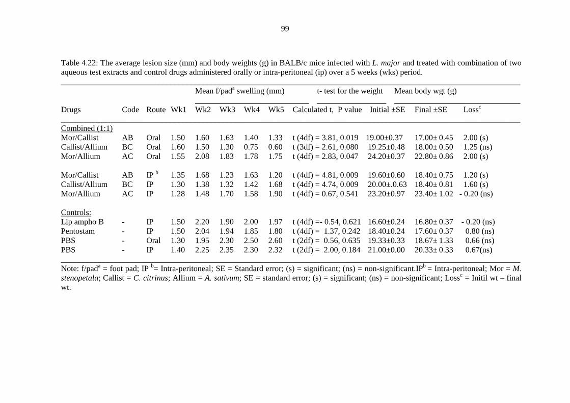
99
Table 4.22: The average lesion size (mm) and body weights (g) in BALB/c mice infected with L. major and treated with combination of two aqueous test extracts and control drugs administered orally or intra-peritoneal (ip) over a 5 weeks (wks) period. _________________________________________________________________________________________________________________
Mean f/pada swelling (mm) t- test for the weight Mean body wgt (g) ____________________________ _________________ _______________________________
Drugs Code Route Wk1 Wk2 Wk3 Wk4 Wk5 Calculated t, P value Initial ±SE Final ±SE Lossc _________________________________________________________________________________________________________________ Combined (1:1) Mor/Callist AB Oral 1.50 1.60 1.63 1.40 1.33 t (4df) = 3.81, 0.019 19.00±0.37 17.00± 0.45 2.00 (s) Callist/Allium BC Oral 1.60 1.50 1.30 0.75 0.60 t (3df) = 2.61, 0.080 19.25±0.48 18.00± 0.50 1.25 (ns)
Mor/Allium AC Oral 1.55 2.08 1.83 1.78 1.75 t (4df) = 2.83, 0.047 24.20±0.37 22.80± 0.86 2.00 (s)
Mor/Callist AB IP b 1.35 1.68 1.23 1.63 1.20 t (4df) = 4.81, 0.009 19.60±0.60 18.40± 0.75 1.20 (s) Callist/Allium BC IP 1.30 1.38 1.32 1.42 1.68 t (4df) = 4.74, 0.009 20.00±.0.63 18.40± 0.81 1.60 (s) Mor/Allium AC IP 1.28 1.48 1.70 1.58 1.90 t (4df) = 0.67, 0.541 23.20±0.97 23.40± 1.02 - 0.20 (ns)
Controls: Lip ampho B - IP 1.50 2.20 1.90 2.00 1.97 t (4df) =- 0.54, 0.621 16.60±0.24 16.80± 0.37 - 0.20 (ns) Pentostam - IP 1.50 2.04 1.94 1.85 1.80 t (4df) = 1.37, 0.242 18.40±0.24 17.60± 0.37 0.80 (ns) PBS - Oral 1.30 1.95 2.30 2.50 2.60 t (2df) = 0.56, 0.635 19.33±0.33 18.67± 1.33 0.66 (ns) PBS - IP 1.40 2.25 2.35 2.30 2.32 t (2df) = 2.00, 0.184 21.00±0.00 20.33± 0.33 0.67(ns) _________________________________________________________________________________________________________________
Note: f/pada = foot pad; IP b= Intra-peritoneal; SE = Standard error; (s) = significant; (ns) = non-significant.IPb = Intra-peritoneal; Mor = M. stenopetala; Callist = C. citrinus; Allium = A. sativum; SE = standard error; (s) = significant; (ns) = non-significant; Lossc = Initil wt – final wt.

100
When combined extracts of M. stenopetala and C. citrinus (AB) were administered intra-
peritoneally to treat L. major infected BALB/c mice, the foot pad lesion sizes tended to
increase and decrease in an alternating pattern as the treatment progressed (Figure 4.19).
However, on average, combination AB led to a significant decrease in the footpad lesions
(P < 0.05) when compared to the controls (Table 4.23). Intra-peritoneal administration of
combination C. citrinus and A. sativum (BC), resulted to the footpad lesion sizes being
maintained at a constant in the first four weeks of treatment and when compared with
controls, the difference was significant (P < 0.05) as indicated in Table 4.23 and Figure
4.19.
Blend of M. stenopetala and A. sativum aqueous extracts (AC ip) did not reduce the lesion
sizes and when compared with PBS controls, the difference was statistically not significant
(P > 0.05). Further comparisons of combinations AB(ip), BC(ip), AC(ip), liposomal
amphotericin B, pentostam, PBS(ip) and PBS(oral) using one way ANOVA also showed
that the difference was significant (F (df 6, 28) = 4.806, P = 0.002 ). Tukey’s post hoc test
showed that both combinations AB(ip) and BC(ip) differed significantly from both PBS
(oral) and PBS (ip) (P < 0.05) but not significantly different from both pentostam and
liposomal amphotericin B (P > 0.05). Combination AC (ip) was not significantly different
from all the control drugs (P > 0.05).

101
Table 4.23: Summary of paired sample t test analysis to compare aqueous blends (1:1 ratio) of the test extracts on the mean lesion sizes in treated BALB/c mice. _________________________________________________________________________ Calculated Tabulated Blend vs control t value t value, df P value _________________________________________________________________________ AB oral/PBS oral - 2.412 2.78, 4 0.073
BC oral/PBS oral - 2.325 2.78, 4 0.081
AC oral/PBS oral - 1.492 2.78, 4 0.210
AB ip/PBS ip - 3.277 2.78, 4 0.031
BC ip/PBS ip - 2.858 2.78, 4 0.046
AC ip/PBS ip - 2.347 2.78, 4 0.079
AB ip/Pento ip - 3.790 2.78, 4 0.019
BC ip/Pento ip - 2.914 2.78, 4 0.044
AC ip/Pento ip - 0.893 2.78, 4 0.423
AB ip/Lip amphB ip - 3.415 2.78, 4 0.027
BC ip/Lip amphB ip - 2.942 2.78, 4 0.042
AC ip/Lip amphB ip - 2.262 2.78, 4 0.087
_________________________________________________________________________ AB = M. stenopetala and C. citrinus; BC = C. citrinus and A. sativum; AC = M. stenopetala and A. sativum; PBS = Phosphate buffered saline; Pento = Pentostam and Lip amphB = Liposomal amphotericin B; ip = intra peritoneally.

102
Figure 4.19: The footpad swelling after ip treatment of L. major-infected BALB/c mice with aqueous extracts blends (1:1) of M. stenopetala and C. citrinus (AB); C. citrinus and A. sativum (BC); M. stenopetala and A. sativum (AC); Pento = Pentostam; lip amph B = Liposomal amphotericin B, and PBS = phosphate buffered saline.
The loss of body weights in BALB/c mice that were treated with combinations AB (oral),
AB (ip), BC (ip) and AC(oral), was statistically significant (P < 0.05), over the treatment
period. In contrast, BALB/c mice treated with the control drugs liposomal amphotericin B,
pentostam and PBS saline, loss of body weight was non significant (P > 0.05).
Combination BC (oral) and AC (ip) also led to insignificant weight loss (P > 0.05) in
infected BALB/c mice and this compared closely with the controls.
4.3.1.4 Effects of combining two methanolic extracts on lesion sizes and body weights
Oral methanolic extracts blends of A. sativum and C. citrinus (FG) and C. citrinus and M.
stenopetala (GH) reduced the foot pad lesion sizes from week three of treatment as
compared to oral PBS which was associated with a steady lesion size increase (Figure
4.20). The efficacy of these two blends FG and GH showed a significant reduction of
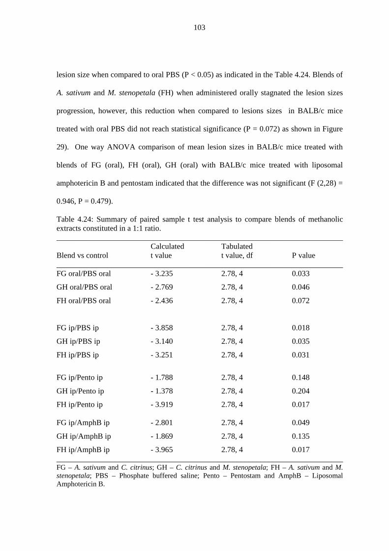
103
lesion size when compared to oral PBS (P < 0.05) as indicated in the Table 4.24. Blends of
A. sativum and M. stenopetala (FH) when administered orally stagnated the lesion sizes
progression, however, this reduction when compared to lesions sizes in BALB/c mice
treated with oral PBS did not reach statistical significance (P = 0.072) as shown in Figure
29). One way ANOVA comparison of mean lesion sizes in BALB/c mice treated with
blends of FG (oral), FH (oral), GH (oral) with BALB/c mice treated with liposomal
amphotericin B and pentostam indicated that the difference was not significant (F (2,28) =
0.946, P = 0.479).
Table 4.24: Summary of paired sample t test analysis to compare blends of methanolic extracts constituted in a 1:1 ratio. _________________________________________________________________________ Calculated Tabulated Blend vs control t value t value, df P value _________________________________________________________________________ FG oral/PBS oral - 3.235 2.78, 4 0.033
GH oral/PBS oral - 2.769 2.78, 4 0.046
FH oral/PBS oral - 2.436 2.78, 4 0.072
FG ip/PBS ip - 3.858 2.78, 4 0.018
GH ip/PBS ip - 3.140 2.78, 4 0.035
FH ip/PBS ip - 3.251 2.78, 4 0.031
FG ip/Pento ip - 1.788 2.78, 4 0.148
GH ip/Pento ip - 1.378 2.78, 4 0.204
FH ip/Pento ip - 3.919 2.78, 4 0.017 FG ip/AmphB ip - 2.801 2.78, 4 0.049
GH ip/AmphB ip - 1.869 2.78, 4 0.135
FH ip/AmphB ip - 3.965 2.78, 4 0.017 _________________________________________________________________________ FG – A. sativum and C. citrinus; GH – C. citrinus and M. stenopetala; FH – A. sativum and M. stenopetala; PBS – Phosphate buffered saline; Pento – Pentostam and AmphB – Liposomal Amphotericin B.

104
Figure 4.20: The foot pad swelling after oral treatment of L. major-infected BALB/c mice with methanolic extracts blends (ratio 1:1) of A. sativum and C. citrinus (FG); A. sativum and M. stenopetala (FH); C. citrinus and M. stenopetala (GH); pentostam (pento), liposomal amphotericin B (Lip amph B) and phosphate buffered saline (PBS).
Blends of A. sativum and M. stenopetala (FH) administered intra peritoneally reduced the
lesion sizes steadily from week two onwards (Figure 4.21) and its efficacy was
significantly different from that of pentostam and liposomal amphotericin B (P < 0.05) as
shown in Table 4.24. Combination FG (ip) caused a slight reduction in lesion sizes from
week four onwards with its efficacy being significantly different from that of liposomal
amphotericin B (P < 0.05) but not significantly different from pentostam (P > 0.05).
Combination GH (ip) caused a reduction in lesion size and its efficacy was significantly
from PBS (ip) (P < 0.05) but not significantly different from both pentostam and liposomal
amphotericin B (P > 0.05) as indicated in Table 4.24. One way ANOVA however
indicated a low non significance difference (F (6,28) = 2.345, p = 0.058) between the mean
lesion sizes of FG(ip), FH(ip), and GH(ip) and those of liposomal amphotericin B,
pentostam, PBS (ip) and PBS (oral).

105
Table 4.25: The average lesion size (mm) and body weights (g) in BALB/c mice infected with L. major and treated with combinations of two methanolic test extracts and Leishmania drugs administered orally or intra-peritoneal (ip) over a 5 weeks (wks) period. ___________________________________________________________________________________________________________ Man f/pada swelling (mm) t-test for body wgts Mean body wgt (g) ____________________________ _________________ _____________________________ Drugs Code Route Wk1 Wk2 Wk3 Wk4 Wk5 Calculated t, P value Initial ± SE Final ± SE Lossc ___________________________________________________________________________________________________________ Combined (1:1) Mor/Callist FG Oral 1.35 1.67 1.96 2.05 2.15 t (3df) = 3.00, 0.058 19.25±0.25 18.25± 0.63 1.00 (ns)
Callist/Allium GH Oral 1.37 1.64 1.74 1.74 2.24 - 20.33± 0.67 19.33± 0.67 1.00 (n/c) Mor/Allium FH Oral 1.40 1.73 2.06 2.03 2.06 t (2df) = 1.00, 0.423 18.67± 0.33 18.33± 0.33 0.34 (ns)
Mor/Callist FG IP b 1.40 1.75 1.80 1.90 1.77 t (3df) = 1.00, 0.391 19.00± 0.58 18.50± 0.50 0.50 (ns) Callist/Allium GH IP 1.50 1.64 1.90 1.77 1.80 t (2df) = 5.00, 0.038 19.00± 2.08 17.33± 1.76 1.67 (s) Mor/Allium FH IP 1.45 1.85 1.62 1.45 1.59 t (2df) = 2.00, 0.184 18.33± 0.33 18.00± 0.58 0.33 (ns) Controls:
Amphotericin B - IP 1.50 2.20 1.90 2.00 1.97 t (4df) = -0.54, 0.621 16.60±0.24 16.80± 0.37 - 0.20 (ns) Pentostam - IP 1.50 2.04 1.94 1.85 1.80 t (4df) = 1.37, 0.242 18.40±0.24 17.60± 0.40 0.80 (ns) PBS - Oral 1.30 1.95 2.30 2.50 2.60 t (2df) = 0.56, 0.635 19.33±0.33 18.67± 1.33 0.66 (ns) PBS - IP 1.40 2.25 2.35 2.30 2.32 t (2df) = 2.00, 0.184 21.00±0.00 20.33± 0.33 0.67 (ns) ____________________________________________________________________________________________________________
Note: f/pada = foot pad; IPb = Intra-peritoneal; Mor = Moringa stenopetala; Callist = Callistemon citrinus; Allium = Allium sativum; SE = standard error; (s) = significant; (ns) = non-significant; n/c = not computable; Loss c = Initial wt – final wt.

106
Figure 4.21: The foot pad swelling after ip treatment of L. major-infected BALB/c mice with methanolic extracts blends (ratio 1:1) of A. sativum and C. citrinus (FG); A. sativum and M. stenopetala (FH); C. citrinus and M. stenopetala (GH); Pentostam (Pento), Liposomal amphotericin B (Lip amph B), and phosphate buffered saline (PBS).
The methanolic extracts combinations FG (oral), FH (oral), FG (ip) and FH (ip) did not
cause significant body weight loss in BALB/c mice treated using these blends. The
BALB/c mice treated with combined extracts GH (ip) had a significant loss of body
weight (P < 0.05). Two way ANOVA (Linear model: univariate) showed that there were
significant differences between mean body weights of BALB/c mice treated with different
extracts and control drugs (F (df 13, 51) = 17.430, P = 0.0001). The effect of the 5 weeks
treatment period on the body weights of the BALB/c mice was however not significant (P
> 0.05). However, Tukey’s test showed that orally administered blends of extracts FG, FH,
and GH had significant effects (P < 0.05) on the body weights of the mice when compared
to control drugs pentostam and liposomal amphotericin B. Similarly, mice treated with
extracts combinations FG, FH and GH administered intraperitoneally had body weights
that differed significantly (P < 0.05) from those of mice treated with oral PBS at the end of

107
the treatment period. In summary, the length (5 weeks) treatment period had a non
significant influence on the body weights of mice (P = 0.118) while the drug types had
significant effect on the body weights (P = 0.0001).
4.3.1.5 Effects of combining three different aqueous extracts on lesion sizes and
body weights
When the L. major infected BALB/c mice were treated with a combination of three
aqueous test extracts of M. stenopetala, A. sativum and C. citrinus (ACB) at concentrations
of 125µg/ml in a ratio of 2:2:1, their foot pad lesion increased in size as the treatment
progressed and this compared closely with the mice that were treated with PBS controls
(Figure 4.22). The combination of M. stenopetala, C. citrinus and A. sativum (ABC),
however, decreased the lesion sizes from week 3 of treatment and it was comparable to
lesion sizes in mice treated with pentostam. Combination of extracts from C. citrinus, A.
sativum and M. stenopetala, (BCA) decreased the lesion sizes for a short period between
week 2 and week 3 of treatment (Figure 4.22 and Table 4.26).
The mean lesion sizes in the infected BALB/c mice treated with combined extracts ABC,
ACB, and BCA differed significantly from the mean lesion sizes in mice treated with the
controls pentostam, liposomal amphotericin B and the PBS orals (F (df 6, 28) = 6.500, P =
0.0001). However, Tukey post hoc test was carried out in order to compute all the possible
differences between the different pairs. There was a significant difference (P < 0.05)
between the mean lesion sizes in BALB/c mice treated with combination ABC and those
treated with PBS (ip) and PBS (oral). Similarly, there was a significant difference (P <
0.05) between the mean lesion sizes in mice treated with combination BCA and in mice
treated with liposomal amphotericin B, PBS (ip) and oral PBS (Table 4.27). There was no

108
significance difference (P > 0.05) between mean lesion sizes for the mice treated with
combination ACB and mean lesion sizes in mice treated with PBS controls.
Figure 4.22: The foot pad swelling after oral/ip treatment of L. major-infected BALB/c mice with blends of three aqueous extracts (ratio 2:2:1) of M. stenopetala, C. citrinus and A. sativum (ABC); C. citrinus, A. sativum and M. stenopetala (BCA); M. stenopetala, A. sativum and C. citrinus (ACB); Pentostam (Pento), Liposomal amphotericin B (Lip amph B) and oral phosphate buffered saline (PBS oral).

109
Table 4.26: The average lesion size (mm) and body weights (g) in BALB/c mice infected with L. major and treated with combination of three aqueous or methanolic test extracts and control drugs administered orally and intra-peritoneally (ip) interchangeably over a a period of 5 weeks.
________________________________________________________________________________________________________________ Mean f/pad a swelling (mm) t-test for body wgts Mean body wgt (g) Blends ___________________________ _________________ _______________________________ & controls Code Route Wk1 Wk2 Wk3 Wk4 Wk5 Calculated t, P value Initial & SE Final & SE Lossc ________________________________________________________________________________________________________________ Aqueous (2:2:1): Mor/Callist/Alli ABC Oral/IPb1.20 1.43 1.43 1.10 1.75 t (4df) = 0.54, 0.621 23.60±0.75 23.40±0.51 0.20 (ns) Callist/Alli/Mor BCA Oral/IP 0.92 1.24 1.04 1.40 1.24 t (4df) = 1.50, 0.208 24.60±0.60 24.40±0.75 0.20 (ns)
Mor/Alli/Callist ACB Oral/IP 0.92 1.34 1.58 1.80 1.70 t (5df) = 2.74, 0.041 24.70±0.42 23.50±0.62 0.20 (s)
Methanolic (2:2:1): Mor/Callist/Alli FGH Oral/IPb1.50 2.24 3.04 3.64 2.73 t (4df) = 3.50, 0.025 13.60±0.40 14.00± 0.32 - 0.40 (s) Callist/Alli/Mor GHF Oral/IP 1.40 2.25 3.08 3.35 3.50 t (4df) = 0.34, 0.749 13.40±0.24 14.60± 0.87 - 1.20 (ns) Mor/Alli/Callist FHG Oral/IP 1.08 1.96 2.24 3.52 2.70 t (4df) = 0.78, 0.477 14.00± 0.32 14.00± 0.89 0.00 (ns) Controls:
Lip ampho B - IP 1.50 2.20 1.90 2.00 1.97 t (4df) =- 0.54, 0.621 16.60±0.24 16.80± 0.37 - 0.20 (ns) Pentostam - IP 1.50 2.04 1.94 1.85 1.80 t (4df) = 1.37, 0.242 18.40±0.24 17.60± 0.37 0.80 (ns) PBS - Oral 1.30 1.95 2.30 2.50 2.60 t (2df) = 0.56, 0.635 19.33±0.33 18.67± 1.33 0.66 (ns) PBS - IP 1.40 2.25 2.35 2.30 2.32 t (2df) = 2.00, 0.184 21.00± 0.00 20.33± 0.33 0.67 (ns) ________________________________________________________________________________________________________________
Note: f/pada = foot pad; Oral/IPb = Oral and intra-peritoneal administration of extracts & drugs was carried out interchangeably; Mor = M. stenopetala; Callist = C. citrinus; Alli = A. sativum; SE = standard error; Loss c = Initial wt – final wt.

110
Table 4.27: Specific blends of three different extracts that reduced significantly the lesion sizes as indicated by Tukey’s multiple comparison tests. _________________________________________________________________________ Test Mean Standard 95% Confidence Interval blend Control Difference Error P level Lower limit Upper limit _________________________________________________________________________ ABC PBS (oral) - 0.748 0.210 0.020 - 1.414 - 0.082
ABC PBS (ip) - 0.742 0.210 0.022 - 1.408 - 0.076
BCA Amph B (ip) - 0.744 0.210 0.021 - 1.410 - 0.078
BCA PBS (oral) - 0.960 0.210 0.002 - 1.626 - 0.294
BCA PBS (ip) - 0.954 0.210 0.002 - 1.620 - 0.288 _________________________________________________________________________
When the combinations ABC and BCA were used to treat infected mice, there was no
significant difference between the mice initial and final body weights. However, mice
treated with combination ACB were associated with significant body weight loss (paired t
test; t (5df) = 2.74, P < 0.05). Two way ANOVA showed that there were a significant
difference between the body weights of the mice treated with drugs ABC, BCA, ACB,
liposomal amphotericin B, PBS(oral), PBS (ip) and pentostam (F (13, 50) = 28.400, P =
0.0001). Tukey’s post hoc test showed that there was no significant difference between the
mean bodies weights for the groups of mice treated with combinations ABC, BCA and
ACB. However, body weights of mice treated with these combined drugs differed
significantly from those treated with all the control drugs including liposomal
amphotericin B, PBS (oral), PBS (ip) and pentostam ( P < 0.05). The treatment period of 5
weeks did not have a significant influence on the body weights of the mice.

111
4.3.1.6 Effects of blends of three methanolic extracts on lesion sizes and body weights
Figure 4.23 shows that when infected BALB/c mice were treated with a blend of three
methanolic extracts M. stenopetala, C.citrinus and A. sativum (HGF), the sizes of footpad
lesions increased as the treatment progressed and this matched closely with the infected
mice treated with PBS controls. Similarly, methanolic extracts combinations of A. sativum,
C. citrinus and M. stenopetala (FGH) and blends of A. sativum, M. stenopetala, and C.
citrinus (FHG), did not reduce the lesion sizes in BALB/c mice as the treatment
progressed. However, it was noted that blends FGH and FHG caused a sharp decrease in
the footpad lesion sizes between weeks four and five (Figure 4.23). There was no
significant difference (F (6, 28) = 1.430, P = 0.238) between the mean lesion sizes in mice
treated with the methanolic combinations (HGF, FGH, and FHG) and mean lesion sizes
treated with PBS (oral) and PBS (ip). This conclusion was confirmed by the Tukey’s post
hoc tests that indicated lack of statistical significant difference (P > 0.05).
Two way ANOVA showed that there was a significant difference (F (df 13, 48) = 12.348,
P = 0.0001) between the body weights of the mice treated with combinations of three
methanolic extracts (FGH, FHG and GHF) and those treated with pentostam, liposomal
amphotericin B, PBS (ip) and PBS (oral). The treatment period did not have a significant
effect but the type of treatment (drug or extract) had a significant effect (P < 0.05). Tukey
post hoc test showed that use of combinations FGH, FHG, and GHF to treat L. major
infected BALB mice did not affect their body weights significantly. However, the body
weights of mice treated with the controls pentostam, liposomal amphotericin B, PBS (ip),
and PBS (oral) differed significantly from those treated with combinations of methanolic
extracts (P < 0.05).

112
Figure 4.23: The foot pad swelling after ip/oral treatment of L. major-infected BALB/c mice with methanolic extracts blends (2:2:1) of A. sativum, C. citrinus and M. stenopetala (FGH); A. sativum, M. stenopetala and C. citrinus (FHG); M. stenopetala, C. citrinus and A. sativum (HGF); Pentostam (pento); Liposomal amphotericin B (Lip amph B); phosphate buffered saline (PBS-ip and PBS-oral).
4.3.2 Estimation of number of Leishmania parasites in the infected BALB/c mice
splenocytes
The mice were sacrificed and at necropsy, the spleens were weighed and their impression
smears made (Figure 4.24) and the number of amastigotes per 1000 nucleated splenocytes
(Figure 4.25) was enumerated. The spleen index (%), Leishman-Donovan Unit (LDU) and
total Leishman-Donovani Unit (total LDU) and percentage parasite reduction in spleens of
the infected BALB/c were determined.
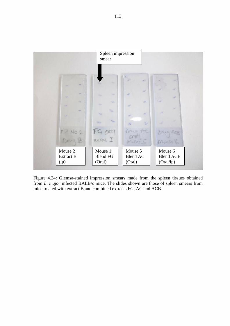
113
Figure 4.24: Giemsa-stained impression smears made from the spleen tissues obtained from L. major infected BALB/c mice. The slides shown are those of spleen smears from mice treated with extract B and combined extracts FG, AC and ACB.
Spleen impression smear
Mouse 2 Extract B (ip)
Mouse 1 Blend FG (Oral)
Mouse 5 Blend AC (Oral)
Mouse 6 Blend ACB (Oral/ip)
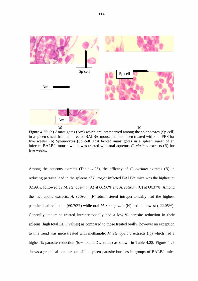
114
(a) (b) Figure 4.25: (a) Amastigotes (Am) which are interspersed among the splenocytes (Sp cell) in a spleen smear from an infected BALB/c mouse that had been treated with oral PBS for five weeks. (b) Splenocytes (Sp cell) that lacked amastigotes in a spleen smear of an infected BALB/c mouse which was treated with oral aqueous C. citrinus extracts (B) for five weeks.
Among the aqueous extracts (Table 4.28), the efficacy of C. citrinus extracts (B) in
reducing parasite load in the spleens of L. major infected BALB/c mice was the highest at
82.99%, followed by M. stenopetala (A) at 66.96% and A. sativum (C) at 60.37%. Among
the methanolic extracts, A. sativum (F) administered intraperitoneally had the highest
parasite load reduction (60.70%) while oral M. stenopetala (H) had the lowest (-22.05%).
Generally, the mice treated intraperitoneally had a low % parasite reduction in their
spleens (high total LDU values) as compared to those treated orally, however an exception
to this trend was mice treated with methanolic M. stenopetala extracts (ip) which had a
higher % parasite reduction (low total LDU value) as shown in Table 4.28. Figure 4.26
shows a graphical comparison of the spleen parasite burdens in groups of BALB/c mice
Am
Sp cell
Am
Sp cell

115
that were treated with single aqueous and methanolic plant extracts. It was noted that the
aqueous C. citrinus extracts (B) had the lowest total LDU values when administered orally
or intra peritoneally (ip) and therefore the extracts had a higher efficacy as compared to the
others. This corresponded to high percentage parasite reduction (Table 4.28 and Figure
4.26). The potential of extracts B in reducing the Leishmania parasites burden in the
spleens of infected BALB/c mice was high and it closely compared to that of the standard
Leishmania drugs pentostam and liposomal amphotericin B.
Table 4.28: The average spleen index ± SE, LDU ± SE and total LDU ± SE for groups of L. major infected BALB/c mice that were treatment with single aqueous and methanolic test extracts, pentostam, liposomal amphotericin B and PBS controls. _________________________________________________________________________ Test extracts Ave spleen Ave total % parasite & controls Route index (%) Ave LDU LDU (× 1000) reduction a _________________________________________________________________________ Aqueous: A (M. stenopetala) oral 0.51 ± 0.14 0.25 ± 0.04 4.06 ± 0.60 66.96 ipb 0.54 ± 0.01 0.57 ± 0.05 10.28 ± 2.02 16.35 B (C. citrinus) oral 0.71 ± 0.21 0.10 ± 0.02 2.09 ± 0.14 82.99 ip 0.53 ± 0.03 0.19 ± 0.13 3.05 ± 2.02 75.18 C (A. sativum) oral 0.42 ± 0.01 0.27 ± 0.01 4.87 ± 0.21 60.37 ip 0.47 ± 0.05 0.33 ± 0.24 6.71 ± 4.80 45.40 Methanolic: F (A. sativum) oral 0.60 ± 0.05 0.22 ± 0.15 4.83 ± 3.20 60.70 ip 0.55 ± 0.05 0.52 ± 0.04 11.31 ± 0.21 07.97 G (C. citrinus) oral 0.49 ± 0.01 0.38 ± 0.16 6.97 ± 2.73 43.29 ip 0.47 ± 0.00 0.50 ± 0.24 8.96 ± 4.38 27.09 H (M. stenopetala) oral 1.31 ± 0.13 0.36 ± 0.07 15.00 ± 1.53 -22.05 ip 1.31 ± 0.31 0.16 ± 0.02 6.36 ± 2.90 48.25 Controls: Pentostam ip 0.73 ± 0.19 0.18 ± 0.08 4.13 ± 1.10 66.40 Lip amph B ip 0.61 ± 0.02 0.24 ± 0.02 4.84 ± 0.38 60.62 PBS ip 0.54 ± 0.04 0.38 ± 0.21 8.74 ± 5.30 28.89 PBS oral 0.56 ± 0.06 0.61 ± 0.22 12.29 ± 4.49 00.00 ________________________________________________________________________ a means that the % was calculated in reference to total LDU for PBS oral which was taken to represent 100% parasite burden; ipb = means intraperitoneal.

116
Computation of the probability level gave a non significant difference between the mean
total LDU for aqueous C. citrinus extract (B) when administered orally or intra
peritoneally and the mean total LDUs for pentostam and liposomal amphotericin B
respectively. There was a significant difference (F (df: 3, 8) = 6.313, P = 0.017) between
the mean total LDU for aqueous oral C. citrinus extracts and the mean total LDU for
control oral PBS. However there was no significant difference between the mean total
LDU for oral aqueous C. citrinus extracts (B) and the mean total LDU of the same extract
B when administered intra peritoneally.
Robust tests of equality of means gave a non significant difference between the mean total
LDUs for extracts A (ip), G (ip), and F (ip) when they were compared to PBS (oral) and
PBS (ip). The efficacies of aqueous M. stenopetala (ip), methanolic C. citrinus (ip), and
methanolic A. sativum (ip) were comparable with that of the control PBS (oral and ip) as
illustrated in Figure 4.26. However, there was no significant difference between the mean
total LDU for the aqueous M. stenopetala (oral), methanolic M. stenopetala (ip),
methanolic C. citrinus (oral), aqueous A. sativum (oral), and methanolic A. sativum (oral)
extracts and that of pentostam and liposomal amphotericin B ( F (6,14) = 0.915, P =
0.512), and the efficacy of these extracts was comaparable to that of pentostam and
liposomal amphotericin B (Figure 4.26). Tukey post hoc test for multiple comparisons of
these extracts also showed differences that were not significant.
The route of the administration of extracts in the infected BALB/c mice seemed to have an
influence on the total LDU values obtained. This was observed for extract H (methanolic
M. stenopetala), whose mean total LDU for oral route and mean total LDU for ip route

117
differed significantly (P = 0.016). For extracts A, B, G and F, the mean total LDUs for
oral routes were lower than ip routes but the difference was not significant ( P > 0.05).
Figure 4.26: Parasite burden in spleens of L. major infected BALB/c mice treated with test extracts, pentostam, liposomal amphotericin B, and PBS. The extracts were A = aqueous M. stenopetala; B = aqueous C. citrinus; G = methanolic C. citrinus; C = aqueous A. sativum extract; F = methanolic A. sativum; H = methanolic M. stenopetala; while the controls were Pento = pentostam; AmphB = liposomal amphotericin B; PBS= phosphate buffered saline.
Orally administered combined aqueous extracts of C. citrinus and A. sativum (BC) and
combined extracts of M. stenopetala and A. sativum (AC) reduced the spleen parasite
burden in infected BALB/c mice by 48.33% to 60.94% while combined aqueous extracts
of M. stenopetala and C. citrinus (AB) reduced the spleen parasite burden by 6.5% (Table
4.29). The higher the total LDU values, the lower the % parasite reduction. Similarly,
drugs AB (ip), AC (ip) and BC (ip) reduced the parasite burden by 36.28% to 74.02 %
which corresponded to total LDU values that ranged from 3.19 ± 1.16 to 7.83 ± 3.24 (×
1000) as shown in Table 4.29.

118
Table 4.29: Shows the average spleen index± SE, LDU± SE and total LDU ± SE for groups of L. major infected BALB/c mice that were treated with blends of both test aqueous and methanolic extracts. _________________________________________________________________________ Blends Ave spleen Ave total % parasite & controls Route Index (%) Ave LDU LDU (× 1000) reductiona _________________________________________________________________________ Blends of two different extracts (1:1) Aqueous: AB (Mor: Cal) oral 0.62 ± 0.07 0.56 ± 0.35 11.49 ± 6.84 6.51 ipb 0.70 ± 0.09 0.33 ± 0.18 7.83 ± 3.24 36.28 BC (Cal: All) oral 0.67 ± 0.00 0.26 ± 0.03 6.35 ± 0.66 48.33 ip 0.50 ± 0.06 0.18 ± 0.06 3.19 ± 1.16 74.04 AC (Mor: All) oral 0.59 ± 0.06 0.20 ± 0.02 4.80 ± 0.95 60.94 ip 0.46 ± 0.04 0.33 ± 0.06 7.22 ± 0.66 41.25 Methanolic: FG (All: Cal) oral 0.52 ± 0.02 0.96 ± 0.11 20.96 ± 0.44 -70.55 ipa 0.50 ± 0.05 0.55 ± 0.05 11.57 ± 0.55 5.86 FH (All: Mor) oral 0.54 ± 0.11 0.53 ± 0.18 12.14 ± 6.53 1.22 ip 0.59 ± 0.11 0.44 ± 0.17 10.48 ± 4.19 14.73 GH (Cal: Mor) oral 0.58 ± 0.03 0.62 ± 0.07 15.10 ± 2.87 -22.86 ip 0.60 ± 0.05 0.56 ± 0.01 13.90 ± 0.24 -13.10 Blends of three differen t extracts (2:2:1) Aqueous: ABC (Mor:Cal:All) oral/ip 0.56 ± 0.11 0.47 ± 0.17 11.11 ± 1.57 9.60 BCA (Cal:All:Mor) oral/ip 0.44 ± 0.02 0.38 ± 0.16 8.73 ± 4.31 28.97 ACB (Mor:All:Cal) oral/ip 0.41 ± 0.01 0.35 ± 0.20 7.21 ± 3.85 41.33 Methanolic: FHG (All:Mor:Cal) oral/ip 0.60 ± 0.07 0.28 ± 0.10 5.34 ± 2.18 56.55 FGH (All:Cal:Mor) oral/ip 1.28 ± 0.05 0.18 ± 0.06 7.63 ± 3.70 37.92 HGF (Mor:Cal:All) oral/ip 1.14 ± 0.22 0.14 ± 0.003 4.43 ± 0.93 63.95 Controls: Pentostam ip 0.73 ± 0.19 0.18 ± 0.08 4.13 ± 1.10 66.40 Lip amhotericin B ip 0.61 ± 0.02 0.24 ± 0.02 4.84 ± 0.38 60.62 PBS ip 0.54 ± 0.04 0.38 ± 0.21 8.74 ± 5.30 28.88 PBS oral 0.56 ± 0.06 0.61 ± 0.22 12.29 ± 4.49 00.00 _________________________________________________________________________ a The percentage was calculated in reference to total LDU for PBS oral which was taken as 100% parasite burden; b ip = intra peritoneal; Mor = M. stenopetala; Cal = C. citrinus; All = A. sativum;
The blends of three different aqueous extracts which included ABC, BCA and ACB
yielded average total LDU values that ranged from 7.21 ± 3.85 to 11.11 ± 1.57 (× 1000)

119
which corresponded to a reduction of parasite burden of 9.60% to 41.33 % as shown in
Table 4.30 above. The efficacy of blends of two different methanolic extracts including
FG, FH and GH, was low with a percentage parasite reduction ranging from -70.55% to
14.73% compared to blends FHG, FGH and HGF that ranged from 37.92 %to 63.95 % .
There was no significant difference between the mean total LDUs for the orally
administered blends of aqueous extracts AB, BC and AC and mean total LDUs of the
blends of three aqueous extracts ABC, BCA and ACB that were administered orally and
intra-peritoneally alternately (F (5, 12) = 1.341, P = 0.312). The efficacy of these two
blends of extracts in vivo tended to be similar. This conclusion agrees with graph
illustration shown in Figure 4.27. It was also noted that there was no significant difference
between mean total LDUs for combinations AB (oral), BC (oral), AC (oral), ABC
(oral/ip), BCA (oral/ip), ACB (oral/ip) and the mean total LDUs for pentostam and
liposomal amphotericin B (F (7,16) = 2.066, P = 0.109).
The difference between the mean total LDUs for combinations AB (ip), BC (ip), AC (ip),
ABC, BCA, ACB and the mean total LDUs for pentostam and liposomal amphotericin B
was significant (P < 0.05). The Games-Howell post hoc test for these combined extracts
showed that the difference between the mean total LDUs for combination BC (ip) and that
of ABC (oral/ip) was significant (P = 0.025). Similarly, the mean total LDUs for
combination ABC (oral/ip) and that of pentostam differed significantly (P = 0.037). The
efficacy of blends of ABC was therefore significantly low. There was no significant
difference between the mean total LDUs for combinations AB (ip), BC (ip), AC (ip),
BCA, ACB with those pentostam and liposomal amphotericin B meaning that their

120
efficacy was close. In order to investigate on the effect of the route of drug administration
into BALB/c mice, the mean total LDUs for the pairs AB (oral) and AB (ip); BC (oral)
and BC (ip); AC (oral) and AC (ip); PBS (oral) and PBS (ip) were compared. Graphically
the mean total LDUs for these pairs seemed different (Figure 4.27), however, the
difference was not significant.
Figure 4.27: Parasite burden in spleens of infected BALB/c mice following treatment with blends of aqueous extracts. AB = M. stenopetala & C. citrinus; BC = C. citrinus & A. sativum; AC = M. stenopetala & A. sativum; ABC = M. stenopetala, C. citrinus & A. sativum; BCA = C. citrinus, A. sativum & M. stenopetala; ACB = M. stenopetala, A. sativum & C. citrinus. The blends of two extracts were administered either orally or intra-peritoneally (ip); the blends of three aqueous extracst were administered orally and intraperitoneally alternately.
Blends of three different methanolic extracts at ratio of 2:2:1 and concentrations of
125µg/ml were associated with low total LDUs when compared to the blends of two
different methanolic extracts (Figure 4.28). There was no significant difference between
mean total LDUs of combinations FHG, FGH and HGF and mean total LDUs for
pentostam and liposomal amphotericin B (F (4, 10) = 1.416, P = 0.298). The efficacy of

121
three combined methanolic extracts when administered orally and intra peritoneally
interchangeably, was close to that of the drugs pentostam and liposomal amphotericin B.
There was also no significant difference between the mean total LDUs for both orally and
intra peritoneally administered methanolic blends of FG, FH and GH and those for both
the oral PBS and ip PBS (F (7, 16) = 2.164, P = 0.095). However, Tukey post hoc test
showed that the mean total LDU values for the FG (oral) and PBS (ip) differed
significantly (P = 0.042). The mean total LDUs values for the extracts administered orally
(FG, FH, and GH) were not significantly different from the mean total LDUs for the same
extracts when administered intra peritoneally (P > 0.05).
Figure 4.28: Parasite burden in spleens of infected BALB/c mice followed by treatment with blends of methanolic extracts. FG = A. sativum & C. citrinus; FH = A. sativum & M. stenopetala; GH = C. citrinus & M. stenopetala; FHG = A. sativum, M. stenopetala & C. citrinus; FGH = A. sativum, C. citrinus & M. stenopetala; HGF = M. stenopetala, C. citrinus & A. sativum. Blends of two extracts were administered either orally or intra-peritoneally (ip); the blends of three extracts were administered orally and intra-peritoneally alternately.

122
4.4 Culturing of tissues obtained from infected and treated BALB/c mice
In order to verify whether the treated BALB/c mice were still possessing viable
Leishmania amastigotes in their visceral organs and the infected footpads, the mice were
sacrificed 5 weeks post-infection using 100μl pentobarbitone sodium (Sagatal®). At
necropsy, portions of their spleens, livers and infected foot pads were cultured at 25oC in
Schneider’s Drosophila medium supplememnted with 20% heat inactivated FBS, 100U/ml
penicillin, 500μg/ml streptomycin and 250μg/ml 5 – fluorocytosine arabinoside.
Viable L. major amastigotes were recovered from tissues obtained from infected footpads
of BALB/c mice at 5 weeks post infection and 4 weeks post treatment with extracts of M.
stenopetala, C. citrinus, A. sativum, pentostam, liposomal amphotericin B and PBS. The
amastigotes recovered were active one day post culture (Table 4.30). Active promastigotes
were evident at 6-7 days post culture for the mice treated with extracts A, B and C. For the
BALB/c mice treated with PBS, active amastigotes were evident in spleen cultures after 4
to 5 days. No promastigotes were recovered from the liver tissues obtained from the
treated BALB/c mice and cultured for at most seven days. The control drugs pentostam
and liposomal amphotericin B cleared the L major amastigotes in both the liver and spleen
since no promastigotes were recovered from these organs (Table 4.30).
The footpad tissues from BALB/c mice that were orally or intra peritoneally treated with
extracts F, G, and H were positive for viable amastigotes at 1 day post culture. Only spleen
tissues obtained from BALB/c mice treated with extracts F(ip) and G(ip) yielded
promastigotes at 4 days post culture. Spleen tissues from infected mice treated with oral
extracts F, G and H had no observable amastigotes upto 7 days post culture. No

123
promastigotes were recovered from the liver tissues obtained from mice treated with oral
or ip methanolic extracts.
Table 4.30: The cultures of tissues from L. major infected BALB/c mice that were previously under treatment. ________________________________________________________________________ Test extracts Days post culture & controls Tissue Route 0 1 2 3 4 5 6 7 ________________________________________________________________________ A Spleen Oral -a - - - - - - +b ip - - - - - - - - Liver Oral - - - - - - - - ip - - - - - - - - Footpad Oral - + + + + + + + ip - + + + + + + + B Spleen Oral - - - - - - + + ip - - - - - - - + Liver Oral - - - - - - - - ip - - - - - - - - Footpad Oral - + + + + + + + ip - + + + + + + + C Spleen Oral - - - - - - + + ip - - - - - - + + Liver Oral - - - - - - - - ip - - - - - - - - Footpad Oral - + + + + + + + ip - + + + + + + + PBS Spleen Oral - - - - + + + + ip - - - - - + + + Liver Oral - - - - - - - - ip - - - - - - - - Footpad Oral - + + + + + + + ip - + + + + + + + Pentostam Spleen ip - - - - - - - - Liver ip - - - - - - - - Footpad ip - + + + + + + + Amph B Spleen ip - - - - - - - - Liver ip - - - - - - - - Footpad ip - + + + + + + + ________________________________________________________________________ -a = No active L. major promastigotes were observed; +b = active L. major promastigotes were observable.

124
All the spleen tissues obtained from infected BALB/c mice that were treated with blends
of two extracts including AB, BC, AC, FG, FH and GH were positive for L. major
amastigotes (Table 4.31). Among the aqueous blends of two ip AB (M. stenopetala & C.
citrinus) and both oral and ip BC (C. citrinus & A. sativum) took 6 to 7 days of culture for
amastigotes to be evident. The BALB/c mice that had been treated with blends of three
extracts including ABC, BCA, ACB, FGH, FHG and HGF had their spleen tissues become
positive for L. major by the 4th day of culture with an exceptional of combination ABC
and ACB which were positive on the 6th day of culture (Table 4.31). The infected mice
treated with control drugs pentostam and liposomal amphotericin B, had spleen tissues that
were negative for L. major amastigotes for the eight days they were observed. This
implied that the standard Leishmania drugs inhibited the visceralizing of L. major
amastigotes in the spleens of BALB/c mice.
The cultured tissues from infected footpads became positive for in their 1st day of culture
for all the BALB/c mice treated with blends of three extracts or PBS or control drugs
(pentostam and liposomal amphotericin B). Liver tissues obtained from mice treated with
the extracts or PBS or control drugs were negative for L. major amastigotes throughout the
eight days of culture.

125
Table 4.31: Cultures of spleen tissues obtained from L. major infected BALB/c mice that had been treated with different blends of extracts. ________________________________________________________________________
Blends Days post culture for spleen tissues
& controls Route Ratio 3 4 5 6 7 ________________________________________________________________________ AB Oral 1:1 -a +b + + + ip 1:1 - - - + + BC Oral 1:1 - - - + + ip 1:1 - - - - + AC Oral 1:1 - - + + + ip 1:1 - + + + + FG Oral 1:1 - + + + + ip 1:1 - - + + + FH Oral 1:1 - + + + + ip 1:1 - - + + + GH Oral 1:1 - + + + + ip 1:1 - - + + + ABC Oral/ip 2:2:1 - - - + + BCA Oral/ip 2:2:1 - + + + + ACB Oral/ip 2:2:1 - - - + + FHG Oral/ip 2:2:1 - + + + + FGH Oral/ip 2:2:1 - + + + + HGF Oral/ip 2:2:1 - + + + + PBS oral none - + + + + PBS ip none - - + + + Pentostam ip none - - - - - Amph B ip none - - - - - ________________________________________________________________________ -a = No active L. major promastigotes were observed; +b = active L. major promastigotes were observable.
126
CHAPTER FIVE: DISCUSSION, CONCLUSIONS AND RECOMMENDATIONS
5.1 DISCUSSION
5.1.1 Introduction
Presently, a huge number of researches have been carried out to extract various natural
products in order to test for their antimicrobial activity. This is because there has been an
alarming increase in the incidence of new and reemerging infectious diseases. Another
concern is the development of resistance to the standard drugs in current clinical use, their
non-availability as well as the high costs of the new generation of drugs with limited
effective span. These issues in the drug world have resulted in increase in morbidity and
mortality. A number of plants have been documented for their biological and antimicrobial
properties. This section, discusses the results obtained when both aqueous and methanolic
crude extracts of A. sativum , M. stenopetala and C. citrinus plants were tested against L.
major in vitro and in vivo.
5.1.2 The in vitro efficacy of test single extracts in inhibiting L. major promastigotes
The present study shows that the crude extract from A. sativum (garlic) has the potential of
inhibiting L. major promastigotes in vitro at concentrations of 5 mg/ml and 3 mg/ml for
aqueous and methanolic extracts respectively. This observation agrees with previous
reports on the potential of garlic extracts in destroying Leishmania promastigotes in vitro.
Aqueous garlic extracts were previously demonstrated to induce a 76.58% killing
(apoptosis) of L. major promastigotes in vitro (Khademvatan et al., 2011b). Similarly, in
their study on assessment of anti-cutaneous leishmaniasis activity of some Sudanese
plants, Khalid et al. (2005) observed that methanolic garlic extracts inhibited in vitro
growth of L. major at different concentrations and that the extracts’ activity compared
closely to that of standard drug pentostam. Records show that garlic extracts possess anti

127
protozoan activity (Nok et al., 2001). When L. major promastigotes are exposed to
aqueous garlic extracts in culture, the parasites undergo some physiological changes that
eventually lead to a programmed cell death (apoptosis) whose main features include cell
shrinkage and DNA fragmentation among others (Khademvatan et al., 2011b). Garlic has
been reported to be rich in allicin (allyl-S-thiosulfinate) and other sulfur containing
compounds which includes diallyl sulfide, diallyl disulfide, diallyl trisulfide, and ajoene.
The different health benefits of garlic have been attributed to these sulfur containing
organic compounds (Islam et al., 2011). Ajoene has been reported to have a direct
significant inhibition of Leishmania growth in vitro (Ledezma et al., 2002).
From the literature, it seems that, little research has been carried out on anti-protozoal
properties of C. citrinus extracts. Majority of the investigations on medicinal properties of
C. citrinus extracts have been focusing on antibacterial potential. The C. citrinus flowers
extracts have been reported to be potent inhibitors of bacterial growth (Cock, 2008a). It
was observed from the present study that C. citrinus crude aqueous and methanolic
flowers extracts were effective against L. major promastigotes in vitro at concentrations
ranging from 1 to 5 mg/ml. The essential oils from C. citrinus when tested against some
species of bacteria have been reported to have low MIC values that ranged between 0.31
and 2.5 mg/ml (Oyedeji et al. 2009). In the present study, the crude methanolic extracts of
C. citrinus had MIC of 1.0 mg/ml against L. major promastigotes and this was in
agreement with the range of MICs reported by Oyedeji et al. (2009). Similarly, both
aqueous and methanolic extracts of Callistemon viminalis were found to exhibit MICs of
1.6 mg/ml against Staphylococcus aureus in vitro (Delahaye et al., 2009) which was also
in the range of 1.0 mg/ml obtained for crude methanolic C. citrinus extracts in the present

128
study. Seyydnejad et al. (2010) observed that the MIC of C. citrinus methanolic and
ethanolic leaves extracts against several species of bacteria ranged between 5 and 40
mg/ml which again agreed with the MIC of 5 mg/ml for aqueous C. citrinus extracts
against L. major observed in the present study. In some other studies however, ethanolic
extracts of C. citrinus leaves have been reported to show low MIC values ranging from
0.02 to 0.31 mg/ml against several species of bacteria (Chitemerere & Mukanganyama,
2011). The variation in the effective concentration as indicated in the different studies was
probably due to the differences in the methods of herbal extractions and sources of the test
plant materials.
The current study further established that aqueous crude extracts of M. stenopetala were
also active against L. major in vitro at concentration of 3 mg/ml while methanolic crude
extracts were active at ≥ 5 mg/ml. Relatively high concentrations of M. stenopetala
extracts were needed to inhibit the survival of L. major in vitro. A previous research has
reported that a concentration of 0.3 g/ml of methanolic bark extract and 2.0 g/ml of leaf
extract of the M. stenopetala inhibits bacteria that cause mastitis in vitro (Biffa, 2005). It
seems therefore that M. stenopetala extracts require a larger concentration to be effective
and this agrees with a recent study in which aqueous and ethanolic leaf extracts of
Moringa oleifera were demonstrated to inhibit all the enteropathogens at concentrations of
20 mg/ml (Oluduro, 2012). The in vitro antileishmanial potential of both aqueous and
methanolic crude extracts of M. stenopetala observed in the current study confirms an
earlier report by Mekonnen & Gassesse (1998) in Nordos et al. (2011) where it was
demonstrated that the M. stenopetala extracts exhibits antileishmanial activity. The species
Moringa is rich in glucosinolates which are a class of organic compounds that contain

129
sulfur and nitrogen and are derived from glucose and an amino acid. They contribute to the
bitter tastes of some plants including moringa, cabbage, turnip, mustard, kale and others.
Natural glucosinolates are usually hydrolysed to natural isothiocyanates (syn: mustard oil),
a process that involves enzyme myrosinase. Young leaves of M. stenopetala contain higher
levels of glucosinolates. Glucosinolates have been reported to possess antimicrobial,
anticancer and antioxidant properties (Bellostas et al., 2010). Nibret & Wink (2010)
demonstrated that the main essential oils in M. stenopetala were benzyl isothiocyanate
(54%) and isobutyl isothiocyanate (16%) and they were demonstrated to have significant
potency as trypanocidal agents. The leishmanicidal property of aqueous and methanolic
M. stenopetala extracts observed in the current study probably could have been attributed
to the glucosinolates and isothiocyanates content present in this plant.
The efficacy of the test extracts was compared with that of the positive control standard
Leishmania drugs pentostam and liposomal amphotericin B. As deduced from the results,
liposomal amphotericin B was a stronger anti-leishmanial drug in vitro when compared to
pentostam. This observation closely agrees with several research findings reported
previously. Berman & Wyler (1980) reported that promastigotes of both L. tropica and L.
donovani were unaffected by a concentration of ≤ 20 µg/ml of pentostam but the two
species of Leishmania were completely eliminated by exposure to 0.25 µg/ml of
amphotericin B in vitro. According to Polonio & Efferth (2008), liposomal amphotericin B
yields high cure rates in pentostam-unresponsive patients and its liposomal formulations
increases their efficacy due to their enhanced drug-targeting effect and reduces the drug’s
toxicity. Incorperating the drug into liposomes increases the intracellular concentration of
the Leishmania drug in the target cells hence increasing the parasite killing in vivo (Alvin

130
et al. (1978) in Berman & Wyler, 1980). The Schneider’s Drosophila medium was used as
negative control also and it supported maximum survival of the L. major promastigotes in
vitro. This was expected because this medium supports the growth of Leishmania
promastigotes and amastigotes as described by Hendricks et al., 1979. The efficacy of test
extracts was higher than that of Schneider’s Drosophila medium.
5.1.3 The effect of combining different test extracts (blends) on in vitro survival of
L. major
Combination therapy as a protocol of treatment is often adopted for many infectious
diseases particularly in cases where the infectious agent fails to respond to the
monotherapeutic regimen. Several studies on leishmaniases indicate that combination of
antileishmanial drugs improves their efficacy and reduces resistance, the dosage required
and toxicity levels of the drugs (Nyakundi et al., 1994; Melaku et al., 2007; Sundar et al.,
2008). Following combination of drugs, synergistic, additive or antagonistic effects may
ensue. In the present study, the combination ratios of M. stenopetala and C. citrinus
extracts (blend AB) that showed synergistic interaction (ratios 1:1 and 2:8) and additive
interaction (ratios 8:2, 6:4, 4:6, and 1:9) tended to be associated with marked inhibition of
L. major promastigotes survival in vitro. In particular, blend AB in a ratio of 1:1 led to a
100% inhibition of L. major promastigotes. Probably the M. stenopetala glucosinolates
and the essential oils of the C. citrinus were more efficacious at equal ratios. According to
Tahany (2010), additive and synergistic effects are advantageous while antagonistic
effects are disadvantageous. The blending of M. stenopetala and A. sativum crude aqueous
extracts (blend AC) in different ratios showed additive (ratios 5:5 and 2:8) and synergistic
(ratio 1:9) interactions and these ratios were similarly associated with low in vitro survival
of L. major promastigotes. Moderate additive interaction ensued for combination of C.
131
citrinus and A. sativum (blend BC) at ratio 1:1. These observations are in line with those
of Gathirwa et al. (2008), who reported that combinations of herbal extracts that were used
for the treatment of malaria, and had marked synergistic and additive interactions, were
more effective and were associated with high anti-plasmodial activity.
The methanolic blends had a similar trend. Combined methanolic extracts of A. sativum
and C. citrinus (blend FG) exhibited additive interactions for majority of the ratios. For
combined extracts of A. sativum and M. stenopetala (blend FH), as the content of A.
sativum increased, the interaction tended to shift from weak additive to strong antagonistic
interaction and vice versa. The C. citrinus and M. stenopetala methanolic extracts
combination (blend GH) exhibited marked synergistic interaction. Additive and
synergistic interactions corresponded to low survival of the L. major promastigotes in
vitro.
5.1.4 Cytotoxicity of the test extracts against L. major promastigotes and vero cells
As observed in the current study, the test extracts had IC50 levels of ≥ 297.75µg/ml and L.
major promastigotes’ viabilities ranging from 52.55 to 80.84%. In comparison, pentostam
and liposomal amphotericin B had IC50 levels of 0.26 and 0.82µg/ml, and promastigotes
viability of 18.41% and 12.22% respectively. Although the standard drugs were effective
against Leishmania promastigotes, they have been reported to be highly toxic to patients
and they require a protracted administration (Martinez & Marr, 1992). Cytotoxic assays
using vero cells also showed that the test extracts were generally less toxic compared to
the pentostam and liposomal amphotericin B. Many drugs used for leishmaniasis treatment
are highly toxic (Santos et al., 2008) and this study confirms that pentostam and liposomal
132
amphotericin B are toxic compared to the extracts tested. The continued use of the
contemporary leishmaniasis drugs despite their toxicity is mainly due to lack of an
alternative. The use of herbal materials can be a cheapera and available alternative.
Both the C. citrinus aqueous and methanolic extracts showed a dose-dependent cytotoxic
effect in which higher doses were more toxic and vice versa as illustrated by the linear
trend line graphs. Methanolic extracts of C. citrinus have recently been reported to have
low toxicity towards the aquatic brine shrimp, Artemia franciscana nauplii (Cock, 2008b).
Similarly, Firoz et al. (2011) demonstrated that ethanol extracts of Callistemon lanceolatus
(synonym: C. citrinus) results to high mortality in albino wistar rats of body weight 150-
250 g at a dose of 3g/kg. Extracts of C. citrinus are rich in essential oils that include 1,8-
cineole and alpha pinene (Oyedeji et al., 2009) and according to Koul et al. (2008), the
terpenoid constituents of essential oils can be moderately toxic to mammals. 1,8-cineole
was shown to have a toxicity level of LD50 2480 mg/kg when administered orally in
experimental rats (Koul et al., 2008). The moderate in vitro cytotoxicity of C. citrinus
extracts to vero cells observed in the present study was probably due to its essentials oils.
Previous reports have shown that aqueous A. sativum extracts possess cytotoxic effects on
L major promastigotes with an IC50 of 37 mg/ml (Gharavi et al., 2011). In the current
study however, the IC50 of aqueous and methanolic A. sativum extracts were 0.58 mg/ml
and 0.86 mg/ml respectively in the same duration of 48 hours. The variation could be
attributed to the source and the primary processing of the garlic used since Gharavi and
colleagues used fresh garlic while the present study used dry garlic. Garlic has sulphur
containing compounds, all of which have medical benefits. Among these compounds are

133
allicin (diallyl thiosulfinate) which is the most abundant and it gives garlic the
characteristic odor.
The aqueous extracts of M. stenopetala (IC50 of 1306µg/ml) were less toxic to vero cells
than the methanolic extracts (IC50 of 912µg/ml) although statistically the difference was
insignificant. According to Mekonnen et al. (2005), ethanol extract of the leaves and seeds
from Moringa stenopetala contain toxic substances that are extractable with organic
solvents or are formed during the process of extraction with these solvents. This could
explain the slight toxicity observed for methanolic moringa extracts in the present study.
As expected, the viability of the vero cells was influenced by the toxicity of the extracts.
The lower the toxicity of the test extracts, the higher was the mean viability (%) of vero
cells after exposure to the extracts and vice versa.
5.1.5 Effects of the test extracts on infection rates and multiplication of L. major
amastigotes in BALB/c mice macrophages
The protozoan parasite L. major infects mononuclear phagocytes, and therefore the control
of infection depends on adequate activation of the infected macrophages to kill the
parasites and inhibit their replication (James, 1995). Reliable leishmanicidal activity must
be complemented with use of intracellular amastigotes in macrophages (Chan-Bacab &
Pena-Rodriguez, 2001). In this regard, the efficacy of the test aqueous and methanolic
extracts in inhibiting the replication of amastigotes in infected BALB/c peritoneal
macrophages was quantified by calculating their infection rate (IR) and multiplication
indices (MI). Based on the IRs and MIs, the C. citrinus aqueous crude extracts had a
parasite inhibition rate of 12% while the methanolic crude extracts had 5%, both at a
concentration of 125µg/ml. In comparison, pentostam and liposomal amphotericin B had
134
parasite inhibition rate of 20% and 78% respectively at a concentration of 12.50µg/ml.
There was a significant difference between the efficacy of the test crude extracts and that
of the Leishmania drugs (P < 0.05). This observation indicates that C. citrinus extracts
which are known for their antibacterial and anti-fungal potential (Delahaye et al., 2009)
also possess anti-leishmanial activity. According to Oyedeji et al. (2009), C. citrinus and
C. viminalis leaves are known to be rich in 1, 8-cineole (eucalyptol) and alpha pinene
essential oils. Both alpha pinene and 1, 8-cineole are non polar terpenes (Delahaye et al.,
2009). Since linalool is a monoterpene that has a strong anti-leishmanial activity (Polonio
& Efferth, 2008), there is a high possibility that eucalyptol and alpha pinene may also have
anti-leishmanial activity and may have contributed to the efficacy of the test C. citrinus
flowers extracts. According to Cock (2012), it has been postulated that the terpenes in the
leaves of Callistemons have been responsible for their efficacy in traditional medicines.
At a concentration of 125µg/ml, the aqueous and methanolic crude leaf extracts of A.
sativum had IR of 51% and 46% respectively in vitro which were close to IR of pentostam
that ranged from 38% to 67% at concentrations of 50µg/ml to 12.5µg/ml. A similar trend
was true for MI. This observation shows that dry garlic extracts inhibits infection of
macrophages by L. major amastigotes. Previous studies have shown that presence of dry
garlic extract in the culture medium resulted in a significantly lower proportion of infected
BALB/c mice peritoneal macrophages (Gamboa-Leon et al., 2007). Similarly, Ghazanfari
et al. (2006) demonstrated that a single dose of 20 mg/kg garlic extract administered intra-
peritoneally alters the number of peritoneal macrophages for at least two weeks in BALB/c
mice. Furthermore intra-peritoneal injection of garlic extracts augment parasite engulfment
and destruction of intracellular amastigotes by host macrophages.

135
Studies conducted else where (Mekonnen et al., 1999) indicated that dried M. stenopetala
leaf acetone extracts were inactive against L. donovani amastigotes. Some years later,
studies indicated that ethanol extracts of M. stenopetala leaves and roots have the potential
in controlling Trypanosoma brucei and Leishmania donovani in vitro experiments (Bosch,
2004). The present study continues to demonstrate that aqueous and methanolic extracts of
M. stenopetala dried leaves have inhibition rates against L. major amastigotes in peritoneal
macrophages of 32% and 17% respectively at a concentration of 125µg/ml. The current
study further demonstrates that the difference between the IR of aqueous M. stenopetala
dried leaf extracts and that of the control drugs pentostam and liposomal amphotericin B
was significant (p < 0.05). In summary therefore, aqueous and methanolic crude extracts of
M. stenopetala dried leaves are active against the L. major amastigotes in vitro.
When the test extracts were compared with the controls, IR of macrophages by L. major
amastigotes in plain RPMI - 1640 medium (negative control) was 84.67± 2.96%. This
agrees with Berman & Wyler (1980) who observed that L. tropica and L. donovani
amastigotes in human macrophages multiplied about three fold in six days when grown in
RPMI - 1640 medium in absence of antileishmanial agents. The IRs of liposomal
amphotericin B and pentostam (positive controls) both at concentration 50µg/ml were 6%
and 14% respectively. The trend was similar for MIs. This observation was close to
Wabwoba et al. (2010) results in which the IRs of liposomal amphotericin B and
pentostam at 100µg/ml were 9% and 11% respectively. In the present study however,
although the difference between MI for liposomal amphotericin B and that of pentostam at
concentration of 50µg/ml was statistically not significant, the in vitro efficacy of liposomal

136
amphotericin B in suppressing the amastigotes multiplication was higher than that of
pentostam.
The combined aqueous extracts (blends AB, AC, and BC) at ratio 1:1 and at
concentrations of 125µg/ml had a low efficacy against L. major amastigotes but a higher
efficacy against L. major promastigotes as indicated by the viability and infection rates of
promastigotes and amastigotes respectively. This was in line with Berman & Wyler (1980)
observations that the sensitivity of amastigotes and promastigotes to antileishmanial agents
tend to differ. The variation in sensitivity is probably inherent or attributable to immune
mechanisms that are taking place in the macrophage in which the amastigotes are engulfed
in the phagolysosome (Berman and Wyler, 1980). The efficacy of blend AC with an in
vitro inhibition rate of 16% against amastigotes in peritoneal macrophages was higher than
those of blends AB and BC which were at < 4%. The present study is in line with a report
by Yousefi et al. (2009), in which combination of Alkana tincturia and Peganum harmala
crude extracts in a ratio of 1:1 (10 µg:10 µg) had a better in vitro effect at low dose against
L. major as compared to the effect of separated extracts.
The methanolic blends (FG, FH, and GH) in the ratios ratio 1:1 and each at concentrations
of 125µg/ml had IRs that ranged from 48% to 73% and MIs that had a similar trend. These
methanolic blends had amastigotes’ inhibition rates that ranged from 14% to 43%.
Therefore, the efficacy of the methanolic extracts blends was generally better than that of
their counterparts’ aqueous blends probably due to the additive and synergistic interactions
of the methanolic combinations. The efficacy of the methanolic combinations was

137
significantly (P < 0.05) lower than that of liposomal amphotericin B but significantly
better (P < 0.05) than that of the RPMI-1640 medium.
The blending of three aqueous extracts (blends ABC, ACB, and BCA) in the ratio of 2:2:1
at concentrations of 125µg/ml reduced the L. major amastigotes infections in peritoneal
macrophages by 17%, 14% and 19% respectively. Similarly, blending of methanolic
extracts (blends FHG, HGF, and FGH) reduced the L. major amastigotes infection by
41%, 52% and 49% respectively at same ratio and concentrations. Pentostam and
liposomal amphotericin B reduced the L. major amastigotes by 84% and 93% respectively
at concentration of 50µg/ml. The combinations of three methanolic extracts were more
effective than the aqueous combinations. When the efficacy of blends comprised of two
extracts was compared to the efficacy of blends comprised of three extracts, it followed
that, the efficacy of the triple combinations was much improved. This supports the
traditional medicine practices in many African communities where several herbs are given
as a mixture. Such an observation was previously reported by Gathirwa et al. (2008) study
in which they demonstrated the usefulness of combining several plants in herbal drugs, as
a good practice of traditional health practitioners.
5.1.6 In vitro stimulation of Nitric Oxide (NO) production by the test extracts in
peritoneal macrophages
Production of nitric oxide (NO) by infected macrophages has been reported to be one of
the mechanisms that enhance the killing of Leishmania parasites in the phagocyte
(Gamboa-Leon et al., 2007). In this study, it was observed that crude methanolic extracts
of C. citrinus, stimulated production of approximately 20µM of NO at concentrations of
6.25µg/ml. It was observed in the present study that C. citrinus crude extracts had

138
inhibitory effects against amastigotes in peritoneal macrophages in vitro. The NO
produced might have played a role in the amastigote inhibition. Other studies have shown
that treatment of pre-infected murine macrophages with 15 ng/ml of linolool-rich essential
oil (monoterpene) reduces the interactions between the macrophages and the parasite by
50%, and that it was also associated with an increased NO production (Polonio & Efferth,
2008). Previous studies have also reported that essential oils in the extracts of C. citrinus
and C. viminalis contain linolool component (Oyedeji et al., 2009). This would therefore
suggest that the NO production associated with C. citrinus methanolic extracts in the
present study possibly was contributed by their linolool fractions. Aqueous C. citrinus
crude extracts were however observed to induce negligible production of NO by peritoneal
murine macrophages yet the extracts had inhibitory effects against the amastigotes in vitro.
About 20 µM of NO production was also triggered by aqueous Garlic extracts (C) at a
concentration of 100µg/ml. This observation agreed with a study by Gamboa-leon et al.
(2007) which reported that aqueous extracts of dry garlic alone was able to induce a
significant threefold increase in NO production from cultures of bone marrow derived
macrophages and that the NO was associated with parasite killing. The present
observation also agreed with a report by Wabwoba et al. (2010) in which aqueous garlic
triggered production of 26 µM and 60 µM of NO at concentrations of 31.25µg/ml and
250µg/ml respectively from peritoneal macrophages in BALB/c mice. Garlic methanolic
extracts (F) also stimulated about 20µM of NO at concentration that ranged between 6.25
and 100µg/ml. An immuno-modulator is a substance, which stimulates or suppresses the
immune system. Medicinal plants have been reported to be rich sources of immune
modulators. Garlic has been shown to have immune-modulatory activity when tested

139
against Leishmania (Kyo et al., 2001) and therapy with a fresh aqueous garlic extracts has
been found to possess significant control of L. major infection. Garlic extracts have been
reported to induce macrophage recruitment and increase phagocytosis and the killing of L.
major by macrophages in vivo (Ghazanfari et al., 2006). It has been suggested that garlic
extracts stimulate T cells to produce interferon gamma (IFN-γ) which in turn stimulate
macrophage NO production. It has been demonstrated that aqueous garlic extracts at a
dose of 37mg/ml induces the IFN-γ gene in L. major infected BALB/c mice macrophages
(Gharavi et al., 2011). New generations of synthetic drugs including tucaresol that are
immune modulators have also been shown to possess the potential of treating
leishmaniasis (Smith et al., 2000). Tucaresol is a cancer and viral drug that enhances Th-1
response and the production of IL-12 and IFN-γ in mice and humans. Such drugs are
expensive for poor population and since leishmaniasis is prevalent in poverty stricken
areas, medicinal plants that have immuno-modulatory potential could be a cheaper
alternative.
The aqueous extracts of M. stenopetala (A) induced negligible amount of NO while
methanolic M. stenopetala extracts (H) induced about 10µM of NO at concentrations that
ranged between 6.25 and 100µg/ml. This implies that different types of extracts from the
same plant may have different biological activities. M. stenopetala leaves have been
reported to contain glucosinolates which are precursors of a wide range of bioactive
compounds (Fahey. 2005). Glucosinolates are soluble in water and can be rapidly
converted (hydrolysed) to isothiocyanates by enzyme myrosinase. Isothiocyanates have a
wide range of medical importance. Further investigation is necessary to verify whether

140
glucosinolates and isothiocyanates trigger NO production in macrophages infected with L.
major amastigotes.
Combined aqueous extracts, that triggered ≤ 20 µM of NO production from infected
macrophages included M. stenopetala & C. citrinus (blend AB), C. citrinus & A. sativum
(blend BC) and M. stenopetala, C. citrinus & A. sativum (blend ABC). Similarly three
combinations of methanolic extracts which included A. sativum and M. stenopetala (blend
FH), M. stenopetala, C. citrinus and A. sativum (blend HGF) as well as A. sativum, C.
citrinus, and M. stenopetala (blend FGH) stimulated the BALB/c mice peritoneal
macrophages to release ≤ 20 µM of NO. These combinations were associated with a
relatively low to slightly above average multiplication indices (MIs) for amastigotes in
peritoneal macrophages that ranged from 33.48% to 72%. Probably it is the NO production
that lowered the survival of the amastigotes in the peritoneal macrophages.
5.1.7 In vivo efficacy of test extracts against L. major using BALB/c mice
Majority of the natural products being tested for their antileishmanial activity are
secondary metabolites derived from plants. Several products from such metabolites that
have been reported to show leishmanicidal activity include quinones, alkaloids, terpenes,
saponins, phenolic derivatives among others. According to Davis & Kedzierski (2005),
most of these compounds have not met the requirements for drug development due to lack
of in vivo activity or high cytotoxicity which require a further chemical modifications to
the basic compounds. Results from animal models may be suggestive of the pathological
features and immunological responses that may take place in humans when exposed to
Leishmania parasites. The aim of using the animal model is to find a drug that can be
administered orally, be effective in a short course (< 10 days) and have no indication of
141
toxicity at highest doses tested, 100 mg/kg (Gupta & Nishi, 2011). It is on this basis that in
vivo assays were carried out to test the methanolic and aqueous extracts of M. stenopetala,
A. sativum, and C. citrinus against L. major in BALB/c mice.
5.1.7.1 Effects of single aqueous and methanolic test extracts on lesion sizes and body
weights in BALB/c mice
Sixty percent of the L. major infected BALB/c mice that were treated with oral aqueous M.
stenopetala extracts (A) either had their footpad lesion sizes reduce or stabilize from week
four of treatment. When administered intra peritoneally, moringa extracts stabilized the
lesion sizes from week three of treatment. Several properties of M. stenopetala extracts
which could contribute to speedy wound healing include having a lot of nutrients
especially proteins which are required to make new tissues. Moringa extracts are also
known to be immune builders, antiulcers and antibacterial which are necessary for tissue
healing. According to Hukkeri et al (2006), ethanolic extracts of Moringa oleifera possess
wound healing properties.
Seventy five percent of the infected mice treated with oral aqueous C. citrinus extracts (B)
had their lesion sizes reduce significantly when compared to those treated with PBS (oral).
Intra peritoneal administration of extracts B also limited the footpad lesion sizes from
week three of treatment onwards and this compared closely with pentostam and liposomal
amphotericin B controls but differed significantly from PBS (ip). Callistemons extracts
have been reported to be strong antibacterial agents and this would have contributed a lot
in inhibiting the bacterial effects in the lesion wounds hence accelerating their healing.
Similarly, oral C. citrinus extracts (G) had efficacy that was not significantly different

142
from that of pentostam and liposomal amphotericin B hence it had the potential of
reducing footpad lesions sizes in BALB/c mice.
About 50% of the mice treated with aqueous A. sativum (C) had their lesion sizes reduce
significantly. Reduction of lesion sizes implied that the wounds were healing. Garlic
extracts have previously been reported to cause re-epithelialization of exposed wounds and
they increase the number of loosely packed collagen and maturation of collagen bundles
(Ejaz et al., 2009). The efficacy of oral methanolic garlic extracts (F) compared closely
with that of liposomal amphotericin B and pentostam and this observation was in line with
Wabwoba et al. (2010) study in which methanolic garlic extracts reduced the foot pad
swelling significantly after 28 days.
Only aqueous M. stenopetala extracts (oral and ip) and aqueous C. citrinus (ip) led to a
significant loss of body weight. Cutaneous leishmaniasis is a painful condition and pain
may have interfered with the feeding behaviour of the mice hence their weights lose. This
observation agreed with a previous study which showed that treatment of groups of mice
with 600mg/kg and 750 mg/kg of aqueous M. stenopetala extracts resulted to a non
significant increase in their body weights (Gebreselassie et al., 2011).The aqueous garlic
did not affect the mice body weights and this agreed with Wabwoba et al. (2010) who
observed that the weight gain in experimental animals which were treated orally and
intraperitoneally with aqueous garlic was not statistically significant. The methanolic
extracts (F, G, and H) did not cause a significant weight gain in BALB/c mice after a five
week period of treatment with the extracts in the present study.

143
5.1.7.2 Effects of blends of aqueous or methanolic test extracts on lesion sizes and
body weights in BALB/c mice
In vivo studies indicated that there was a significant reduction of foot pad lesion sizes in L.
major infected BALB/c mice that were treated with an oral blend of aqueous extracts of C.
citrinus and A. sativum in a ratio of 1:1 (BC), when compared to the mice treated with the
controls PBS (ip and oral), pentostam, and liposomal amphotericin B. Similarly, the lesion
sizes in groups of mice treated with oral aqueous extracts of M. stenopetala and C. citrinus
(blend AB) as well as M. stenopetala and A. sativum (blend AC) were not significantly
different from those of the controls. The descending order of efficacy in reducing the
lesion sizes in L. major infected BALB/c mice was blends BC, AB and AC for both oral
and ip routes of test extracts administration.
The blends that contained C. citrinus aqueous extracts (B) tended to be more effective. The
additive and synergistic interaction at ratio 1:1 for blends BC, and AB respectively may
have contributed to their efficacy in reducing the lesion sizes. Previously, combination
therapy has been reported to be effective against leishmaniasis. Combination of topically
applied granulocyte macrophage colony stimulating factor (GM-CSF) as an adjuvant and
intravenous pentavalent antimonial (meglumine antimoniate) at a dose of 20 mg/kg of
body weight per day for 20 days, has previously been reported to heal cutaneous
leishmaniasis ulcers due to L. braziliensis (Almeida et al., 2005). GM-CSF is a
lymphokine released by sensitized T cells and it activates macrophages to kill intracellular
parasites. According to Gharavi et al. (2011), a combination of aqueous A. sativum and
Glucantine was much more effective in decreasing lesion size than either glucantine or
aqueous garlic alone and that the combination triggered a Th-1 type of immune response.

144
Leishmaniases tend to be endemic in poverty stricken tropical areas where the residents
are likely to address it through herbal therapy. The rationale of the present study was that,
in majority of the Kenyan communities, herbal therapy comprise of a combination of
several plant materials, administered concurrently without consideration of a clear dose for
each herb. The blends tested in vivo were of equal dosage (ratio 1:1) but at concentrations
that represented the individual extracts minimum inhibitory concentration against the
parasites in vitro. The efficacy of the combined extracts in equal dose was relatively good
in majority of the blends.
Orally administered methanolic extracts (FG, FH and GH) had a slight efficacy in reducing
the footpad lesion sizes in BALB/c mice when compared to the PBS controls and the oral
aqueous combinations (AB, BC, and AC). A combination of methanolic A. sativum and
M. stenopetala extracts (FH) administered intra peritoneally into L. major infected mice
had a higher efficacy than orally administered FH, probably because the former was
associated with additive interaction. Intra peritoneal administration of plant extracts have
previously been reported to be more effective than oral administration (Gathirwa et al.,
2008). Similarly, a blend of methanolic extracts of C. citrinus and M. stenopetala (GH)
when administered orally and intraperitoneally reduced the lesion size significantly
probably because the two different extracts upon blending in a ratio 1:1 interacted
synergistically in vitro (SFIC = 0.782).
For triple combination of extracts M. stenopetala, C. citrinus, and A. sativum (ABC) in a
ratio of 2:2:1, lesion sizes decreased significantly in the first three weeks of treatment,
when compared to the controls PBS (oral) and PBS (ip). Therefore, it followed that the

145
addition of A. sativum (C) to combined extracts AB (ratio 1:1) slightly lowered the
efficacy of the later. The blend of BCA (ratio 2:2:1) had a less efficacy compared to oral
drug BC (ratio 1:1) suggesting that inclusion of M. stenopetala extracts (A) reduced the
efficacy. Similarly, inclusion of aqueous C. citrinus extracts (B) in the blend AC (1:1) to
constitute ACB (2:2:1), reduced the later’s efficacy. These observations imply that when
one is using a herbal therapy that is made up of several herbs, it would be important to
establish what combination of herbs is more effective putting into considerations the ratios
and types of the herbs combined. Inclusion of some herbs into the herbal concoctions may
nullify the efficacy of the mixture as deduced from the present study. Like in the
combinations of three aqueous extracts, the inclusion of the third extract to constitute the
triple aqueous combination seemed to decrease the efficacy of the final blend in reducing
the footpads lesion size when compared to the efficacy of a combination of two aqueous
extracts. The present study established that a 1:1 ratio combination of two different
extracts was more efficacious in reducing the foot pads lesion sizes.
5.1.7.3 Effects of test extracts on parasite burden in BALB/c mice spleens
In order to determine spleen parasitic load, total LDU units (number of amastigotes /1000
spleen nuclei × spleen weight in mg × (2 × 10⁵) were determined as described by Bradley
& Kirkley (1977). The percentage parasite load reduction of the individual aqueous
extracts of oral M. stenopetala (extract A), oral C. citrinus (extract B), ip C. citrinus
(extract B), and oral A. sativum (extract C) in BALB/c mice spleens were 66.96%,
82.99%, 75.18%, 60.37% respectively compared to an average of 63.51% for the standard
drugs pentostam and liposomal amphotericin B. This was an important observation of
plant extracts that have antilesishmanial activity yet less toxic as indicated by their high
IC50 values. Their less toxicity levels seemed to support why bulbs of A. sativum and

146
leaves of M. stenopetala are used as foods in many parts of the world. C. citrinus leaves
extracts are used as a hot drink popularly referred to as the ‘tea’ in Jamaica for gastro
enteritis, diarrhea and skin infections. Callistemon flowers are sucked for their nectar and
making sweet drinks in Australia.
The efficacy of the individual test extracts observed in the present study was in line with
several studies that have been done previously. Anti-leishmanial potency of M. stenopetala
extracts has been documented previously by very few researchers (Nordos et al. 2011).
Previously, Mekonnen et al. (1999) reported on in vitro inactivity of dried M. stenopetala
leaves acetone extracts against L. donovani and T. cruzi, however, the present study
observed that both aqueous and methanolic extracts of dried M. stenopetala leaves had in
vitro and in vivo inhibitory effects against L. major. The present efficacy though, of
Moringa extracts was dose-dependent where high doses were more effective. Similarly, in
vivo assays on aqueous M. stenopetala extracts (A) showed a reduction of the parasite load
in the spleens of BALB/c mice by 66.96% when administered orally while the methanolic
M. stenopetala extracts (H) reduced by -22.05% via the same route. High doses of herbal
treatment can be disadvantageous especially for the highly toxic extracts. M. stenopetala
leaves however are used as a vegetable in South Ethiopia and so leaf extract may not be
toxic. A recent study has reported that aqueous extracts of M. stenopetala are non-toxic in
experimental mice even at high doses instead they lead to a significant gain of weight
(Gebreselasie et al., 2011).
Majority of the drug test studies on C. citrinus and other Callistemon species have tended
to concentrate on the antimicrobial activity. Methanolic extracts of C. citrinus have been

147
reported previously to be effective inhibitors of a broad spectrum of gram positive and
gram negative bacteria in vitro (Cock, 2008a; Seyydnejad et al., 2010). Both aqueous and
methanolic extracts of C. viminalis are very effective in inhibiting human intestinal
bacteria (Delahaye et al., 2009). Carbon tetrachloride leaves extracts of C. citrinus have
been documented to show significant antibacterial activity (Islam et al., 2010). Essential
oils including 1,8 cineole and α-pinene obtained from C. citrinus, C. viminalis and C.
comboynensis have shown broad spectrum activity against gram positive and gram
negative bacteria (Oyedeji et al., 2009; Abdelhardy & Aly, 2012). The current study
therefore shows that other than being active antibacterial agents, aqueous and methanolic
extracts from C. citrinus are also antileishmanial both in vitro and in vivo using BALB/c
mice. The results of this study may be the first report indicating that C. citrinus aqueous
and methanolic extracts are leishmanicidal. Most herbal preparations in traditional
medicine are aqueous and therefore the current observation supports the use of C. citrinus
extracts as a hot local drink in Jamaica where it is referred to as ‘the tea’ as a remedy for
gastro enteritis, diarrhea and skin infections.
A. sativum extracts have been documented as having significant anti leishmanial
compounds which enhance the phagocytic and killing activities of host macrophages
(Ghazanfari et al., 2006; Gamboa-Leon et al., 2007). Aqueous garlic extracts have the
ability to stimulate the interferon gamma (IFN-γ) genes in L. major infected macrophages
hence promoting the destruction of engulfed amastigotes (Gharavi et al., 2011).
The aqueous blends (1:1) with the highest efficacy in terms of total LDUs and percentage
reduction rates in BALB/c mice were those of C. citrinus and A. sativum (BC-ip) as well

148
as those of M. stenopetala and A. sativum (AC-oral), with corresponding parasite reduction
rates of 74.04% and 60.94% respectively. As observed, the efficacies of these two blends
compared closely with those of the standard drugs pentostam (66.40%) and liposomal
amphotericin B (60.62%). Blends BC and AC showed additive interactions of the
individual extracts in the combination and this may have been responsible for the
inhibition activity of parasites in the spleens of infected BALB/c mice. The methanolic
blends FG and FH interacted additively while blend GH interacted synergistically,
however the blends in vivo efficacy was poor and it compared closely with the negative
control PBS (oral). This observation requires further investigations. Occurrences of
additive and/or synergistic interactions are some of the advantages of combination therapy
(Tahany et al., 2010). The aqueous blends were more efficacious than their counterparts
the methanolic blends. This observation supports the common procedure of aqueous herbal
application in many Kenyan communities.
The results obtained showed that the most effective combinations of three were HGF and
FGH with percentage parasite reduction of 63.95% and 56.55% respectively that compared
closely to those of the standard drugs pentostam (66.40%) and liposomal amphotericin B
(60.62%). The blends of three different extracts had a higher efficacy than the
corresponding blends of two extracts in terms of total LDU values. Since the combination
of two extracts was in the ratio of 1:1 (drugs AB, BC, and AC) while the combination of
three was in the ratio of 2:2:1 or 1:1:1/2 (drugs ABC, BCA, and ACB), it followed that for
aqueous extracts, the addition of the third extract decreased the efficacy of lowering the
parasite load (%) except for drug ABC whose efficacy increased slightly by 3.09 % only.
In contrast, for the methanolic blends in the ratio 1:1 (FG, FH, and GH), the addition of the

149
third extract to constitute drugs FGH, FHG, and GHF, seemed to increase the efficacy as
indicated by higher parasite load reduction (%). These observations suggest that use of
several herbs in traditional herbal medications may or may not increase the efficacy. It
depends on the particular herbs that are being combined.
5.1.7.4 Effects of the route of test extract administration during treatment of the
infected BALB/c mice
The route of extract administration in the infected BALB/c mice seemed to have an
influence on the efficacy of the single and combined extracts. The single extracts A, B, C,
F, and G with an exception of extract H had higher efficacy in terms of percentage parasite
reduction when they were administered orally. There are several standard effective oral
leishmaniasis drugs that include Miltefosine which has been used against visceral
leishmaniasis at a dose of 2.5 mg/kg/day for 28 days (Patil et al., 2012). In contrast, the
combined extracts AB, BC, FG, FH and GH with an exception of a blend of AC resulted
to higher percentage parasite reduction when administered intra peritoneally. This agreed
with Monzote et al. (2006) study in which oral administration of essential oil from
Chenopodium ambrosioides was less effective than intraperitoneal administration of the
same in retarding leishmaniasis in BALB/c mice.
5.1.7.5 Establishment of clearance of L. major amastigotes in treated BALB/c mice
Active L. major promastigotes were evident 1 day post culture from tissues obtained from
infected footpads of BALB/c mice treated with all single extracts, blends, control drugs
and PBS. This observation suggested that the L. major amastigotes tend to remain viable at
the site of inoculation, in this case the footpads, even when the infected mice are treated
with standard drugs. The current observation also meant that Leishmania parasites take a

150
long time to clear from the site of inoculation. The standard drugs could not completely
clear the amastigotes in 5 weeks even when the lesion seemed healed. This observation
was in line with a previous report that indicated Leishmania donovani amastigotes which
do not cause cutaneous lesion could be retained in the site of inoculation for upto ten
months before visceralizing in hamsters (Anjili et al., 1996).
Majority of the spleen cultures of sacrificed mice that had been treated with single aqueous
and methanolic extracts were positive of L. major promastigotes on the 6th to 7th day of
culture. The same trend was observed for spleen tissues from mice treated with blends AB
(ip), BC (oral) and BC (ip). Majority of the spleen tissues from mice treated with blends
of two and three different extracts were positive for L. major promastigotes on the 4th day
which was comparable to the mice treated with PBS. Spleen tissues from mice treated with
control drugs pentostam and amphotericin B, were positive for L. major promastigotes on
8th day post culture. This suggested that standard drugs had inhibited the survival of L.
major amastigotes in the spleens of BALB/c mice. This was supported by the observation
that they had low total LDU values. This observation also supports that treating
leishmaniasis even by using standard drugs can be very difficult and that it may require a
prolonged treatment. The presence of amastigotes in the spleens of infected BALB/c mice
confirms an earlier report which indicated that L. major causes lesions at the site of
inoculation and then it undergoes visceralization into the internal organs which includes
spleens and livers (Trotter et al., 1980).
There were no active forms of L. major that were recovered from liver tissues obtained
from any of the infected mice that were treated with any of the single or combined extracts

151
or the control drugs pentostam and liposomal amphotericin B after 8 days of culture in
Schneider’s Drosophila medium. This suggested that L. major amastigotes probably infect
the spleen more than the liver. Alternatively, liver tissues may be requiring more days of
culture for the active forms to be evident.
5.2 CONCLUSIONS
5.2.1 In vitro inhibition activity of extracts against promastigotes and amastigotes;
The individual crude aqueous and methanolic test extracts of M. stenopetala, C. citrinus
and A. sativum inhibited the survival of L. major in culture with MICs that ranged from 3
to 5mg/ml while methanolic C. citrinus extracts had an MIC of 1 mg/ml. In comparison,
liposomal amphotericin B and pentostam had MICs of 0.00625 and 0.0125 mg/ml
respectively.
At concentration of 125µg/ml, aqueous and methanolic extracts of M. stenopetala, C.
citrinus, and A. sativum were active against L. major amastigotes in BALB/c mice
peritoneal macrophages, when applied as individual extracts or in blends.
Blends of aqueous and methanolic extracts comprising of three different extracts from M.
stenopetala, C. citrinus, and A. sativum at concentrations of 125µg/ml and at 2:2:1 ratio
inhibited L. major amastigotes in vitro more when compared to blends of two different
extracts at the same concentration and at 1:1 ratio.
Aqueous extracts of A. sativum (C) and methanolic extracts of C. citrinus (G) at
concentrations of 100µg/ml and 6.25µg/ml respectively triggered production of 20µM of
NO in peritoneal BALB/c macrophages in vitro. Therefore, these extracts acted as

152
immunomodulators by stimulating the host macrophages to kill the amastigotes. Blends
containing these extracts including AB, BC, ABC, FH, HGF, and FGH also acted as
immunomodulators in peritoneal macrophages.
5.2.2 In vitro toxicity of the extracts against promastigotes and vero cells;
The aqueous and methanolic extracts of M. stenopetala, C. citrinus, and A. sativum and
their blends were relatively less toxic on promastigotes and the vero cells in vitro as
compared to pentostam and liposomal amphotericin B at the same concentration. Low IC50
levels corresponded to low viabilities of promastigotes and vero cells in vitro.
5.2.3 Interaction of individual extracts in combination (blend effect):
Blending of aqueous or methanolic extracts of M. stenopetala, C. citrinus, and A. sativum
in concentrations or ratios that elicited synergistic or additive interactions of the
constituent individual extracts of the blend, lowered the viability of L. major
promastigotes and hence their low survival in vitro.
5.2.4 Efficacy of the extracts on L. major induced cutaneous lesions.
Orally administered aqueous and methanolic A. sativum (C and F respectively) extracts
reduces the lesion sizes of L. major infected BALB/c mice. Similarly administered
methanolic C. citrinus (G) extracts reduces lesion sizes in L. major infected mice.
Blends of two extracts including C. citrinus and A. sativum (BC), and M. stenopetala and
C. citrinus (AB) were effective in significantly reducing L. major caused food pad lesions
in infected BALB/c mice when administered orally or intraperitoneally.
153
Majority of the blends of three different aqueous or methanolic extracts of M. stenopetala,
A. sativum, and C. citrinus in a ratio of 2:2:1 were not effective in reducing the foot pad
lesions in L. major infected BALB/c mice. Their efficacy compared closely to the negative
control infected mice that were treated with PBS.
5.2.5 L. major amastigotes burden in the spleens of treated mice.
Orally and intra peritoneally administered C. citrinus aqueous extracts (B) caused the
highest percentage parasite reduction in the spleens (high in vivo efficacy) of L. major
infected BALB/c mice.
The blends with a high in vivo efficacy in reducing amastigotes burden in the spleens of L.
major infected BALB/c mice were ip BC (aqueous extracts of C. citrinus and A. sativum at
ratio 1:1) and ip/oral HGF (methanolic extracts of M. stenopetala, C. citrinus and A.
sativum at ratio 2:2:1) with percentage parasite reduction of 74.04%, and 63.95%
respectively.
5.2.6 Summary.
In summary therefore, the crude methanolic extracts of C. citrinus at 1mg/ml and the
blend of M. stenopetala and C. citrinus (AB) at 1:1 ratio, displayed high in vitro anti
promastigote activity. Aqueous and methanolic extracts of A. sativum at 125µg/ml and a
blend of methanolic extracts of M. stenopetala, C. citrinus, and A. sativum (HGF) at 2:2:1
had a high anti-amastigote activity in peritoneal macrophages where they also elicited
immunomodulatory activity. Garlic and bottlebrush extracts and blends BC (C. citrinus

154
and A. sativum), and AB (M. stenopetala and C. citrinus) reduced L. major caused lesions
significantly. All the aqueous and methanolic extracts of M. stenopetala, C. citrinus, and
A. sativum were less toxic compared to pentostam and liposomal amphotercin B. The
blends in which synergistic and additive interactions of the individual extracts occurred,
were more efficacious against L. major in vitro and in vivo.
5.3 RECOMMENDATIONS
1. The Kenyan Ministry of Health should semsitize the public on the antileishmanial
potential and low toxicity of aqueous crude extracts of M. stenopetala, C. citrinus,
& A. sativum which are cheap and locally available. The ministry should then
organize for appropriate verifications and possibility of ultimately using the herbs
in controlling CL in leishmniases endemic rural areas where access to
contemporary drugs is low.
2. Kenyatta University through the Chandaria Business Incubation Center and other
stakeholders should consider developing natural products based on these results for
the management of CL in poverty stricken leishmnaiases endemic rural areas of
Kenya.
3. Use of garlic in the diets should be emphasized in leishmaniasis endemic foci
because garlic are immuno-modulators and they can stimulate immunity to control
leishmaniases.
4. The use of Moringa stenopetala as an antileishmanial agent and as a nutrient
source should be emphasized in leishamniases endemic Baringo County of Kenya.
This can be done through awareness campaigns through the relavant stakeholders
in areas around Lake Baringo Islands where Moringa plant grows naturally;
155
5.4 SUGGESTIONS FOR FURTHER RESEARCH
1. The efficacy of the crude extracts should also be tested in primates.
2. The active ompounds of the crude extracts that showed antileishmanial activity
should be investigated.
3. Topical applications of the crude extracts should be prepared and tested against L.
major in BALB/c mice since the crude extracts seemed to reduce parasite burden in
the spleen more than at the foot pad (infection site).
4. The low efficacy of the blends of three extracts in reducing the L. major caused
foot pad lesions in BALB/c mice when compared to the blends of two should be
investigated.
5. The methanolic blends of A. sativum and C. citrinus (FG), and A. sativum and M.
stenopetala (FH) showed in vitro additive interaction and a high antipromastigotes
activity while the blend of C. citrinus and M. stenopetala (GH) interacted
synergistically and had a high in vitro anti promastigote efficacy, however the two
blends in vivo efficacy against spleen amastigotes and in reducing the foot pad
lesions was poor and it compared closely with that of the negative control PBS
(oral). This observation may require further investigations.
6. The orally administered aqueous blend of C. citrinus and A. sativum extracts (BC)
at 1:1 ratio is potent in reducing CL lesions in BALB/c mice and further tests
should be done in primates.

156
REFERENCES
Abdelhady, M. I. & Aly, H. A. H. (2012). Antioxidant and antimicrobial activities of Callistemon comboynensis essential oils. Free Radicals and Antioxidants; 2 (1): 37 – 41.
Ahmad, I., Mehmood, Z. & Mohammad, F. (1998). Screening of some Indian medicinal plants for their antimicrobial properties. Journal of Ethnopharmocology; 62: 183 – 193.
Ali, N., Shah, S. W. A. & Ahmad, B. (2010). Calcium channel blocking of fruits of Callistemon citrinus. Journal of the chemical Society of Pakistan; 33 (2): 245 – 248.
Ali, N., Ahmed, G., Shah, S. W. A., Shah, I., Ghias, M. & Khan, I. (2011). Acute toxicity, brine shrimp cytotoxicity and relaxant activity of fruits of Callistemon citrinus Curtis. BMS Complementary and alternative medicine; 11: 99, doi: 10.1186/1472-6882-11-99.
Almeida, R. P., Brito, J., Machado, P. L., De Jesus, A. R., Schriefer, A., Guimaraes, L. H. & Carvalho, E. M. (2005). Successful treatment of refractory cutaneous leishmaniasis with GM-CSF and Antimonials. American Journal of Tropical Medicine & Hygiene; 73 (1): 79-81.
Alvar J.,Valez ID., Bern C., Herrero, M., Desjeux, P., Cano, J., Jannin, J., den Boer, M., WHO leishmaniasis control team. (2012). Leishmaniasis worldwide and global estimates of its incidences. PLoS ONE; 7 (5): e 35671. Doi: 10.1371/journal.pone. 0035671.
Anez, N., Carrasco, H. & Parada H. (1999). Acute Chaga’s disease in western Venezuela: A clinical seroparesitologic and epidemiological study. American Journal of tropical medicine & hygiene; 60: 215 – 222.
Anjili, C. O., Mbati, P. A., Mwangi, R. W., Githure, J. I. & Koech, D. K. (1996). A simple method of maintaining, detecting and recovering virulent Leishmania donovani in hamsters. Acta Tropica; 60 (4): 263 – 267.
Arnault, I. & Auger, J. (2006). Seleno-compounds in garlic and onions. Journal of Chromatography; 1112 (1-2): 23 – 30.
Augusti, K. T. (1996). Therapeutic Values of onion (Allium cepa L.) and garlic (Allium sativum L. Indian Journal of Experimental Biology; 34: 634 - 640.
Barragan, P., Lopez-Velez, R., Olmo, M. & Podzamczer, D. (2010). Visceral Leishmaniasis treated with Antimonials/ Paromomycin followed by Itraconazole/Miltefosine after standard therapy failures in a HIV- Infected Patient. American Journal of Tropical Medicine & Hygiene; 83 (1): 10 – 12.
157
Beach R, Kiilu G, Hendricks LD, Oster CN, Leeuwenburg J. (1984). Cutaneous leishmaniasis: transmission of Leishmania major to man by the bite of naturally infected Phlebotomus duboscqi. Transactions of the Royal Society Tropical Medicine and Hygiene.; 78:747–751.
Bellostas, N., Sorensen, J. C., Nikiema, A., Sorensen, H., Pasternak, D. & Kumar, S. (2010). Glucosinolates in leaves of Moringa species grown and disseminated in Niger. African Journal of Agricultural Research; 5 (11): 1338 – 1340.
Berenbaum, M. C. (1978). A method for testing for synergy with any number of agents. Journal of Infectious Diseases 137: 122 – 130.
Berman, J. D. & Lee, L. S. (1984). Activity of antileishmanial agents against amastigotes in human derived macrophages and in mouse peritoneal macrophages. Journal of Parasitology; 70: 220 – 225.
Berman, J. D. & Wyler, D. J. (1980). An in vitro model for investigation of chemotherapeutic agents in leishmaniasis. The Journal of Infectious Diseases; 142 (1): 83 – 86.
Biffa, D. (2005). In vitro antimicrobial activities of bark and leaf extracts of Moringa stenopetala against mastitis causing bacterial pathogens. Ethiopian Pharmaceutical Journal; 23: 15 – 22.
Block, E. (1985). The Chemistry of garlic and onions. Scientific American; 252(3): 114 – 119.
Bolton, S., Null, G. & Troetel, W. M. (1982). The medical uses of garlic-fact and fiction. The Journal of the American Pharmacists Association. ; 22: 40 – 43.
Borrelli, F., Capasso, R. & Izzo, A. A. (2007). Garlic (Allium sativum L): Adverse effects and drug interactions in humans. Molecular Nutrition & Food Research; 51: 1386 – 1397. Doi 10.1002/mnfr.200700072.
Bosch, C. H.(2004). Moringa stenopetala (Baker. F.) Cufod. [Internet] Record from Protabase. Grubben, G. T. H. & Denton, O. A. (Editors). PROTA (Plant Resources of Tropical Africa/ Ressources Vegetales de l’ Afrique tropicale), Wageningen, Netherlands.
Bradley, D.J. & Kirkley, J. (1977). Regulation of Leishmania population within the host. The variable course of Leishmania donovani infections in mice. Clinical Experimental Immunology; 30: 119 – 129.
Cellini, L., Di Camphi, E., Masuli, M., Di Bartolomeo, S. & Allocati, N. (1996). Inhibition Of Helicibacter Pylori By Garlic Extract (Allium sativum). FEMS Immunology and Medical Microbiology; 13(4): 273 – 277.
Chakravarty, J. & Sundar, S. (2010). Drug Resistance in Leishmaniasis. Journal Global Infectious Disease; 2: 167 – 176.

158
Chan-Bacab, M. J. & Pena-Rodriguez, L. M. (2001). Plant Products with leishmanicidal activity. Natural Product Reports; 18: 674 – 688.
Chitemerere, T. A. & Mukanganyama, S. (2011). In vitro antibacterial activity of selected medicinal plant from Zimbabwe. The African Journal of Plant Science and Biotechnology; 5 (1): 1 – 7.
Chulay, J. D. & Bryceson, A. D. M. (1983). Quantification of amastigotes in smears of splenic aspirates from patients with visceral leishmaniasis. American Journal of Tropical Medicine and Hygiene; 32: 3475 – 3479.
Cock, I. E. (2008a). Antibacterial activity of selected Australian native plant extracts. The Internet Journal of Microbiology; 4 (2).
Cock, I. E. (2008b). Assessment of the toxicity of selected Australian native plant extracts using the Artemia franciscana nauplii bioassay. The Internet Journal of Toxicology; 5(2): doi: 10.5580/226a.
Cock, I. E. (2012). Antimicrobial activity of Callistemon citrinus and Callistemon salignus methanolic extracts. Pharcognosy Communications; 2(3): 50 -57.
Cowan, M. M. (1999). Plant products as antimicrobial agents. Clinical Microbiology Reviews; 12: 564 – 582.
Croft, S. L., Sundar, S. & Fairlamb, A. H. (2006a). Drug resistance in leishmaniasis. Clinical Microbiology Reviews; 19 (1): 111 – 126.
Croft, S. L., Seifert, K. & Yardley, V. (2006b). Current scenario of drug development for leishmaniasis. Indian journal of Medical Research; 123 (3): 399 – 410.
Davis, A. J. & Kedzierski, L. (2005). Recent advances in antileishmanial drug development. Current Opionin in Investigational Drugs; 6 (2): 163 – 168.
Delahaye, C., Rainford, L., Nicholson, A., Mitchell, S., Lindo, J. & Ahmad, M. (2009). Antibacterial and antifungal analysis of crude extracts from leaves of Callistemon viminalis. Journal of Medical and Biological Sciences; 3(1): ISSN 1934 – 7189.
Delorenzi, J. C., Attias, M., Gattass, C. R., Andrade, M., Rezende, C., Da Cunha Pinto, A., Henriques, A. T., Bou-Habib, D. C. & Saraiva, E. M. (2001). Anti-Leishmanial Activity of an Indole Alkaloid From Peschiera australis. Antimicrobial Agents Chemotherapy; 45: 1349 – 1354.
Dongmo, B. N., Dongmo, P. M. J., Ngoune, L. T., Kwazou, N. L., Zollo, P. H. A. & Menut, C. (2010). Antifungal activities of essential oils of some Camerounian Myrtaceae on Aspergillus flavus Link ex. Fries. Asian Journal of Experimental Biological Sciences; 1 (4): 907 – 914.

159
Dutta, A., Mandal, G., Mandal, C. & Chatterjee, M. (2007). In vitro antileishmanial activity of Aloe vera leaf exudates: A pontential herbal therapy in leishmaniasis. Glycoconjugate Journal; 24: 81 – 86.
Eilert, U., Walters, B. & Nahrstedt, A. (1980). Antibiotic principles of seeds of M. oleifera and M. stenopetala Lam. Planta Med; 39: 235.
Ejaz, S., Chekarova, I., Cho, J. W., Lee, S. Y., Ashraf, S. & Lim, C. W. (2009). Effect of aged garlic extract on wound healing: a new frontier in wound management. Drug and chemical toxicology; 32(3); 191 – 203. Doi: 10. 1080/01480540902862236.
El-Hassan, A.M. & Zijlstra, E. E. (2001). Leishmaniasis in Sudan: Mucosal Leishmaniasis. Transactions of the Royal Society of Tropical Medicine and Hygiene; 95 (Suppl): S19 – 26.
El-Safi, S. H., Murphy, A. G., Bryceson, A. D. M. & Neal, R. A. (1990). A double-blind clinical trial of the treatment of cutaneous leishmaniasis with paromomycin ointment. Transaction of the Royal Society of Tropical Medicine & Hygiene; 84: 690 – 691.
Fahey, J. W. (2005). Moringa oleifera: A review of the medicinal evidence for its nutritional, therapeutic and prophylactic properties: Part I. Trees for life Journal; 1: 5.
Fehri, B., Aiache, J. M., Korbi, S., Monkni, M., Ben, M. S., Memmi, A., Hizaoui, B. & Bouker, T. (1991). Toxic effects induced by the repeated administration of Allium sativum L. Journal of Pharmacology Belgium; 46: 363 – 374.
Ferreira, I. C. P., Lonardoni, M. V. C., Leon, L. L., Filho, L. G., Pinto, L. H. B. & deOliveira, A. J. B. (2004). Anti-leishmanial activity of alkaloidal extract from Aspidosperma ramiflorum. Memorias do Instituto Oswaldo Cruz; 99 (3).
Firoz, N., Bharatesh, K., Nilesh, P., Vijay, G., Tabassum, S. & Nilofar, N. (2011). Cardioprotective activity of ethanolic extract of Callistemon lanceolatus leaves on doxorubicin cardiomyopathy in rats. Bangladesh Journal of Pharmacology; 6 (1): 38 – 45.
Freitas-Junior, L. H., Chatelain, E., Kim, H. A., Siqueira-Neto, J. L. (2012). Visceral Leishmaniasis treatment: What do we have, What do we need and how to deliver it? International Journal for parasitology: Drugs and Drug Resisitance; 2: 11 – 19.
Gamboa-Leon, R., Paraguai de Souza, E., Borja-Cabrera, G. P., Santos, F. N., Myashiro, L. M., Pinheiro, R. O., Dumonteil, E. and Palatnik-de-Sousa, C. B. (2006). Immunotherapy against visceral leishmaniasis with the nucleoside hydrolase-DNA vaccine of Leishmania donovani. Vaccine; 24: 4863-4873.
Gamboa-Leon, M. R., Aranda-Gonzalez, I., Mut-Martin, M., Garcia-Miss, M. R. & Dumonteil, E. (2007). In vivo and in vitro control of Leishmania mexicana due to garlic- induced NO production. Scandinavian Jouurnal of Immunology; 66: 508 – 514.

160
Gathirwa, J. W., Rukunga, G. M., Njagi, E.N.M., Omar, S. A., Guantai, A. N., Tolo, F. M., Kimani, C. W., Muthaura, C. N., Kirira, P. G., Ndunda, T. N., Amalemba, G., Mungai, G. M. & Ndiege, I. O. (2008). The in vitro anti-plasmodial and in vivo anti-malarial efficacy of combinations of some medicinal plants used traditionally for treatment of malaria by the Meru Community in Kenya. Journal of Ethno-pharmacology; 115: 223 – 231.
Gebreselassie, D., Mekonnen, Y., Gebru, G., Ergete, W. & Huruy, K. (2011). The effects of Moringa stenopetala on blood parameters and histopathology of liver and kidney in mice. Ethiopian Journal of Health Development; 25(1): 51 – 57.
Githure, J. I., Anjili, C. O., Ngumbi, P. M., Mwanyumba, P. M., Lugalia, R., Koech, D. K. and Kinoti, G. K. (1995). Isolation and characterization of flagellates from rodents and canids in Masinga, Machakos District, Kenya. African Journal of Health Sciences; 2: 372-375.
Gicheru, M. M., Olobo, J. O., Anjili, C. O., Orago, A. S., Modabber, F. and Scott, P. (2001). Vervet monkeys vaccinated with killed Leishmania major parasites and interleukin -12 develop a type I immune response but are not protected against challenge infection. Infection and Immunity; 69(1): 245-251.
Grogyl, M., Thomason, T. N. & Franke, E. D. (1992). Drug Resistance: Its Implication In Systemic Chemotherapy Of Cutaneous And Muco-cutaneous Leishmaniasis Disease. American Journal Of Tropical Medicine & Hygiene; 47: 117 – 126.
Ghazanfari, T., Hassan, Z. M., Ebtekar, M., Ahmadiani, A., Naderi, G. & Azar, A. (2000). Garlic induces a shift in cytokine pattern in Leishmania major- infected balb/c mice. Scandinavian Journal of Immunology; 52 (5): 491 – 495.
Ghanzafari, T., Hassan, Z. M. & Khamesipou, A. (2006). Enhancement of Peritoneal Macrophage Phagocytic against Leishmania Major by Garlic (Allium Sativum) treatment. Journal of Ethnopharmacology; 103(3): 333 – 337.
Gharavi, M. J., Nobakhr, M., Khademvatan, S. H., Bandani, E., Bakhshayesh, M. 7 Roozbehani, M. (2011). The effect of garlic extract on expression of IFN-γ and INos genes in macrophages infected with Leishmania major. Iranian Journal of Parasitology; 6 (3): 74 – 81.
Gupta, S., Thapar, M. M., Wernsdorfer, W. H., Bj ӧrkman, A. (2002). In vitro interaction of artemisinin with atavaquone, quinine and mefloquine against Plasmodium falciparum. Antimicrobial Agents and Chemotherapy; 46: 1510 – 1515.
Gupta, S. & Nishi, S. (2011). Visceral leishmaniasis: Experimental models for drug discovery. Indian Journal of Medical Research; 133: 27 – 39.
Haidaris, C. G. & Bonventre, P. F. (1983). Efficacy of Combined Immunostimulation And Chemotherapy In Experimental Visceral Leishmaniasis. American Journal of Tropical Medicine & Hygiene; 32: 286 – 295.

161
Hendricks, L. D. & Wright, N. (1979). Diagnosis of Cutaneous Leishmaniasis by In Vitro Cultivation of Saline Aspirates In Schneider’s Drosophila Medium. American Journal of Tropical Medicine & Hygiene; 28: 962 - 964.
Hollzmuller, P., Sereno, D., Cavalevra, M., Mangot, L., Daulounde, S., Vinceneau, P. & Lemesre, J. L. (2002). Nitric Oxide Mediated Proteasome Dependent Oligonucleosomal DNA Fragmentation In L. Amazonensis Amastigotes. Infection Immunology; 70: 3727 – 3735.
Hukkeri, V.I., Nagathan, C. V., Karadi, R.V. and Patil, B. S. (2006). Antipyretic and wound healing activities of Moringa oleifera Lam in rats. Indian Journal of Pharmaceutical Science; 68: 124 – 126.
Islam, M. R., Ahamed, R., Rahman, Md. O., Akbar, M. A., Al-Amin, M., Alam, K. D. & Lyzu, F. (2010). In Vitro Antimicrobial Activities of Four Medicinally Important Plants in Bangladesh. European Journal of Scientific Research; 39 (2): 199 – 206.
Islam, M. S., Kusumoto, Y. & Abdulla Al-Mamun, M. (2011). Cytotoxicity and cancer (HeLa) killing effeicacy of aqueous garlic (Allium sativum) extract. Journal of Scientific Research; 3(2): 375-382.
Jaafari, M. R., Bavarsad, N., Bazzaz, B. S. F., Samiei, A., Soroush, D., Ghorbani, S., Heravi, M. M. L. & Khamesipour, A. (2009). Effect Of Topical Liposomes Containing Paromomycin Sulfate in the course of L. Major infection in Susceptible Balb/c Mice. Antimicrobial Agents and Chemotherapy; 53 (6): 2259 – 2265.
James, S. L. (1995). Role of nitric oxide in parasitic infections. Microbiological Reviews; 59: 533 – 547.
Jayaram Panicker, C. K. (2007).Textbook of Medical Parasitology (6th Edition). Jaypee Brothers Medical Publishers (P) Ltd, New Delhi.
Joy, P. P., Thomas, J., Mathew, S. & Skaria, B. P. (2001). Medicinal Plants. Tropical Horticulture Volume 2. (eds. Bose, T. K., Kabir, J., Das, P. & Joy, P. P.). Naya Prokash, Calcutta, 449 – 632.
Kassi, M., Kassi, M., Afgan, A. K., Rehman, R. and Kasi, P. M. (2008). Marring Leishmaniasis: The stigmatization and the impact of cutaneous leishmaniasis in Pakistan and Afganistan. PLoS Neglected Tropical Diseases; 2(10): e259.doi: 10.1371/journal.pntd.0000259.
Khademvatan, S., Gharavi, M., Rahim, F. & Saki, J. (2011a). Miltefosine-induced apoptotic cell death on L. major and L. tropica strains. Korean Journal of Parasitology; 49(1): 17 – 23.
Khademvatan, S., Saki, J., Gharavi, J. M. & Rahim, F. (2011b). Allium sativum extract induces apoptosis in Leishmania major (MRHO/IR/75/ER) promastigotes. Journal of Medicinal Plants Research; 5 (16): 3725 – 3732.

162
Khalid, F. A., Abdalla, N. M., Mohomed, H. E. O., Toum, A. M., Magzoub, M. M. A. & Ali, M. S. (2005). In vitro assessment of anti-cutaneous leishmaniasis activity of some Sudanese plants. Turtiye Parazitoloji Dergesi © Turkish Society for Parasitology; 29 (1): 3 – 6.
Kigondu, E. V. M., Rukunga, G. M., Keriko, J. M., Tonui, W. K., Gachirwa, J. W., Kirira, P. G., Irungu, B., Ingonga, J. M. & Ndiege, I. O. (2009). Anti-Parasitic Activity And Cytotoxicity Of Selected Medicinal Plants From Kenya. Journal of Ethnopharmacology; 123(3): 504 – 509.
Kimber, C. D., Evans, D. A., Robinson, B. L. & Peters, W. (1981). Control of yeast contamination with 5-fluorocytosine in the in vitro cultivation of Leishmania spp. American Journal of Tropical Medicine & Parasitology; 75: 453 – 454.
Koff, A. B. & Rosen, T. (1994). Treatment of cutaneous leishmaniasis. Journal of American Academy of Dermatology; 31: 693 – 708.
Kolaczinski, J., Reithinger, R., Worku, D., Ochieng, A., Kisimiro, J., Kabatereine, N. & Brooker, S. (2008). Risk Factors of Visceral Leishmaniasis in East Africa: A Case-control study in Pokot Territory of Kenya and Uganda. International Journal of Epidemiology. 37(2): 344 – 352.
Koul, O., Walia, S. & Dhaliwal, G. S. (2008). Essential oils as green pesticides: Potential and constraints. Biopesticides International; 4(1): 63 – 84.
Kutish, G. F. & Janovy, Jr. J. (1981). Inhibition of in-vitro macrophage digestion capacity by infection with Leishmania donovani (Protozoa: Kinetoplastida). The Journal of Parasitology; 67: 457-462.
Kyo, E., Uda, N., Kasugar, S. & Itakura, Y. (2001). Imunomodulatory effects of aged garlic extract. The Journal of Nutrition; 131(3): 10755 – 10795.
Ledezma, E., Jorquera, A., Bendezu, H., Vivas, J. & Perez, G. (2002). Antiproliferative and leishmanicidal effect of ajoene on various Leishmania species: ultrastructural study. Parasitology Research; 88: 748 – 753.
Martinez, S. & Marr, J. J. (1992). Allopurinol In The Treatment Of American Cutaneous Leishmaniasis. The New England Journal of Medicine; 326: 741 – 744.
Mebrahtu, Y., Lawyer, P., Githure, J. I., Were, J. B., Muigai, R., Hendricks, L., Leewenburg, J., Koech, D. and Roberts, C. (1989). Visceral leishmaniasis unresponsive to Pentostam caused by Leishmania tropica in Kenya. The American Society of tropical Medicine and Hygiene; 41: 289-294.
Mekonnen, Y., Yardley, V., Rock, P & Croft, S. (1999). In vitro anti-trypanosomal activity of Moringa stenopetala leaves and roots. Phytotherapy Resources; 13: 538 – 539.

163
Mekonnen, Y. & Drager, B. (2003). Glucosinolates in Moringa stenopetala. Planta Medica; 69: 380 – 382.
Mekonnen, N., Houghton, P. & Timbrell, S. (2005). The toxicity of extracts of plant parts of Moringa stenopetala in HEPG2 cells in vitro. Phytotherapy Resources; 19(10): 870 – 875.
Melaku, Y., Collin, S. M., Keus, K., Gatluak, F., Ritmeijer, K. Davidson, R. N. (2007). Treatment of Kala-azar in South Sudan using a 17 day regimen of Sodium stibogluconate combined with Paromomycin: a retrospective comparison with 30 day sodium stibogluconate monotherapy. American Journal of Tropical Medicine & Hygiene; 77: 89 – 94.
Molyneux, D. H. and Ashford, R. W. (1983). The Biology of Trypanosoma and Leishmania parasites of man and domestic animals. International publications service. Taylor and Francis (Eds) Inc, New York: 6- 245.
Monzote, L., Montaivo, A. M., Scull, R., Miranda, M. & Abreu, J. (2006). Activity , toxicity, and analysis of resistance of essential from Chenopodium ambrosioides after intrapertoneal, oral and intralesional administration in Balb/c mice infected with Leishmania amazonensis: a preliminary study. Biomedicine Pharmacotherapy; 61(2-3): 148 – 153.
Monzote, L., Montaivo, A. M., Scull, R., Miranda, M. & Abreu, J. (2007). Combined effect of the essential oil from Chenopodium ambrosioides and antileishmanial drugs on promastigotes of L. amazonensis. Revista do Instituto de Medicina Tropical de Sao Paulo; 49(4): DOI 10.1590/s0036-46652007000400012.
Mosmann, T. (1983). Rapid Colorimetric Assay For Cellular Growth And Survival: Application To Proliferation And Cytotoxicity Assays. Journal of Immunological Methods; 16: 55 – 63.
Narendhirakannan, R. T. & Rajeswari, K. (2010). In vitro Antioxidant properties of three varieties of Allium sativum L. extracts. E. Journal of Chemistry; 7(SI): S573 – S579.
Nazreen, S., Kaur, G., Alam, M. M., Haider, S., Shafi, S., Hamid, H. & Alam, M. S. (2011). Hypoglycaemic activity of Callistemon lanceolatus leaf ethanolic extracts in streptozotocin induced diabetic rats. Pharmacology Online; 1: 799 – 808.
Ngure, P. K., Kimutai, A., Nganga, Z. W., Rukunga, G. & Tonui, W. K. (2009a). A Review of Leishmaniasis in Eastern Africa. Journal of Nanjing Medical University; 23 (2): 79 – 86.
Ngure, P. K., Tonui, W. K., Ingonga, J., Mutai, M., Kigondu, E., Nganga, Z., Rukunga, G. & Kimutai, A. (2009b). In Vitro Anti-Leishmanial Activity Of Extracts Of Warbugia ugandensis (Canallaceae), A Kenyan Medicinal Plant. Journal of Medicinal Plants Research; 3(2): 61 – 66.

164
Ngumbi, P. M., Kaburi, J. C., Anjili, C. O. & Haas, F. (2010). Phlebotomus (Larroussius) orientalis (Diptera: Psychodidae) as a probable secondary vector of visceral leishmaniasis in Kenya. Journal of Vector Borne Diseases; 47: 58 – 60.
Nibret, E. & Wink, M. (2010). Trypanocidal and antileukemia effects of the essential oils of Hegenia abyssinica, Leonotis ocymifolia, Moringa stenopetala and their main individual constituents. Phytomedicine; 17 (12): 911 – 920.
Nilforoushzadeh, M. A., Jaffrey, F., Moradi, S., Derakhshan, R. & Haftbaradaran, E. (2007). Effect of topical honey application along with intralesional injection of glucantime in the treatment of leishmaniasis. BMC Complementary and Alternative Medicine; 7: 7 – 13.
Nolan, T. J. & Farrel, J. P. (1987). Experimental Infections of the Multimammate Rat (Mastomys natalensis) with L. Donovani and L. Major. American Journal of Tropical Medicine & Hygiene; 36(2): 264 – 269.
Nok, A. J., Williams, S. & Onyenekwe, P. C. (1996). Allium sativum induced death of African trypanosomes. Parasitololgy Research; 82 (70): 634 – 637.
Nordos, A., Makonnen, E. & Debella, A. (2011). Effects of crude extracts and fractions of Moringa stenopetala (Baker f.) Cufodontis leaves in normoglycemic and alloxan-induced diabetic mice. African Journal of Pharmacology; 5(20): 2220 – 2225.
Nyakundi, P.M., Wasunna, K.M., Rashid, J.R., Gachihi, G.S., Mbugua, J., Kirigi, G. & Mutinga, J. (1994). Is one year follow-up justified in kala-azar post-treatment? East African Medical Journal; 71(7): 453 – 459.
Oluduro, A. O. (2012). Evaluation of antimicrobial properties and nutritional potentials of Moringa oleifera Lam. leaf in South-western Nigeria. Malaysian Journal of Microbiology; 8(2): 59 – 67.
Oyedeji, O. O., Lawal, O. A. Shode, F. O. & Oyedeji, A. O. (2009). Chemical composition and Antibacterial activity of the essential oils of Callistemon citrinus and Callistemon viminalis from South Africa. Molecules; 14(6): 1990 – 1998.
Pal, S. K., Mukherjee, P. K., Saha, K., Pal, M. & Saha, B. P. (1995). Antimicrobial Action Of The Leaf Extract Of Moringa oleifera Lam. Ancient Science of Life; 14(3): 197 – 199.
Pamplona-Roger, C. D. (2000). Garlic Heals and Efficiently Prevents Many Diseases. Encyclopedia of Medicinal Plants. Education and Health Library. Editorial Safeliz S. L. Madrid, Spain. 230 – 233.
Patil, R. S., Patil, M. S., Kshirsagar, S. S., Chaudhari, P. S., Bayas, J. P. & Oswal, R. J. (2012). Synthetic and natural products against leishmaniasis: A Review. World Journal of Public Health Sciences; 1(1): 7 – 22.

165
Perkins, P. V., Githure, J. I., Mebrahtu, Y., Kiilu, G., Anjili, C., Ngumbi, P.S., Nzovu, J. , Oster, C. N., Whitmire, R. E., Leeuwenburg, J., et al., (1988). Isolation of Leishmania donovani from Phlebotomus martini in Baringo district, Kenya. Transactions of the Royal Society of Tropical Medicine and Hygiene; 82 (5):695-700.
Polat, U. (2010). The effects on metabolism of glucosinolates and their hydrolysis products. Journal of Biological and Environmental Sciences; 4(10): 39 – 42.
Polonio, T. & Efferth, T. (2008). Leishmaniasis: Drug resistance and natural products (Review). International Journal of Molecular Medicine; 22: 277 – 286.
Powolny, A. A., & Singh, V. S. (2008). Multitargeted prevention and thrapy of cancer by diallyl trisulfide and related Allium vegetable derived organosulfur compounds. Cancer letters; 269 (2): 305 – 314.
Ramachandran, S., Fontanille, P., Pandey, A. & Larroche, C. (2006). Gluconic acid: Properties, applications and microbial production. A review, Food Technology and Biotechnology;44 (2): 185 – 195.
Rasoanaivo, P., Wright, C.W., Willcox, M. L. & Gilbert, B. (2011). Whole plant extracts versus single compounds for treatment of malaria: synergy and positive interactions. Malaria Journal; 10 (1): S4, doi: 10.1186/1475-2875-10-S1-S4.
Ray, R. Y., Tsou, S. C. S., Lee, T. C., Chang, L. C., Kuo, G. Lai, P. Y. & Sorensen, H. (2007). Metabolic effects in rapeseed (Brassica napus L.) seedlings after root exposure to glyphosate. Pesticide Biochemistry and Physiology; 89: 220 – 229.
Ridley, M. J. (1987). Pathology. In Peters, W. and Killick-Kendrick, R (eds). The Leishmaniases in Biology and Medicine, vol. 2. Orlando: Academic Press, 666-695.
Salgado, B. S., Monteiro, L. N. & Rocha, N. S. (2011). Allium species poisoning in dogs and cats. Journal of Venomous Animals and Toxins including Tropical Diseases; 17 (1): DOI 10:1590/S1678-91992011000100002.
Sampath Kumar, K. P., Debjit, B., Chiranjib, Pankaj, T. & Rakesh, K. (2010). Allium sativum and its health benefits: An overview. Journal of Chemical and Pharmaceutical Research; 2(1): 135 – 146.
Santos, D. O., Coutinho, C. E. R., Bottino, C. G., Viera, R. T., Nascimento, S. B., Bourguignon, S. C., Castro, H. C., Madeira, M. F., Bernardino, A., Corte-Real, S., Pinho, R.T. & Rodrigues, C. R. (2008). Leishmaniasis treatment – a challenge that remains: a review. Parasitology Research; 103: 1 – 10.
Scott, P., Eaton, A., Gause, W. C. & Hondwicz, B. (1996). Early IL-4 production does not predict susceptibility to Leishmania major. Experimental Parasitology., 84(2): 178 – 187.

166
Seyydnejad, S. M., Niknejad, M., Darabpoor, I. & Motamedi, H. (2010). Antibacterial Activity of Hydroalcoholic Extract of Callistemon citrinus and Albizia lebbeck. American Journal of Applied Sciences; 7 (1): 13 – 16.
Shapiro, T. A., Fahey, J. W., Wade, K. L., Stephenson, K. K. & Talalay, P. (2001). Chemoprotective glucosinolates and isothiocyanates of broccoli sprouts: metabolism and excretion in humans. Cancer Epidemiology, Biomarkers & Prevention; 10(5): 506 – 508.
Singh, V. K. & Singh, D. K. (2008). Pharmacological effects of garlic (Allium sativum L). ARBS Annual Review of Biomedical Sciences; 10:6 – 26.
Sixsmith, D. G., Watkins, W. M., Chulay, J. D., Spencer, H. C. (1984). In vitro antimalarial activity of tetrahydrofoliate dehydrogenase inhibitors. American Journal of Tropical Medicine and Hygiene; 33: 772 – 776.
Smith, A. C., Yardley, V., Rhodes, J. & Croft, S. L. (2000). Activity of the novel immuno-modulatory compound tucaresol against experimental visceral leishmaniasis. Antimicrob. Agents Chemother; 44: 1494 – 1498.
Soulsby, E.J. (1982). Helminths, Arthropods and Protozoa of Domesticated Animals. Bailliere Tindall, Eastbourne, East Sussex; 547.
Soto-Mancipe, J., Grogyl, M. & Berman, J. D. (1993). Evaluation of Pentamidine for the treatment of cutaneous leishmaniasis in Colombia. Clinical Infectious Diseases; 16(3): 417 – 425.
Sundar, S., Rai, M., Chakravarty, J., Agarwal, D., Agrawal, N., Vaillant, M., Olliaro, P. & Murray, H. W. (2008). New treatment approach in Indian visceral leishmaniasis: single –dose liposomal amphotericin B followed by short- course oral miltefosine. Clinical. Infectious Diseases; 47: 1000 – 1006.
Sundar, S., Sinha, P. K., Rai, M., Verma, D. K., Nawin, K., Alam, S. Chakravarty, J., Vaillant, N., Verma, N., Pandey, K., Kumari, P., Lai, C. S., Arora, R., Sharma, B., Ellis, S., Strub-Wourgaft, N., Balesgaram, M., Olliaro, P., Das, P. & Modabber, F. (2011). Comparison of short-course multidrug treatment with standard therapy for visceral leishmaniasis in India: an open-label, non inferiority, randomized controlled trial. Lancet; 377: 477 – 486.
Tahany, M. A. A., Hegazy, A. K., Sayed, A. M., Kabiel, H. F., El-Alfy, T & El- Komy, S. M. (2010). Study on combined antimicrobial activity of some biologically active constituents from wild Moringa peregrine Forssk. Journal of Yeast and Fungal Research; 1(1): 15 – 24.
Tonui, W. K. (2006). Situational Analysis of Leishmaniases Research in Kenya. African Journal of Health Sciences; 13: 7 – 21.
Trotter, E. R., Peters, W. & Robinson, B. L. (1980). The experimental chemotherapy of leishmaniasis, VI: The development of rodent models for cutaneous infection with L.
167
major and L. mexicana amazonensis. Annals of Tropical Medical Parasitology; 74 (2): 127 – 138.
TWAS (2008). Research Grants Programme: Africa’s Moringa Tree. TWAS Newsletter; 20(3): 36 – 38.
Uliana, S.R., Nelson, K., Beverley, S.M., Carmargo, E.P. and Floeter-winter, L.M. (1994). Discrimination amongst Leishmania by polymerase chain reaction and hybridization with small subunit ribosomal DNA derived with oligonucleotides. Journal of Eukaryotes Microbiology; 44: 324 – 330.
Wabwoba, B., Anjili, C. O., Ngeiywa, M. M., Ngure, P. K., Kigondu, E. M., Ingonga, J. & Makwali, J. (2010). Experimental Chemotherapy with Allium Sativum (Liliaceae) Methanolic Extract in Rodents infected with Leishmania major and Leishmania donovani. Journal of Vector Borne Diseases; 47: 160 – 167.
WHO (1994). World Health Organization, Report Consultative Meeting: Leishmania/ HIV Co-infection., Rome 6 – 7 September, Geneva: 1 – 14.
Yang, Y.C., Choi, H.Y., Choi, W. S., Clark, J. M. & Ahn, Y.J. (2004). Ovicidal and adulticidal activity of Eucalyptus globulus leaf oil terpenoids against Pediculus humanus capitis (Anoplura: Pediculidae). Journal of Agricultural and Food Chemistry; 52(9): 2507 – 11.
Yousefi, R., Ghaffarifar, F. & Asl, A. D. (2009). The effect of Alkanna tincturia and Peganum harmala extracts on Leishmania major (MRHO/IR/75/ER) in vitro. Iranian Journal of Parasitology; 4(1): 40 – 47.
Zijlstra, E. E., Musa, A. M., Khalil, E. A. G., El- Hassan, I. M. & El- Hassan, A. M. (2003). Post Kala-azar Dermal Leishmaniasis. Lancet; 3: 87 – 98.
Websites: http://rpmedia.ask.com/wikipedia/commons/8/83/Leishmania_LifeCycle.gif http://wikipedia.org/wiki/ File: Glucosinolates, allyl isothiocyanates, Alpha-Pinene, Eucalyptol

168
APPENDICES
Appendix I: Aproval of Research Proposal

169
Appendix II: Research Authorization
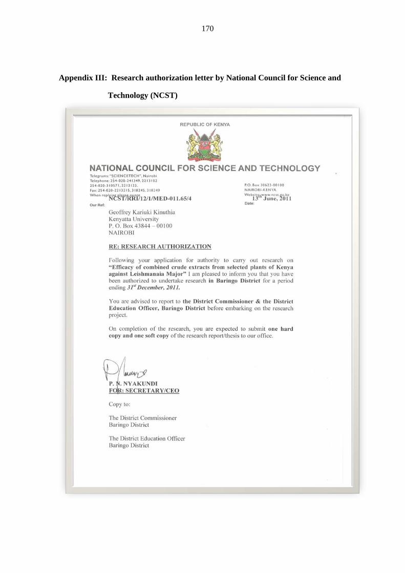
170
Appendix III: Research authorization letter by National Council for Science and
Technology (NCST)

171
Appendix IV: Research Permit by NCST