Embed Size (px)
Citation preview
1
Effects of topical application of CHF6467, a mutated form of human nerve growth factor, on skin wound
healing in diabetic mice.
*A. Giuliani, *L. Lorenzini, *V. A. Baldassarro, M. Pannella, M. Cescatti, M. Fernandez, G. Alastra, A. Flagelli,
G. Villetti, B. P. Imbimbo, L. Giardino, L. Calzà
University of Bologna, Ozzano Emilia, Italy
*these authors contributed equally to the study
Affiliations:
AG Department of Veterinary Medical Science, University of Bologna, Italy
LL Department of Veterinary Medical Science and Interdepartmental Center for Industrial Research in Life
Sciences and Technologies University of Bologna, Italy
VAB Interdepartmental Center for Industrial Research in Life Sciences and Technologies University of
Bologna, Italy
MP Fondazione IRET, Ozzano Emilia, Italy
MF Department of Veterinary Medical Science, University of Bologna, Italy
MC Fondazione IRET, Ozzano Emilia, Italy
GA Interdepartmental Center for Industrial Research in Life Sciences and Technologies University of
Bologna, Italy
AF Interdepartmental Center for Industrial Research in Life Sciences and Technologies University of
Bologna, Italy
GV Chiesi Farmaceutici, Parma, Italy
BPI Chiesi Farmaceutici, Parma, Italy
LG Department of Veterinary Medical Science, University of Bologna, Italy; Interdepartmental Center for
Industrial Research in Life Sciences and Technologies, University of Bologna, Italy; Fondazione IRET,
Ozzano Emilia, Italy
This article has not been copyedited and formatted. The final version may differ from this version.JPET Fast Forward. Published on September 18, 2020 as DOI: 10.1124/jpet.120.000110
at ASPE
T Journals on January 1, 2022
jpet.aspetjournals.orgD
ownloaded from

2
LC Department of Pharmacy and Biotechnology, University of Bologna, Italy; Interdepartmental Center for
Industrial Research in Life Sciences and Technologies, University of Bologna, Italy; Fondazione IRET,
Ozzano Emilia, Italy
This article has not been copyedited and formatted. The final version may differ from this version.JPET Fast Forward. Published on September 18, 2020 as DOI: 10.1124/jpet.120.000110
at ASPE
T Journals on January 1, 2022
jpet.aspetjournals.orgD
ownloaded from

3
Running title: CHF6467 for diabetic wound treatment
Correspondence and requests for materials should be addressed to:
Laura Calzà, MD (ORCID ID https://orcid.org/0000-0002-4426-8477)
CIRI-SDV, University of Bologna
Via Tolara di Sopra 41/E,
40064 Ozzano dell’Emilia (BO), Italy
Email: [email protected]
Number of text pages: 18
Number of tables: 2 (+ 3 in supplementary materials)
Number of figures: 8 (+ 1 in supplementary materials)
Number of references: 77
Number of abstract words: 248
Number of introduction words: 747
Number of discussion words: 1490
List of Abbreviations: Nonstandard abbreviations are defined in the text.
HE, Haemotoxylin and Eosin staining; NGF, nerve growth factor; p75, p75 neurotrophin receptor; PECAM-1,
Platelet endothelial cell adhesion molecule-1; PGP9.5, Protein gene product 9.5; TrkA, neurotrophic tyrosine
kinase receptor A.
Recommended section assignment: Endocrine and Diabetes
This article has not been copyedited and formatted. The final version may differ from this version.JPET Fast Forward. Published on September 18, 2020 as DOI: 10.1124/jpet.120.000110
at ASPE
T Journals on January 1, 2022
jpet.aspetjournals.orgD
ownloaded from
4
ABSTRACT
Nerve growth factor (NGF) is the protein responsible for the development and maintenance of sensory skin
innervation. Given the role of appropriate innervation in skin healing, NGF has been indicated as a possible pro-
healing treatment in pathological conditions characterized by nerve ending loss, such as chronic ulcers in
diabetes, however its use as a therapeutic agent is limited by its hyperalgesic effect. We tested the effect of
topical application of the non-algogenic NGF derivative hNGFP61S/R100E in two models of skin ulcer induced
in dbdb diabetic mice, investigating healing time, skin histology, reinnervation and angiogenesis using
morphological and molecular approaches. We showed that the topical administration of CHF6467, a
recombinant human NGF in which an amino acid substitution (R100E) abolished the hyperalgesic effect usually
associated with NGF, accelerates skin repair in experimental wounds (full-excision and pressure ulcer) induced
in diabetic mice (dbdb). CHF6467-induced acceleration of wound healing was accompanied by increased re-
epithelization, re-innervation and re-vascularization, as assessed by histology, immunohistochemistry and image
analysis. Bioinformatic analysis of differentially expressed genes and signaling pathways in the wound tissues
showed that Akt-mTOR was the most regulated pathway. In spite of the transdermal absorption leading to
measurable, dose-dependent increases in CHF6467 plasma levels, no systemic thermal or local mechanical
hyperalgesia was observed in treated mice. When tested in vitro in human cell lines, CHF6467 stimulated
keratinocyte and fibroblast proliferation and tube formation by endothelial cells. Collectively, these results
support a possible use of CHF6467 as a pro-healing agent in skin lesions in diabetes.
SIGNIFICANCE STATEMENT
Topical application of CHF6467 accelerates re-innervation, neoangiogenesis and wound healing in diabetic mice
in both full-thickness skin excision and pressure ulcer models through the Akt/mTOR pathway, and does not
induce hyperalgesia.
This article has not been copyedited and formatted. The final version may differ from this version.JPET Fast Forward. Published on September 18, 2020 as DOI: 10.1124/jpet.120.000110
at ASPE
T Journals on January 1, 2022
jpet.aspetjournals.orgD
ownloaded from

5
INTRODUCTION
Wound healing is a complex biological process involving different cell types which interact in a defined spatial
and temporal sequence (Rodrigues et al. 2016). When a skin wound does not repair within an established time
window, the lesion becomes a “chronic ulcer”, a phenomenon usually associated with co-morbidities such as
diabetes, obesity, cardiovascular diseases, or simply aging (Jaul et al. 2018), and as complication of surgical
wounds (Nenna et al. 2019). In diabetic patients, the incidence of chronic non-healing wounds is fairly high,
with an estimated 2% of this population in developed countries suffering from non-healing injuries, while
complications cause 90% of limb amputations (Rodrigues et al. 2016). Moreover, the quality of the repaired
skin, and the occurrence of wound relapse, are still open issues (Martinengo et al. 2019). The problem of chronic
skin wounds is further complicated by the worldwide increase in diabetes prevalence, contributing to 9% of
global mortality (Shaw et al. 2010).
Despite the efforts made to establish guidelines for acute and chronic skin wound treatment (Powers et al. 2016;
Westby et al. 2017; Blume and Wu 2018; Everett and Mathioudakis 2018), only three FDA-approved therapies
are available: two are skin substitutes, and a recombinant human platelet-derived growth factor (PDGF-BB,
becaplermin, Regranex®). Given that 50% of diabetic ulcers fail to heal (Lebrun et al. 2010), new solutions are
needed to shorten healing time, guarantee healing in severe, non-healing lesions, and prevent ulcer relapse where
possible.
One target for new products is the improvement of endogenous regeneration (Wells and Watt 2018) through
growth factors (Werner and Grose 2003). Originally described as a “neurotrophic factor” by Rita Levi-
Montalcini (Levi-Montalcini 1987), Nerve Growth Factor (NGF) supports a number of physiological processes
in the human body (Levi-Montalcini 1987). In the peripheral nervous system, the development of thinly
myelinated Aδ or unmyelinated C-fibers which innervate the skin is an NGF-dependent process (Indo 2010),
and half of the body’s nociceptive sensory neurons remain dependent on NGF in adulthood (Snider and
McMahon 1998). Notably, diabetic skin is characterized by reduced epidermal innervation (McCarthy et al.
1995; Kennedy et al. 1996), a factor considered to be one of the main cause of diabetic neuropathy (Ebenezer
and Polydefkis 2014) (Ebenezer and Polydefkis, 2014) supporting chronic ulcers (Laverdet et al. 2015).
NGF is now considered a “pleiotropic molecule” (Aloe and Calzà 2004). Inflammatory and immune cells
produce NGF and express NGF high- (trkA) and low- (p75) affinity receptors, while inflammation enhances the
This article has not been copyedited and formatted. The final version may differ from this version.JPET Fast Forward. Published on September 18, 2020 as DOI: 10.1124/jpet.120.000110
at ASPE
T Journals on January 1, 2022
jpet.aspetjournals.orgD
ownloaded from

6
synthesis of NGF in various tissues (Calzà et al. 1997) and in fibroblasts, epithelial, endothelial, and immune
cells (Prencipe et al. 2014; Gostynska et al. 2019). NGF has been indicated as an angiogenic molecule which
stimulates nitric oxide synthase (NOS) and vascular endothelial growth factor (VEGF) production (Calzà et al.
2001), and induces a variety of effects on endothelial cells by autocrine and/or paracrine mechanisms (Nico et al.
2008; Gostynska et al. 2019). In the skin microenvironment, NGF appears to be a major player in the
communication between sensory neurons and skin cells (Chéret et al. 2013).
A role of endogenous NGF in wound healing has been suggested by in vitro, animal, and human studies (Werner
and Grose 2003; Kawamoto and Matsuda 2004; Chéret et al. 2013), which have led to the approval of NGF eye
drops for the treatment of corneal neurotrophic ulcers (Sacchetti et al. 2020), and which have demonstrated the
role of NGF as a peripheral pain mediator, particularly in inflammatory pain states (Pezet and McMahon 2006;
Lewin et al. 2014).
In order to distinguish the positive properties of NGF from its detrimental effects, several trkA-binding agents
have been developed (Carleton et al. 2018; Bagal et al. 2019). The rare autosomal recessive neuropathy known
as hereditary sensory and autonomic neuropathy type V (HSAN V) is caused by a mutation in the NGF gene
(R100W), and HSAN V homozygous patients display a congenital indifference to painful events, with deficits in
peripheral nociceptors (Einarsdottir et al. 2004). Similarly, replacement of the arginine residue at position 100
with a serine residue has been shown to abolish NGF-induced hyperalgesia without affecting downstream trkA-
mediated signaling (Testa et al. 2019). The additional replacement of the proline residue at position 61 of NGF
with a serine residue leads to a non-algogenic derivative of hNGF (hNGFP61S/R100E, CHF6467) (Severini et
al. 2017).
The aim of this study was to test the efficacy of topical CHF6467 in two skin ulcer models in diabetic mice.
Healing time, repaired skin histology, reinnervation and angiogenesis were investigated using morphological and
molecular approaches.
MATERIALS AND METHODS
Animals and group composition
This article has not been copyedited and formatted. The final version may differ from this version.JPET Fast Forward. Published on September 18, 2020 as DOI: 10.1124/jpet.120.000110
at ASPE
T Journals on January 1, 2022
jpet.aspetjournals.orgD
ownloaded from

7
All animal protocols described herein were carried out according to the European Community Council
Directives (2010/63/EU), complied with the ARRIVE guidelines and the NIH Guide for the Care and Use of
Laboratory Animals, and were approved by the Ministry of Health (authorization no. 391/2017-PR). Ten eleven-
week-old genetically diabetic C57BL/KsJ-m+/+Leprdb (db/db) male mice were included in the experiments
(Charles River Laboratories -Calco-Lecco). The animals were housed with food pellets and water ad libitum, and
a dark-light cycle of 12 hours. Glycemia was measured prior to treatment, the day after the final treatment and
prior to sacrifice in non-fasting animals, between 9 and 10AM (Contour XT, Bayer, Basel, Switzerland).
For skin excision lesions, the following groups were included (N=8 each, according to the power analysis of
sample size calculation based on a pilot experiment):
- db/db, wound + vehicle
- db/db, wound + CHF6467 1µg/cm2
- db/db, wound + CHF6467 10µg/cm2
- db/db, wound + CHF6467 30µg/cm2
- db/db, wound + moNGF, 10µg/cm2
- db/db, wound + hNGF, 10µg/cm2
For pressure ulcers, the following groups were included (N=15 each, according to the power analysis of sample
size calculation based on a pilot experiment):
- db/db, wound + vehicle
- db/db, wound + CHF6467 1µg/cm2
- db/db, wound + CHF6467 10µg/cm2
- db/db, wound + CHF6467 100µg/cm2
Intact mice were also included for histology and immunohistochemistry experiments. For each model, mice were
randomly assigned to the experimental groups following lesion induction.
On the day of sacrifice, mice were deeply anesthetized and skin samples (1cm × 1cm) taken from the area of the
wound. For the skin excision study, 4 samples were collected from each group for immunohistochemistry and 4
samples were collected for histology. For the pressure ulcer study, a 6 mm skin area was taken using an
excisional punch (wound area). A 6 mm area of intact skin was collected for an exploratory tumorigenic study.
For the pathway array experiments, 6 mice from each group were used.
Drugs, surgery, wound dressing and monitoring
This article has not been copyedited and formatted. The final version may differ from this version.JPET Fast Forward. Published on September 18, 2020 as DOI: 10.1124/jpet.120.000110
at ASPE
T Journals on January 1, 2022
jpet.aspetjournals.orgD
ownloaded from

8
The following compounds were included in the study: CHF6467, provided by Chiesi (produced by AGC
Biologics, Heidelberg, Germany); human -NGF, recombinant, E.coli. (hrNGF, N-245, Alomone, Jerusalem,
Israel), and mouse NGF, recombinant (moNGF, 1156-NG, R&D System). The CHF6467 production process is
completely devoid of additives of animal or human origin, and the purification process has incorporated
technological advances, including precursor and multimodal chromatography resins. The recombinant NGF
protein was expressed in E. coli, refolded from inclusion bodies, purified, and proteolytically processed to NGF,
as previously described (Patent PCT/EP2019/060733). The vehicle was a solution of purified water, acetic acid
20mM, and L-methionine 20mM, pH5.5.
For the skin excision model, a 6mm circular full-thickness wound was created by dermal punch biopsy on the
midback of the animals, and the wound area immediately covered with a Tegaderm dressing (Tegaderm Roll -
3M Health Care, St.Paul, MN, USA). A 26-gauge needle was used to infuse 50l of medication through the
Tegaderm into the wound bed. Compounds and vehicle were administered daily for a total of 7 days from the
time of surgery onwards (Fig. 1A) (Landi et al. 2003), and animals were monitored daily for dressing integrity
and infection. The Tegaderm was changed weekly in all animals until wound healing was complete. Photographs
were taken to monitor the wounds and the area was measured using Nis-Elements AR 3.2 software (Nikon
Corporation, Tokyo).
Pressure ulcers were induced using two magnetic disks of 12 mm diameter and a thickness of 5.0 mm, with an
average weight of 2.4 g and 1000 G magnetic force (Algamagnetic, Italy). A skin fold was gently raised and
placed between the two magnets. This procedure left a 1.0 cm skin bridge between the magnets creating about 50
mm Hg compressive pressure between the two plates (Stadler et al. 2004). Three ischemia-reperfusion (I/R)
cycles were used, where a single I/R cycle consisted of a period of 12 h for the application of the magnets
followed by a rest period of 12 hours without magnets. Application of the compounds (60μl in each ulcer) began
following wound curettage (at day 3 after the last I/R cycle) and was continued daily until 50% closure at the
same dosage (day 14), then twice a week (40 μl day 17, 20 μl from day 19) until 100% closure at the same
dosage (Fig. 5A). Treatment of the wound following surgical curettage consisted of removal of the exudate by
gentle application of sterile gauze soaked in sterile saline solution. The wounds were dressed with Tegaderm
which was replaced as above. The lesions were monitored via visual inspection by two independent operators to
establish day of closure, without Tegaderm removal.
This article has not been copyedited and formatted. The final version may differ from this version.JPET Fast Forward. Published on September 18, 2020 as DOI: 10.1124/jpet.120.000110
at ASPE
T Journals on January 1, 2022
jpet.aspetjournals.orgD
ownloaded from
9
Hyperalgesia
Thermal hyperalgesia was evaluated in freely moving animals by Hargreave's Method using the Thermal Plantar
Test Instrument (Ugo Basile, Comerio, Varese) on day 0 (before surgery) and day 7 (the day after the final NGF
application). The mean of four latency to paw retraction measurements was used for statistical analysis.
Local mechanical hyperalgesia was evaluated using the Electronic Von Frey apparatus (Bioseb, Vitrolles Cedex,
France) fitted with an elastic spring tip at the edge of the wound on a gently constricted animal. The mean of
four measurements were used for the statistical analysis.
Total NGF plasmaassay
The Human Adipokine Magnetic Bead Panel 2 kit (HADK2MAG-61K, EMD Millipore Corporation, Billerica,
MA, USA) was used to quantify the hNGF in the plasma samples using ×MAP technology and a MAGPIX
Luminex platform. The assay was performed according to the manufacturer’s specifications, with some in-house
modifications. In brief, following incubation of the bead population with plasma samples (25 l) overnight at
RT, the beads were washed and incubated, initially with detection antibody solution for 1h at RT, then with the
streptavidin–phycoerythrin conjugated solution for 30 min RT. After washing, the beads were resuspended in
100 l of Drive fluid and read on the MAGPIX instrument. The data was analyzed using xPONENT 4.2 ®
software and the results expressed as pg/mL. We obtained values within the dynamic range of the standard curve
(from 10000 to 0.128 pg/mL) for all the samples. Standard curves had a correlation coefficient (R2) value >0.98.
The detection limit of NGF- was 0.3-0.7 pg/mL.
With this kit, a very small amount of mouse NGF- was quantified in the vehicle group due to antibody species
cross-reactivity. Moreover, since the recombinant mouse NGF- used for treatment is a homodimer of two 120
amino acid polypeptides which shares approximately 90% amino acid homology with human NGF-β, NGF was
also quantified in moNGF--treated mice.
Immunohistochemistry and quantitative analysis
The samples collected for histology were fixed in paraformaldehyde 4% (w/v) and picric acid saturated aqueous
solution in Sörensen buffer 0.1 M pH 7, embedded in paraffin, sectioned at 4m, and stained using hematoxylin
This article has not been copyedited and formatted. The final version may differ from this version.JPET Fast Forward. Published on September 18, 2020 as DOI: 10.1124/jpet.120.000110
at ASPE
T Journals on January 1, 2022
jpet.aspetjournals.orgD
ownloaded from

10
and eosin (HE). The samples collected for immunohistochemistry were rapidly removed and fixed as above for
24h, then washed for at least 48h in 5% sucrose in 0.1 M phosphate buffer. Cryostat sections (14m thick) were
incubated overnight at 4°C with the primary antibodies: Laminin (Rabbit, Sigma, 1:1000), PGP9.5 (Rabbit,
Boheringer, 1:2000), and PECAM-1 (Goat, R&D systems, 1:150). After rinsing in PBS for 20 min, the sections
were incubated with the secondary antisera conjugated with Rhodamine Red™-X-conjugated - affinity-pure
Donkey anti-Rabbit IgG (Jackson Immunoresearch) or secondary DyLight 488 Donkey anti-Goat IgG (Thermo
Fisher Scientific), diluted in 0.3% Triton / PBS. The sections were then rinsed in PBS mounted in glycerol
containing 1,4-phenylendiamine (0.1 g/L).
Immunofluorescence images were captured using a Nikon Eclipse E600 microscope equipped with the Q
Imaging Retiga-2000RV digital CCD camera (Q Imaging, Surrey, BC, Canada) and a z-axis motorized stage.
Analyses were performed using the Nis-Elements AR 3.2 software, by applying the same procedure to all
images under comparison. Briefly, five z-stakes every 2 m were collected from each image (300x500 m), and
the maximum intensity projection was used to calculate the immunoreactive area. For all morphological
analyses, five images and two levels/animal sampled in the epidermal papillae at the center of the repaired ulcer
were analyzed in each animal. All analyses were performed blindly. The immunoreactive area was calculated as
area/fraction (percentage of Laminin/PECAM-1 and PGP9.5 over 400x300m area). The measurement of
repaired epidermal thickness was performed on histological sections (4m thick, HE) in the same area. The
mean value of five measurements/section and two sections for animal was used for the statistical analysis. In
preparing the figures, the immunofluorescence images were occasionally contrasted. Where this is the case, the
same procedure was applied to all images under comparison.
RT-PCR and STRING analysis
Samples for RT2 Profiler PCR Arrays (Qiagen) were collected from the full-thickness skin excision experiment
at the core of the lesion (6 mm diameter) and RNA was extracted from all animals (6 animals per group),
quantified and pooled.
Pooled RNAs were retrotranscribed using the RT2 First Strand Synthesis Kit (Qiagen). Each pooled group was
tested using a single PCR array, using the CFX96 real time PCR instrument (BioRad).
This article has not been copyedited and formatted. The final version may differ from this version.JPET Fast Forward. Published on September 18, 2020 as DOI: 10.1124/jpet.120.000110
at ASPE
T Journals on January 1, 2022
jpet.aspetjournals.orgD
ownloaded from
11
Qiagen mouse angiogenesis, extracellular matrix and adhesion protein, neurotrophins and growth factors were
used to profile the expression of 250 genes. The possible tumorigenesis effect was also investigated using the
dedicated array. The relative gene expression was calculated using the 2-(ΔΔCq) comparative method.
The dedicated Qiagen online data analysis software for the relative quantification of gene expression was used to
perform the analysis and generate the graphs. The differentially expressed genes identified were then plotted in
the STRING network analysis software (v.10; http://string-db.org/) to analyze the functional interactions
between their biological functions.
Cell culture, immunocytochemistry and image analysis
To test the CHF6467 activity in skin cell lines, adult human primary epidermal keratinocytes (HEKa, ATCC®
PCS-200-011™), human skin fibroblast cell lines (BJ, ATCC® CRL-2522™), and human umbilical vein
endothelial cells (HUVECp, umbilical vein endothelial cells pooled from multiple isolates, Invitrogen C-015-5C,
Thermo Fisher Scientific, Waltham, MA, USA) were used and cultured according to the respective conventional
protocols (Gostynska et al., 2020).
For the cell proliferation assay, cells were seeded in 96-well plates (BJ, 3.5 × 10³ cells/well, FBS 2%; HEK, 2 ×
103 cells/well, no FBS) and treated with CHF6467 at 0, 50, 100 or 200 ng/ml, changing the medium every
second day. Every two days, for 8 or 10 total days for HEK and BJ cells respectively, cells were detached by
trypsinization and counted using a Bürker chamber (3 wells per time point, 2 counts per well).
HUVECs were used for the tube formation assay. Cells were seeded overnight at 9 × 10⁴ cells/well in 24-well
plates in culture medium containing 0, 50, 100 or 200 ng/ml of CHF6467. The next day, the cells were detached
by Trypsin/EDTA and seeded on Geltrex™ LDEV-free matrix (Thermo Fisher Scientific) in culture medium
containing the same concentration of CHF6467. Tube formation analysis was performed after 8 hours. Cells
were fixed, washed, incubated with blocking solution for 1 hour and then overnight at 4°C with primary
antibody (rabbit actin, Santa Cruz, 1: 200). After 2 washes in PBS, cells were incubated with secondary antibody
(Donkey anti-rabbit Alexa Fluor 488, Molecular Probes, 1:500) and Hoechst nuclear staining at 37°C for 30
minutes, and finally washed twice in PBS. Cultures were analyzed using the cell-based High Content Screening
technology (CellInsight™ CX5 HCS Platform, Thermo Scientific). The angiogenesis algorithm of the
CellInsight software permits detection of all cells present in the well by identification of the nuclei (Hoechst
staining), and each cell body by structural protein staining (actin). The angiogenesis index is automatically
This article has not been copyedited and formatted. The final version may differ from this version.JPET Fast Forward. Published on September 18, 2020 as DOI: 10.1124/jpet.120.000110
at ASPE
T Journals on January 1, 2022
jpet.aspetjournals.orgD
ownloaded from

12
calculated as follows: angiogenesis index = (1000 * total area of connected tubes) / total image area = (% of the
image area covered by connected tubes) * 10.
HUVEC and BJ cells cultured in high-glucose conditions and treated with CHF6467 200 ng/ml for 48 hours
were used for Western Blot analysis.
Western Blot
Western blot analysis was performed to quantify the Akt protein expression in cell cultures and skin. For skin
tissue samples, the analysis was performed on the same samples used for RT-PCR and STRING analysis, and
from intact skin. The skin tissue was homogenized in RIPA buffer and protease inhibitor (1x cocktail inhibitor
Sigma, 1 mM PMSF, 10 mM sodium fluoride, 1mM sodium orthovanadate) with a homogenization ratio 1:8
(mg:µL) and centrifuged at 12000 g for 20 min at 4°C. Cells were detached from the culture plates, centrifuged,
and the pellet resuspended in RIPA buffer and protease inhibitor. Total protein concentration was estimated
using a standard colorimetric method. For each experiment, the same quantity of proteins was loaded (tissues 25
µg; HUVECs, 15 µg; BJ, 0.5 µg) and the marker protein (Precision Plus Protein Standards, Bio-Rad) added.
A solution of Leammli / β-mercaptoethanol was added to each sample, and after heating treatment (100 °C, 5
minutes), the proteins were resolved in 4-20% Mini-PROTEAN TGX Stain-Free Gels (Bio-Rad) and transferred
to Amersham Protran 0.45µm Nitrocellulose Blotting Membrane (Bio-Rad). After blocking in Tris Buffer Saline
solution containing 1% Tween20 (TBST) and 2.5% BSA, membranes were incubated with the primary antibody
(rabbit pan-Akt, Cell Signaling -Leiden, The Netherlands- 1:1000; mouse β-actin, Santa Cruz Biotechnology -
Dallas, Texas-, 1:150) overnight at 4°C. After washing three times in TBST, the membranes were incubated with
HRP-conjugated secondary antibodies (swine anti-rabbit, Dako, 1:5000; swine anti-mouse, Dako, 1:5000) and
HRP-conjugated protein for marker visualization (Precision Protein SrepTactin HRP-conjugate, Bio-Rad,
1:10000) for 1 hour at RT. The membranes were then washed three times with TBST. The immunoreactive
signal was detected by incubating the membranes with Clarity Western ECL Substrate (Bio-Rad) for 5 minutes
at RT in darkness and using the BioRad Chemi DOC MP imaging systems.
Western blot signals were measured by densitometry using ImageJ (Fiji) software. The Akt signals were
normalized on β-actin in all the analyzed samples. Akt quantification was further normalized on intact skin
values to obtain the variation index (intact skin = 1). For cell cultures samples, normalized Akt values were used
for the analysis. Western blot signals were measured by densitometry using ImageJ (Fiji) software.
This article has not been copyedited and formatted. The final version may differ from this version.JPET Fast Forward. Published on September 18, 2020 as DOI: 10.1124/jpet.120.000110
at ASPE
T Journals on January 1, 2022
jpet.aspetjournals.orgD
ownloaded from

13
Statistical analysis
Data is expressed as mean+SEM. Statistical comparison of the rate of skin ulcer healing over time between
treatment groups was performed with the log-rank test that compares the hazard functions of the different groups
at each observed event time (Kaplan–Meier analysis). The Holm-Sidak test was used for multiple comparisons
versus a control group (vehicle) (SigmaPlot 11, Systat Software Inc., San Jose, CA, USA). Histological and
immunohistochemical results are reported as mean ± SEM. Prism software (GraphPad Software, San Diego, CA,
USA) was used for the statistical analyses of this data, and for graph generation. The Student’s t-test or one-way
ANOVA were used to analyze the data. Results were considered significant when the probability of their
occurrence as a result of chance alone was less than 5% (p < 0.05).
RESULTS
PC12 in vitro assay
CHF6467 biological activity in PC12 cells was compared to that of moNGF and hrNGF using the neurite
elongation test (Radio et al. 2008), performed by HCS measuring the average neurite length per cell, total neurite
length per cell, and the percentage of cells showing a neurite equal to or longer than the cell body length. Results
are presented in Fig. 1, supplementary materials. Initially, all NGF treatments were compared to the vehicle
group and, from this analysis, only the most effective concentration for each NGF treatment was selected for
further analysis (Fig. 2, supplementary materials). Compared to the vehicle treated group, all effective NGF
treatment doses showed a significantly higher mean neurite length (moNGF, P = 0.0394; hrNGF, P = 0.0196;
CHF6467, P = 0.0033) and mean total neurite length (moNGF, P = 0.0338; hrNGF, P = 0.0006; CHF6467, P =
0.0211). Only CHF6467 was found to significantly increase the percentage of cells showing long neurites (P =
0.0367), compared to the vehicle.
Animal monitoring
Animals were monitored for glycemia levels and body weight (Tables I and II). All diabetic mice showed very
high blood glucose levels. No differences between the treatment groups were observed at the different times,
either in the full-thickness excision or pressure ulcer model.
This article has not been copyedited and formatted. The final version may differ from this version.JPET Fast Forward. Published on September 18, 2020 as DOI: 10.1124/jpet.120.000110
at ASPE
T Journals on January 1, 2022
jpet.aspetjournals.orgD
ownloaded from

14
Full-thickness excision skin ulcer
Wound healing
The study schedule is shown in Fig 1A. Pictures of the wounds from representative mice from the vehicle,
CHF6467, moNGF and hrNGF 10g/cm2 groups are shown in Fig. 1B. The time course of mean percentage
reduction in ulcer area compared to baseline is given in Fig. 1C, and shows that all NGF treatments significantly
(two-way ANOVA, p < 0.0001) accelerated ulcer healing compared to vehicle (post-hoc test vs vehicle:
CHF6467 1g/cm2 p<0.0001; CHF6467 10g/cm2 p<0.0001; CHF6467 30g/cm2 p<0.0001; moNGF 10g/cm2
p=0.0001; hrNGF 10g/cm2 p<0.0001). The day-of-closure in individual mice is given in Table I, supplementary
materials. At Day 29, complete healing was observed in all animals treated with hrNGF 10 g/cm2 or CHF6467
30 g/cm2. Fig. 1D shows the proportion of animals not completely healed over time, with moNGF, hrNGF and
CHF6467 at 30 g/cm2 significantly accelerating the healing process compared to the vehicle.
Pain threshold
Thermal hyperalgesia was evaluated before skin lesion induction (d0) and after the final NGF application (d7).
Results are shown in Fig. 2A. A significant reduction in pain threshold was observed in hrNGF-treated animals.
No differences were observed in the other groups.
Total NGF plasma levels
Blood was collected 24 hours after the final application of NGF and at sacrifice (see Fig. 1A). Results are shown
in Fig. 2B. The kit used to quantify NGF includes a capture monoclonal antibody that recognizes the epitope
Ala 46-Asn 62, thus we quantified the total NGF, i.e. mouse endogenous NGF (since human antibody cross
reacts with mouse NGF) and the CHF6467 present in the plasma of the treated animals.
A dose-dependent, transient increase in total NGF plasma level is observed in CHF6467-treated mice, reaching
an average value of 350 pg/ml at d7. An increase in total NGF plasma levels is also observed in the moNGF-
treated group. This is not surprising, since the recombinant mouse NGF-β used for treatments shares
approximately 90% homology with human NGF-β at amino acid level (Ullrich et al. 1983). This increase is
reversable, as indicated by the low levels of total NGF at sacrifice (12.80 ± 3.3 pg/ml). Very low levels of
This article has not been copyedited and formatted. The final version may differ from this version.JPET Fast Forward. Published on September 18, 2020 as DOI: 10.1124/jpet.120.000110
at ASPE
T Journals on January 1, 2022
jpet.aspetjournals.orgD
ownloaded from
15
endogenous NGF- were detected in the vehicle-treated mice (11.0 ± 1.2 pg/ml), due to human antibody cross-
reactivity with the mouse protein.
Histology and immunohistochemistry
The entire area of the skin ulcer, including a 5 mm margin of intact skin, was excised, embedded in paraffin and
serially sectioned from the border to the centrum. The sections were then stained (HE), and representative low-
power images taken at the different levels of the wound are shown in Fig. 3A (border of the lesion) and 3D (core
of the lesion). High-power micrographs show the re-epithelization process at the wound border (Fig. 3B), where
the epidermis migrating tongue (MET) and the extensive granulation tissue in the derma below the epidermal
layer, characterized by inflammation, cell proliferation, matrix deposition and angiogenesis, are evident (Fig.
3E). Re-epithelization has been evaluated by measuring the epidermal layer thickness in the central area of the
repaired wound. Non-healing wounds, as monitored in Table I (supplementary) were excluded from the analysis.
Representative images from intact, vehicle- and CHF6467 30g/cm2-treated mice are presented in 3 F-H,
respectively, and the results are shown in Fig 3I. CHF6467 induced a dose-dependent thickening of the
epidermal layer and a comparable effect was observed after treatment with matching doses of both moNGF and
hrNGF. In treated mice, the basal layer of the epidermis was characterized by hypercellularity of the basal and
spinous layers, possibly reflecting cell proliferation. Moreover, the dermis was also thicker and strongly stained,
suggesting a higher extracellular matrix deposition, and showed the presence of skin annexes (glands and hair
follicles).
Skin reinnervation was analyzed by immunostaining for the protein PGP9.5. The anatomy of the skin
innervation is presented in Fig. 3C, where PGP9.5 –IR fibers in the intact mouse skin can be seen in the
subcutaneous, deep cutaneous and sub-epidermal fibers. Radial fibers can be seen between the deep-
subcutaneous and subependymal plexus, while the sub-epidermal plexus supplies the free epidermal nerve
endings to the epidermis.
Fig. 3J-L illustrates PGP9.5-IR at sacrifice in intact, vehicle, and CHF6467 30g/cm2-treated mice, respectively.
Results from the morphometric analysis are presented as an immunoreactive percentage area in Fig. 3M. No
differences in skin innervation were present between the intact animals, CHF6467 (all dosages) and moNGF
groups, and there is no evidence of innervation restoration in vehicle-treated mice. On the contrary, a
hyperinnervation is observed using hrNGF.
This article has not been copyedited and formatted. The final version may differ from this version.JPET Fast Forward. Published on September 18, 2020 as DOI: 10.1124/jpet.120.000110
at ASPE
T Journals on January 1, 2022
jpet.aspetjournals.orgD
ownloaded from

16
The effect of topical NGF application on angiogenesis was estimated using laminin as basal membrane marker,
thus marking the outer endothelial cell membrane. Representative images are given in Fig. 3N-P, while the
results are shown in Fig. 3Q. CHF6467 30g/cm2 induces a significant increase in laminin-IR, as hrNGF,
possibly reflecting angiogenesis in progress.
Transcriptome analysis and validation
The aim of this experiment was to explore molecular mechanisms supporting the positive effect of CHF6467 on
wound healing in diabetic mice, focusing on inflammation, extracellular matrix deposition, innervation, and
angiogenesis. For this purpose, an exploratory strategy was used to map mRNA alterations of 252 key genes
involved in angiogenesis, ECM, and growth factors (GFs) in the wound at 50% of the repair process (see Table
III, supplementary material, for the full list). According to the data analysis software, the Thbs1 gene was used
as the housekeeping gene. The results for angiogenesis (A), extracellular matrix (B) and growth factor (C) genes
are presented in Fig 4 as a scatter plot analysis. Compared to vehicle-treated mice, the CHF6467 30g/cm2
treatment down-regulated several genes, including Akt, Ccl2 (chemokine (C-C motif) ligand 2), Ctgf (connective
tissue growth factor), Hif1a (hypoxia-inducible factor 1-alpha), Mmp14 (matrix metalloproteinases-14), Thbs2
(thrombospondin 2), Tnc (Tenascin C), and up-regulated Mmp9 (matrix metalloproteinases-9).
We then analyzed the array data by STRING software analysis for pathway discovery, considering all the
differentially expressed genes identified (web-software STRING 10 and Gene Ontology databases) to predict
protein–protein interactions. We adopted stringent inclusion criteria by including genes whose expression
changes are greater than the selected boundary (≥ 3). Analysis criteria were also stringent: confidence = 0.9
(highest, KEGG database); 1st shell. The results are shown in Fig. 4D, indicating a strong cluster around Akt-
mTOR, which also includes Hifa and Hsp90.
In order to confirm the Akt mRNA down-regulation by the CHF6467 compared to vehicle-treated mice, we
quantified the Akt protein in the same samples. Values were normalized on the intact skin of the same animals.
Western blot analysis confirmed that CHF6467 treatment blocked the ulcer-mediated increase of Akt (Student’s
t-test, p = 0.0049, Fig. 4E-F).
Pressure ulcer
Wound healing
This article has not been copyedited and formatted. The final version may differ from this version.JPET Fast Forward. Published on September 18, 2020 as DOI: 10.1124/jpet.120.000110
at ASPE
T Journals on January 1, 2022
jpet.aspetjournals.orgD
ownloaded from

17
The schedule of the experiment is presented in Fig. 5A. Representative images from the experimental groups are
shown in Fig. 5B. “Day of closure” was established based on visual inspection and photo analysis by two
independent operators (see Table II, supplementary materials for individual data). The clinical diagnosis of “non-
healing wound” was confirmed by histological analysis of the specimens. The log-rank analysis of the healing
curves indicated that all three doses (1, 10 and 100 g/cm2) of topical CHF6467 significantly accelerated the
healing process compared to vehicle (Fig. 5C), with 35 out of 36 skin lesions (97%) healed at Day 28 in the 10
g/cm2 group.
Pain threshold
Thermal hyperalgesia results are shown in Fig. 5D. A significant reduction in paw withdrawal latency was
observed in CHF6467 1g/cm2- compared to vehicle-treated mice. No hyperalgesia to thermal stimuli was
observed in the other groups. Local mechanical hyperalgesia was also evaluated the day after the final test
compound application (on day 15). No hyperalgesia to locally applied mechanic stimuli was observed in the
CHF6467-treated groups (data not shown).
Histology and immunohistochemistry
Samples of the skin containing the area of the ulcer, including a margin of intact skin, were excised, embedded
in paraffin, serially sectioned to obtain the “equator” of the repaired ulcer and stained for HE. A representative
low-power image at the ulcer “equator” at sacrifice is shown in Fig. 6A. The histological structure of the
epidermis, derma and hypodermis of the intact skin (peripheral parts) is shown, including the ulcer area
(rectangle), where the epidermis is visible, but the derma is still incompletely restored, as indicated by the
prevalence of collagen fibers and the paucity of dermal annexes (glands and hair follicles). The re-epithelization
was analyzed by measuring the epidermal thickness at the equator of the lesioned area. Representative images in
vehicle- and CHF6467-treated animals and respective results are shown in Fig. 6B-F. The histological analysis
confirmed the clinical data (Table II, supplementary), showing that the new epidermal layer is complete in 18
out of 30 ulcers in vehicle-treated mice; in 29 out of 36 ulcers in CHF6467 1g/cm2-treated mice; in 35 out of 36
ulcers in CHF6467 10g/cm2-treated mice, and in 32 out of 36 ulcers in CHF6467 100g/cm2-treated mice. The
thickness of the newly formed epidermis sampled at the lesion equator progressively increases in a dose-
dependent trend in CHF6467-treated mice compared to vehicle, resulting as being significantly thicker in
CHF6467 100g /cm2-treated mice (Student’s t-test, vehicle vs CHF6467 100g/cm2, P=0.0347).
This article has not been copyedited and formatted. The final version may differ from this version.JPET Fast Forward. Published on September 18, 2020 as DOI: 10.1124/jpet.120.000110
at ASPE
T Journals on January 1, 2022
jpet.aspetjournals.orgD
ownloaded from

18
The effect of topical CHF6467 application on nerve ending re-growth in the repaired skin has been analyzed as
PGP9.5-IR in the epidermal papillae of the repaired area. Representative images and results from the
morphometric analysis are given in Fig. 6G-K, respectively. A slight dose-dependent increase in innervation is
observed (one-way ANOVA, F (3, 63) = 3.716, P=0.016), resulting in a significant effect in CHF6467 100g
/cm2–treated mice.
The effect of topical CHF6467 application on angiogenesis was estimated using PECAM-1 (CD31) as a marker.
Representative images and results from the morphometric analysis are shown in Fig. 6L-P. An increase in
capillary density, as evaluated by PECAM-1-IR, is observed in the dermis of CHF6467-treated mice (one-way
ANOVA, F (3, 63) = 15.48, P < 0.0001), resulting in a significant effect in CHF6467 10g /cm2 and CHF6467
100g /cm2–treated mice.
CHF6467 effect on in vitro population doubling of HEK and BJ cells and angiogenesis assays in HUVEC
cells
The aim of these experiments was to investigate the effect of CHF6467 on three different cell types involved in
the wound healing process, i.e. keratinocytes, fibroblasts and endothelial cells, considering proliferation and in
vitro angiogenesis as respective readouts. We used three extensively used human cell lines, i.e. HEK
keratinocytes, BJ fibroblasts, and HUVEC endothelial cells. Fig. 7 A and B illustrate HEK cultures in the
investigated experimental conditions. CHF6467 induced a concentration-dependent increase in cell proliferation
up to the 192-hour time point (two-way ANOVA, treatment F(3, 100) p < 0.0001, time F(4, 100) p < 0.0001,
interaction F(12, 100) p < 0.0001) compared to the respective vehicle, and at the last time-point, all tested
CHF6467 concentrations resulted in increased cell numbers compared to the vehicle-treated group (Dunnett’s
post-hoc; 50, 100 and 200 ng/ml, p < 0.0001) (Fig. 7C).
Fig. 7D and E illustrate BJ cultures in the investigated experimental conditions. CHF6467 induced a
concentration-dependent increase in cell proliferation in fibroblasts compared to the respective vehicle up to the
240-hour time point in culture (two-way ANOVA, treatment F(3, 100) p < 0.0001, time F(4, 100) p < 0.0001,
interaction F(12, 100) p < 0.0001), and at the last time point, only the highest concentration of CHF6467 (200
ng/ml) produced an increase in cell number compared to cells exposed to vehicle (Dunnett’s post-hoc, p <
0.0001) (Fig. 7F).
This article has not been copyedited and formatted. The final version may differ from this version.JPET Fast Forward. Published on September 18, 2020 as DOI: 10.1124/jpet.120.000110
at ASPE
T Journals on January 1, 2022
jpet.aspetjournals.orgD
ownloaded from

19
The effect of CHF6467 on angiogenesis was tested on HUVEC cells stained for actin, using the conventional
angiogenic assay protocol for cell growth and analysis. DAPT 5M was used as reference compound. Fig. 7 G
and H illustrate the effect of CHF6467 at the highest tested dose on angiogenesis (H) compared to vehicle-
treated cells (G). The results indicate robust pro-angiogenic effects of CHF6467 (Fig. 7I; one-way ANOVA, F(4,
5) = 13.89, p = 0.0064), whose response at 100 and 200nM (Dunnett’s post-test, compared to vehicle, 100 ng/ml,
p = 0.006; 200 ng/ml, p = 0.0051) was similar to the DAPT reference compound (Dunnett’s post-hoc, compared
to vehicle, p = 0.0062).
To further support the mechanism identified by the bioinformatic analysis and confirmed by the protein analysis
in the skin, we also quantified the Akt protein in BJ and HUVEC cell high-glucose cultures treated with vehicle
or the highest dose of CHF6467 for 48 hours (200 ng/ml). BJ cultures (Fig. 8A) show a decrease in Akt protein
quantity when treated with CHF6467 (Student’s t test, p = 0.0071; Fig. 8B-C), while the same treatment in
HUVEC (Fig.8D) resulted in no change in protein level (Fig. 8E-F).
Potential tumorigenic effect: exploratory study
To explore whether topical application of CHF6467 and the stimulating effect on cell proliferation activate
aberrant, cancer-related pathways, we performed a pathway-focused gene expression analysis on tissue samples
obtained immediately after CHF6467 discontinuation (day 14), corresponding to 50-70% of the repair process
(see Table III, supplementary material, for the list of the investigated genes). The expression analysis was
performed considering only the mean expression of B2m, or Acly, Xrcc4, Casp2, Stmn1, Igfbp3 as house-keeping
genes, according to the Qiagen data analysis software recommendation. In both cases, the expression level of
only one of the 84 genes (Ercc3) was down-regulated at the 100 g/cm2 concentration of CHF6467 (Fig. 2,
supplementary materials), while the expression of the other genes was unchanged.
DISCUSSION
In this study, we showed that the topical application of CHF6467, a non-algogenic derivative of human NGF,
accelerates skin repair in experimental wounds induced in diabetic mice. The main results of the study can be
This article has not been copyedited and formatted. The final version may differ from this version.JPET Fast Forward. Published on September 18, 2020 as DOI: 10.1124/jpet.120.000110
at ASPE
T Journals on January 1, 2022
jpet.aspetjournals.orgD
ownloaded from

20
summarized as follows: (i) CHF6467 accelerates wound healing in both the full-thickness skin excision model
and in the model of pressure ulcer induced by repeated ischemia-reperfusion cycles; (ii) topical CHF6467
application does not induce systemic thermal or local mechanical hyperalgesia; (iii) CHF6467 improves re-
epithelization, re-innervation and re-vascularization, as assessed by histology, immunohistochemistry and image
analysis; (iv) the underlying molecular mechanisms involve the Akt-mTOR pathway; (v) when tested in vitro in
human cell lines, CHF6467 stimulates keratinocyte and fibroblast proliferation, and tube formation by
endothelial cells.
The study was performed in dbdb mice, the most widely used mouse model of type 2 diabetes mellitus (Alpers
and Hudkins, 2011). These mice develop progressive sensory loss, electrophysiological impairments and skin
innervation loss (De Gregorio et al. 2018), as well as hyperalgesia to thermal and mechanical stimuli (Shi et al.
2013; Tang et al. 2019), aspects which reflect at least some of the features observed in human diabetic
neuropathy (Yorek 2016).
Wound healing in dbdb mice includes all phases of the process, i.e. inflammation (phase 1); proliferation
(including re-epithelization), granulation tissue formation mainly by fibroblasts, neovascularization (phase 2),
and remodeling, with scar maturation and reorganization of the extracellular matrix (phase 3). However, the
repair process is much slower in dbdb than in db/-, particularly in wounds covered by Tegaderm as in our
experimental conditions (Sullivan et al. 2007), making this mouse model very appropriate for testing pro-healing
compounds.
We observed that the topical application of CHF6467 accelerates wound healing in both ulcer models in dbdb
mice. Given the different pathogenic mechanisms supporting the ulcer (surgery and ischemia/reperfusion,
respectively), and the differences in ulcer severity, we used two different application schemes. In the full-
thickness ulcer, in which the lesion diameter is around 6 mm, we applied CHF6467 for 7 consecutive days
starting immediately following lesion induction, therefore during the inflammatory phase of the process. Whilst
in the pressure ulcers in which the lesion diameter is around 12 mm after debriding, we applied CHF6467 for 12
consecutive days, then every 2 days up to day 26, therefore in the inflammatory, but also in the proliferative
phase of the process.
In the full-thickness skin excision model, the healing curve in the CHF6467-treated animal begins to differ from
the other groups at 11 days post-lesion, and healing is complete in almost all animals treated with 10 and 30
This article has not been copyedited and formatted. The final version may differ from this version.JPET Fast Forward. Published on September 18, 2020 as DOI: 10.1124/jpet.120.000110
at ASPE
T Journals on January 1, 2022
jpet.aspetjournals.orgD
ownloaded from

21
g/cm2 of CHF6467 at 18 days post-lesion. Histological and immunohistochemical analysis reveals that NGF
treatment (with native mouse and human forms of NGF and CHF6467) improves re-epithelization, re-
innervation and angiogenesis in the repaired skin, confirming the previously described effects of the biologically
active 2.5S NGF subunit on wound healing in full-thickness excision lesions in diabetic mice (Muangman et al.
2004).
To confirm the effectiveness of CHF6467 in skin wound healing in diabetic mice, we tested three different
dosages (1, 10, 100g/cm2) in a pressure ulcer model based on ischemia-reperfusion cycles (Stadler et al. 2004;
Huggenberger and Detmar 2011), a model which has also been used to test new pharmacological and non-
pharmacological treatments in dbdb mice(Duscher et al. 2015; Danigo et al. 2015; Cronk et al. 2015). Topical
CHF6467 application accelerated the healing rate and time of ulcer repair, stimulating re-epithelization, re-
innervation and angiogenesis processes in this model also.
Notably, none of the CHF6467 doses decreased the pain threshold, measured 24 hours after the final application,
despite the dose-dependent increase in total plasma NGF levels compared to baseline, an increase which is
transient and which normalized 22 days after CHF6467/NGF treatment withdrawal. On the contrary, an increase
in pain sensitivity was observed in hrNGF-treated mice, allowing us to conclude that transdermal absorption of
the mutated NGF CHF6467 does not induce pain threshold variations for thermal stimuli after multiple dose
administration.
From these experiments, we can conclude that the topical application of CHF6467, a modified human
recombinant NGF that does not elicit hyperalgesia at therapeutic doses, accelerates wound healing in diabetic
mice. Improved re-innervation of the wound area may be a supporting mechanism: indeed, a central rationale for
using NGF to promote healing in diabetes stems from a feature of the disease known as “small-fiber neuropathy”
and characterized by a reduced number of nerve endings in the epidermis (Ebenezer and Polydefkis 2014).
Appropriate skin innervation plays a preeminent role in wound healing (Kiya and Kubo 2019), and a reduction
in neurotrophic support to peripheral neurons has been described in diabetic skin (Sima 2003). NGF is the
neurotrophin responsible for establishing sensory innervation of the skin during development, and for its
maintenance in adulthood (Indo 2010). Thus, the topical application of NGF has been considered as a possible
therapy for accelerating skin wound healing in pressure ulcers (Aloe 2004) and post-chronic vasculitis ulcers
(Tuveri et al. 2000).
This article has not been copyedited and formatted. The final version may differ from this version.JPET Fast Forward. Published on September 18, 2020 as DOI: 10.1124/jpet.120.000110
at ASPE
T Journals on January 1, 2022
jpet.aspetjournals.orgD
ownloaded from

22
Several cell types in the skin are NGF-sensitive (Sofroniew et al. 2001) and may be involved in the positive
effect on wound healing exerted by the topical application of CHF6467. CHF6467, when applied in vitro,
induces a concentration-dependent increase in keratinocyte and fibroblast proliferation. This confirms recent
results demonstrating that moNGF stimulates in vitro keratinocyte proliferation and migration, and reverses the
impairment induced by high d-glucose concentrations in the culture medium (Gostynska et al. 2019).
NGF plays a role in angiogenesis, as suggested by in vivo studies in several tissues under physiological and
pathological conditions. It also promotes VEGF synthesis and secretion (Calzà et al. 2001; Li et al. 2018;
Ahluwalia et al. 2018); indeed, an NGF defect has been associated with impaired angiogenesis and defective
mucosa repair (Ahluwalia et al. 2018). A protective and proangiogenic role of NGF on endothelial cells has been
also described in vitro (Ahluwalia et al. 2017; Lazarovici et al. 2018), and we demonstrated that both moNGF
and CHF6467 increase endothelial cell proliferation and in vitro angiogenesis in a concentration-dependent
manner, as well as restoring the tube formation capacity disrupted by high glucose concentrations in the culture
medium (Gostynska et al. 2019).
Given the substantial hypertrophy observed in repaired skin epithelium, and the disputed role of NGF in cancer
(Demir et al. 2016), we also explored the regulation of 80 cancer-related genes at 50% of the wound healing
process in the pressure ulcer model. The only down-regulated gene in mice treated with the highest CHF6467
dose is Ercc3, a gene involved in DNA repair but also in hair follicle physiology (Yu et al. 2012).
Wound healing involves dozens of molecules which interact with the different cell types concerned. We
therefore adopted an exploratory, bioinformatic strategy to identify possible primary molecular pathways
involved in the pro-healing effect of CHF6467, comparing gene expression regulation in CHF6467 and vehicle-
treated mice in the full-thickness excision model at 50% of the repair process, when ECM deposition,
keratinocyte proliferation and angiogenesis are occurring simultaneously. From the analysis of 240 genes related
to angiogenesis, ECM, and growth factors, we observed that matrix metallopeptidase 9 (Mmp9), an enzyme
involved in tissue remodeling and required for the recruitment of endothelial stem cells during angiogenesis
(Heissig et al. 2002), is up-regulated in CHF6467- mice compared to vehicle-treated. Conversely, Akt1, Ccl2,
Ctgf, HIF1a, Mmp14, Thbs2 and Tnc were all down-regulated by CHF6467 treatment. Notably, a hyperactivity
of these genes seems to play a role in pathological wound healing in diabetes and other conditions (Bornstein et
al. 2000; Botusan et al. 2008; Liu et al. 2012; Ishikawa et al. 2015; Catrina and Zheng 2016; Zigrino et al. 2016;
Abu El-Asrar et al. 2018; Gale et al. 2018; Kunkemoeller et al. 2019).
This article has not been copyedited and formatted. The final version may differ from this version.JPET Fast Forward. Published on September 18, 2020 as DOI: 10.1124/jpet.120.000110
at ASPE
T Journals on January 1, 2022
jpet.aspetjournals.orgD
ownloaded from

23
The bioinformatic analysis performed by STRING software indicates that the Akt-mTOR pathway is involved in
the pro-healing effects of CHF6467, as also confirmed by protein analysis, and fibroblasts are the cells in which
this regulation occurs. Akt and mTOR are considered survival and cellular growth in response to lesions. NGF,
via trkA, activates PI3K/Akt and ERK/MAPK signaling pathways and the downstream mTOR, and this pathway
mediates several NGF effects, such as endothelial cell invasion and cord formation during development (Park et
al. 2007), angiogenesis in subchondral bone (Yu et al. 2019), and neuroprotection (Elsherbiny et al. 2019).
Moreover, NGF-enhanced secretion of VEGF is also mediated by the ERK1/2 and PI3K/Akt pathways (Wang et
al. 2016). An impairment of the Akt-mTOR pathway has been indicated as a possible cause of wound healing
impairment in diabetic mice (Huang et al. 2015; Jere et al. 2019), and transient pharmacological activation of
the PI3K-Akt-mTOR signaling axis has been considered a novel clinical intervention strategy to accelerate
wound (ulcer) healing (Squarize et al. 2010).
In conclusion, we showed that the topical application of CHF6467, a non-algogenic hrNGF derivative,
accelerates skin wound healing in diabetic mice, in both full-thickness excision and pressure ulcer models, also
improving angiogenesis and re-innervation, without inducing hyperalgesia. We suggest that the Akt/mTOR
pathway may be activated by CHF6467.
ACKNOWLEDGMENTS
Dr. Natalia Gostynska is gratefully acknowledged for the BJ experiments. All datasets are available to the
corresponding author upon reasonable request.
AUTHOR CONTRIBUTIONS
Participated in research design: Calzà L., Giardino L. Imbimbo B.P., Villetti G.,
This article has not been copyedited and formatted. The final version may differ from this version.JPET Fast Forward. Published on September 18, 2020 as DOI: 10.1124/jpet.120.000110
at ASPE
T Journals on January 1, 2022
jpet.aspetjournals.orgD
ownloaded from

24
Conducted experiments: Alastra G., Cescatti M., Fernandez M., Flagelli A., Giuliani A., Lorenzini L., Pannella
M.,
Contributed new reagents or analytic tools: Villetti G.
Performed data analysis: All Authors
Wrote or contributed to the writing of the manuscript: Baldassarro V. A., Calzà L., Giardino L., Imbimbo B.P.,
Villetti G.
References
Abu El-Asrar AM, Mohammad G, Allegaert E, Ahmad A, Siddiquei MM, Alam K, Gikandi PW, De Hertogh G,
Opdenakker G (2018) Matrix metalloproteinase-14 is a biomarker of angiogenic activity in proliferative
diabetic retinopathy. Mol Vis 24:394–406
Ahluwalia A, Jones MK, Hoa N, Tarnawski AS (2017) NGF protects endothelial cells from indomethacin-
induced injury through activation of mitochondria and upregulation of IGF-1. Cell Signal 40:22–29 .
https://doi.org/10.1016/j.cellsig.2017.08.006
Ahluwalia A, Jones MK, Hoa N, Zhu E, Brzozowski T, Tarnawski AS (2018) Reduced NGF in Gastric
Endothelial Cells Is One of the Main Causes of Impaired Angiogenesis in Aging Gastric Mucosa. CMGH
6:199–213 . https://doi.org/10.1016/j.jcmgh.2018.05.003
Aloe L (2004) Nerve growth factor, human skin ulcers and vascularization. Our experience. In: Progress in Brain
Research. Elsevier, pp 515–522
Aloe L, Calzà L (2004) NGF and related molecules in health and disease: Preface. In: Progress in Brain
Research. p 544
Bagal SK, Omoto K, Blakemore DC, Bungay PJ, Bilsland JG, Clarke PJ, Corbett MS, Cronin CN, Cui JJ, Dias
R, Flanagan NJ, Greasley SE, Grimley R, Johnson E, Fengas D, Kitching L, Kraus ML, McAlpine I,
Nagata A, Waldron GJ, Warmus JS (2019) Discovery of Allosteric, Potent, Subtype Selective, and
Peripherally Restricted TrkA Kinase Inhibitors. J Med Chem 62:247–265 .
This article has not been copyedited and formatted. The final version may differ from this version.JPET Fast Forward. Published on September 18, 2020 as DOI: 10.1124/jpet.120.000110
at ASPE
T Journals on January 1, 2022
jpet.aspetjournals.orgD
ownloaded from
25
https://doi.org/10.1021/acs.jmedchem.8b00280
Blume P, Wu S (2018) Updating the Diabetic Foot Treatment Algorithm: Recommendations on Treatment Using
Advanced Medicine and Therapies. Wounds 30:29–35
Bornstein P, Kyriakides TR, Yang Z, Armstrong LC, Birk DE (2000) Thrombospondin 2 modulates collagen
fibrillogenesis and angiogenesis. J Investig Dermatology Symp Proc 5:61–66 .
https://doi.org/10.1046/j.1087-0024.2000.00005.x
Botusan IR, Sunkari VG, Savu O, Catrina AI, Grünler J, Lindberg S, Pereira T, Ylä-Herttuala S, Poellinger L,
Brismar K, Catrina SB (2008) Stabilization of HIF-1α is critical to improve wound healing in diabetic
mice. Proc Natl Acad Sci U S A 105:19426–19431 . https://doi.org/10.1073/pnas.0805230105
Calzà L, Giardino L, Giuliani A, Aloe L, Levi-Montalcini R (2001) Nerve growth factor control of neuronal
expression of angiogenetic and vasoactive factors. Proc Natl Acad Sci U S A 98:4160–4165 .
https://doi.org/10.1073/pnas.051626998
Calzà L, Giardino L, Pozza M, Micera A, Aloe L (1997) Time-course changes of nerve growth factor,
corticotropin-releasing hormone, and nitric oxide synthase isoforms and their possible role in the
development of inflammatory response in experimental allergic encephalomyelitis. Proc Natl Acad Sci U
S A 94:3368–3373 . https://doi.org/10.1073/pnas.94.7.3368
Carleton LA, Chakravarthy R, van der Sloot AM, Mnich K, Serrano L, Samali A, Gorman AM (2018)
Generation of rationally-designed nerve growth factor (NGF) variants with receptor specificity. Biochem
Biophys Res Commun 495:700–705 . https://doi.org/10.1016/j.bbrc.2017.11.003
Catrina SB, Zheng X (2016) Disturbed hypoxic responses as a pathogenic mechanism of diabetic foot ulcers.
Diabetes Metab Res Rev 32:179–185 . https://doi.org/10.1002/dmrr.2742
Chéret J, Lebonvallet N, Carré JL, Misery L, Le Gall-Ianotto C (2013) Role of neuropeptides, neurotrophins,
and neurohormones in skin wound healing. In: Wound Repair and Regeneration. Wound Repair Regen, pp
772–788
Cronk SM, Kelly-Goss MR, Ray HC, Mendel TA, Hoehn KL, Bruce AC, Dey BK, Guendel AM, Tavakol DN,
Herman IM, Peirce SM, Yates PA (2015) Adipose-Derived Stem Cells From Diabetic Mice Show
Impaired Vascular Stabilization in a Murine Model of Diabetic Retinopathy. Stem Cells Transl Med
4:459–467 . https://doi.org/10.5966/sctm.2014-0108
Danigo A, Nasser M, Bessaguet F, Javellaud J, Oudart N, Achard JM, Demiot C (2015) Candesartan restores
pressure-induced vasodilation and prevents skin pressure ulcer formation in diabetic mice. Cardiovasc
This article has not been copyedited and formatted. The final version may differ from this version.JPET Fast Forward. Published on September 18, 2020 as DOI: 10.1124/jpet.120.000110
at ASPE
T Journals on January 1, 2022
jpet.aspetjournals.orgD
ownloaded from

26
Diabetol 14:26 . https://doi.org/10.1186/s12933-015-0185-4
De Gregorio C, Contador D, Campero M, Ezquer M, Ezquer F (2018) Characterization of diabetic neuropathy
progression in a mouse model of type 2 diabetes mellitus. Biol Open 7: .
https://doi.org/10.1242/bio.036830
Demir IE, Tieftrunk E, Schorn S, Friess H, Ceyhan GO (2016) Nerve growth factor & TrkA as novel therapeutic
targets in cancer. Biochim. Biophys. Acta - Rev. Cancer 1866:37–50
Duscher D, Neofytou E, Wong VW, Maan ZN, Rennert RC, Inayathullah M, Januszyk M, Rodrigues M,
Malkovskiy A V., Whitmore AJ, Walmsley GG, Galvez MG, Whittam AJ, Brownlee M, Rajadas J,
Gurtner GC (2015) Transdermal deferoxamine prevents pressure-induced diabetic ulcers. Proc Natl Acad
Sci U S A 112:94–99 . https://doi.org/10.1073/pnas.1413445112
Ebenezer G, Polydefkis M (2014) Epidermal innervation in diabetes. In: Handbook of Clinical Neurology.
Elsevier B.V., pp 261–274
Einarsdottir E, Carlsson A, Minde J, Toolanen G, Svensson O, Solders G, Holmgren G, Holmberg D, Holmberg
M (2004) A mutation in the nerve growth factor beta gene (NGFB) causes loss of pain perception. Hum
Mol Genet 13:799–805 . https://doi.org/10.1093/hmg/ddh096
Elsherbiny NM, Abdel-Mottaleb Y, Elkazaz AY, Atef H, Lashine RM, Youssef AM, Ezzat W, El-Ghaiesh SH,
Elshaer RE, El-Shafey M, Zaitone SA (2019) Carbamazepine Alleviates Retinal and Optic Nerve Neural
Degeneration in Diabetic Mice via Nerve Growth Factor-Induced PI3K/Akt/mTOR Activation. Front
Neurosci 13:1089 . https://doi.org/10.3389/fnins.2019.01089
Everett E, Mathioudakis N (2018) Update on management of diabetic foot ulcers. Ann. N. Y. Acad. Sci.
1411:153–165
Gale JD, Jensen J, Berman G, Freimuth W, Li G, Pleil A, Kutty M, Rosenthal A, Boswell CB, Noah V. EM,
Young L (2018) A Placebo-controlled Study of PF-06473871 (Anti-Connective Tissue Growth Factor
Antisense Oligonucleotide) in Reducing Hypertrophic Skin Scarring. Plast Reconstr Surg - Glob Open
6:e1861 . https://doi.org/10.1097/gox.0000000000001861
Gostynska N, Pannella M, Rocco ML, Giardino L, Aloe L, Calzà L (2019) The pleiotropic molecule NGF
regulates the in vitro properties of fibroblasts, keratinocytes and endothelial cells: implications for wound
healing. Am J Physiol Physiol ajpcell.00180.2019 . https://doi.org/10.1152/ajpcell.00180.2019
Heissig B, Hattori K, Dias S, Friedrich M, Ferris B, Hackett NR, Crystal RG, Besmer P, Lyden D, Moore MAS,
Werb Z, Rafii S (2002) Recruitment of stem and progenitor cells from the bone marrow niche requires
This article has not been copyedited and formatted. The final version may differ from this version.JPET Fast Forward. Published on September 18, 2020 as DOI: 10.1124/jpet.120.000110
at ASPE
T Journals on January 1, 2022
jpet.aspetjournals.orgD
ownloaded from

27
MMP-9 mediated release of Kit-ligand. Cell 109:625–637 . https://doi.org/10.1016/S0092-8674(02)00754-
7
Huang H, Cui W, Qiu W, Zhu M, Zhao R, Zeng D, Dong C, Wang X, Guo W, Xing W, Li X, Li L, Tan Y, Wu
X, Chen L, Fu X, Luo D, Xu X (2015) Impaired wound healing results from the dysfunction of the
Akt/mTOR pathway in diabetic rats. J Dermatol Sci 79:241–251 .
https://doi.org/10.1016/j.jdermsci.2015.06.002
Huggenberger R, Detmar M (2011) The cutaneous vascular system in chronic skin inflammation. In: Journal of
Investigative Dermatology Symposium Proceedings. J Investig Dermatol Symp Proc, pp 24–32
Indo Y (2010) Nerve growth factor, pain, itch and inflammation: Lessons from congenital insensitivity to pain
with anhidrosis. Expert Rev. Neurother. 10:1707–1724
Ishikawa K, Yoshida S, Kobayashi Y, Zhou Y, Nakama T, Nakao S, Sassa Y, Oshima Y, Niiro H, Akashi K,
Kono T, Ishibashi T (2015) Microarray analysis of gene expression in fibrovascular membranes excised
from patients with proliferative diabetic retinopathy. Investig Ophthalmol Vis Sci 56:932–946 .
https://doi.org/10.1167/iovs.14-15589
Jaul E, Barron J, Rosenzweig JP, Menczel J (2018) An overview of co-morbidities and the development of
pressure ulcers among older adults. BMC Geriatr. 18
Jere SW, Houreld NN, Abrahamse H (2019) Role of the PI3K/AKT (mTOR and GSK3β) signalling pathway
and photobiomodulation in diabetic wound healing. Cytokine Growth Factor Rev. 50:52–59
Kawamoto K, Matsuda H (2004) Nerve growth factor and wound healing. In: Progress in Brain Research.
Elsevier, pp 369–384
Kennedy WR, Wendelschafer-Crabb G, Johnson T (1996) Quantitation of epidermal nerves in diabetic
neuropathy. Neurology 47:1042–1048 . https://doi.org/10.1212/WNL.47.4.1042
Kiya K, Kubo T (2019) Neurovascular interactions in skin wound healing. Neurochem. Int. 125:144–150
Kunkemoeller B, Bancroft T, Xing H, Morris AH, Luciano AK, Wu J, Fernandez-Hernando C, Kyriakides TR
(2019) Elevated thrombospondin 2 contributes to delayed wound healing in diabetes. Diabetes 68:2016–
2023 . https://doi.org/10.2337/db18-1001
Landi F, Aloe L, Russo A, Cesari M, Onder G, Bonini S, Carbonin PU, Bernabei R (2003) Topical Treatment of
Pressure Ulcers with Nerve Growth Factor: A Randomized Clinical Trial. Ann Intern Med 139: .
https://doi.org/10.7326/0003-4819-139-8-200310210-00006
Laverdet B, Danigo A, Girard D, Magy L, Demiot C, Desmoulière A (2015) Skin innervation: Important roles
This article has not been copyedited and formatted. The final version may differ from this version.JPET Fast Forward. Published on September 18, 2020 as DOI: 10.1124/jpet.120.000110
at ASPE
T Journals on January 1, 2022
jpet.aspetjournals.orgD
ownloaded from

28
during normal and pathological cutaneous repair. Histol. Histopathol. 30:875–892
Lazarovici P, Lahiani A, Gincberg G, Haham D, Fluksman A, Benny O, Marcinkiewicz C, Lelkes PI (2018)
Nerve growth factor-induced angiogenesis: 1. Endothelial cell tube formation assay. In: Methods in
Molecular Biology. Humana Press Inc., pp 239–250
Lebrun E, Tomic-Canic M, Kirsner RS (2010) The role of surgical debridement in healing of diabetic foot
ulcers. Wound Repair Regen 18:433–438 . https://doi.org/10.1111/j.1524-475X.2010.00619.x
Levi-Montalcini R (1987) The nerve growth factor 35 years later. Science (80- ) 237:1154–1162 .
https://doi.org/10.1126/science.3306916
Lewin GR, Lechner SG, Smith ESJ (2014) Nerve growth factor and nociception: From experimental
embryology to new analgesic therapy. Handb Exp Pharmacol 220:251–282 . https://doi.org/10.1007/978-
3-642-45106-5_10
Li X, Li F, Ling L, Li C, Zhong Y (2018) Intranasal administration of nerve growth factor promotes
angiogenesis via activation of PI3K/Akt signaling following cerebral infarction in rats. Am J Transl Res
10:3481–3492
Liu ZH, Chen LL, Deng XL, Song HJ, Liao YF, Zeng TS, Zheng J, Li HQ (2012) Methylation status of CpG
sites in the MCP-1 promoter is correlated to serum MCP-1 in type 2 diabetes. J Endocrinol Invest 35:585–
589 . https://doi.org/10.3275/7981
Martinengo L, Olsson M, Bajpai R, Soljak M, Upton Z, Schmidtchen A, Car J, Järbrink K (2019) Prevalence of
chronic wounds in the general population: systematic review and meta-analysis of observational studies.
Ann. Epidemiol. 29:8–15
McCarthy BG, Hsieh ST, Stocks A, Hauer P, Macko C, Cornblath DR, Griffin JW, McArthur JC (1995)
Cutaneous innervation in sensory neuropathies: Evaluation by skin biopsy. Neurology 45:1848–1855 .
https://doi.org/10.1212/WNL.45.10.1848
Muangman P, Muffley LA, Anthony JP, Spenny ML, Underwood RA, Olerud JE, Gibran NS (2004) Nerve
growth factor accelerates wound healing in diabetic mice. Wound Repair Regen 12:44–52 .
https://doi.org/10.1111/j.1067-1927.2004.012110.x-1
Nenna A, Nappi F, Dougal J, Satriano U, Chello C, Mastroianni C, Lusini M, Chello M, Spadaccio C (2019)
Sternal wound closure in the current era: the need of a tailored approach. Gen. Thorac. Cardiovasc. Surg.
67:907–916
Nico B, Mangieri D, Benagiano V, Crivellato E, Ribatti D (2008) Nerve growth factor as an angiogenic factor.
This article has not been copyedited and formatted. The final version may differ from this version.JPET Fast Forward. Published on September 18, 2020 as DOI: 10.1124/jpet.120.000110
at ASPE
T Journals on January 1, 2022
jpet.aspetjournals.orgD
ownloaded from

29
Microvasc. Res. 75:135–141
Park MJ, Kwak HJ, Lee HC, Yoo DH, Park IC, Kim MS, Lee SH, Chang HR, Hong S Il (2007) Nerve growth
factor induces endothelial cell invasion and cord formation by promoting matrix metalloproteinase-2
expression through the phosphatidylinositol 3-kinase/Akt signaling pathway and AP-2 transcription factor.
J Biol Chem 282:30485–30496 . https://doi.org/10.1074/jbc.M701081200
Pezet S, McMahon SB (2006) Neurotrophins: Mediators and modulators of pain. Annu. Rev. Neurosci. 29:507–
538
Powers JG, Higham C, Broussard K, Phillips TJ (2016) Wound healing and treating wounds Chronic wound care
and management. J. Am. Acad. Dermatol. 74:607–625
Prencipe G, Minnone G, Strippoli R, De Pasquale L, Petrini S, Caiello I, Manni L, De Benedetti F, Bracci-
Laudiero L (2014) Nerve Growth Factor Downregulates Inflammatory Response in Human Monocytes
through TrkA. J Immunol 192:3345–3354 . https://doi.org/10.4049/jimmunol.1300825
Radio NM, Breier JM, Shafer TJ, Mundy WR (2008) Assessmentof chemical effects on neurite outgrowth in
PC12 cells using high content screening. Toxicol Sci 105:106–118 . https://doi.org/10.1093/toxsci/kfn114
Rodrigues BT, Vangaveti VN, Malabu UH (2016) Prevalence and Risk Factors for Diabetic Lower Limb
Amputation: A Clinic-Based Case Control Study. J Diabetes Res 2016: .
https://doi.org/10.1155/2016/5941957
Sacchetti M, Lambiase A, Schmidl D, Schmetterer L, Ferrari M, Mantelli F, Allegretti M, Garhoefer G (2020)
Effect of recombinant human nerve growth factor eye drops in patients with dry eye: A phase IIa, open
label, multiple-dose study. Br J Ophthalmol 104:127–135 . https://doi.org/10.1136/bjophthalmol-2018-
312470
Severini C, Petrocchi Passeri P, Ciotti MT, Florenzano F, Petrella C, Malerba F, Bruni B, D’Onofrio M, Arisi I,
Brandi R, Possenti R, Calissano P, Cattaneo A (2017) Nerve growth factor derivative NGF61/100
promotes outgrowth of primary sensory neurons with reduced signs of nociceptive sensitization.
Neuropharmacology 117:134–148 . https://doi.org/10.1016/j.neuropharm.2017.01.035
Shaw JE, Sicree RA, Zimmet PZ (2010) Global estimates of the prevalence of diabetes for 2010 and 2030.
Diabetes Res. Clin. Pract. 87:4–14
Shi TJS, Zhang MD, Zeberg H, Nilsson J, Grünler J, Liu SX, Xiang Q, Persson J, Fried KJ, Catrina SB,
Watanabe M, Århem P, Brismar K, Hökfelt TGM (2013) Coenzyme Q10 prevents peripheral neuropathy
and attenuates neuron loss in the db-/db- mouse, a type 2 diabetes model. Proc Natl Acad Sci U S A
This article has not been copyedited and formatted. The final version may differ from this version.JPET Fast Forward. Published on September 18, 2020 as DOI: 10.1124/jpet.120.000110
at ASPE
T Journals on January 1, 2022
jpet.aspetjournals.orgD
ownloaded from

30
110:690–695 . https://doi.org/10.1073/pnas.1220794110
Sima AAF (2003) New insights into the metabolic and molecular basis for diabetic neuropathy. Cell. Mol. Life
Sci. 60:2445–2464
Snider WD, McMahon SB (1998) Tackling pain at the source: New ideas about nociceptors. Neuron 20:629–632
Sofroniew M V., Howe CL, Mobley WC (2001) Nerve growth factor signaling, neuroprotection, and neural
repair. Annu. Rev. Neurosci. 24:1217–1281
Squarize CH, Castilho RM, Bugge TH, Gutkind JS (2010) Accelerated wound healing by mTOR activation in
genetically defined mouse models. PLoS One 5: . https://doi.org/10.1371/journal.pone.0010643
Stadler I, Zhang RY, Oskoui P, Whittaker MS, Lanzafame RJ (2004) Development of a simple, noninvasive,
clinically relevant model of pressure ulcers in the mouse. J Investig Surg 17:221–227 .
https://doi.org/10.1080/08941930490472046
Sullivan KA, Hayes JM, Wiggin TD, Backus C, Su Oh S, Lentz SI, Brosius F, Feldman EL (2007) Mouse
models of diabetic neuropathy. Neurobiol Dis 28:276–285 . https://doi.org/10.1016/j.nbd.2007.07.022
Tang Z, Cao F, Zhang H, Tang J, Li H, Zhang Y, Feng B, Wang H (2019) Peripheral pain is enhanced by
insulin-like growth factor 1 and its receptors in a mouse model of type 2 diabetes mellitus. J Diabetes
11:309–315 . https://doi.org/10.1111/1753-0407.12841
Testa G, Mainardi M, Morelli C, Olimpico F, Pancrazi L, Petrella C, Severini C, Florio R, Malerba F, Stefanov
A, Strettoi E, Brandi R, Arisi I, Heppenstall P, Costa M, Capsoni S, Cattaneo A (2019) The NGFR100W
mutation specifically impairs nociception without affecting cognitive performance in a mouse model of
hereditary sensory and autonomic neuropathy type V. J Neurosci 39:9702–9715 .
https://doi.org/10.1523/JNEUROSCI.0688-19.2019
Tuveri M, Generini S, Matucci-Cerinic M, Aloe L (2000) NGF, a useful tool in the treatment of chronic
vasculitic ulcers in rheumatoid arthritis. Lancet 356:1739–1740 . https://doi.org/10.1016/S0140-
6736(00)03212-8
Ullrich A, Gray A, Berman C, Dull TJ (1983) Human β-nerve growth factor gene sequence highly homologous
to that of mouse. Nature 303:821–825 . https://doi.org/10.1038/303821a0
Wang J, He C, Zhou T, Huang Z, Zhou L, Liu X (2016) NGF increases VEGF expression and promotes cell
proliferation via ERK1/2 and AKT signaling in Müller cells. Mol Vis 22:254–263
Wells JM, Watt FM (2018) Diverse mechanisms for endogenous regeneration and repair in mammalian organs.
Nature 557:322–328
This article has not been copyedited and formatted. The final version may differ from this version.JPET Fast Forward. Published on September 18, 2020 as DOI: 10.1124/jpet.120.000110
at ASPE
T Journals on January 1, 2022
jpet.aspetjournals.orgD
ownloaded from
31
Werner S, Grose R (2003) Regulation of wound healing by growth factors and cytokines. Physiol. Rev. 83:835–
870
Westby MJ, Dumville JC, Soares MO, Stubbs N, Norman G (2017) Dressings and topical agents for treating
pressure ulcers. Cochrane Database Syst. Rev. 2017
Yorek MA (2016) Alternatives to the Streptozotocin-Diabetic Rodent. In: International Review of Neurobiology.
Academic Press Inc., pp 89–112
Yu M, Bell RH, Ho MM, Leung G, Haegert A, Carr N, Shapiro J, McElwee KJ (2012) Deficiency in nucleotide
excision repair family gene activity, especially ERCC3, is associated with non-pigmented hair fiber
growth. PLoS One 7:34185 . https://doi.org/10.1371/journal.pone.0034185
Yu X, Qi Y, Zhao T, Fang J, Liu X, Xu T, Yang Q, Dai X (2019) NGF increases FGF2 expression and promotes
endothelial cell migration and tube formation through PI3K/Akt and ERK/MAPK pathways in human
chondrocytes. Osteoarthr Cartil 27:526–534 . https://doi.org/10.1016/j.joca.2018.12.007
Zigrino P, Brinckmann J, Niehoff A, Lu Y, Giebeler N, Eckes B, Kadler KE, Mauch C (2016) Fibroblast-
Derived MMP-14 Regulates Collagen Homeostasis in Adult Skin. J Invest Dermatol 136:1575–1583 .
https://doi.org/10.1016/j.jid.2016.03.036
This article has not been copyedited and formatted. The final version may differ from this version.JPET Fast Forward. Published on September 18, 2020 as DOI: 10.1124/jpet.120.000110
at ASPE
T Journals on January 1, 2022
jpet.aspetjournals.orgD
ownloaded from

32
FOOTNOTES
FUNDING.
The work was supported by Chiesi Farmaceutici, Parma, Italy. The sponsor/funder provided the CHF6467 and
editorial assistance, and contributed to the study design.
This article has not been copyedited and formatted. The final version may differ from this version.JPET Fast Forward. Published on September 18, 2020 as DOI: 10.1124/jpet.120.000110
at ASPE
T Journals on January 1, 2022
jpet.aspetjournals.orgD
ownloaded from

33
Legend to the figures
Figure 1
Full-thickness excision lesion: schedule of the experiment and healing curves.
A. Schedule of the experiment. B. Representative photos of the ulcers at different post-lesion times in vehicle,
CHF6467, moNGF, hrNGF, 10g/cm2-treated dbdb mice. C. Ulcer areas in the experimental groups, expressed
as a percentage of the skin lesion area at day 0. Statistical analysis: two-way ANOVA indicates a time effect (F
(50,434)=417.3, p<0.0001), a treatment effect (F (5,434)=21.45, p<0.0001), and an interaction (F
(50,434)=1.638, p=0.0055). D. Kaplan-Meier curve expressing the non-healing probability in the experimental
groups.
Figure 2
Pain threshold and NGF plasma levels in full-thickness excision lesion.
A. Thermal hyperalgesia measured before the first topical treatment (d0) and immediately after the final
application (d7) in all experimental groups. Statistical analysis: Student’s t-test, *p<0.05. B. Total NGF plasma
level 2 hours (day 7) and 21 days (day 28) after the last topical NGF application. The horizontal lines refer to
NGF plasma levels in vehicle-treated mice. Statistical analysis: one-way ANOVA and Dunnett’s post-hoc test,
***p<0.001
Figure 3
Histological and immunohistochemical analysis of the repaired skin in the full-thickness excision model.
A, D: HE staining of the skin biopsy at experimental day 8, tangential to the lesion (A) and at the lesion equator.
(D); B: HE staining showing the epithelial cell migration at the lesion border (epidermis migrating tongue) and
(E) the underlying extensive granulation and angiogenesis. C: PGP-9.5 immunostaining in intact skin,
illustrating the skin innervation anatomy.
F-I: representative HE stained section of the repaired skin in intact (F), vehicle (G), CHF6467 30g/cm2-treated
dbdb mice (H), and morphometric evaluation of epidermal thickness (I, grey horizontal bar corresponds to
intact).
This article has not been copyedited and formatted. The final version may differ from this version.JPET Fast Forward. Published on September 18, 2020 as DOI: 10.1124/jpet.120.000110
at ASPE
T Journals on January 1, 2022
jpet.aspetjournals.orgD
ownloaded from

34
J-M: representative micrographs of PGP 9.5-IR fibers in repaired skin in intact (J), vehicle (K), CHF6467
30g/cm2-treated dbdb mice (L), and morphometric evaluation of PGP 9.5-positive percentage area (M, white
empty circle corresponds to intact).
N-Q: representative micrographs of laminin-IR in repaired skin in intact (N), vehicle (O), CHF6467 30g/cm2-
treated dbdb mice (P), and morphometric evaluation of laminin-positive percentage area (Q, white empty circle
corresponds to intact).
Statistical analysis: one-way ANOVA and Dunnett’s post-hoc test, *p<0.05, **p<0.01, ****p<0.0001.
Bars: A,D 500m; B,E 50m; C, F-P 100m.
Figure 4
RT2 Profiler PCR arrays analysis.
A-C: Scatter plot representation of gene expression analysis of RT2 Profiler PCR arrays for specific pathways:
angiogenesis (A), extracellular matrix and adhesion molecules (B) and neurotrophins and growth factors (C)
analyzed in the samples taken from the core of lesions of dbdb mice treated with CHF 30 µg/cm2 vs dbdb mice
treated with vehicle, at 50% of closure. The table of the differentially expressed genes (fold of change > 3) is
included in each graph.
D: STRING software analysis of protein interactions. Proteins coded by the differentially expressed genes were
included in the analysis and a 1st shell of interacting proteins was added.
E-F: Ulcer-induced variation expression of Akt protein levels quantified (E). Quantification was performed by
Western Blot on samples taken from core of ulcers (6 mm) of animals treated with vehicle or CHF6467 (n = 6)
and on intact skin of the same animals (n = 3) (F). Akt quantification was normalized on b-Actin levels for each
sample and the obtained Akt levels of ulcers were further normalized on intact skin values to obtain the variation
index (intact skin = 1, horizontal dotted line in graph E).
Statistical analysis. Student’s t-test, ** p < 0.01.
Figure 5
Pressure ulcer lesion: schedule of the experiment and healing curve.
A. Schedule of the experiment. B. Representative photos of the ulcers at different post-lesion times in vehicle,
CHF6467 1, 10, 30g/cm2-treated dbdb mice. C. Kaplan-Meier curve expressing the non-healing probability in
This article has not been copyedited and formatted. The final version may differ from this version.JPET Fast Forward. Published on September 18, 2020 as DOI: 10.1124/jpet.120.000110
at ASPE
T Journals on January 1, 2022
jpet.aspetjournals.orgD
ownloaded from

35
the experimental groups. D. Thermal hyperalgesia measured at day 15 in all experimental groups. Statistical
analysis: one-way ANOVA and Dunnett’s post-hoc test, *p<0.05
Figure 6
Histological and immunohistochemical analysis of the skin in the pressure ulcer model.
A: HE staining of the skin biopsy at experimental day 28, showing the central area (dotted square) considered for
morphometry.
B-F: representative HE stained section of the repaired skin in vehicle (B), CHF6467 1 (C), 10 (D), 100g/cm2 -
(E) treated dbdb mice, and morphometric evaluation of epidermal thickness (F).
G-K: representative micrographs of PGP 9.5-IR fibers in vehicle (G), CHF6467 1 (H), 10 (I), 100g/cm2 -(J)
treated dbdb mice, and morphometric evaluation of PGP 9.5-positive percentage area (K).
L-P: representative micrographs of PECAM-1-IR in repaired skin in vehicle (L), CHF6467 1 (M), 10 (N),
100g/cm2 -(O) treated dbdb mice, and morphometric evaluation of PECAM-1-positive percentage area (P).
Statistical analysis: one-way ANOVA and Dunnett’s post-hoc test, *p<0.05, **p<0.01, ****p<0.0001.
Bars: A,D 500m; B,O 50m.
Figure 7
Effect of CHF6467 exposure in human cell cultures.
A-C: Representative images of HEK cells exposed to vehicle (A) or CHF6467 200 ng/ml (B) at 192 hours in
cultures and quantification of cell number at 5 different time points (every 48 hours) of cells exposed to vehicle
or CHF6467 at three different concentrations (50, 100, 200 ng/ml; C). Scale bar: 20 µm.
D-F: Representative images of BJ cells exposed to vehicle (D) or CHF6467 200 ng/ml (F) at 240 hours in
cultures and quantification of cell number at 6 different time points (every 48 hours) of cells exposed to vehicle
or CHF6467 at three different concentrations (50, 100, 200 ng/ml; F). Scale bar: 20 µm.
G-I: Representative images (cell-based HCS montage representing the whole well) of HUVEC cells exposed to
vehicle (G) or CHF6467 200 ng/ml (H), and quantification of angiogenesis index at 8 hours of cells exposed to
vehicle or CHF6467 at three different concentrations (50, 100, 200 ng/ml; F). Scale bar: 200 µm.
Statistical analysis. Two-way ANOVA (C, F) or one-way ANOVA (I) followed by Dunnett’s post-hoc. Asterisks
represent differences compared to vehicle-treated cultures (** p < 0.01; **** p > 0.0001).
This article has not been copyedited and formatted. The final version may differ from this version.JPET Fast Forward. Published on September 18, 2020 as DOI: 10.1124/jpet.120.000110
at ASPE
T Journals on January 1, 2022
jpet.aspetjournals.orgD
ownloaded from

36
Figure 8
Akt protein quantification in cell cultures.
A-C: Akt protein levels quantification in BJ cells high-glucose cultures (A) exposed to vehicle or CHF6467 200
ng/ml (B) by Western Blot (C).
D-F: Akt protein levels quantification in HUVEC high-glucose cultures (D) exposed to vehicle or CHF6467 200
ng/ml (E) by Western Blot (F).
Scale bar: 10 µm.
Statistical analysis. Student’s t-test, ** p < 0.01.
This article has not been copyedited and formatted. The final version may differ from this version.JPET Fast Forward. Published on September 18, 2020 as DOI: 10.1124/jpet.120.000110
at ASPE
T Journals on January 1, 2022
jpet.aspetjournals.orgD
ownloaded from

37
Table I.
Body weight and glycemia of animals included in the full-thickness excision study
Groups, N=8 Body weight, day -1, gr Body weight, day 26, gr Glycemia day -1, g/ml Glycemia day 26, g/ml
Intact 42.4 + 1.7 43.1 + 1.5 434 + 33 562 + 29
Vehicle 38.9 + 0.4 36.5 + 1.3 406 + 35 552 + 30
CHF6467, 1g/cm2 40.3 + 1.4 38.9 + 1.2 398 + 37 512 + 45
CHF6467, 10g/cm2 39.8 + 1.2 35.8 + 2.3 482 + 27 585 + 17
CHF6467, 30g/cm2 38.9 + 1.6 37.9 + 1.6 360 + 64 578 + 14
moNGF, 10g/cm2 38.4 + 1.7 38.9 + 1.6 391 + 36 471 + 45
hNGF, 10g/cm2 39,1 + 8,9 35.4 + 1,8 404 + 60 573 + 17
This article has not been copyedited and formatted. The final version may differ from this version.JPET Fast Forward. Published on September 18, 2020 as DOI: 10.1124/jpet.120.000110
at ASPE
T Journals on January 1, 2022
jpet.aspetjournals.orgD
ownloaded from

38
Table II.
Body weight and glycemia of animals included in the pressure ulcer study
Groups Body weight, day -1, gr Body weight, day 26, gr Glycemia day -1, g/ml Glycemia day 26, g/ml
Vehicle, N=15 38.5 + 1.1 35.1 + 1.3 339 + 31 553 + 19
CHF6467, 1mg/cm2, N=18 39.0 + 0.7 37.3 + 1.0 361 + 29 575 + 11
CHF6467, 10mg/cm2,
N=18
40.6 + 0.9 37.3 + 1.0 330 + 24 527 + 32
CHF6467, 100mg/cm2,
N=12
39.4 + 1.1 37.0 + 1.2 364 + 41 518 + 42
This article has not been copyedited and formatted. The final version may differ from this version.JPET Fast Forward. Published on September 18, 2020 as DOI: 10.1124/jpet.120.000110
at ASPE
T Journals on January 1, 2022
jpet.aspetjournals.orgD
ownloaded from

This article has not been copyedited and formatted. The final version may differ from this version.JPET Fast Forward. Published on September 18, 2020 as DOI: 10.1124/jpet.120.000110
at ASPE
T Journals on January 1, 2022
jpet.aspetjournals.orgD
ownloaded from

This article has not been copyedited and formatted. The final version may differ from this version.JPET Fast Forward. Published on September 18, 2020 as DOI: 10.1124/jpet.120.000110
at ASPE
T Journals on January 1, 2022
jpet.aspetjournals.orgD
ownloaded from

This article has not been copyedited and formatted. The final version may differ from this version.JPET Fast Forward. Published on September 18, 2020 as DOI: 10.1124/jpet.120.000110
at ASPE
T Journals on January 1, 2022
jpet.aspetjournals.orgD
ownloaded from

This article has not been copyedited and formatted. The final version may differ from this version.JPET Fast Forward. Published on September 18, 2020 as DOI: 10.1124/jpet.120.000110
at ASPE
T Journals on January 1, 2022
jpet.aspetjournals.orgD
ownloaded from
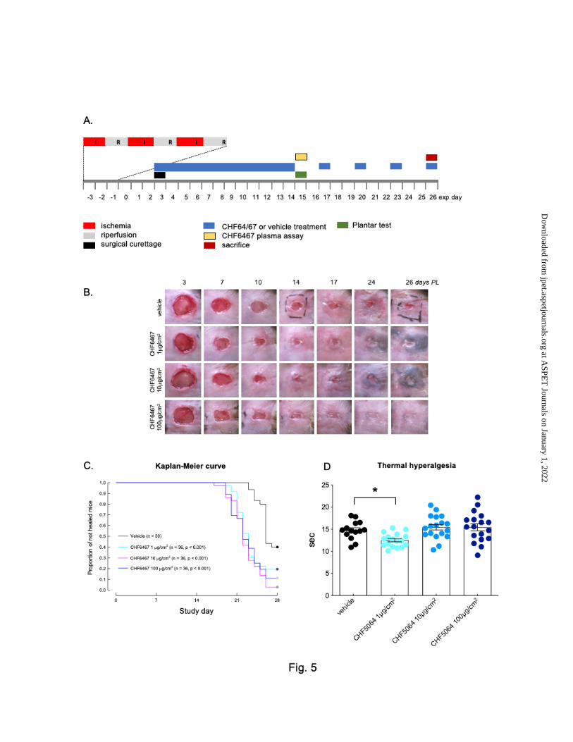
This article has not been copyedited and formatted. The final version may differ from this version.JPET Fast Forward. Published on September 18, 2020 as DOI: 10.1124/jpet.120.000110
at ASPE
T Journals on January 1, 2022
jpet.aspetjournals.orgD
ownloaded from

This article has not been copyedited and formatted. The final version may differ from this version.JPET Fast Forward. Published on September 18, 2020 as DOI: 10.1124/jpet.120.000110
at ASPE
T Journals on January 1, 2022
jpet.aspetjournals.orgD
ownloaded from

This article has not been copyedited and formatted. The final version may differ from this version.JPET Fast Forward. Published on September 18, 2020 as DOI: 10.1124/jpet.120.000110
at ASPE
T Journals on January 1, 2022
jpet.aspetjournals.orgD
ownloaded from

This article has not been copyedited and formatted. The final version may differ from this version.JPET Fast Forward. Published on September 18, 2020 as DOI: 10.1124/jpet.120.000110
at ASPE
T Journals on January 1, 2022
jpet.aspetjournals.orgD
ownloaded from