Embed Size (px)
Citation preview
Topical Antimicrobial and Bandaging Effects on Equine Distal
Limb Wound Healing
Douglass B. Berry, II, DVM
Thesis submitted to the Faulty of the Virginia Polytechnic Institute and State University in partial
fulfillment of the requirements for the degree of
Masters of Science in
Biomedical and Veterinary Sciences
Kenneth E. Sullins, DVM, MS, Chair
Nathaniel A. White II, DVM, MS
Rick D. Howard, DVM, PhD
May 4, 2001
Leesburg, Virginia
Keywords: Antimicrobials, Silver sulfadiazine, Povidone-iodine, Wound Healing, Horses,
Bandaging

ii
Topical Antimicrobial and Bandaging Effects on Equine Distal
Limb Wound Healing by
Douglass B. Berry II, DVM
Committee Chairman: Kenneth E. Sullins, DVM, MS
Professor, Marion duPont Scott Equine Medical Center
Virginia-Maryland Regional College of Veterinary Medicine
(ABSTRACT)
The objective of this study was to determine if topical antimicrobials silver sulfadiazine
and povidone-iodine ointment increase rates of healing of equine distal limb wounds that heal by
second intention. Second, to determine the effect of bandaging with these topical antimicrobials.
Six healthy adult horses were used to create thirty-six, 2.5 cm2 standardized full-thickness
metacarpal/tarsal skin wounds. Each wound was exposed to a single treatment: 1.0 % silver
sulfadiazine cream bandaged (SSD-B), 1.0 % silver sulfadiazine slow release matrix bandaged
(SDX-B), 1.0% silver sulfadiazine slow release matrix not bandaged (SDX-NB), povidone-
iodine ointment bandaged (PI-B), untreated control bandaged (C-B) and untreated control not
bandaged (C-NB) until healing. Wound area, granulation tissue area and perimeter were
measured with planimetry software from digital images obtained at each observation. Exuberant
granulation tissue was excised when present. The days until healing, rate of healing parameter,
rates of contraction and epithelialization were compared among groups using pair-wise analysis
of least square means.
iii
The healing parameters and mean days to healing did not statistically differ between
groups. Analysis of percent wound contraction and rate of epithelialization between groups was
similar. Mean number of days to healing ranged from 83 (PI-B and C-B) to101 (SSD-B). All
bandaged wounds produced exuberant granulation tissue requiring excision compared to none of
the unbandaged. The identified rates of epithelialization and wound contraction found
insignificant differences between antimicrobial treated versus untreated wounds. Similarly, rates
of epithelialization and wound contraction found insignificant differences between bandaged
versus unbandaged wounds. Topical povidone-iodine and silver sulfadiazine did not increase
rates of healing under bandage.
The 1.0% silver sulfadiazine slow release matrix not bandaged (SDX-NB) adhered well
to dry wounds. Silver sulfadiazine slow-release matrix provides does not impede wound healing
and provides good adherence to dry wounds not amenable to bandaging.
iv
Acknowledgements
This project was possible through the support of the Virginia Horse Industry Board.
I would like to thank David Hoberman for statistical assistance, the volunteers and
nursing staff of the Marion duPont Scott Equine Medical Center and my committee
members.
v
TABLE OF CONTENTS PAGE NO.
ACKNOWLEDGEMENTS iv
LIST OF FIGURES & TABLES vi
LIST OF ABBREVIATIONS vi 1.0 INTRODUCTION TO WOUND HEALING AND HORSES 1
1.1. Clinical Incidence 3
1.2. Physiological Differences 3
1.3. Wound Healing Kinetics 6
1.4. Topical Medications 7
2.0 EXPERIMENTAL DESIGN
2.1. Horses 10
2.2. Wound Creation 10
2.3. Assignment of Treatment Groups 11
2.4. Assessment of Healing 12
2.5. Statistical Analysis 13
3.0 EXPERIMENTAL RESULTS
3.1. Subjective Assessment 15
3.2. Days to Healing 15
3.3. Effect of Bandaging 15
3.4. Epithelialization 16
3.5. Contraction 16
4.0 DISCUSSION
4.1. Effect of Topical Antimicrobials 17
4.2. Effect of Exuberant Granulation Tissue 18
4.3. Effect of Bandaging 19
4.4. Clinical Application and Further Research 20
5.0 LITERATURE CITED 21
6.0 VITA 27
vi
List of Figures Page # Figure 1. Mean area of each treatment group versus time 24 Figure 2. Wound Image 25
List of Tables Table 1. Summary of Treatment Groups 11 Table 2. Mean values for wound healing in the six treatment groups 26
List of Abbreviations
Co. Company IU International Units IV intravenous IM intramuscular kg kilogram mg milligram PO per os

1
1.0 Introduction to Wound Healing and Horses Wounds of the distal equine limb are of concern due to loss of performance and potential
loss of commercial value (Cochrane 1997). Compared with wounds of the trunk, distal limb
wounds have a longer preparatory phase characterized by greater wound retraction, slower rates
of epithelialization and earlier cessation of wound contraction (Jacobs et al. 1984, Wilmink et al.
1999a). Anatomic location, specifically, body versus distal limb, contributes to physiologic
differences in organization of the cellular inflammatory response have been found between the
horse and pony to explain some of this phenomenon (Wilmink et al. 1999a, Wilmink et al.
1999b). Recent investigations focus on the roles of epidermal growth factor, fibroblastic growth
factor-Β1 and the matrix metalloproteinases regulation of fibroplasia (Cochrane 1997,
Knottenbelt 1997). These studies suggest that the persistence of inflammation contributes to
longer healing times in the horse compared to the pony. These findings support earlier research,
which demonstrated that horses have inherent dermal and subcutaneous precursors predisposing
them to exuberant granulation tissue formation (Chvapil et al. 1979). Exuberant granulation
tissue delays healing by inhibiting wound contraction and subsequent epithelialization and
eventually resulting in a larger scar (Jacobs et al. 1984, Lees et al. 1989, Fretz et al. 1983).
Where infection contributes to the prolonged lag phase of wound healing, topical antibacterial
therapy may improve the quality and rate of wound healing. Topical medications may alter the
wound environment by antimicrobial activity, corticosteroid-induced reduction of fibroplasia or
promotion of epithelialization (Stashak 1991b). Limited controlled studies have been performed
on horses.
Because wound healing is a dynamic process, a primary goal should be management of
local factors to provide an environment that promotes acceptable wound healing. Requirements
2
for this include microcirculation for oxygenation and nutrition, moisture, increased temperature,
neutral to slightly acidic pH, and a bacterial population less than 105 organisms per gram of tissue
(Wilmink et al. 1999a, Peacock Jr. 1984). A bandage with a topical antimicrobial is a commonly
used means of providing this environment. Silver sulfadiazine cream (1.0%) has been reported
to increase the rate of epithelialization in human burn patients, mice and pigs (Lansdown et al.
1997, Kjolseth et al. 1994, Geronemus et al. 1979) when compared to other topical medications
including povidone-iodine.
Two antimicrobial preparations used commonly in equine wound management are 10%
povidone-iodine ointment and 1% silver sulfadiazine cream. A 1% silver sulfadiazine slow-
release matrix has recently developed that adheres to the wound bed when dry and can remain in
place on unbandaged wounds. The topical application of either antimicrobial provides broad
spectrum of antibacterial and antifungal activity in vitro. In vivo studies evaluating silver
sulfadiazine, neomycin, bacitracin, polymyxin combinations or iodine containing ointments in
murine and porcine models have shown varying degrees of improved wound healing primarily
through increased epithelialization (Kjolseth et al. 1994, Geronemus et al. 1979). The
assessment of these treatments in the equine assessment have not been reported. Controlled
trials to examine the effect of commonly used topical antimicrobials in equine wounds would be
beneficial (Stashak 1991b).
The objective of this study was to evaluate rates of contraction, epithelialization and days
to healing of the equine distal limb wounds treated with either of two topical preparations of
1.0% silver sulfadiazine or 10% povidone iodine compared to untreated controls. The additional
influence of bandaging of with these antimicrobials on granulation tissue formation was
evaluated.
3
1.1 Clinical Incidence
Distal limb wounds are of common concern to the horse owner primarily from temporary
performance restriction and potential loss of commercial value (Cochrane 1997). In a large,
multicentered retrospective study of 122,642 cases presented to nineteen veterinary colleges in a
four-year period, 2% of all cases required wound treatment and 61% of these wounds were
treated with debridement only and healed by second intention (Lindsay 1990). This was because
of significant tissue loss, degree of contamination or both. Distal limb wounds requiring second
intention healing therefore presents a management concern because once the protective dermal
barrier is lost, the remaining tissue may become susceptible to infection (Peacock Jr. 1984,
Stashak 1991b). In another retrospective report of 452 cases, a 52% infection rate occurred in
clean contaminated orthopedic wounds (Macdonald et al. 1994). This degree of morbidity
makes management of distal limb wounds an important clinical concern.
1.2 Physiological Differences
Compared with wounds of the trunk, distal limb wounds have a longer preparatory phase
of second intention healing characterized by greater wound retraction, slower rates of
epithelialization and earlier cessation of wound contraction (Jacobs et al. 1984, Wilmink et al.
1999a). Regional anatomic and physiologic differences in organization of the cellular
inflammatory response explain some of this phenomenon (Wilmink et al. 1999b, Wilmink et al.
1999a). Most recent investigations focus on the roles of epidermal growth factor, fibroblastic
growth factor-Β1 and the matrix metalloproteinases abilities to regulate fibroplasia (Knottenbelt
1997, Cochrane 1997). These studies suggest the persistence of inflammation contributes to the
prolonged healing times between the pony and the horse. This supports earlier research which
4
determined horses have inherent connective tissue precursors, which activate in the dermis and
subcutaneous tissues predisposing them to the formation of exuberant granulation tissue (Chvapil
et al. 1979). The result of uninhibited fibroplasia is exuberant granulation tissue which delays the
healing process by inhibiting epithelialization and contraction subsequently resulting in a larger
granulation epithelium scar (Jacobs et al. 1984, Lees et al. 1989, Fretz et al. 1983).
The goal of wound healing by second intention is to repair the tissue defect through
granulation tissue formation, which allows a matrix for epithelial cell migration. Tissue repair
requires a good blood supply and an adequate inflammatory response, which attracts
macrophages and fibroblasts that are necessary for clearance of debris and initiation of repair.
These cells are supported by the vascular endothelium (Silver 1982).
During the initial response to injury, platelet accumulation provides chemotactic
attraction through platelet derived growth factor. Macrophages and other leukocytes are
activated and infiltrate the wound. This initial inflammatory phase of wound healing including
debridement of necrotic tissue and foreign debris is crucial for initiation of the repair phase.
Once this macrophage directed phagocytosis clears the tissue debris, perivascular pleuropotential
mesenchymal cells differentiate into fibroblasts and myofibroblasts. These cells produce
collagen, protein polysaccharide, and glycoprotein ground a process which reaches a peak
around the fifth day after wounding (Chvapil et al. 1991, Stashak 1991a). If the inflammation
does not subside, prolonged stimulation of these cells will promote continued, and in the case of
the equine leg wound, excessive fibroplasia.
Recent research in horses focused on exogenous cytokine therapy. This used activated
macrophage supernatant to influence the wound environment (Wilson et al. 1996) in an attempt
to inhibit excessive fibroplasia. Though there was an inhibition of the equine fibroblasts in vitro,

5
in ponies little effect was observed when wounds were exposed to this growth factor milieu. To
be effective second intention healing relies heavily upon the acute inflammatory phase following
injury (Chvapil et al. 1991, Knottenbelt 1997). This was recently demonstrated in a comparison
between the histologic aspects of second-intention wound healing in the horse versus the pony
(Wilmink et al. 1999c). In that study, there was a difference between the horse and pony
inflammatory response as measured by presence of leukocytes and the differentiated fibroblast,
the myofibroblast. Furthermore there was reduced and prolonged inflammatory response in the
horse compared to the pony. This finding correlated with the clinical presence of purulent
exudate on the wound surface but the reason for the prolonged inflammation in the horse was
unclear. In a second part of this work (Wilmink et al. 1999c), the researchers determined it was
the degree of organization of myofibroblasts that determined the extent and rate of wound
contraction pony. This result supports the notion that once effective inflammation has occurred,
the repair and maturation processes can initiate rapid and efficient extracellular matrix
reorganization, wound contraction and epithelialization (Stashak 1991b). Persistent stimulation
of the wound that occurs with tissue infection, necrotic tissue, foreign body, movement, and
iatrogenic irritants placed on the wound leads to chronic inflammation that encourages the
development of proliferating granulation tissue and delayed wound healing (Blackford &
Blackford 1995).
During the proliferative phase of wound healing rapid reduction in wound size is due to
contraction of actin containing myofibroblasts. The rates of contraction vary according to wound
location on the horse (Wilmink et al. 1999c). The clinical observation suggests the rate of
healing of equine limb wounds is approximately half that of body wounds (Walton & Neal 1972,
Jacobs et al. 1984). The physiologic explanation of this is not fully understood but is centered on
6
regional inherent differences in wound contraction. Contraction ceases when epithelialization is
complete or when tension in the surrounding skin exceeds the exertion by the myofibroblasts
(Swaim 1980). However, once the myofibroblasts encounter tensile forces which can not be
overcome, epithelialization must be relied upon to cover the remaining granulation bed.
Optimally, second intention wound healing contracts rapidly during the rapid healing phase,
approximately days 14 through 35 in a horse, to minimize amount of epithelialization, which is
often referred to as the scar epithelium (Jacobs et al. 1984). The differences in wound
contraction appears to be determined by local environments such as oxygenation, moisture,
temperature rather than by inherent contraction capacity of the cells (Wilmink et al. 1999b).
1.3 Wound Healing Kinetics
All cutaneous wounds heal by two independent processes, contraction and
epithelialization. Wound contraction reduces the perimeter by centripetal movement of dermis
and epidermis and occurs as long as the contractive forces of the wound myofibroblasts exceed
the reactive forces of the surrounding skin on the wound edge (Lees et al. 1989, Jacobs et al.
1984). Epithelialization is the process that keratocytes proliferate and migrate to cover the
surface of the cutaneous defect. The sum of these processes determines the rate of wound
healing.
Second intention skin healing in horses and occurs in an ordered process and has been
mathematically computed following first-order differential equation (Mcgrath & Simon 1983,
Madison & Gronwall 1992, Schumacher et al. 1992). This process progresses at a rate
proportional to the amount of area remaining to contract in a logarithmic linear rate.
Schumacher deduced that equine lower limb wound contraction could be described by using a bi-

7
exponential mathematically derived rate constant while epithelialization, which is an advancing
process, is described by a monoexponential equation. Madison and Gronwall used a similar
exponential equation and concluded wound contraction is a complex process and may not be
adequately described by simple mathematics equations.
Before these studies, rates of wound healing were derived from a linear regression
equations that analyzed the square root of the area (Snowden 1981, Fretz et al. 1983, Bertone et
al. 1985). The researchers determined a rate of wound healing but not the components of
contraction and epithelialization, therefore, making comparison to later studies difficult. Using
this earlier method, the overall rate of second intention healing in the pony was determined to be
0.56mm/day(is this area or linear) for open surgically created wounds on the dorsal fetlock of
ponies (Fretz et al. 1983). This is similar to limb wounds of humans 0.58mm/day (Snowden
1981). These pony wounds were treated with excision of excessive granulation tissue only and
rate of healing was a constant during the rapid phase of wound closure. Fretz et al. determined
the rate of healing was dependent on the treatment applied to the wound with reference to
bandaging alone. However, there was no effort made to minimize infection other than
prevention of contamination by the varied treatment bandages.
1.4 Topical Medications
In attempts to reduce exuberant granulation tissue formation, application of topical
medications either alter the wound environment by suppression of opportunistic bacterial
infection, reduction of mitotic activity or promotion of epithelialization through fat-soluble
vitamin supplementation. Many topical therapies have been applied to control the inflammatory
8
response leading to delayed healing but limited controlled studies with antimicrobials have been
evaluated on horses.
Topical wound preparations have been studied on the horse to promote increased rates of
wound healing, decrease the incidence of exuberant granulation tissue and produce an optimal
cosmetic result (Bertone et al. 1985, Madison et al. 1991, Blackford et al. 1991, Bigbie et al.
1991, Barber 1989, Wilson et al. 1996, Woollen et al. 1987, Yvorchuk-St. Jean et al. 1995).
Often, this employs the use�of a topical antimicrobial for prevention of wound infection therefore
avoiding systemic antibiotic toxicity and expense. This does require avoidance of local toxicity
or contact dermatitis in the antimicrobial preparation. Among the three published studies using
antimicrobials in horses, two of the three assessed the effect of a topical antimicrobial
combination with the presence of a glucocorticosteroid (Barber 1989, Blackford et al. 1991,
Woollen et al. 1987). Two additional, but not studied, antimicrobial preparations used commonly
in equine wound management are 10% povidone-iodine ointment and 1% silver sulfadiazine
cream or a recently developed slow-release silver sulfadiazine matrix. The topical application of
either antimicrobial provides broad antibacterial and antifungal spectrum in vitro �and is
economical for routine use. The in vivo studies evaluating silver sulfadiazine, neomycin,
bacitracin, polymyxin combinations or iodine containing ointments in murine and porcine
models have shown varying degrees of improved wound healing primarily through increased
epithelialization (Geronemus et al. 1979, Kjolseth et al. 1994) but in vivo assessment of their
effect on healing has not been determined in the horse. Based on the widespread use, controlled
trials to examine the effect of topical antimicrobials on equine wound healing are necessary
(Stashak 1991b).
9
The clinical rationale for topical antimicrobial therapy is suppression of opportunistic
bacterial tissue damage which decreases rates of wound healing thereby increasing morbidity and
cost. Because wound healing is a dynamic process, a primary goal should be management of
local factors to provide an environment that promotes acceptable wound healing. Requirements
for this include a wound environment that provides microcirculation for oxygenation and
nutrition, moisture, increased temperature, neutral to slightly acidic pH, and a bacterial
population less than 105 organisms per gram of tissue (Peacock Jr. 1984, Knottenbelt 1997). A
bandage with a topical antimicrobial is a common means of providing this environment.
Beneficial effects from 1.0% silver sulfadiazine cream have been reported in human burn
patients, mice and pigs (Lansdown et al. 1997, Kjolseth et al. 1994, Geronemus et al. 1979) by
increasing the rate of epithelialization when compared to other topical medications, including
povidone-iodine, and untreated controls.
The experimental hypothesis is to determine if equine distal limb wounds treated with
either 1.0% silver sulfadiazine and bandaging demonstrated increased rates of second-intention
healing pertaining to the equine distal limb
10
2.0 Experimental Design 2.1 Horses
The Institutional Animal Care and Use Committee approved the protocol used in this
study. Six adult geldings (4 Thoroughbreds, 1 Warmblood, 1 Quarter horse) 5 to 11 years (mean
9.2) old and weighing 527 to 645 kg (mean, 593.5) were used. The horses were vaccinated
routinely (Equiloid®, Ft. Dodge Laboratories, Ft. Dodge, IA.) and received ivermectin
(Eqvalan®, Merck & Co. AgVet division, Rahway, NJ.) (0.2 mg/kg of body weight, PO). The
horses were grouped in pairs and housed in covered stalls with an adjoining small dry-lot
paddock. All horses were allowed free choice timothy/orchard grass hay and one kilogram of a
concentrate (Priority 10®, Southern States Co-op, Cooperative Milling, Gettysburg, PA) per day.
2.2 Wound Creation
Each horse received procaine penicillin (Pfi-Pen G®, Pfizer Animal Health, New York,
NY.) (22,000 IU/kg, IM) preoperatively then twice daily and phenylbutazone (phenylbutazone
tablets, Vedco, St. Joseph, MO.) (4.4mg/kg, PO) once daily for 72 hours. Following a 12-hour
fast, each horse was pre-medicated with xylazine hydrochloride (Sedazine®, Ft. Dodge
Laboratories, Ft. Dodge, IA.) (0.5 mg/kg, IV) and guaifenesin (5% IV to effect). Anesthesia was
induced with ketamine hydrochloride (Ketaset®, Ft. Dodge Laboratories, Ft. Dodge, IA.) (2.2
mg/kg IV) and maintained with the intravenous combination of xylazine, ketamine and
guaifenesin (500mg, 2000mg, and 50 grams respectively) in 1 liter of sterile saline administered
to effect.
The dorsomedial metacarpi and metatarsi were aseptically prepared with chlorhexidine
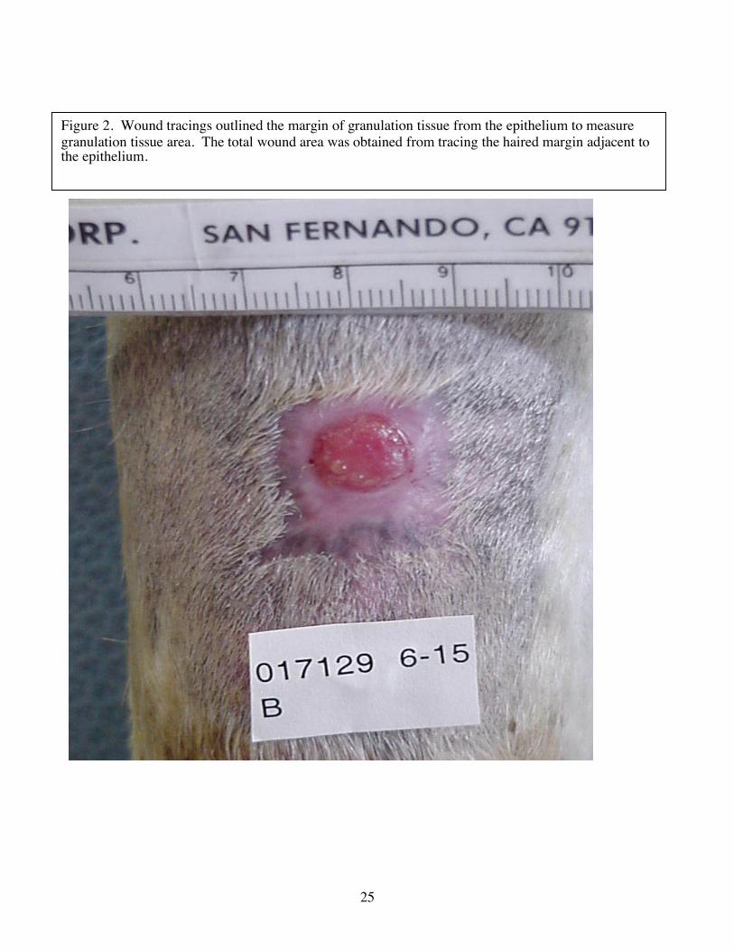
gluconate and water. A 6.25-cm2 full-thickness skin wound was created on each proximal and

11
distal metacarpus and on each proximal metatarsus avoiding the underlying long or common
extensor tendons for a total of six wounds per horse (Figure 2). Following a 2.5-cm flexible
square stainless steel template with a #10 scalpel blade created each standardized wound. The
tissue within the template was excised with scissors to the level of the periosteum. Permanent
tattoo ink was injected intradermally through predrilled holes in the template 10 mm from and
parallel to the margin of the wound (Wilmink et al. 1999a).
2.3 Assignment of Treatment Groups
One of six treatments was administered to each of the wounds on each horse. The
treatments (Table 1) included 1.0 % silver sulfadiazine cream (Silvadene®, Hoechst-Marion-
Roussel, Kansas City, MO.) bandaged (SSD-B), 1.0 % silver sulfadiazine slow release matrix
(Silvadex SR, Buford Biomedical Inc, Frederick, MD.) bandaged (SDX-B), 1.0% silver
sulfadiazine slow release matrix not bandaged (SDX-NB), 10% povidone-iodine ointment
(Biozide® gel, Performance Products Inc, St. Louis, MO.) bandaged (PI-B), untreated control
bandaged (C-B) and untreated control not bandaged (C-NB). Treatments were continued until
complete epithelialization of the wound had occurred. The bandaged wounds were covered with
a rayon-polyethylene non-adherent dressing (Release®, Johnson & Johnson, Arlington, TX.) and
elastic adhesive tape (Elastikon®, Johnson & Johnson, Arlington, TX.)
Table 1. Summary of Treatment Groups Treatment Groups Explanation
All treatments were applied to each horse
Treatment applications rotated between the six locations
SDX-NB silver sulfadiazine slow-release matrix without bandage SSD-B silver sulfadiazine cream under bandage SDX-B silver sulfadiazine slow-release matrix under bandage PI-B povidone-iodine under bandage C-NB untreated control without bandage C-B untreated control without bandage

12
The elastic tape was wrapped until pressure was firm. Bandages were changed and each
wound was photographed on the first postoperative day, every other day for 17 days then every
third day thereafter until the wound had healed. Wound treatments consisted of 500 mg total
weight of each medication applied to the non-adherent pad. The SDX-NB group had equal
amount of preparation applied with a clean tongue depressor and massaged into the wound bed
or eschar. The SDX-NB group was further evaluated on ability of the medication to adhere to
the wound bed.
2.4 Assessment of Wound Healing
Wounds were considered to have healed when visible epithelium covered the wound.
Wounds were considered to have exuberant granulation tissue when the periphery of the
granulation bed was above the surrounding level of the skin or advancing the epithelium. When
exuberant granulation tissue was present, it was excised to the level of the surrounding skin or
migrating epithelium with a # 10 scalpel blade (Howard et al. 1993).
Photographs were taken with a digital camera (Sony Mavica® MVC-FD83, Sony Corp,
Tokyo, Japan.) through a macro lens at a 22-cm focal distance after wiping the wound clean of
any exudate and clipping any hair interfering with identification of the wound margin and prior
to any exuberant granulation tissue excision. A horizontal metric scale was attached just
proximal to the wound to provide a calibration reference. A label was placed below the wound
to identify wound location, date and horse. The total wound area, wound perimeter and
granulation area was determined using planimetry software (NIH Image, version 1.62. Available
at: http://rsb.info.nih.gov/nih-image. May 1999.)

13
Each digital image was displayed on a 40-cm color monitor (Apple Multiple Scan® 17
display, Apple Computer Inc, Cupertino, CA.). Each image was scaled to fit the window before
the program was calibrated to a 1.0 cm distance from the metric scale in the image. The wound
area and perimeter were determined by tracing the hair margin of the wound periphery. The
granulation tissue area was determined by tracing the cursor around the margin between
granulation tissue and epithelium when present. Each wound was measured three times, and the
average of the three was recorded. The epithelial area was calculated by subtracting the
granulation tissue area from the wound area. The area of the epithelium and total wound area
were equal when wound healing was complete.
Wound contraction was expressed as a percentage of the wound area from the maximal
wound size3 following wound creation. The healing parameter describes the linear advance of
the wound margin toward the wound center for comparison of healing rate between differently
shaped wounds. It was calculated by dividing the final wound area by the average of the initial
and final wound perimeter (Gilman 1990). The rate of epithelialization was derived from the
slopes of the linear regression for plots of epithelial area versus time (Jacobs et al. 1984). For
analysis of contraction during the rapid phase from 20 to 62 days (Geronemus et al. 1979), the
natural log of the wound area was taken as the unit of analysis. Linear regression was then used
to identify the rate constant for each treatment group.
2.5 Statistical Analysis.
The treatment groups were randomized in a Latin square design balanced for wound
location on each horse. For each treatment group, Fisher’s square least square means
comparisons for analysis of variance was performed for days to healing, wound healing
14
parameter, rate of epithelialization and rate constant for contraction between groups. For all
analyses, P<0.05 was considered significant. Statistical software (PROC GLM and PROC
REG, SAS Institute Inc, Cary, NC.) was used for all analyses.

15
3.0 Experimental Results 3.1 Subjective Assessment
Horses remained comfortable and did not disturb their wounds. All wounds underwent
linear expansion in the first weeks followed by rapid exponential contraction (Figure 1). Each
retained the original square periphery to slightly stellate with a round, central granulation bed
until complete epithelialization. The wound tattoos were only visible 29 of 36 sites. Therefore,
were not useful in determining calculations. This did not affect the ability to accurately measure
the wounds through out the protocol.
3.2 Days to Healing
Mean days to complete healing, healing rate parameter, number of times granulation
tissue was excised and percent wound healing attributable to contraction, for all treatment groups
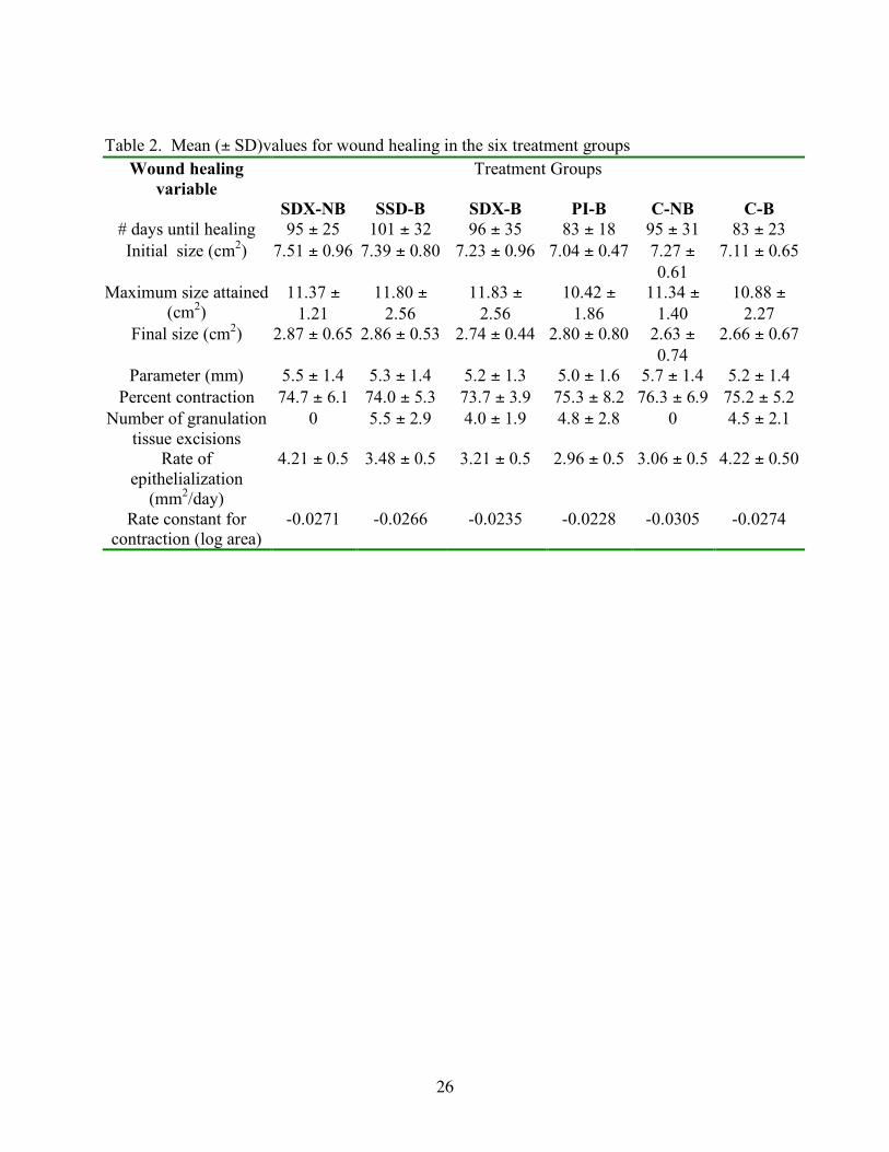
are shown in Table 2. The mean days to complete healing for the treatment groups ranged from
83 days for C-B and PI-B to 101 days for SSD-B with a combined mean for all groups of 92
days. Significant differences were not detected in healing, healing parameter and percent wound
contraction treatment groups and controls. All the wounds of horse 3 healed significantly faster
(mean 57 days) than the remaining five horses (P=0.01). Horse 3 was the smallest of the group;
527 kg vs group mean of 593.5 kg.
3.3 Effect of Bandaging
All bandaged wounds eventually produced exuberant granulation tissue that
required excision. The unbandaged wounds became covered with a dry eschar beginning
14-21 days after wounding and did not produce exuberant granulation tissue. The eschar

16
remained in place and reduced in size as contraction and epithelialization progressed until
it fell off when epithelialization was visibly complete. Among the bandaged wounds, the
mean number of times the exuberant granulation tissue was excised was 5 (range 1-9).
These wounds would remain moist then progress to purulent exudate prior to becoming
exuberant, regardless of treatment. Following exuberant granulation tissue excision,
wound surface appearance returned to moist and clean at the next bandage change.
3.4 Epithelialization
Epithelium was first present in 15 of the 36 wounds by day 7 following wounding. There
were no differences between groups for initiation of epithelialization. The rate of
epithelialization ranged from 2.96+/-0.50 mm2/day for PI-B to 4.21+/-0.50 mm2/day for C-NB.
These calculated differences were not determined to be of sufficient statistically significance
(P=0.09)(power 0.39).
3.5 Contraction
Maximal wound size was attained during the second week after which the wounds in
each group contracted to 75 percent of their maximal size when healed. Significant differences
in wound area were not present in all measurements of wound areas between any treatment
groups. The slope from the exponential regression model from the maximal wound size through
the rapid phase of contraction ending at 62 days post wounding ranged from –0.023 for PI-B to –
0.030 for C-NB.
17
4.0 Discussion
In this study, topical medication when compared to controls did not affect the rate of
healing of the distal limb wounds. The increased rate of epithelialization (Kjolseth et al. 1994,
Geronemus et al. 1979) or decreased rate of wound contraction (Leitch et al. 1993) with 1.0%
silver sulfadiazine found in other species was not observed in these horses.
This discussion of these findings are related to experimental, small wounds with easily
managed wounds. Larger wounds will have variables not present in these small controlled
wounds. Further, larger granulating areas will have more tendency to become exuberant as the
time required to complete contraction is prolonged.
Lastly, the results of this study could not identify a positive or detrimental effect of the
treatments employed. Retrospective evaluation determined the sample size was not sufficient
enough to account for the small differences present in healing rates between groups. Though
trends exist for a difference to be present between PI-B and C-B compared to SSD-B, the small
population size may have contributed in preventing us from disproving the null hypothesis.
4.1 Effect of Topical Antimicrobials
The rationale for application of topical antimicrobials is prophylaxis against and
treatment of infection in compromised skin. These preparations allow high local antimicrobial
efficacy while avoiding systemic toxicity and are most effective in the earlier stages of healing
prior to a solid granulation bed. Our experimental protocol was designed to identified the effect
of medication, particularly 1.0 % silver sulfadiazine, and not designed to maximize the
18
antimicrobial effects of the individual preparations. Which, the proposed application frequency
for burn wounds is twelve hours (Kaye 2000).
Importantly, a negative effect from either 1.0% silver-sulfadiazine or 10% povidone-
iodine medication was absent. We believe the use of either is advisable in the clinical situation
where wounding is less controlled. However, prolonged application of a bandage or either
antimicrobial tested under bandage was associated with exuberant granulation tissue formation.
Therefore, if a bandage were not necessary for wound protection or mechanical stabilization
based on location, the topical use of the silver sulfadiazine slow-release matrix could provide
antimicrobial protection.
4.2 Effect of Exuberant Granulation Tissue
Recurrent exuberant granulation tissue production specific to the bandaged groups has
been reported (Fretz et al. 1983, Howard et al. 1993, Theoret et al. 2001). The reason for the
exuberant granulation tissue formation was not the design of this study and therefore was not
determined. It is proposed that treatments that decrease the ambient oxygen tension stimulate
angiogenesis through a variety of cytokines and growth factors promoting fibroplasia leading to
exuberant granulation tissue (Howard et al. 1993, Knighton et al. 1981, Theoret et al. 2001).
This may have occurred in our study when the three preparations are placed under bandage
simulating a semi-occlusive type dressing. A second observation from this study is the
accumulation of mild fibrinous exudate under these bandages reflective of persistent
inflammation. This proteinaceous covering over developing granulation tissue remains intimately
associated with contain profibrotic mediators encouraging unimpeded growth. This is a direct
result of bandaging, as the unbandaged groups would desiccate into a dry eschar. To best assess
19
the effect of the antimicrobial treatments, this study design implemented unbandaged SDX-NB
and C-NB groups.
4.3 Effect of Bandaging
The bandages were applied for uniform delivery of the treatments without adhering to the
wound sites and providing protection from environmental contamination or secondary trauma.
The bandages did confine the fibrinous exudate to the immediate area of the wound. That effect
appears significantly related to the production of exuberant granulation tissue formation. All
bandaged wounds produced exuberant granulation tissue.
The protocol in this study was patterned from previous reports to closely mimics typical
clinical management of granulating wounds (Howard et al. 1993, Blackford & Blackford 1995)
Subsequently, the every third day bandage change could have permitted enough exudate
accumulation to affect the environment and facilitate exuberant granulation tissue formation
(Fretz et al. 1983, Howard et al. 1993). Further studies designed with similar and varying
bandage change protocols could facilitate understanding the complex intra-cellular
communication differences in wound healing of similar wounds. In comparison with
unbandaged wounds, once the granulation bed was established in an unbandaged wound,
superficial contamination appears to play a minimal role in the course of healing. The absence
of overt infection characterized by reddened granulation tissue and noticeable purulent exudate is
supportive. The conclusion of the effect of bandaging is that the wound healing components of
contraction and epithelialization would be impeded by the continual production of exuberent
granulation tissue. Any medication requiring bandaging will need to consider theses effects
before claiming efficacy.

20
4.4 Clinical Application and Further Research
Further studies should continue to focus on the efficacy of other antimicrobial
preparations for equine distal limb wound healing. The use of silver containing dressings
continue to promise antibacterial properties and a silver-chloride nylon wound dressings may
provide antibacterial activity without the deleterious effects of bandaging previously outlined
(Adams et al. 1999).
From the results of this study we determined the application of either silver sulfadiazine
or povidone-iodine does not improve the healing compared to controls; that may not hold true in
other uncontrolled wounds that are contaminated. In infected wounds, the silver sulfadiazine
slow-release paste may offer convenient antimicrobial action without bandaging.
The lack of benefit from bandaging demonstrated herein cannot be universally applied to
naturally-occurring equine wounds. Bandages are necessary to protect the underlying tissue until
granulation tissue formation provides an opportunity to determine the best course of further
treatment.

21
5.0 Literature Cited
Adams AP, Santschi EM, Mellencamp MA (1999) Antibacterial effects of a silver chloride-coated nylon wound dressing. Veterinary Surgery 28, 219-225.
Barber SM (1989) In American Association of Equine Practicioners, Vol. 35 Boston, pp. 107-
116. Bertone AL, Sullins KE, Stashak TS et al. (1985) Effect of wound location and the use of topical
collagen gel on exuberant granulation tissue formation and wound healing in the horse and pony. Am J Vet Res 46, 1438-44.
Bigbie RB, Schumacher J, Swaim SF et al. (1991) Effects of amnion and live yeast cell
derivative on second-intention healing in horses. Am J Vet Res 52, 1376-82. Blackford JT, Blackford LW (1995) Wound Management. In: The Horse: Diseases and Cinical
Management. Kobluck CN, Ames TR, Goer RJ (eds). W.B. Saunders, Philadelphia, pp. 513-525.
Blackford JT, Blackford LW, Adair HS (1991) In American Association of Equine Practicioners,
Vol. 37, pp. 71-81. Chvapil M, Holubec H, Chvapil T (1991) Inert wound dressing is not desirable. J Surg Res 51,
245-52. Chvapil M, Pfister T, Esclanda S et al. (1979) Dynamics of healing of skin wounds in the horse
as compared with the rat. Exp Mol Pathol 13, 83-90. Cochrane CA (1997) Models in vivo of wound healing in the horse and the role of growth
factors. Veterinary Dermatology 8, 259-272. Fretz PB, Martin GS, Jacobs KA et al. (1983) Treatment of exuberant granulation tissue in the
horse: Evaluation of four methods. Vet Surg 12, 137-140. Geronemus RG, Mertz PM, Eaglstein WH (1979) Wound healing: the effects of topical
antimicrobial agents. Archives of Dermatology 115, 1311-1314. Gilman TH (1990) Parameter for measurement of wound closure. Wounds 2, 95-101. Howard RD, Stashak TS, Baxter GM (1993) Evaluation of occlusive dressings for management
of full-thickness excisional wounds on the distal portion of the limbs of horses. Am J Vet Res 54, 2150-4.
Jacobs KA, Leach DH, Fretz PB et al. (1984) Comparative aspects of the healing of excisional
wounds on the leg and body of horses. Veterinary Surgery 12, 83-90.

22
Kaye ET (2000) Topical Antibacterial Agents. Infectious Disease of North America 14, 321-338. Kjolseth D, Frank JM, Barker JH et al. (1994) Comparison of the effects of commonly used
wound agents on epithelialization and neaovascularization. Journal of the American College of Surgeons 179, 305-312.
Knighton DR, Silver LA, Hunt TK (1981) Regulation of wound-healing angiogenesis-effect of
oxygen gradients and inspired oxygen concentration. Surgery 90, 262-270. Knottenbelt DC (1997) Equine wound management: are there significant differences in healing
at different sites on the body? Veterinary Dermatology 8, 273-290. Lansdown ABG, Sampson B, Laupattarakasem P et al. (1997) Silver aids healing in the sterile
skin wound: experimental studies in the laboratory rat. British Journal of Dermatology 137, 728-735.
Lees MJ, Fietz PB, Bailey JB et al. (1989) Second-intention wound healing. Compendium on
Continuing Education 11, 857-864. Leitch IO, Kucukcelebi A, Robson MC (1993) Inhibition of wound contraction by topical
antimicrobials. Aust N Z J Surg 63, 289-293. Lindsay WA (1990) Principles of Wound Healing. In: Current Practice of Equine Surgery. White
II NA, Moore JN (eds). J.B.Lippincott Company, Philadelphia, pp. 123-151. MacDonald DG, Morley PS, Bailey JV et al. (1994) An examination of the occurrence of
surgical wound infection following equine orthopaedic surgery (1981-1990). Equine Vet J 26, 323-6.
Madison JB, Gronwall RR (1992) Influence of wound shape on wound contraction in horses. Am
J Vet Res 53, 1575-8. Madison JB, Hamir AN, Ehrlich HP et al. (1991) Effects of a proprietary topical medication on
wound healing and collagen deposition in horses. Am J Vet Res 52, 1128-31. McGrath M, Simon R (1983) Wound Geometry and the kinetics of wound contraction. Platic and
Reconstructive Surgery 72, 66-72. Peacock Jr. EE (1984) Wound Repair, WB Saunders, Philadelphia, Schumacher J, Brumbaugh G, Honnas C et al. (1992) Kinetics of healing of grafted and
nongrafted wounds on the distal portion of the forelimbs of horses. Am J. Vet. Res 53, 1568-1574.

23
Silver I (1982) Basic physiology of wound healing in the horse. Equine Veterinary Journal 14, 7-15.
Snowden JM (1981) Wound contraction, A quantitative interpretation.[part 2]. Aust. J. Exp.
Biol. Med.Sci. 59, 203. Stashak TS (1991a) Principles of Wound Healing. In: Equine Wound Management. (eds).
Lea&Febiger, Malern, PA, pp. 11. Stashak TS (1991b) Selected factors that affect wound healing. In: Equine Wound Management.
(eds). Lea & Febiger, Malvern, PA, pp. 22-24, 36-51. Swaim S (1980) Surgery of Traumatized Skin, WB Suanders and Co., Philadelphia, Theoret CL, Barber SM, Moyana TN et al. (2001) Expression of transforming growth factor B1,
B3, and basic fibroblastic growth factor in full-thickness skin wounds of equine limbs and thorax. Vet Surg 30, 269-277.
Walton G, Neal P (1972) Observations on wound healing in the horse. The role of wound
contraction. Equine Vet J 4, 93-97. Wilmink JM, Stolk PW, van Weeren PR et al. (1999a) Differences in second-intention wound
healing between horses and ponies: macroscopic aspect. Equine Vet J 31, 53-60. Wilmink JM, Van Weeren PR, Nederbragt H et al. (1999b) Wound healing by second intention:
contraction capacity of fibroblasts of horses and ponies. Veterinary Surgery 28, 217. Wilmink JM, van Weeren PR, Stolk PW et al. (1999c) Differences in second-intention wound
healing between horses and ponies: histological aspects. Equine Vet J 31, 61-7. Wilson DA, Adelstein EH, Keegan KG et al. (1996) In vitro and in vivo effects of activated
macrophage supernatant on distal limb wounds of ponies. Am J of Vet Res 57, 1220-1224.
Woollen N, DeBowes RM, Leipold HW et al. (1987) In American Association of Equine
Practioners, Vol. 33 New Orleans, pp. 569-576. Yvorchuk-St. Jean K, Gaughan E, St. Jean G et al. (1995) Evaluation of a porous bovine
collagen membrane bandage for management of wounds in horses. American Journal of Veterinary Research 56, 1663-1667.

24
Figure 1. Mean area of each treatment group versus time. The insignificant differences
in area during the first two weeks after wounding, the preparatory phase, were followed by a
uniform contraction phase.
Wound Area
0
200
400
600
800
1000
1200
1400
0 3 7 11 15 20 32 44 50 62 68 80 89 95 101
104
110
116
125
134
146
Days After Wounding
Are
a m
m2
SDX-NBSSD-BSDX-BPI-BC-NBC-B
25
Figure 2. Wound tracings outlined the margin of granulation tissue from the epithelium to measure granulation tissue area. The total wound area was obtained from tracing the haired margin adjacent to the epithelium.
26
Table 2. Mean (± SD)values for wound healing in the six treatment groups
Wound healing variable
Treatment Groups
SDX-NB SSD-B SDX-B PI-B C-NB C-B # days until healing 95 ± 25 101 ± 32 96 ± 35 83 ± 18 95 ± 31 83 ± 23 Initial size (cm2) 7.51 ± 0.96 7.39 ± 0.80 7.23 ± 0.96 7.04 ± 0.47 7.27 ±
0.61 7.11 ± 0.65
Maximum size attained (cm2)
11.37 ± 1.21
11.80 ± 2.56
11.83 ± 2.56
10.42 ± 1.86
11.34 ± 1.40
10.88 ± 2.27
Final size (cm2) 2.87 ± 0.65 2.86 ± 0.53 2.74 ± 0.44 2.80 ± 0.80 2.63 ± 0.74
2.66 ± 0.67
Parameter (mm) 5.5 ± 1.4 5.3 ± 1.4 5.2 ± 1.3 5.0 ± 1.6 5.7 ± 1.4 5.2 ± 1.4 Percent contraction 74.7 ± 6.1 74.0 ± 5.3 73.7 ± 3.9 75.3 ± 8.2 76.3 ± 6.9 75.2 ± 5.2
Number of granulation tissue excisions
0 5.5 ± 2.9 4.0 ± 1.9 4.8 ± 2.8 0 4.5 ± 2.1
Rate of epithelialization
(mm2/day)
4.21 ± 0.5 3.48 ± 0.5 3.21 ± 0.5 2.96 ± 0.5 3.06 ± 0.5 4.22 ± 0.50
Rate constant for contraction (log area)
-0.0271 -0.0266 -0.0235 -0.0228 -0.0305 -0.0274

27
6.0 Curriculum Vitae
Douglass Boone Berry II
Department of Large Animal Clinical Sciences
Virginia-Maryland Regional College of Veterinary Medicine
Phase II, Duckpond Drive (0442), Blacksburg VA 24061
Office (540) 231-4621 Fax (540) 231-1676 [email protected]
I. Education: 9/91-6/95 Auburn University. Doctor of Veterinary Medicine.
8/88-6/91 University of Kentucky. Animal Science major
II. Professional Experience: 7/01-present Clinical Instructor in Large Animal Surgery/Emergency Critical Care. Virginia-
Maryland Regional College of Veterinary Medicine. Blacksburg, Virginia. 7/98-6/01 Resident in Equine Surgery. Marion duPont Scott Equine Medical Center, Virginia-
Maryland Regional College of Veterinary Medicine. Leesburg, Virginia. 7/97-6/98 Clinical Fellowship in Large Animal Medicine and Surgery. Oregon State University College of Veterinary Medicine. Corvallis, Oregon. 10/96-6/97 Resident Veterinarian Castleton Farm. Lexington, Kentucky. Associate of Dr. H. Steve Conboy. Responsible for over 230 Standardbred
broodmares and their offspring, stallion collection and processing of semen and elective minor surgical procedures.
7/95-9/96 Resident Veterinarian Walmac International. Lexington, Kentucky.
Resident veterinarian for a large Thoroughbred breeding farm. Responsible for broodmare, stallions and offspring overall herd health program. Duties involved; foaling oversight, broodmare obstetrics, sales preparation, and maintenance of 20 two-year-olds in training.
28
III. Veterinary Related Responsibilities: 10/00 Paddock Official, Morven Park Steeplechase, Leesburg, VA.
4/00 Chief Veterinary Official, Fair Hill Advanced Horse Trials at Menfelt, Frederick, MD.
9/99 Chief Veterinary Official, Fair Hill Advanced Horse Trials at Menfelt, Frederick, MD.
4/99 Chief Veterinary Official, Fair Hill Advanced Horse Trials at Menfelt, Frederick, MD.
10/98 Course Official, Morven Park Steeplechase, Leesburg, VA. IV. Research Funded:
2000 Topical Antimicrobial Effects on Equine Wound Healing Principal Investigators, D.B. Berry II, K.E. Sullins
Funded July 2000 by Virginia Horse Industry Board $4,330 Completed May 2001.
1997 Age Dependence of Horses for Immunization for Viral Infectious Disease. Principal Investigator, T. Chambers PhD., Co-investigators include D.B. Berry Funded April 1997 by A.Q.H.A. Research $12,130. Completed April 1998.
V. Refereed Publications:
Berry II DB, Lyons ET, Drudge H, et al. Observations in Horses on the Effects of Ivermectin Treatment on Strongyle Egg Production and Larval Development. Journal of the Helminthological. Society of Washington. 1993;60(1):89-92.
VII. Proceedings:
Holland RE, Conboy HS, Berry II DB et al. Age dependence of foal vaccination for equine influenza. Proceedings of the Eighth International Conference on Equine Infectious Diseases. 1998; pg.190.
Conboy HS, Berry II DB, Chambers T. and Holland RB. Failure of Foal Seroconversion Following
Equine Influenza Vaccination. Proceedings of the 43rd Annual Convention of the A.A.E.P. 1997; pg. 22-23.
VIII. Abstracts:
Berry II DB and Sullins, KE. Topical Antimicrobial and Bandaging Effects on Equine Distal Limb
Wound Healing. Proceeding of the 11th Annual A.C.V.S. Veterinary Symposium. 2001 (accepted)

29
IX. Academic Honors: 1995 Lloyds of London Scholarship recipient. Auburn University 1994 Dean's List. Auburn University 1994 East Alabama V.M.A. Leadership Award recipient. Auburn University 1991 Oswald Research and Creativity Program award recipient. University of Kentucky 1988 Wayland Rhodes Scholarship recipient. University of Kentucky X. Teaching Experience:
A. Professional Curriculum OSUCVM
VM 724 Large Animal Surgery 1998 2 lecture hours
XI. Graduate School Presentations:
2000 Mechanisms Contributing to Chronic Wound Healing. Fall seminar
Collateral Ligament Desmitis of the Equine Tarsus (7 cases). Case series
1999 Peri-operative Analgesia for Equine Orthopedic Surgery. Fall seminar
Equine Wound Contraction and Epithelialization. Spring seminar
Surgical Management of Intra-Uterine Cysts in a Mare. Case report
1998 Equine Wound Physiology. Fall seminar
Thoracic Vertebral Compression Fracture in a Foal. Case report
XII. Affiliations and Professional Associations: American Veterinary Medical Association American Association of Equine Practitioners American College of Veterinary Surgeons
Wound Healing Society
XIII. Continuing Education: Association for the Study of Internal Fixation. Equine Basic Internal Fixation Course. 1999 American Veterinary Medical Association Annual Convention. 1997 American Association of Equine Practitioners Annual Convention. 1996 Mid-American Veterinary Convention. 1995 Ultrasound Short-Course by Dr. Norman Rantanen. 1995 XIV. State Veterinary License: Kentucky, Maryland