Embed Size (px)
Citation preview

Journal of the Korean Society of Dyers and Finishers
Vol. 19, No. 5, pp. 30~36(2007. 10)
256 / www.ksdf.or.kr
〈Research Paper〉
Effects of Protective Colloids on the Formation of Polyurea Microcapsules
Eungmin Lee1, Heain Kim and Soomin Park*
1Daewoo International/ISM, Kangseo-Gu, Busan, 1746-1, Korea
Organic Material System Engineering, Pusan National University, Busan, 609-735, Korea
(Received: August 27, 2007/Revised: September 11, 2007/Accepted: October 17, 2007)
Abstract― Cypermethrin-containing polyurea microcapsules were prepared by interfacial polymerization using
aromatic 2,4-toluene diisocyanate(TDI) and Ethylene diamine(EDA) as wall forming materials. The effects of the
protective colloids of polyvinylalcohol(PVA) and gelatin were investigated through experimentation. The mean
size of the polyurea microcapsules was smaller and the surface morphology of the PVA was much smoother than
gelatin. In addition the release behavior was much more controlled and better sustained. As the concentration of
protective colloid increased, the wall membrane of the polyurea microcapsules became more stable, the thermal
stability of the wall membrane increased, the mean particle size became smaller, and the particle distribution was
more uniform. The release behavior of the core material changed according to the concentration. As the gelatin
concentration was increased, a more controlled and sustained release behavior was observed. However, in the
case of PVA, the increase of PVA concentration lead to a more rapid release rate.
Keywords: microcapsule, polyurea, PVA, gelatin, release behavior
1. Introduction
Polymeric microcapsules are under wide
investigation for practical uses such as pharma-
ceutical dosages, fragrant materials1), pesticides,
ink, and so on. Cypermethrin is a kind of
pesticide2) which has a variety of applications.
It controls a wide spectrum of insects, parti-
cularly Lepidoptera in cereals, citrus, forestry,
soybeans, tomatoes, fruits, vegetables, vines,
tobacco, cotton, coffee and other crops. It is
yellowish brown, viscous, and semi-solid at
ambient temperature and is highly soluble in
an organic solvent such as cyclohexane.
Microencapsulation methods can be divided
into three major categories : a physical method,
a phase separation method, and an interfacial
reaction method. Among these methods, the
interfacial reaction method is the most feasible
and facile method for introducing immiscible
oil-water interfaces. This method can which
affect the physical and chemical properties of
*Corresponding author. Tel: +82-51-510-2412; Fax: +82-51-512-8175; e-mail: [email protected]
the final microcapsules3-7)
.
During a microencapsulation process that uses
the interfacial reaction method, monomers in
the saving oil and water phase diffuse into
the oil-water interface where they react with
each other to form a polymer membrane8-10).
In general, O/W emulsion is more useful in
forming microcapsules containing oily active
materials as their core, and it can be achieved
by mechanically dispersing the oil phase into
water continuous phase11). When O/W emulsion
droplets are formed, the external water-soluble
monomers diffuse across the water phase and
react with the internal oil-soluble monomer to
form a polymer membrane. As the reaction pro-
gresses, the membrane gradually strengthens
and thickens to form a hard wall.
So the final properties of microcapsules greatly
depend on the stability of o/w emulsions to
stand mechanical agitation, the membrane of
the droplets breaking up, and aggregations taking

Effects of Protective Colloids on the Formation of Polyurea Microcapsules 31 KSDF
J. of the Korean Soc. of Dyers and Finishers, Vol. 19, No. 5(2007.10) / 257
place between the membranes of the respective
droplets.
In our research, we focused on the microen-
capsulation of pesticide oils and cypermethrin.
The interfacial reaction based on the polyme-
rization of TDI and EDA was also investi-
gated to evaluate the influences of the pro-
tective colloids on microencapsulation.
2. Experimental
2.1 Materials
The monomers used in this experiment to
form the microcapsule wall in the oil and
water phase were ethylenediamine(EDA) and
2,4-toluene-diisocyanate(TDI). Both were purchased
from Junsei chemicals Co., Ltd., Japan. Cyper-
methrin was obtained from LG chemical Co.,
Ltd., Korea, and its solvent, cyclohexane, was
purchased from Junsei chemicals Co., Ltd.,
Japan. Finally polyvinylalcohol(PVA, DP ; 500)
was purchased from Yakuri pure chemicals
Co., Ltd., Japan. and they were used as pro-
tective colloids. All the purchased chemicals
and the reagents were used without any
further purification.
2.2 Preparation of the microcapsules
Polyurea microcapsules were formed by carrying
out an interfacial polymerization reaction in
an O/W emulsion between TDI dissolved in
cyclohexane and EDA dissolved in water. The
oil phase formed the dispersed phase. All
reactions took place in ambient temperature.
An organic solution, with 8.6g of TDI in
cyclohexane and dissolved 0.05mol of EDA in
distilled water, was used as wall-forming
materials. In addition 0.2g of cypermethrin
was used as the core material, and PVA and
gelatin in different concentrations were used
as the protective colloid. The O/W emulsion
was formed by adding the organic solution
200ml aqueous solution containing protective
colloid, and then the mixture was stirred for
three minutes. The EDA solution was then
added the O/W emulsion after stirring for
fifteen minutes to prevent agglomeration between
the microcapsules. The temperature was increased
to 70℃ to facilitate the reaction and was main-
tained at 70℃ for three hours. The microcapsule
suspension was cooled and washed with 10%
ethanol to remove the unreacted isocyanates
on the surface, and then filtered and dried in
a vacuum oven at 25℃ for twenty four hours.
2.3 Characterization of the Microcapsule
The infrared spectra of the core material
and microcapsules was observed using a com-
puterized Nicolet Impact 400D Fourier trans-
form infrared(FTIR) spectrophotometer. A thermo-
gravimetric analysis(TGA) was carried out on
a DSC Mettler TA-3000(Thermal Science PL-STA).
Each sample was heated at the rate of 10℃
/min up to 500℃ under a constant N2 flow.
The mean particle size and distribution of the
microcapsules were measured with a particle
analyzer (Galai CIS-100, Galai Production Ltd.,
Israel). The shape and morphology of the micro-
capsules were examined using a scanning
electron microscope(SEM, Hitachi S-4200, Japan).
Finally the release behavior of the micro-
capsules was observed with a UV-Vis spectro-
photometer(UV-1601, Japan).
3. Results and Discussion
3.1 FT-IR Spectra
The FTIR spectra of pure TDI as the wall
forming material, with cypermethrin as the
core material and the microcapsules, are pre-
sented in Fig. 1. As seen in Fig. 1, the spectra
show adsorption bands at 3400cm-1 for the
N-H stretching, and 1700cm-1
for the C=O
stretching of in the urea formation. The IR
spectra also indicates a completion of the reaction
of TDI with EDA by a disappearance of
2260cm-1 of NCO by pure TDI, and the appearance
of NH absorption band. In particular, the
over CH stretching vibration is shown at
2900cm-1
for the aliphatic methylene group of

KSDF 32 Eungmin Lee⋅Heain Kim⋅Soomin Park
258 / www.ksdf.or.kr
Wave numbers (cm-1)
1000200030004000
(a)
(b)
(C)
Fig. 1. FT-IR spectra of pure TDI⒜, cypermethrin⒝, and polyurea microcapsules⒞.
diamines and C=O adsorption bands.
Therefore it can be confirmed that the core
and wall materials of the microcapsules con-
sisted of polyurea and cypermethrin.
3.2 Particle Size Distribution
Fig. 2 and 3 show the mean particle size of
polyurea microcapsules containing cyper-
methrin in the presence of a protective
colloids solution from different concentrations
of gelatin solution and PVA. As shown in
Figure 2, when the concentration of gelatin is
increased from 0.1% to 2%, the mean particle
size of the microcapsules is uneven. That is to
say, in this case the particle size of the thinner
(0.1, 0.5%) side of the gelatin appears smaller
than the thick (1.0, 2.0%) side, and part-
icularly in a solution of 0.5% there appears
the smallest mean particle size. This is related
to the stability of the emulsion globules and
the initial membrane in the beginning of the
reaction. In the case of a thinner solution,
when the initial membrane was formed using
polyurea, the stability of the membrane was
so weak that it couldn't bear the mechanical
stirring and finally the membrane broke into
pieces or aggregation took place between the
emulsion globules. So, the smaller particles
might have been membrane fragments broken
by the stirring.
In Fig. 3, according to the concentration of
PVA, the mean particle size of polyurea micro-
capsules decreased from 7.15 to 5.68㎛ gradually.
This is also related to the fact that, in addition
to what was explained above, PVA is more
suitable to make small particles than gelatin.
A lot of hydroxy groups of PVA interact
with H2O and these hydrogen bonds affect a
rapid reaction with NCO and NH2 to decrease
the interaction between unbounded water and
NCO. The initial membrane is more stable
and stronger. So consequently smaller and
more uniform particles can be formed rather
than those formed using gelatin.
The particle size distribution of polyurea
microcapsules prepared using 0.2% and 4.0% PVA
Gelatin conc.(%)
1 2
Mea
n pa
rtic
le s
ize
(um
)
11
12
13
14
15
16
17
18
19
20
Fig. 2. Mean particles size of polyurea microcapsules prepared with different gelatin concentrations.
PVA conc. (%)
0 1 2 3 4
Mea
n pa
rtcl
e si
ze ( μ
m)
5
6
7
8
Fig. 3. Mean particles size changes of polyurea microcapsules prepared with different PVA con-centrations.
Effects of Protective Colloids on the Formation of Polyurea Microcapsules 33 KSDF
J. of the Korean Soc. of Dyers and Finishers, Vol. 19, No. 5(2007.10) / 259
solutions are shown in Fig. 4. As the concen-
tration of PVA in the solution increased, the
size distribution became narrower. This is caused
by the fact that the addition of a high concen-
tration of PVA emulsion resulted in strong
membrane walls. This was due to a rapid
polycondensation caused by a lot of hydroxyl
groups which protected the interaction between
unbounded water and NCO.
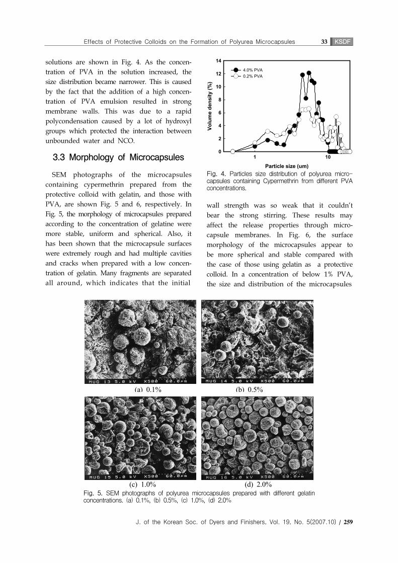
3.3 Morphology of Microcapsules
SEM photographs of the microcapsules
containing cypermethrin prepared from the
protective colloid with gelatin, and those with
PVA, are shown Fig. 5 and 6, respectively. In
Fig. 5, the morphology of microcapsules prepared
according to the concentration of gelatine were
more stable, uniform and spherical. Also, it
has been shown that the microcapsule surfaces
were extremely rough and had multiple cavities
and cracks when prepared with a low concen-
tration of gelatin. Many fragments are separated
all around, which indicates that the initial
(a) 0.1% (b) 0.5%
(c) 1.0% (d) 2.0%Fig. 5. SEM photographs of polyurea microcapsules prepared with different gelatin concentrations. (a) 0.1%, (b) 0.5%, (c) 1.0%, (d) 2.0%
Particle size (um)
1 10
Volu
me
dens
ity (%
)
0
2
4
6
8
10
12
14
4.0% PVA0.2% PVA
Fig. 4. Particles size distribution of polyurea micro-capsules containing Cypermethrin from different PVA concentrations.
wall strength was so weak that it couldn't
bear the strong stirring. These results may
affect the release properties through micro-
capsule membranes. In Fig. 6, the surface
morphology of the microcapsules appear to
be more spherical and stable compared with
the case of those using gelatin as a protective
colloid. In a concentration of below 1% PVA,
the size and distribution of the microcapsules

KSDF 34 Eungmin Lee⋅Heain Kim⋅Soomin Park
260 / www.ksdf.or.kr
(a) 0.2% (b) 0.5%
(c) 2.0% (d) 4.0%Fig. 6. SEM photographs of polyurea microcapsules prepared with different PVA concentrations. (a) 0.2%, (b) 0.5%, (c) 2.0%, (d) 4.0%
were not uniform, namely the bigger micro-
capsules and the smaller microcapsules were
mixed together. This can support the previous
results for the particle size distribution. In
other words, as the PVA concentration becomes
higher, the particle size distribution becomes
more uniform.
3.4 Thermal properties
TGA diagrams show the thermal stability of
the microcapsules prepared from gelatin and
PVA. Fig. 7 and 8 show the TGA diagrams
of the microcapsules prepared from the two
different protective colloids using gelatin and
PVA, respectively. All samples showed that
the first weight loss of about 80% occurred in
the temperature range from 220℃ to 340℃, and
the second weight loss of 7 to 10% occurred,
in the concentration of gelatin, up to 500℃.
The first weight loss was considered to be
due to the melting of the polymer wall
followed by the decomposition of the cyper-
methrin core materials. The residual weight of
each sample became greater with the increase
in the concentration of gelatin. This is related to
the membrane structure and physical properties.
As the concentration of gelatin increased the
portion of CONH in gelatin and OH in PVA,
which interacted with the free water, became
greater, As s result this increases the frequency
of the reaction rate between -NCO and -NH2
to form urea linkages during microencapsulation.
This leads to the formation a stronger mem-
brane structure, resulting in thermal resistance.
These diagrams also indicate that the residual
weight of the microcapsules prepared using
gelatin is relatively higher than those prepared
using PVA. It is supposed that the micro-
capsule with gelatin contains a relatively small
amount of liquid core material (the weight loss
is the same as in the case of the concen-
tration of gelatin) and lots of fragments from
breakage due to the weak formation of the
initial membrane. In order words, the liquid/
solid ratio becomes lower and the resultant
residual weight was much slighter in TGA.

Effects of Protective Colloids on the Formation of Polyurea Microcapsules 35 KSDF
J. of the Korean Soc. of Dyers and Finishers, Vol. 19, No. 5(2007.10) / 261
Temperature (oC)
100 200 300 400 500
Res
idua
l wei
ght (
%)
0
20
40
60
80
1000.1% gelatin0.5% gelatin1.0% gelatin2.0% gelatin
Fig. 7. TGA diagrams of polyurea microcapsules prepared with from different gelatin concentrations.
3.5 Release Properties
Release properties were investigated by releasing
relative amounts of cypermethrin through a
dialysis tube using a UV-visible spectrophoto-
meter. 7mg of cypermethrin loaded polyurea
microcapsules were suspended in 10ml of
hexane and then put into the dialysis tube
(Seamless Cellulose Tubing, Visking Company,
Japan). The dialysis tube was placed into a
250ml bottle with 100ml of hexane, and the
media was left while the temperature was
kept constantly at 50℃. At specific time intervals,
a sample was taken from each media, and the
released amount of cypermethrin was determined
by the UV-visible spectrophotometer at 250nm.
Fig. 9 shows the release behavior of the
polyurea microcapsules containing cypermethrin
formed using a gelatin solution for a protective
Tme (hr)
0 5 10 15 20 25
Rel
ease
d C
yper
met
hrin
(%)
0
20
40
60
80
100
0.1% gelatin0.5% gelatin1.0% gelatin2.0% gelatin
Fig. 9. Effect of gelatin concentration on the release rate of Cypermethrin from polyurea microcapsules.
Temperature (oC)
100 200 300 400 500
Res
idua
l wei
ght (
%)
0
20
40
60
80
1000.2% PVA 0.5% PVA2.0% PVA 4.0% PVA
Fig. 8. TGA diagrams of polyurea microcapsules prepared with different PVA concentrations.
colloid by polycondensation. It shows, as assumed
from the results of the SEM photographs and
examinations, that the release behavior through
the microcapsules, membrane becomes more
controlled and sustained with the increase of
the gelatin concentration. These are related to
the pores or cracks on the membrane surface.
The microcapsules are unstable in a concen-
tration of under 1.0% gelatin because these
cause the formation of pores or cracks on the
membrane's surface. Therefore the apparent
faster release rate in the beginning is due to
the cypermethrin diffusing through the holes.
The release behavior of microcapsules prepared
with a PVA solution is shown in Fig. 10.
These microcapsules , however, are in reverse
order to those in the gelatin solution. As
shown in the release profile, the release rate
through the microcapsule's membrane, according
Time(hr)0 20 40 60 80 100 120 140 160
% C
yper
met
hrin
rele
ased
0
20
40
60
80
100
0.2% PVA 0.5% PVA 2.0% PVA4.0% PVA
Fig. 10 . Effect of PVA concentration on the release rate of Cypermethrin from polyurea microcapsules.

KSDF 36 Eungmin Lee⋅Heain Kim⋅Soomin Park
262 / www.ksdf.or.kr
to time, becomes faster in relation to the con-
centration of PVA. This is related with the
particle size properties, namely in a high con-
centration, as the mean particle size is smaller
the distribution is more uniform. Thus the core
material could be easily released due to a
higher specific area. In opposition to low con-
centration, the broad size distribution and some-
what large particles decreased the release rate
by their relatively lower specific area.
It is confirmed that the wall membranes pre-
pared using a PVA solution for the protective
colloid showed a more controlled and sustained
release behavior. This may be due to the
stability and strength of the internal structure
as explained before.
4. Conclusions
Cypermethrin-containing polyurea micro-
capsules were prepared using interfacial poly-
merization with TDI and EDA, and their chara-
cterizations were investigated to evaluate the
effects of the protective colloid. The mean
particle size of polyurea microcapsules was
smaller and the surface morphology was much
smoother when PVA was used rather than
gelatin. In addition, the release behavior was
much more controlled and better sustained.
From the results of TGA and SEM it was
abserved that polyurea microcapsules prepared
using a protective colloid from PVA had a
better particle size distribution, morphology
and a longer release property compared to
those prepared using gelatin.
Reference
1. K. Hong, S. Park, Melamine Resin Micro-
capsules Containing Fragrant Oil, Materials
Chemistry and Physics, 58(2), 128-131(1999).
2. K. Hirech, S. Payan, G. Carnelle, L. Brujes
and J. Legrand, Microencapsulation of an
insecticide by interfacial polymerisation,
Powder Technology, 130, 324-330(2003).
3. Peihong Ni, Mingzu, Nianxi Yan, Effect of
operating variables and monomers on the
formation of polyurea microcapsules, Journal
of Membrane Science, 103, 51-55(1995).
4. S. K. Yadav, Kartic C. Khilar and A. K.
Suresh, Release rates from semi-crystalline
polymer microcapsules formed by interfacial
polycondensation, Journal of Membrane Science,
125(2), 213-218(1997).
5. D. Saihi, I. Vroman, S. Giraud and S. Bour-
bigot, Microencapsulation of ammonium
phosphate with a polyurethane shell part I:
Coacervation technique, Reactive and Functional
Polymers, 64(3), 127-138(2005).
6. Nihal Sarier and Emel Onder, The manu-
facture of microencapsulated phase change
materials suitable for the design of ther-
mally enhanced fabrics, Thermochimica Acta,
452(2), 149-160(2007).
7. J. Kubota, N. Hirabayashi, A. Kato and H.
Yoshizawa, Microreactor utilized preparation
of monodispersed polymeric microcapsules
by urea/formaldehyde phase separation
method, Colloids and Surfaces A: Physicochemical
and Engineering Aspects, 302, 320-325(2007).
8. K. Hong, S. Park, Preparation and Charate-
rization of Polyurea Microcapsules with
Different Diamines, Materials Research Bulletin,
34(6), 963-969(1999).
9. K. Hong, S. Park, Preparation of Polyurea
Microcapsules with Different Composition
Ratios, Materials Science and Engineering, A272(2),
418-421(1999).
10. K. Hong, S. Park, Morphologies and Release
Behavior of Polyurea Microcapsules from
Different Polyisocyanates, Journal of Materials
Science, 34(1), 3161-3164(1999).
11. S. K. Yadav, K. C. Khilar, A. K. Suresh,
Preparation of Polyurea Microcapsules con-
taining Ovalbumin, AIChE Journal, 42(9), 2616-
2626(1996).