Embed Size (px)
Citation preview

/. exp. Biol. 158, 181-198 (1991) 181Printed in Great Britain © The Company of Biologists Limited 1991
EFFECTS OF EXERCISE, HYPOXIA AND FEEDING ON THEGASTROINTESTINAL BLOOD FLOW IN THE ATLANTIC
COD GADUS MORHUA
BY MICHAEL AXELSSON AND REGINA FRTTSCHE*
Comparative Neuroscience Unit, Department of Zoophysiology, University ofGoteborg, PO Box 25059, S-4O0 31 Goteborg, Sweden
Accepted 26 March 1991
Summary
Cardiac output, ventral and dorsal aortic blood pressure, heart rate, and coeliacand mesenteric artery blood flow were recorded simultaneously in the Atlanticcod, Gadus morhua L., at rest, during exercise, during hypoxia and after feeding.
In the resting unfed animals, coeliac artery blood flow was 4.1±0.8mJmin~1kg~1 and mesenteric artery blood flow was 3.5±l.lmlmin~1kg~1 (mean±S.E.M., 7V=10); together, these flows represent approximately 40% of the cardiacoutput.
Exercise or exposure to hypoxia resulted in increased visceral vascularresistance, leading to reductions in the coeliac and mesenteric artery blood flows.
Coeliac and mesenteric blood flows were increased 24 h after feeding and thecoeliac and systemic vascular resistances decreased in comparison with theprefeeding values. Phentolamine did not affect the gastrointestinal artery bloodflow, but produced a significant decrease in the mesenteric and systemic vascularresistance.
Treatment with bretylium and phentolamine revealed differences between thecoeliac and the mesenteric vasculature regarding the control mechanisms duringhypoxia and during exercise and feeding. During hypoxia, an adrenergic control ofthe gastrointestinal vasculature with both nervous and humoral components wasfound, whereas during exercise and after feeding an additional non-adrenergicmechanism controlling gut blood flow was demonstrated.
Introduction
The general mechanisms involved in the control of the heart and vasculature infish in vivo are reasonably well understood (for references, see Holeton andRandall, 1967; Daxboeck and Holeton, 1978; Randall and Daxboeck, 1982;Axelsson and Nilsson, 1986; Gehrke and Fielder, 1988; Fritsche and Nilsson,1990), but there are still relatively few reports on the regional distribution of the
*To whom reprint requests should be sent.
words: Gadus morhua, teleost, gastrointestinal blood flow, exercise, hypoxia, feeding.

182 M. AXELSSON AND R. FRITSCHE
cardiac output or the control mechanisms involved during exercise and hypoxia(Laurent et al. 1983; Nilsson, 1983; Farrell, 1984).
Regulation of the total systemic vascular resistance in the Atlantic cod (Cadusmorhua) and rainbow trout (Oncorhynchus mykiss [=Salmo gairdneri]) is me-diated via tonically active adrenergic nerves, with little or no contribution fromcirculating catecholamines (Smith, 1978; Smith et al. 1985; Axelsson and Nilsson,1986). During exercise, cardiac output increases as a result of an increase in bothheart rate and stroke volume, while the systemic resistance either decreases(rainbow trout) or remains virtually unchanged (Atlantic cod) (Kiceniuk andJones, 1977; Daxboeck, 1981; Randall and Daxboeck, 1982; Axelsson and Nilsson,1986). Hypoxic exposure, however, induces an elevation of the total systemicresistance in the Atlantic cod without any change in cardiac output (Fritsche andNilsson, 1989). The change in systemic vascular resistance during exercise andhypoxia is mediated via adrenergic nerves and, during hypoxia, also to a certainextent via circulating catecholamines (Kiceniuk and Jones, 1977; Randall andDaxboeck, 1982; Axelsson and Nilsson, 1986; Fritsche and Nilsson, 1990).
The systemic vascular circuit consists of at least two distinct parts: the visceraland the somatic circulation. The reactions of the visceral circulation duringexercise and hypoxia, or after feeding, have not been studied extensively in fish.There are some indications of a redistribution of cardiac output away from the gutvasculature during exercise in the rainbow trout (Daxboeck, 1981). Changes in thegastrointestinal vascular resistance may also affect the systemic blood pressure infish, as suggested in the study by Axelsson et al. (1989) on the sea raven(Hemitripterus americanus).
In mammals, the diffuse adrenergic sympathetic nervous discharge seen duringexercise increases the vascular resistance in the gastrointestinal circulation, whilethe local metabolically induced vasodilatation in the working muscles is unaffectedby this discharge (Orwell et al. 1964; Donald, 1980). Therefore, the fraction of thecardiac output reaching the gut is reduced during exercise (Rushmer et al. 1961;Vatner et al. 1971).
Feeding has been shown to cause an increased gastrointestinal blood flow inmammals, but whether this is due to an increased cardiac output or a selectiveincrease in visceral blood flow is not clear (for a discussion, see Fara, 1984). In thesea raven (Hemitripterus americanus) it was shown that the coeliac artery bloodflow increased as a result of feeding and that visceral vascular resistance was, atleast in part, controlled by an cr-adrenoceptor-mediated mechanism (Axelssonetal 1989).
The control of the visceral blood flow and resistance, at least in mammals, ismediated largely by noradrenergic nerves. However, the rapidly increasingnumbers of reported non-adrenergic, non-cholinergic (NANC) neurotransmittersand hormones of the gut raise the question of a control of the gut circulation bynon-adrenergic agents, e.g. neuropeptides (Nilsson and Holmgren, 1989).
The present study has the following major objectives: first, to measure thegastrointestinal blood flow in resting, unrestrained fish and to determine the r o f l

Gastrointestinal blood flow in the cod 183
of circulating catecholamines and adrenergic nerves in the control of gut bloodflow; and second, to study the effects of exercise, hypoxia and feeding on visceralblood flow.
Materials and methodsThe study was carried out on Atlantic cod, Gadus morhua L., of both sexes,
with a body mass of 550-1050 g and a body length of 38-45 cm. The fish were keptin well-aerated, recirculating sea water at 10-11° C. The animals were acclimatedto the departmental seawater system for 1 week before surgery and used within 2weeks of capture. The fish were never fed in captivity. The exercise and feedingexperiments were performed in August-November 1989 and the hypoxia exper-iments in May-June 1990.
Surgical and preparative procedures
The fish were anaesthetized in MS 222 (tricaine methane sulphonate; 100 mg I"1,Sigma), until breathing movements ceased, and transferred to an operating table,where aerated sea water containing the anaesthetic (50 mg I"1) was passed over thegills throughout the surgery. A cannula (PE50) filled with heparinized(lOOi.u. mP1) 0.9% NaCl was inserted into the afferent branchial artery of thethird gill arch for measurement of ventral aortic blood pressure (PVA) and heartrate (fii), and for taking blood samples. A second cannula was occlusively insertedinto the efferent branchial artery of the same gill arch for measurement of dorsalaortic pressure (PDA) and injection of adrenergic antagonists. Both cannulae weresecured with skin sutures.
In order to measure cardiac output (Q=ventral aortic blood flow), the ventralaorta was exposed via an incision immediately anterior to the base of the pectoralfins. The ventral aorta was carefully dissected free from the surrounding tissuewithout rupturing the pericardium, and a cuff-type Doppler flow probe(2.5-3.0 mm i.d., single crystal, P. Pohl International Inc.) was placed around theventral aorta.
In the cod, the gastrointestinal tract is supplied with blood via the coeliac andthe mesenteric arteries. These arteries branch off the right suprabranchial arteryas the coeliaco-mesenteric artery, which divides into the two main visceralarteries: the coeliac artery supplies the stomach-liver region including the pyloriccaeca, while the mesenteric artery supplies part of the stomach and the intestine.
To record coeliac and mesenteric artery blood flow (<7COA> ?MCA) the animal wasplaced on its right side and an incision was made between the pectoral fin and thepelvic fin. The coeliac and mesenteric arteries were freed from the surroundingtissue, taking great care not to damage the nerves running along each of thesevessels. Both vessels were tightly fitted with a cuff-type Doppler flow probe(1.3-1.6mm i.d., single crystal, P. Pohl International Inc.). The leads from the
A ) w probes were tunnelled to the outside just behind the right pectoral fin, and

184 M. AXELSSON AND R. FRITSCHE
each was secured with two skin sutures. The Doppler flow probes were connectedto a Doppler flow meter (Iowa University).
The cannulae were attached to Honeywell pressure transducers (model156PC06GW2). Calibration of the transducers was made electrically via speciallyconstructed preamplifiers, and the electrical calibration was occasionally checkedusing a static water column. The pressure and flow signals were suitably amplifiedand displayed on a Grass Polygraph recorder system (model 7D). /k was derivedfrom the phasic blood pressure (PVA) signals via a Grass 7P44 tachograph, andexpressed as beats min"1. Q, <?COA> <7MeA and stroke volume (Vs) are expressed inkHz Doppler shift, or percentage changes from control values.
After surgery, the animals were transferred to the experimental chambers andallowed to recover for at least 24 h before any experiments were conducted.During this time the effects of anaesthesia and handling wore off and thecardiovascular parameters reached steady levels (Smith et al. 1985).
Experimental protocol
The experimental protocol used is similar to that used in earlier studies of codcardiovascular physiology (Smith et al. 1985; Axelsson and Nilsson, 1986; Fritscheand Nilsson, 1990). Three different groups of animals were used to study theeffects of exercise, hypoxia and feeding on the blood supply to the gastrointestinalcanal.
Sequential injection of drugs was used to abolish the influence of the adrenergicnerves and circulating catecholamines. This procedure allows comparisons of thedifferent variables of each individual before and after a certain treatment.Bretylium tosylate (a gift from the Wellcome Foundation) was used to abolish theeffects of adrenergic nerves on the circulatory system (Smith et al. 1985).
Since the primary objective of this study was to determine gut blood flowcontrol, i.e. vasomotor events in the visceral vasculature, further elucidation of,for example, cardiac function using ^3-adrenoceptor antagonists was not made(see, however, Fritsche and Nilsson, 1990).
Phentolamine methanesulphonate (Ciba-Geigy) was used to abolish the remain-ing cr-adrenoceptor-mediated adrenergic vasomotor control (i.e. control viacirculating catecholamines). The drugs were dissolved in 0.9% NaCl. Injectionswere made through the dorsal aortic cannula and the injected volume waslml kg"1. The selectivity of the drugs in the doses used has been established instudies of both cod (Smith et al. 1985; Axelsson and Nilsson, 1986; Fritsche andNilsson, 1990) and toad (Wahlqvist and Campbell, 1988).
Exercise
After surgery, the animals were transferred to a water channel (see Axelssonand Nilsson, 1986) and left to recover for at least 24h. The water in the channelwas steadily replaced (21min~1) from the departmental seawater system. Thetemperature was kept between 10 and 11 °C in all experiments.
The experiment was started by recording resting variables in untreated animaj

Gastrointestinal blood flow in the cod 185
{untreated rest) of PVA, PDA, Q, <7MeA> <7COA and/k, and a blood sample (0.3ml)was taken for later catecholamine analysis. The water flow in the swim channel wasthen started and the speed adjusted to 2/3 body lengths s"1 (Axelsson and Nilsson,1986). The exercise period lasted lOmin and during that time the cardiovascularvariables were recorded (untreated exercise). At the end of this period anotherblood sample was taken for catecholamine analysis.
After the first experiment, the fish was slowly injected (over 10-20 min) withbretylium tosylate (lOmgkg"1) via the efferent branchial artery. The fish was leftto recover for another 24 h, during which time the side-effects of bretylium woreoff, leaving a blockade of the adrenergic nerves only (Smith et al. 1985; Axelssonand Nilsson, 1986; Axelsson, 1988). At this point the resting variables (bretyliumrest) were again recorded and another exercise period was then performed, andthe cardiovascular variables (bretylium exercise) were recorded. A blood samplefor catecholamine analysis was also taken at the end of this exercise period.
Phentolamine methanesulphonate (2mgkg- 1) was then injected into thebretylium-treated fish. Four hours later, when the recorded variables were stable(phentolamine rest), the fish was subjected to a final exercise bout during whichrecordings of the variables were made (phentolamine exercise), and a final bloodsample for catecholamine analysis was taken at the end of the exercise period.
Hypoxia
The experiments were conducted using the water channel described by Fritscheand Nilsson (1989). The water in the channel was replaced at a rate ofapproximately 11 min"1 from the departmental water system at a temperature of10-11 °C. Oxygen tension in the water channel was continuously recorded by anoxygen electrode placed in front of the fish and connected to a RadiometerPHM71 and Grass polygraph recorder system model 7D.
Control values of PVA, PDA, / H , Q, q-MeA and qcoA were recorded at thebeginning of each experiment (untreated normoxia). During that time a bloodsample (0.3 ml) was also taken for later catecholamine analysis. Hypoxic waterwas prepared by bubbling N2 through a barrel of water. The fish was made hypoxicby switching two three-way stopcocks so that water entered the experimental boxfrom the barrel. The hypoxic period lasted 8min, during which time thecardiovascular variables were recorded (untreated hypoxia). The oxygen tension inthe water was rapidly reduced (within lmin) to a constant level of 4.0-5.3 kPa(=PWQ2~30-40 mmHg). At the end of the hypoxic period another blood sample(0.3 ml) was taken for later analysis of catecholamines. The hypoxic period endedby switching to normoxic water.
After the first experiment, the fish was slowly injected (over 10-20 min) withbretylium tosylate (lOmgkg^1) via the efferent branchial artery. After 24 h ofrecovery from the side-effects of bretylium, the cardiovascular variables wereagain recorded (bretylium normoxia), and then another period of exposure tohypoxia was performed and the cardiovascular variables (bretylium hypoxia) were
recorded.

186 M. AXELSSON AND R. FRITSCHE
Phentolamine methanesulphonate (2mgkg-1) was then injected into thebretylium-treated fish and 4h later the variables were recorded {phentolaminenormoxia), and then the fish was subjected to a final period of hypoxia(phentolamine hypoxia).
Feeding
After surgery, the animals were transferred to the aquarium in which thefeeding experiments were performed. The water in the aquarium was continuouslyreplaced ^ lmin" 1 ) from the departmental seawater system, and the temperaturewas kept at 10-11° C during the experiments.
After the recovery period, control values of PVA, PDA, Q, ^M e A, ^COA and/kwere recorded (prefeeding). The fish were then lightly anaesthetized by addingMS 222 (100 mgP1) to the water. When the animal had lost the righting reflex,approximately 25-35 g kg"1 body mass of rainbow trout (Oncorhynchus my kiss)was introduced into the stomach of the cod using a pair of forceps. The water in theaquarium was changed and the fish regained the righting reflex within a fewminutes.
Twenty-four hours after feeding, the variables were again recorded (postfeed-ing) and the animals were then injected with phentolamine (2mgkg~1). Fourhours after the phentolamine injection, final recordings of PVA, PDA, Q, qMeA,
and fa were made {phentolamine postfeeding).
Calibration of the Doppler flow probes
In 10 animals, an in situ calibration of the mesenteric and coeliac artery flowprobes was performed at the end of the experiment. The animals were killed andthe flow probes were calibrated in situ after securing an inflow cannula in the rightsuprabranchial artery and outflow cannulae posterior to the flow probes in boththe coeliac and the mesenteric artery. To ensure structural similarity of the vessel,i.e. a 'tight fit' of the flow probe also during calibration, the outflow cannula wasraised to about 15-20cm, which produced the necessary counter-pressure.Heparinized cod blood (Hct=5-8%), was used to calibrate the probes. The datawere then analyzed by linear regression analysis. The flow velocities measuredwith the Doppler technique showed linear correlation (r>0.98±0.01; yV=10) withvolume flow; the technique therefore provides an adequate measure of flow rate.
Analysis of catecholamines
Plasma levels of the catecholamines noradrenaline and adrenaline weremeasured using high performance liquid chromatography (HPLC) with electro-chemical detection, as described by Hallman et al. (1978). The blood samples wereimmediately mixed with 20 (A of a solution containing glutathione (0.2 mol P 1 ) andEGTA (0.2 moP1) and centrifuged to remove the blood cells. The plasma sampleswere kept frozen at —80°C for no more than 4 weeks before being processed. Theextraction and analysis were performed as described by Fritsche and Nilsson(1990).

Gastrointestinal blood flow in the cod 187
Data acquisition, calculations and statistics
In addition to the Grass polygraph recordings, a data-acquisition software pack-age (AD/DATA;P. Thore'n, University of Goteborg) was used and all data werefed into an IBM PPC computer. The sampling frequency was set to 2 samples s"1
and on-line mean value calculations over 5-s periods were performed. Data fromindividual experiments were superimposed and presented in graphs as means ±S.E.M., where each point represents a mean value of six samples (=30 s).
The Wilcoxon's sign rank test for paired samples (two-tailed) was used todetermine the statistical significance of the observed effects of exercise, hypoxiaand feeding in combination with the different drug treatments. The level ofsignificance was set to P^O.05 in all experiments and statistically significantchanges are indicated by asterisks and triangles in the figures.
For the statistical tests, a 30-s resting or normoxic period (untreated rest anduntreated normoxia) was compared with a 30-s period at the end of the exerciseand hypoxic period (untreated exercise and untreated hypoxia). After drugtreatment the same comparisons were made; bretylium rest and bretyliumnormoxia were compared with bretylium exercise and bretylium hypoxia, respect-ively, and phentolamine rest and phentolamine normoxia were compared withphentolamine exercise and phentolamine hypoxia, respectively.
To compare the exercise and hypoxic variables before and after each drugtreatment the following comparisons were also made: untreated exercise withbretylium exercise; bretylium exercise with phentolamine exercise; untreatedhypoxia with bretylium hypoxia; bretylium hypoxia with phentolamine hypoxia. Inthe feeding experiments, a 3-min period, 24h after surgery (prefeeding) wascompared with a 3-min period 24 h after feeding (postfeeding) and, finally, a 3-minperiod 4h after the phentolamine injection (phentolamine postfeeding) wascompared with the 24 h postfeeding (postfeeding) period. When variables wereused in more than one comparison in the statistical evaluation, a sequentiallyrejective Bonferroni test (Holm, 1979) was used to eliminate, as far as possible, therisk of discarding any true null hypothesis.
The systemic (Rs), the mesenteric (RMCA) ar>d the coeliac (RQOA) vascular
resistances were calculated as the pressure drop across the vascular bed divided bythe blood flow through the same vascular circuit. For these calculations, threeassumptions were made: (1) PDA equals the blood pressure in the mesenteric andcoeliac artery; (2) central venous blood pressure is zero and does not changesignificantly during the experiment (Kiceniuk and Jones, 1977); (3) there is nochange in the blood viscosity during the experiment. For further discussion aboutvascular resistance calculations, see Greenway (1982).
Results
The recorded values of PVA, PDA, /H compare well with previously recordedvalues of these variables in the cod (Smith et al. 1985; Axelsson and Nilsson, 1986;Axelsson, 1988; Fritsche and Nilsson, 1989, 1990). The mesenteric and coeliac
188 M . AXELSSON AND R . FRTTSCHE
artery blood flow probes were calibrated in 10 animals, and the mean controlblood flow in the coeliac artery was found to be 4.1±0.8mlmin kg ; thecorresponding blood flow in the mesenteric artery was 3.5±l.lmlmin~1kg~1.
Exercise
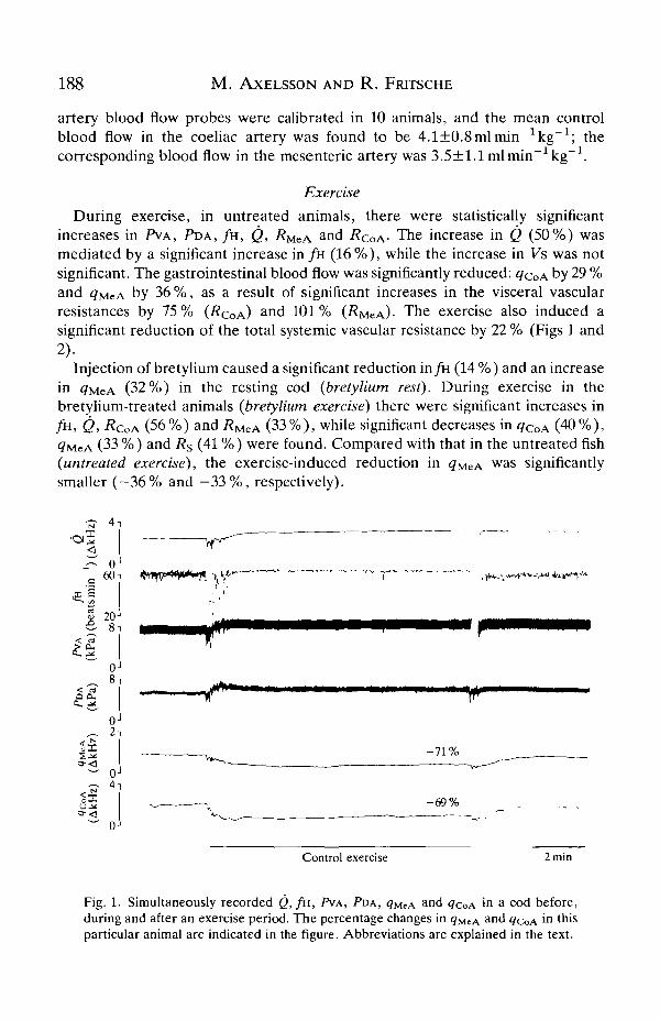
During exercise, in untreated animals, there were statistically significantincreases in PVA, PDA, / H , Q, RMZA
and RCOA- The increase in Q (50%) wasmediated by a significant increase in / H (16%), while the increase in Vs was notsignificant. The gastrointestinal blood flow was significantly reduced: ^COA by 29 %and qMeA by 36 %, as a result of significant increases in the visceral vascularresistances by 75% (RQOA)
a n d 101% (RMCA)- The exercise also induced asignificant reduction of the total systemic vascular resistance by 22 % (Figs 1 and2).
Inj ection of bretylium caused a significant reduction in fa (14 %) and an increasein <7MeA (32 %) in the resting cod (bretylium rest). During exercise in thebretylium-treated animals (bretylium exercise) there were significant increases infi1, Q, RCOA (56%) and i?MeA (33%), while significant decreases in <?CoA (40%),<7MCA (33 %) and Rs (41 %) were found. Compared with that in the untreated fish(untreated exercise), the exercise-induced reduction in <?MeA w a s significantlysmaller ( -36% and - 3 3 % , respectively).
o60
< CO
a a.
2 - 7 1 %
-69%
Control exercise 2min
Fig. 1. Simultaneously recorded Q, / H , PVA, PDA, q^cA and ^COA in a cod before,during and after an exercise period. The percentage changes in <?MCA a nd <7COA in thisparticular animal are indicated in the figure. Abbreviations are explained in the text.
Gastrointestinal blood flow in the cod 189
,i B
6
4
2
0
60
40
20
PVA
t/1 °°S- CO
S?
I"5o
1a£ a
^ • o
as w
unoC
OS J :
6040200
-20
200
-20-40-60-80
20016012080400
20
0-10
- 3 0
- 5 0
VA , ^linnnnnmi
PDA
060
40
20806040200
-20
200
- 2 0- 4 0- 6 0- 8 0
20016012080400
20
0-10
- 3 0
- 5 0
PDA
<7CoA
0
60
40
20806040200
-20
200
-20-40- 6 0- 8 0
20016012080400;
20
0-10
-30
-50
PVA
PDA
Untreatedexercise
Bretyliumexercise
Phentolamineexercise
Fig. 2. A summary of cardiovascular responses to 10 min of exercise in the Atlanticcod, in untreated animals (untreated exercise) in the same animals after treatment withbretylium (bretylium exercise) and after additional phentolamine treatment (phentola-mine exercise), N=8-9. Mean values±s.E.M. are presented. Asterisks indicatestatistically significant (P^0.05) differences compared with rest values. Trianglesindicate statistically significant (P=£0.05) differences compared with the value prior totreatment (bretylium and the subsequent phentolamine treatment). Abbreviations areexplained in the text.

190 M. AXELSSON AND R. FRITSCHE
Following the phentolamine injection there were significant reductions in theresting values (phentolamine rest) of PDA and PVA , while fw increased by 17 %, Qby 21 % and ^MeA by 19 % compared with values in bretylium-treated fish. Duringthe final exercise bout (phentolamine exercise), there were significant decreases inPDA (12%), <jcoA (44%), qMeA (34%) and Rs (44%). At the same time, therewere significant increases in fa (14%), Q (35%), Vs (18%) and RCOA (38%).There were no significant increases in either adrenaline or noradrenaline levelsduring exercise, or in the exercising fish after bretylium injection. Only after thecombined bretylium+phentolamine treatment did the plasma concentration ofadrenaline increase (see Fig. 5).
Hypoxia
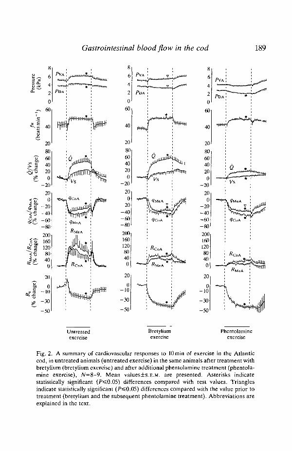
During hypoxia (untreated hypoxia), there were statistically significant increasesin PVA, PDA, Q, RMCA and RCOA- The increase in Q (32 %) was mediated by anincrease in Vs (29%). The gastrointestinal blood flow was significantly reduced:<7coA by 42 % and <?MeA by 62 %. The visceral vascular resistance increasedsignificantly by 159 % in RcoA a n d by 325 % in RMCA- /H and Rs were notsignificantly different from the untreated hypoxia values at the end of the hypoxicperiod (Fig. 3).
After bretylium treatment (bretylium hypoxia), the hypoxia-induced increasesin PVA and PDA seen before treatment were abolished. Instead, PDA wassignificantly decreased at the end of hypoxia after bretylium treatment. Cardiacoutput increased and the gastrointestinal blood flow decreased significantly (both<?MeA and <7coA by 41%) after bretylium treatment. However, there were noincreases in PVMCA and i?coA in the bretylium-treated fish. Hypoxia induced asignificant reduction in the total systemic vascular resistance (by 47%) in theseanimals (Fig. 3).
Following phentolamine treatment, both PVA and PDA were significantlydecreased at the end of the hypoxic period (phentolamine hypoxia). Thesereductions were due to a decrease in the systemic vascular resistance. Both Q andVs remained more or less unchanged throughout the period of hypoxia.decreased significantly during hypoxia (28%), and RMCA as well asdecreased significantly during the same period (by 48 % and 45 %, respectively)(Fig. 3). Both adrenaline and noradrenaline levels increased significantly in theplasma during hypoxia (see Fig. 5).
Feeding
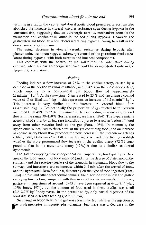
There was a statistically significant postfeeding increase in gut blood flow: qcoAincreased by 72 % and qMeA by 42 %, while RcoA and Rs decreased significantly by3 1 % and 93%, respectively (Fig. 4). Four hours after the injection of the a-adrenoceptor antagonist phentolamine (phentolamine postfeeding), a final record-ing of the variables was performed and statistically significant decreases, comparedto the postfeeding recording, were found in PVA (13%), RMCA (by 14%) andRs (by 0.4%) (Fig. 4). No significant changes in the mesenteric or

Gastrointestinal blood flow in the cod 191
<V
<
o(J
OS
^5
uocccdJ ^o
i ang
e)
cCOJ3o
uacc
806040200
-20
200
-20-40- 6 0-80
600480360240120
0-60
40
0
-40
20
-40
Vs
<7COA
I<7MCA
40
T\
I <7MeA»
Untreatedexercise
Bretyliumexercise
Phentolamineexercise
Fig. 3. A summary of cardiovascular responses to 8 min of exposure to hypoxia in theAtlantic cod in untreated animals (untreated hypoxia) in the same animals aftertreatment with bretylium (bretylium hypoxia) and after additional phentolaminetreatment (phentolamine hypoxia), N=8-12. Mean values±s.E.M. are presented.Asterisks indicate statistically significant (PsSO.05) differences compared with nor-moxic values. Abbreviations are explained in the text.
artery blood flow could be observed after the injection of the ar-adrenoceptorantagonist.
Plasma concentrations of catecholamines in the fish are presented in Fig. 5.

192 M . AXELSSON AND R. FRITSCHE
100 -i
80-
60-
oE
40-
20-
0 J
6 -
0-
f>VA
m
P
<7CoA Vs
Prefeeding
PostfeedingPhentolaminepostfeeding ]
I
0 i
- 2 0 -
- 4 0 -
- 6 0 -
- 8 0 -
-100
-60
50 -r•40 I•30 %
•20 £ .
• io <£,
-o
RMeA
Fig. 4. Effects of feeding on circulatory variables in the cod. Bars show meanvalues+s.E.M., iV=8-13. The flow and resistance values are shown as percentagechanges from the untreated value (prefeeding). Asterisks indicate statistically signifi-cant (P«0.05) differences between untreated (prefeeding) and 24-h postfeeding(postfeeding) and 24-h postfeeding compared to 4 h after phentolamine (phentolamine.postfeeding). Abbreviations are explained in the text.
Discussion
The present investigation is the first to present simultaneous measurements ofthe coeliac and mesenteric artery blood flow (<7COA> 9MCA) in a teleost fish inassociation with exercise, hypoxia and feeding. In this study only relative changesin the ventral aortic blood flow were recorded, and for the discussion a Q value of18-19 ml min"1 kg"1 will be used (Q=19.2±0.9mlmin~1kg"1, Axelsson andNilsson, 1986; 17.3±1.0mlmin"1kg"1, Axelsson, 1986, 19.2±2.3mlmin-1kg"1,Fritsche and Nilsson, 1989).
In the Atlantic cod, the coeliac and mesenteric arteries are of approximately thesame diameter, and estimation of the volume flow in 10 animals by careful in situcalibration of the flow probes revealed similar blood flows in the two vessels (4.1and 3.5 ml min ~~1 kg"1, respectively). This represents about 40 % of cardiac outputin the resting cod, assuming that (2=18-19mlmin~1 kg"1. In a study of the se

Gastrointestinal blood flow in the cod 193
100
p 90
i 80
£ 70
9 60
I 50g 40
30
20
10
Noradrenaline
Adrenaline
URest
U B PExercise
U UNormoxia Hypoxia
Fig. 5. Plasma concentrations of noradrenaline and adrenaline in resting and exercis-ing fish, N=9, and in fish during normoxia, N=8. U, untreated fish; B, fish treated withbretylium; P, fish treated with phentolamine. Values are mean+s.E.M. Asterisksindicate values statistically different (P=£0.05) from rest values (exercise) or normoxiavalues (hypoxia).
raven (Hemitripterus americanus), a prefeeding blood flow of 2.9mlmin~1kg~1
was observed in the coeliac artery, but no measurement of the mesenteric arteryblood flow was performed (Axelsson et al. 1989). However, assuming that total gutblood flow is double that in the coeliac artery (the coeliac and mesenteric arteriesare also of similar diameter in the sea raven), gut blood flow in the sea ravenrepresents approximately 30% of the cardiac output. These values are notdramatically different from the 27-30 % of cardiac output that is directed to thegut circulation via the coeliac and mesenteric arteries in resting, unfed mammals(Greenway, 1982).
Exercise
In the resting fish, injected bretylium produced a 32 % increase in themesenteric artery blood flow, while no significant change in the coeliac arteryblood flow could be detected. The subsequent phentolamine injection produced afurther increase in the mesenteric artery blood flow (19 %) , but again no change inthe coeliac artery blood flow could be detected. These observations are compatiblewith the presence of an adrenergic tonus on the mesenteric vasculature in the cod,with both nervous and humoral components. Since the coeliac vasculature wasunaffected by the adrenergic antagonists, non-adrenergic control of this vascularcircuit is postulated. This contrasts with the conclusions from a study of the searaven, where phentolamine produced a clear increase in coeliac artery blood flow,demonstrating adrenergic control of the coeliac artery vascular bed (Axelssonetal. 1989).
During exercise there was a reduction of the basal flow in the coeliac and

194 M. AXELSSON AND R. FRITSCHE
mesenteric arteries, caused by a marked increase in gut vascular resistance. Thisresistance increase was most pronounced at the onset of the exercise period, and areturn towards the pre-exercise level was seen with time (Fig. 2). This mayrepresent an autoregulatory 'escape reaction', a well-documented phenomenon inmammals, where it is most pronounced in the intestinal circulation but absent inskeletal muscles or adipose tissue (Greenway, 1984). The exact mechanismsbehind this escape reaction are not known, but in mammals it is unaffected by theyS-adrenoceptor antagonists atropine, antihistamines, indomethacin and naloxone(Greenway et al. 1976; Greenway, 1984).
In the cod, bretylium treatment abolished the observed 'escape reaction';instead, the resistance increase in the coeliac and mesenteric artery persistedduring the whole exercise period. This pattern was not altered by the subsequentor-adrenoceptor blockade with phentolamine. If the initial response is an escapereaction of the mammalian type, this finding suggests that an adrenergic nervousmechanism is involved in the development of this phenomenon in the cod.
The visceral vascular resistance also increased during exercise after bretyliumtreatment, causing a reduction in coeliac and mesenteric artery blood flow. Afteradditional phentolamine treatment, only coeliac vascular resistance increasedsignificantly, but the blood flow decreased in both vessels. The control of gut bloodflow during exercise must therefore involve a non-adrenergic mechanism, inaddition to an adrenergic one.
Exercise induced a decrease in the. total systemic vascular resistance in theuntreated fish, which may be explained by hyperaemia in the working muscles dueto released metabolites. This phenomenon, observed and described by Gaskell(1880), has since been extensively studied in mammals (Whipp and Ward, 1982).An exercise-induced decrease in systemic vascular resistance has also beendemonstrated in the rainbow trout (Kiceniuk and Jones, 1977; Randall andDaxboeck, 1982).
In the cod, bretylium treatment markedly potentiated the exercise-inducedreduction in the systemic vascular resistance. This could be explained by anincrease in the adrenergic nervous tone in the vasculature during exercise, whichnormally counteracts the effects of the local metabolite-induced hyperaemia.After bretylium treatment, this vasomotor activity ceases, revealing the full effectof the hyperaemia.
Hypoxia
During normoxia, injected bretylium and phentolamine did not change thevisceral vascular resistance in the cod. These findings suggest the lack ofadrenergic tonus in the gut vasculature in the undisturbed cod.
Although total cardiac output and dorsal aortic blood pressure increased duringhypoxia, blood flow in the coeliac and mesenteric arteries decreased during thesame period. This reduction in blood flow to the gut is due to increased visceralvascular resistance. After bretylium treatment, and also after additional phentola-mine treatment, the systemic vascular resistance decreased during hypoxia
Gastrointestinal blood flow in the cod 195
resulting in a fall in the ventral and dorsal aortic blood pressures. Bretylium alsoabolished the increase in visceral vascular resistance seen during hypoxia in theuntreated fish, suggesting that an adrenergic nervous mechanism controls themesenteric and coeliac vasculature in the cod during hypoxia. However, thegastrointestinal blood flow still decreased during hypoxia, owing to a fall in thedorsal aortic blood pressure.
The actual decrease in visceral vascular resistance during hypoxia afterphentolamine treatment suggests adrenergic control of the gastrointestinal vascu-lature during hypoxia, with both nervous and humoral components.
This contrasts with the control of the gastrointestinal vasculature duringexercise, when a clear adrenergic influence could be demonstrated only in themesenteric vasculature.
Feeding
Feeding induced a flow increase of 72 % in the coeliac artery, caused by adecrease in the coeliac vascular resistance, and of 42 % in the mesenteric artery,which amounts to a postprandial gut blood flow of approximately12 ml min"1 kg"1. At the same time, Q increased by 23 % and, assuming a controlvalue of Q of 18mlmin~1kg~1, this represents an increase of 4.2mlmin~1kg~1.This increase is very similar to the increase in visceral blood flow(4.4 ml min"1 kg"1). Postprandially the proportion of Q directed to the visceraincreased from 40% to 52%. In mammals, the postfeeding increase in gut bloodflow is in the range 30-150% (for references, see Fara, 1984). The hyperaemia isaccomplished either by an increase in cardiac output or by a redistribution of bloodaway from other vascular beds to the gut (Fara, 1984). In mammals, thehyperaemia is localized to those parts of the gut containing food, and an increasein coeliac artery blood flow precedes the flow increase in the mesenteric arteries(Biber, 1974; Gallavan et al. 1980). Further work is needed in fish to establishwhether the more pronounced flow increase in the coeliac artery (72%) com-pared to that in the mesenteric artery (42%) is due to a similar sequentialhyperaemia.
The gastric emptying time is dependent on temperature, food quality, surfacearea of the food, amount of food ingested (and thus the degree of distension of thestomach) and the secretory surface of the stomach. In mammals, blood flow to thestomach and intestine starts to increase within 3-5 min after the arrival of food,and the hyperaemia lasts for 4-6 h, depending on the type of food ingested (Fara,1984). In fish and other ectothermic animals, the digestion rate is low and gastricemptying time is long compared with that in endothermic mammals. In the cod,gastric emptying times of around 12-45 h have been reported at 6-10°C (Tyler,1970; Jones, 1974), but the amount of food used in these studies was small(0.2-2.7% kg"1 body mass). In the present study, only partial digestion of thefood was seen 29 h after feeding (post mortem).
No change in blood flow to the gut was seen in the fed fish after the injection ofcr-adrenoceptor antagonist phentolamine, but there was a decrease in the

196 M. AXELSSON AND R. FRTTSCHE
mesenteric vascular resistance. This contrasts with the results from the sea ravenstudy, where phentolamine caused an increase in the coeliac artery flow fromS.Smlmin^kg"1 (postprandial) to 11.3mlmin~1kg"1 (Axelsson etal. 1989).
In the cod, it has been shown that both substance P and vasoactive intestinalpolypeptide (VIP) increase the gastrointestinal blood flow and that there exists acholinergically mediated vasoconstrictor mechanism in the coeliac artery vascularcircuit (Jensen et al. 1990). In mammals, it has been suggested that cholecystokinin(CCK) may be the active mediator of the mesenteric postprandial hyperaemia(Fara, 1984), but many other gastrointestinal hormones cause vasodilatation in thesmall intestine of mammals, for instance secretin, gastrin, glucagon, VIP, opiateagonists (morphine, endorphins, met-enkephalin), neurotensin, substance P andgastric inhibitory polypeptide (Chou et al. 1984). The large number of putativeneurotransmitters and gut hormones with a potential role in gut vascular controlopens up a wealth of possible mechanisms, and further studies are obviouslyneeded to elucidate the mechanisms involved in the regulation of the gut bloodsupply during exercise, hypoxia and feeding in fish.
It is clear from this study that the control of the gastrointestinal vasculaturevaries with the stimulus. During hypoxia, adrenergic control of the gastrointestinalvasculature with both nervous and humoral components was found, while thecontrol of gut blood flow during exercise and feeding also involves an additionalnon-adrenergic mechanism.
This work was supported by the Swedish Natural Science Research Council, theMagnus Bergvall Foundation and the Lars Hierta Memory Foundation. We wishto thank Ms Gunilla Rydgren for skilful technical assistance with the analyses ofcatecholamines and Mr Uno Larsson for supplying the fish. We are also grateful toDr Susanne Holmgren and Professor Stefan Nilsson for reading and makingvaluable comments on the manuscript.
ReferencesAXELSSON, M. (1988). The importance of nervous and humoral mechanisms in the control of
cardiac performance in the Atlantic cod, Gadus morhua at rest and during non-exhaustiveexercise. /. exp. Biol. 137, 287-303.
AXELSSON, M., DRIEDZIC, W. R., FARRELL, A. P. AND NILSSON, S. (1989). Regulation of cardiacoutput and gut blood flow in the sea raven, Hemitripterus americanus. Fish Physiol. Biochem.6, 315-326.
AXELSSON, M. AND NILSSON, S. (1986). Blood pressure regulation during exercise in the Atlanticcod, Gadus morhua. J. exp. Biol. 126,225-236.
BIBER, B. (1974). Vasodilator mechanisms in the small intestine. Actaphysiol. Scand. 90 (Suppl.401), 1-31.
CHOU, C. C , MANGINO, M. J. AND SAWMILLER, D. R. (1984). Gastrointestinal hormones andintestinal blood flow. In Physiology of the Intestinal Circulation (ed. A. P. Shepherd and D. N.Granger), pp. 121-130. New York: Raven Press.
DAXBOECK, C. (1981). A study of the cardiovascular system of fish (Salmo gairdneri) at rest andduring swimming exercise. PhD thesis, University of British Columbia, Vancouver.
DAXBOECK, C. AND HOLETON, G. F. (1978). Oxygen receptors in the rainbow trout, Salmogairdneri. Can. J. Zool. 56, 1254-1259.

Gastrointestinal blood flow in the cod 197
DONALD, E. D. (1980). Role of autonomic nerves in the cardiovascular response to exercise inthe dog. In Exercise Bioenergetics and Gas Exchange (ed. P. Cerretelli and B. J. Whipp), pp.267-274. Amsterdam: Elsevier, North-Holland Biomedical Press.
FARA, J. W. (1984). Postprandial mesenteric hyperemia. In Physiology of the IntestinalCirculation (ed. A. P. Shepherd and D. N. Granger), pp. 99-106. New York: Raven Press.
FARRELL, A. P. (1984). A review of cardiac performance in the teleost heart: intrinsic andhumoral regulation. Can. J. Zool. 62, 523-536.
FRITSCHE, R. AND NILSSON, S. (1989). Cardiovascular responses to hypoxia in the Atlantic cod,Gadus morhua. Exp. Biol. 48, 153-160.
FRITSCHE, R. AND NILSSON, S. (1990). Autonomic nervous control of blood pressure and heartrate during hypoxia in the cod, Gadus morhua. J. comp. Physiol. B 160, 287-292.
GALLAVAN, R. H., CHOU, C. C , KVIETYS, P. R. AND SIT, S. P. (1980). Regional blood flowduring digestion in the conscious dog. Am. J. Physiol. 238, H220-FL225.
GASKELL, W. H. (1880). On the tonicity of heart and blood vessels./. Physiol., Lond. 4,48-75.GEHRKE, P. C. AND FIELDER, D. R. (1988). Effects of temperature and dissolved oxygen on heart
rate, ventilation rate and oxygen consumption of spangled perch, Leioptherapon unicolor.J. comp. Physiol. 157, 771-782.
GREEN WAY, C. V. (1982). Mechanisms and quantitative assessment of drug effects on cardiacoutput with a new model of the circulation. Pharmac. Rev. 33, 213-251.
GREENWAY, C. V. (1984). Neural control and autoregulatory escape. In Physiology of theIntestinal Circulation (ed. A. P. Shepherd and D. N. Granger), pp. 61-71. New York: Ravenpress.
GREENWAY, C. V., SCOTT, G. D. AND ZINK, J. (1976). Sites of autoregulatory escape of bloodflow in the mesenteric vascular bed. J. Physiol., Lond. 259, 1-12.
HALLMAN, H., FARNEBO, L.-O., HAMBERGER, B. AND JONSON, G. (1978). A sensitive method forthe determination of plasma catecholamines using liquid chromatography withelectrochemical detection. Life Sci. 23, 1049-1052.
HOLETON, G. F. AND RANDALL, D. J. (1967). Changes in blood pressure in the rainbow troutduring hypoxia. J. exp. Biol. 46, 297-305.
HOLM, S. (1979). A simple sequentially rejective multiple test procedure. Scand. J. Statist. 6,65-70.
JENSEN, J., AXELSSON, M. AND HOLMGREN, S. (1990). Effects of substance P and vasoactiveintestinal polypeptide on gastrointestinal blood flow in the Atlantic cod Gadus morhua.J. exp. Biol. 156, 361-373.
JONES, R. (1974). Hie rate of elimination of food from the stomach of haddock,Melanogrammus aeglefinus, cod, Gadus morhua and whiting, Merlangius merlangus. J. Cons.int. Explor. Mer. 35, 225-298.
KICENIUK, J. W. AND JONES, D. R. (1977). The oxygen transport system in trout {Salmogairdneri) during sustained exercise. /. exp. Biol. 69, 247-260.
LAURENT, P., HOLMGREN, S. AND NILSSON, S. (1983). Nervous and humoral control of the fishheart: structure and function. Comp. Biochem. Physiol. 76A, 524-542.
NILSSON, S. (1983). Autonomic Nerve Function in the Vertebrates. Berlin, Heidelberg, NewYork: Springer-Verlag.
NILSSON, S. AND HOLMGREN, S. (1989). Novel neurotransmitter in the autonomic nervous systemof nonmammalian vertebrates. Pharmac. Ther. 41, 257-287.
ORWELL, L. B., BLACKMON, J. R. AND BRUCE, R. A. (1964). Indocyanine green clearance andestimated hepatic blood flow during mild to maximal exercise in upright man. /. clin. Invest.43, 1677-1690.
RANDALL, D. J. AND DAXBOECK, C. (1982). Cardiovascular changes in the rainbow trout (Salmogairdneri Richardson) during exercise. Can. J. Zool. 60, 1135-1140.
RUSHMER, R. F., FRANKLIN, D. L., VAN OTTERS, R. L. AND SMITH, O. A. (1961). Changes inperipheral blood flow distribution in healthy dogs. Circulation Res. 9, 675-687.
SMITH, D. G. (1978). Neural regulation of blood pressure in rainbow trout (Salmo gairdneri)Can. J. Zool. 56, 1678-1683.
SMITH, D. G., WAHLQVIST, I., NILSSON, S. AND ERIKSSON, B.-M. (1985). Nervous control of theblood pressure in the Atlantic cod, Gadus morhua. J. exp. Biol. 117, 335-347.

198 M. AXELSSON AND R. FRITSCHE
TYLER, A. V. (1970). Rates of gastric emptying in young cod. /. Fish. Res. Bd Can. 27,1177-1189.
VATNER, S. F., FRANKLIN, D., HIGGINS, C. B., PATRICK, T., WHITE, S. AND VAN CITTERS, R. L.(1971). Coronary dynamics in unrestrained conscious baboons. Am. J. Physiol. 221,13%-1401.
WAHLQVIST, I. AND CAMPBELL, G. (1988). Autonomic influences on heart rate and bloodpressure in the toad, Bufo marinus, at rest and during exercise. /. exp. Biol. 134, 377-3%.
WHIPP, B. J. AND WARD, S. A. (1982). Cardiopulmonary coupling during exercise. /. exp. Biol.100, 175-193.