Embed Size (px)
Citation preview
Cancer Detection and Prevention 27 (2003) 451–456
Effectiveness of a dynamic breast examination training modelto improve clinical breast examination (CBE) skills
Gregory J. Gerling, MSa,∗, Alicia M. Weissman, MDb,1,Geb W. Thomas, PhDc,2, Edwin L. Dove, PhDd,3
a Department of Industrial Engineering, University of Iowa, Industrial Engineering, 3131 Seamans Center, Iowa City, IA 52242, USAb Family Medicine, University of Iowa, Family Medicine, 01291-A PFP, Iowa City, IA 52242, USA
c Industrial Engineering, University of Iowa, Industrial Engineering, 2404 Seamans Center, Iowa City, IA 52242, USAd Biomedical Engineering, University of Iowa, Biomedical Engineering, 1412 Seamans Center, Iowa City, IA 52242, USA
Accepted 5 September 2003
Abstract
Despite the potential utility of clinical breast examination (CBE), doctors’ palpation skills are often inadequate and difficult to train. CBEsensitivity ranges from 39–59%, in part because current training does not effectively teach tactile skills. To address CBE training limitations,we developed a breast examination training model with 15 dynamically controlled lumps, set to desired hardness within underlying rib andmuscle structures, in a silicone breast. In an experiment of 48 medical students, training with the dynamic model increased lump detectionby 1.35 lumps compared to 0.60 lumps for a traditional breast model (P = 0.008), reduced false positives by−0.70 lumps compared to+0.42 lumps (P = 0.0277), and demonstrated skill transfer with a 1.17 lump detection improvement on the traditional device comparedto only a 0.17 lump detection improvement by traditional device trainees on the dynamic device (P < 0.001). Findings demonstrate theadvantage of the dynamic model over conventional models in training CBE tactile skills.© 2003 International Society for Preventive Oncology. Published by Elsevier Ltd. All rights reserved.
Keywords: Palpation; Breast cancer; Training; Simulation; Tactile; Haptic
1. Introduction
1.1. Background
Breast cancer kills 40,000 women yearly in the UnitedStates, with approximately 200,000 new cases diagnosed in2001[1]. Fortunately, the 5-year survival rate exceeds 98%for tumors under 2.0 cm. Therefore, research has focusedon early detection as one possible means of saving lives.Current screening methods include mammography, selfbreast examination (SBE), and clinical breast examination(CBE).
Evidence supporting the clinical efficacy of CBE emergedfrom the Canadian National Breast Screening Study andfound that when performed in addition to CBE, mammog-raphy did not significantly affect the frequency of cancer di-
∗ Corresponding author. Tel.:+1-319-358-1615; fax:+1-319-335-5669.E-mail address: [email protected] (G.J. Gerling).
1 Tel.: +1-319-384-7765.2 Tel.: +1-319-335-5936.3 Tel.: +1-319-335-5635.
agnosis, stage at diagnosis, detection of interval cancers, orbreast cancer mortality[2]. The Japanese national CBE pro-gram demonstrated a 40% decrease in the age adjusted mor-tality rate in municipalities given intensified CBE screeningcompared to control municipalities[3].
Despite its potential effectiveness[2,3], CBE sensitivityis only 39–59%[4]. It appears that physicians’ skills varywidely and many physicians possess only modest clinicalskills [5]. Often these skills fall short of performing a maxi-mally effective CBE. Many practicing physicians, when sur-veyed, acknowledge a need to increase their competence inCBE. One study reported that 43% of primary care resident,faculty and nurse practitioners lack confidence in their CBEskills [5]. Practitioners may underutilize CBE if they do notfeel proficient in CBE performance.
Performing an accurate CBE involves using a throughsearch pattern, adequate pressure, proper finger positioning,and discriminating a solid mass from surrounding nodularity[6]. Breast palpation is a practiced tactile skill that does notnecessarily correlate with cognitive knowledge about breastcancer detection[7]. Training which emphasizes tactile skillsis known to improve examination sensitivity[8]. Currently,
0361-090X/$30.00 © 2003 International Society for Preventive Oncology. Published by Elsevier Ltd. All rights reserved.doi:10.1016/j.cdp.2003.09.008
452 G.J. Gerling et al. / Cancer Detection and Prevention 27 (2003) 451–456
CBE is taught with live patient volunteers, artificial breastmodels, and training videos. The artificial breast modelsare the only modality that can reliably improve the tactileability to identify masses, since patient volunteers may nothave breast masses and the characteristics of masses that arepresent cannot be controlled.
Artificial breast models are made of silicone embeddedwith foreign objects representing tumors. The models areeffective in improving lump detection[8]. Training with sil-icone models is also known to increase detection of benignlumps in live patients, demonstrating transfer of skills be-tween the models and the clinical examination[9]. Optimallytrained examiners can detect lumps as small as 2–3 mm[10].Retraining and reassessment improve skill retention overtime. After training, mean lump detection by trained partic-ipants has been seen to improve from 44 to 69%[11,12].Careful training and supervision of health professionals hasbeen reviewed in papers and subsequently modeled in hos-pitals [2,13].
Despite advances in tactile training with silicone breastmodels, CBE training in practice falls short. Two inherentcharacteristics of static silicone breast models limit their util-ity. First, the models provide a limited range of difficulty;once the trainee has identified all the lumps in these models,little more can be learned by repeating the examination. Sec-ond, training with static breast models increases sensitivityat the cost of increasing false detections[7,8,11], leading toa high false positive rate.
To address these limitations, we developed and tested anovel dynamic breast examination training device that canpresent the learner with up to fifteen lumps of varying char-acteristics in changing configurations. We built the modelto address two important training issues: (1) range of diffi-culty (by gradually increasing difficulty from a set of larger,harder lumps to smaller, softer lumps) and (2) high false pos-itive rate (by providing tactile feedback through active lumpinflation/deflation beneath the fingertips). This breast exam-ination model was then tested against standard, static breastmodels to determine its ability to train for improved lumpdetection, lowered false detection reports, and improved skilltransfer.
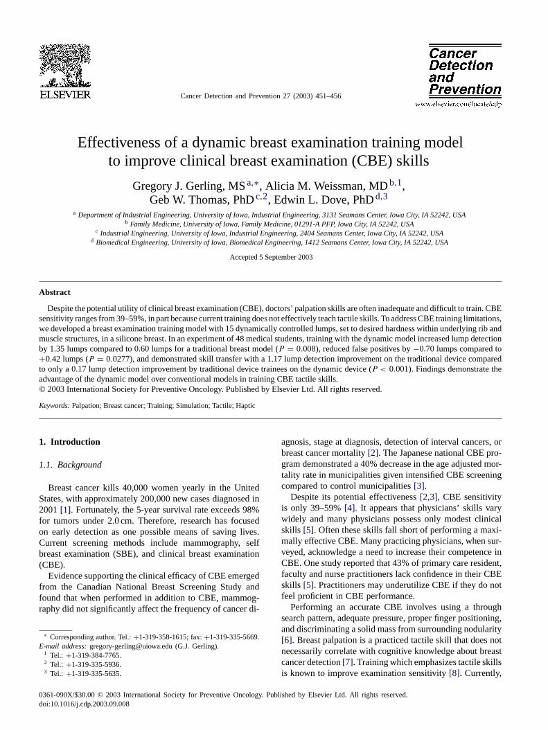
Fig. 1. Breast model comparison, static (left) and dynamic (right) with a 12 in. ruler.
2. Methods
We conducted an experiment to test the effectiveness ofthe dynamic training device in improving the lump detectionand lowering false detections of CBE. We hypothesized thattraining with the dynamic breast model would lead to (1)higher lump detection, without increasing false detections,compared to training with a static silicone breast model; and(2) greater skill transfer to other breast models.
2.1. Breast models
The dynamic training model (Fig. 1) is a prototype sili-cone breast model with 15 lumps that can be individuallyinflated, to known, controllable levels of hardness[14]. Thebreast model includes simulated rib and intercostal mus-cle structures. Lumps vary in size (0.3–1.5 cm), hardness(20–50 durometers), depth of placement (shallow, medium,and deep), and fixedness (fixed and mobile). The siliconematrix is opaque and has a hard silicone backing with em-bedded ribs and entry points of thin, polyethylene tubesleading to the lumps of the same material. Lump hardnessis controlled by an external pressure system, which selec-tively inflates the polyethylene sacs. Lumps are undetectablewhen deflated. The silicone matrix is homogeneous with lit-tle nodularity.
The control breast models (Fig. 1) are round opaque breastmodels with square bases. These static models are composedof a silicone matrix with embedded semi-solid lumps. Thelumps are made of silicone polymers in a cylindrical shape.We used breast models CPM-S and CPM-F from the Mam-matech Corporation, which are widely used to teach andevaluate breast examination skills[15]. The CPM-F modelis firmer, but otherwise the two models are identical, withlump size, hardness, position, and depth being fixed andequal in both Mammacare models. The models have lownodularity.
Engineering tests including stress/strain analysis ofbreast tissue and tumor hardness were taken to ensurethat the firmness of the dynamic and static models weresimilar. Additionally, a small, subjective test with fifteen
G.J. Gerling et al. / Cancer Detection and Prevention 27 (2003) 451–456 453
physicians showed the dynamic simulator was similar tothe Mammatech breast models in simulating real tissue[16].
2.2. Participants and study procedures
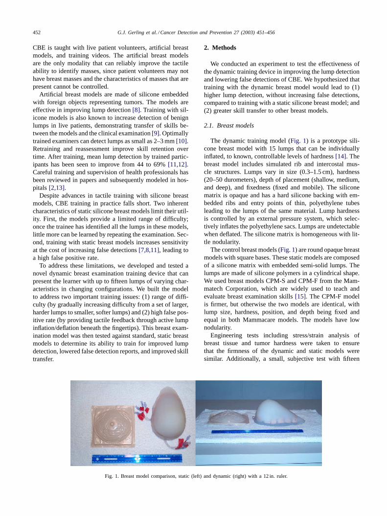
The 48 participants were first through third year medicalstudents at the University of Iowa. They included 30 womenand 18 men aged 22–40 with a mean age of 25. Participantswere assigned to one of eight groups by order of responseto the call for participants. We balanced the eight groups bygender and year in medical school, factors that reflect prioropportunities to practice breast examination techniques. Us-ing eight groups enabled us to vary the order of tests 1 and2 within each pre-test and post-test, to determine possibledifferences based on presentation order. Training groups Aand B were each assigned 15 women and 9 men (8 firstyear, 15 second year, and 1 third year) (Fig. 2). All par-ticipants signed informed consent documents for the study,
Fig. 2. Presentation order of the eight experimental groups.
which was approved by the University of Iowa InstitutionalReview Board.
The experiment included two pre-tests, a training session,a break, and three post-tests (Fig. 2). Pre-test correct, missed,and false positive detection scores were gathered on each ofthe two models, where participants were allotted 2 min toexamine each breast model. Participants reported locationsof lumps and lump properties to the research assistant whorecorded onto a scripted diagram. The 2 min examination in-terval is based on the time normally taken in a breast exam-ination[8]. Because five lumps are fixed in the static model,five lumps with equivalent characteristics (size, hardness,and depth) were consistently inflated in the dynamic train-ing model. Each 15 min training session included discussionand demonstration of recommended search pattern, fingerpressure, part and number of fingers used, finger motion,nodularity effects, breast area coverage, and lump properties.The research assistant provided the training according to de-tailed, written instructions describing the above techniques.
454 G.J. Gerling et al. / Cancer Detection and Prevention 27 (2003) 451–456
With the dynamic model, lumps could be turned on and off,enabling trainees to palpate the same lump at varying hard-ness and distinguish it from background silicone material.Since this was not possible with the static model, partic-ipants in the control group alternately felt areas with andwithout lumps. After the 20 min rest, post-training scoreswere gathered on each of two models with order followingfrom the trainee’s particular group. Because all Mammacarebreast models have identical lump configurations, we usedthe same firm static model (CPM-F) rotated 90◦. The lumppositions and properties were consistently changed in thedynamic training model with similar characteristics to thestatic model in terms of size, hardness, and depth[16]. Toaccount for a possibility of bias in learning how the dynamicmodel operates, a third model (CPM-S) was introduced forthe final post-test.
2.3. Statistical methods
Analysis of variance (ANOVA) techniques were utilizedin comparing the data to determine differences amongstatic and dynamic training groups. The independent vari-able (between-subjects dimension) was the training device(two levels: static, dynamic). The dependent variables(within-subjects dimension) were (1) the tested device(static, dynamic, third model) and (2) test condition (pretestand posttest), giving a repeated-measures design. Thus, wemeasured the correctly detected lumps to obtain preteststatic and dynamic values and posttest static, dynamic, andthird model values. We also measured false detections undereach condition.
We performed three main analyses: lumps detected, falsedetections, and transfer of training for lumps detected. Weintended to look at the differences based on type of training(static or dynamic).
3. Results
All 48 participants completed testing. Because presen-tation order in the first and second tests of pre-tests andpost-tests did not significantly affect the results (compos-ite lump detection: pre-test (F(5, 42) = 0.07, P = 0.80),post-test (F(5, 42) = 0.05, P = 0.83); and composite falsepositives: pre-test (F(5, 42) = 2.45, P = 0.13), post-test(F(5, 42) = 1.17, P = 0.29)), the eight groups were col-lapsed to two groups of 24 participants according to the in-dependent variable (type of training: static or dynamic).
3.1. Lump detection
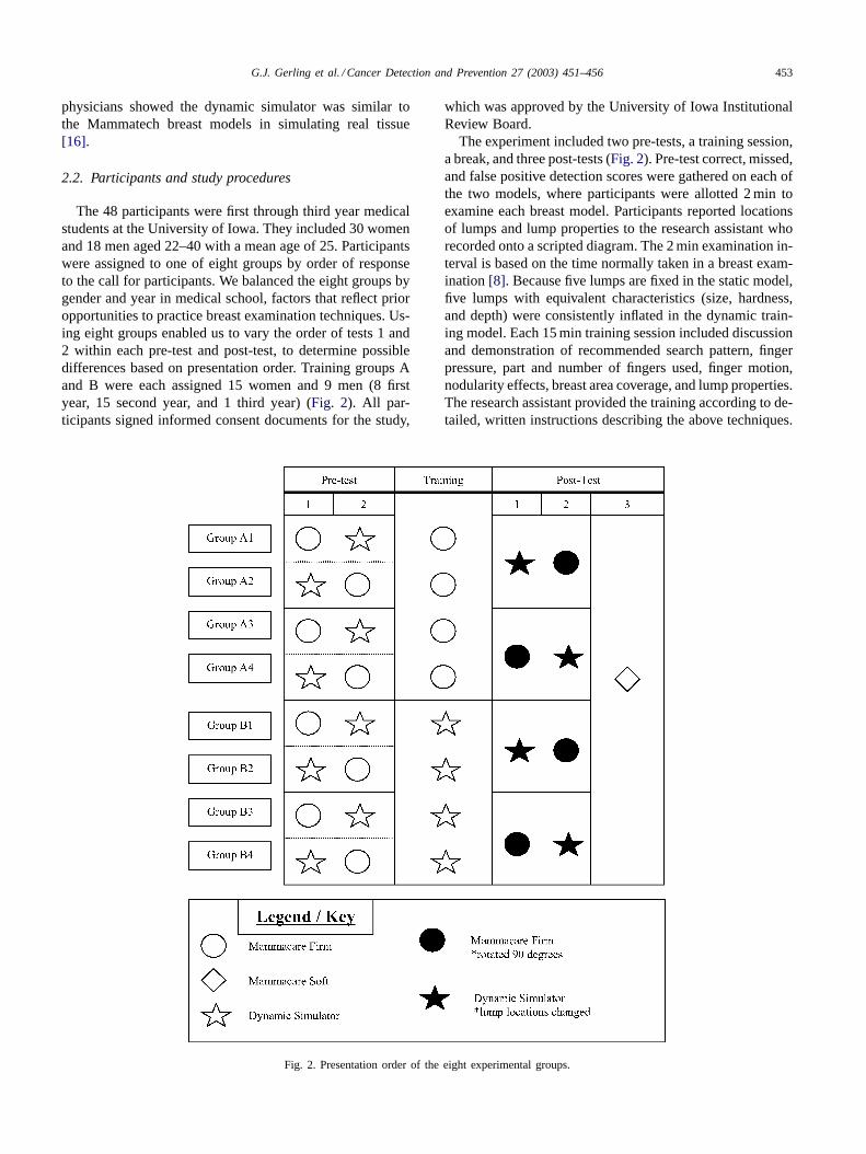
Each participant has two lump detection measures: thepre-test to post-test difference in the number of lumps foundfor (1) the static model and (2) the dynamic model. Compos-ite lump detection improvement was defined as the sum ofthe lump detection improvement on the static model and the
Fig. 3. Composite lump detection increase (static+ dynamic) based upontraining model.
lump detection improvement on the dynamic model. Com-posite lump detection improvement (Fig. 3) was greater af-ter training with the dynamic training model (1.35 lumps,S.D. = 0.92) than after training with the static device (0.60lumps, S.D. = 0.96) (P = 0.008). The participants whotrained on the dynamic model showed a trend toward detect-ing more lumps on the third model introduced at the end ofthe experiment, but this result was not statistically signifi-cant (dynamic model: 3.04 of 5 lumps, S.D. = 1.12; staticmodel: 2.54 lumps, S.D. = 0.88) (P = 0.0927).
3.2. False detections
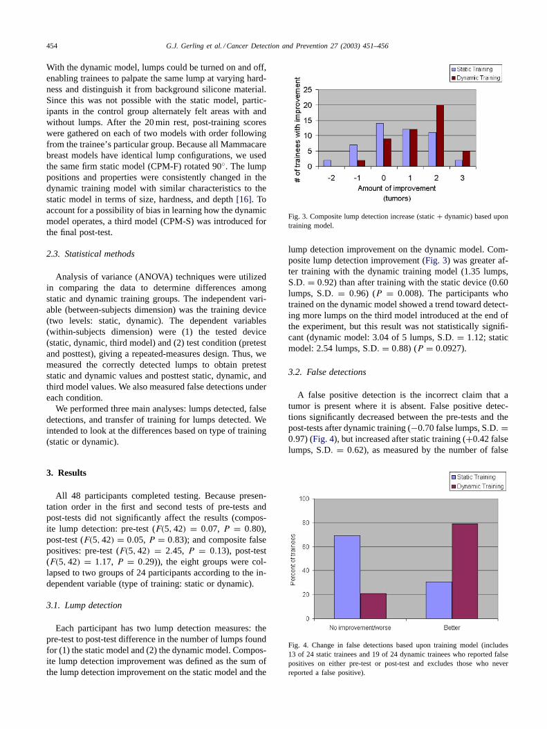
A false positive detection is the incorrect claim that atumor is present where it is absent. False positive detec-tions significantly decreased between the pre-tests and thepost-tests after dynamic training (−0.70 false lumps, S.D. =0.97) (Fig. 4), but increased after static training (+0.42 falselumps, S.D. = 0.62), as measured by the number of false
Fig. 4. Change in false detections based upon training model (includes13 of 24 static trainees and 19 of 24 dynamic trainees who reported falsepositives on either pre-test or post-test and excludes those who neverreported a false positive).
G.J. Gerling et al. / Cancer Detection and Prevention 27 (2003) 451–456 455
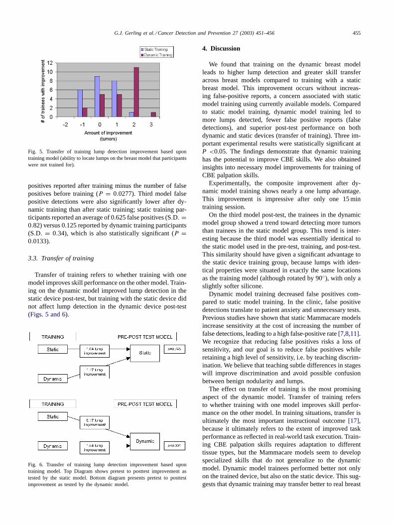
Fig. 5. Transfer of training lump detection improvement based upontraining model (ability to locate lumps on the breast model that participantswere not trained for).
positives reported after training minus the number of falsepositives before training (P = 0.0277). Third model falsepositive detections were also significantly lower after dy-namic training than after static training; static training par-ticipants reported an average of 0.625 false positives (S.D. =0.82) versus 0.125 reported by dynamic training participants(S.D. = 0.34), which is also statistically significant (P =0.0133).
3.3. Transfer of training
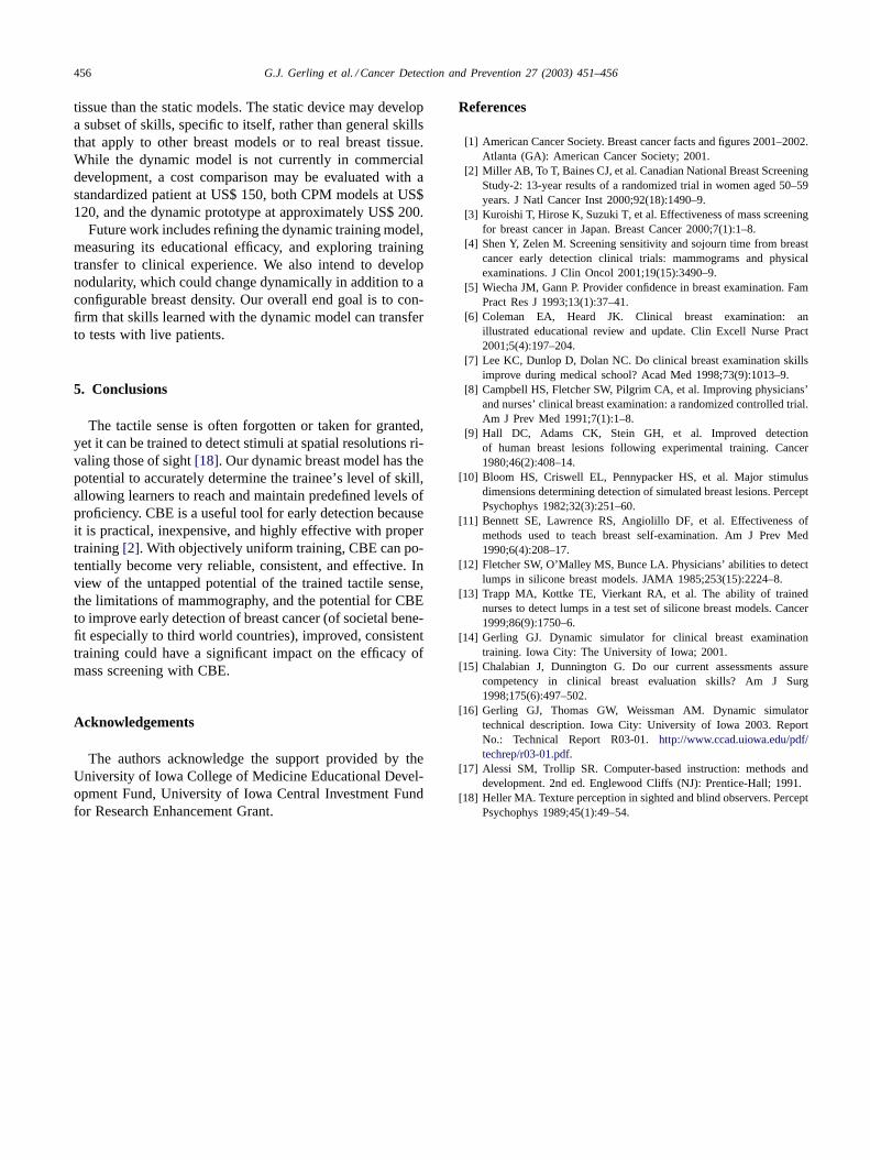
Transfer of training refers to whether training with onemodel improves skill performance on the other model. Train-ing on the dynamic model improved lump detection in thestatic device post-test, but training with the static device didnot affect lump detection in the dynamic device post-test(Figs. 5 and 6).
Fig. 6. Transfer of training lump detection improvement based upontraining model. Top Diagram shows pretest to posttest improvement astested by the static model. Bottom diagram presents pretest to posttestimprovement as tested by the dynamic model.
4. Discussion
We found that training on the dynamic breast modelleads to higher lump detection and greater skill transferacross breast models compared to training with a staticbreast model. This improvement occurs without increas-ing false-positive reports, a concern associated with staticmodel training using currently available models. Comparedto static model training, dynamic model training led tomore lumps detected, fewer false positive reports (falsedetections), and superior post-test performance on bothdynamic and static devices (transfer of training). Three im-portant experimental results were statistically significant atP <0.05. The findings demonstrate that dynamic traininghas the potential to improve CBE skills. We also obtainedinsights into necessary model improvements for training ofCBE palpation skills.
Experimentally, the composite improvement after dy-namic model training shows nearly a one lump advantage.This improvement is impressive after only one 15 mintraining session.
On the third model post-test, the trainees in the dynamicmodel group showed a trend toward detecting more tumorsthan trainees in the static model group. This trend is inter-esting because the third model was essentially identical tothe static model used in the pre-test, training, and post-test.This similarity should have given a significant advantage tothe static device training group, because lumps with iden-tical properties were situated in exactly the same locationsas the training model (although rotated by 90◦), with only aslightly softer silicone.
Dynamic model training decreased false positives com-pared to static model training. In the clinic, false positivedetections translate to patient anxiety and unnecessary tests.Previous studies have shown that static Mammacare modelsincrease sensitivity at the cost of increasing the number offalse detections, leading to a high false-positive rate[7,8,11].We recognize that reducing false positives risks a loss ofsensitivity, and our goal is to reduce false positives whileretaining a high level of sensitivity, i.e. by teaching discrim-ination. We believe that teaching subtle differences in stageswill improve discrimination and avoid possible confusionbetween benign nodularity and lumps.
The effect on transfer of training is the most promisingaspect of the dynamic model. Transfer of training refersto whether training with one model improves skill perfor-mance on the other model. In training situations, transfer isultimately the most important instructional outcome[17],because it ultimately refers to the extent of improved taskperformance as reflected in real-world task execution. Train-ing CBE palpation skills requires adaptation to differenttissue types, but the Mammacare models seem to developspecialized skills that do not generalize to the dynamicmodel. Dynamic model trainees performed better not onlyon the trained device, but also on the static device. This sug-gests that dynamic training may transfer better to real breast
456 G.J. Gerling et al. / Cancer Detection and Prevention 27 (2003) 451–456
tissue than the static models. The static device may developa subset of skills, specific to itself, rather than general skillsthat apply to other breast models or to real breast tissue.While the dynamic model is not currently in commercialdevelopment, a cost comparison may be evaluated with astandardized patient at US$ 150, both CPM models at US$120, and the dynamic prototype at approximately US$ 200.
Future work includes refining the dynamic training model,measuring its educational efficacy, and exploring trainingtransfer to clinical experience. We also intend to developnodularity, which could change dynamically in addition to aconfigurable breast density. Our overall end goal is to con-firm that skills learned with the dynamic model can transferto tests with live patients.
5. Conclusions
The tactile sense is often forgotten or taken for granted,yet it can be trained to detect stimuli at spatial resolutions ri-valing those of sight[18]. Our dynamic breast model has thepotential to accurately determine the trainee’s level of skill,allowing learners to reach and maintain predefined levels ofproficiency. CBE is a useful tool for early detection becauseit is practical, inexpensive, and highly effective with propertraining[2]. With objectively uniform training, CBE can po-tentially become very reliable, consistent, and effective. Inview of the untapped potential of the trained tactile sense,the limitations of mammography, and the potential for CBEto improve early detection of breast cancer (of societal bene-fit especially to third world countries), improved, consistenttraining could have a significant impact on the efficacy ofmass screening with CBE.
Acknowledgements
The authors acknowledge the support provided by theUniversity of Iowa College of Medicine Educational Devel-opment Fund, University of Iowa Central Investment Fundfor Research Enhancement Grant.
References
[1] American Cancer Society. Breast cancer facts and figures 2001–2002.Atlanta (GA): American Cancer Society; 2001.
[2] Miller AB, To T, Baines CJ, et al. Canadian National Breast ScreeningStudy-2: 13-year results of a randomized trial in women aged 50–59years. J Natl Cancer Inst 2000;92(18):1490–9.
[3] Kuroishi T, Hirose K, Suzuki T, et al. Effectiveness of mass screeningfor breast cancer in Japan. Breast Cancer 2000;7(1):1–8.
[4] Shen Y, Zelen M. Screening sensitivity and sojourn time from breastcancer early detection clinical trials: mammograms and physicalexaminations. J Clin Oncol 2001;19(15):3490–9.
[5] Wiecha JM, Gann P. Provider confidence in breast examination. FamPract Res J 1993;13(1):37–41.
[6] Coleman EA, Heard JK. Clinical breast examination: anillustrated educational review and update. Clin Excell Nurse Pract2001;5(4):197–204.
[7] Lee KC, Dunlop D, Dolan NC. Do clinical breast examination skillsimprove during medical school? Acad Med 1998;73(9):1013–9.
[8] Campbell HS, Fletcher SW, Pilgrim CA, et al. Improving physicians’and nurses’ clinical breast examination: a randomized controlled trial.Am J Prev Med 1991;7(1):1–8.
[9] Hall DC, Adams CK, Stein GH, et al. Improved detectionof human breast lesions following experimental training. Cancer1980;46(2):408–14.
[10] Bloom HS, Criswell EL, Pennypacker HS, et al. Major stimulusdimensions determining detection of simulated breast lesions. PerceptPsychophys 1982;32(3):251–60.
[11] Bennett SE, Lawrence RS, Angiolillo DF, et al. Effectiveness ofmethods used to teach breast self-examination. Am J Prev Med1990;6(4):208–17.
[12] Fletcher SW, O’Malley MS, Bunce LA. Physicians’ abilities to detectlumps in silicone breast models. JAMA 1985;253(15):2224–8.
[13] Trapp MA, Kottke TE, Vierkant RA, et al. The ability of trainednurses to detect lumps in a test set of silicone breast models. Cancer1999;86(9):1750–6.
[14] Gerling GJ. Dynamic simulator for clinical breast examinationtraining. Iowa City: The University of Iowa; 2001.
[15] Chalabian J, Dunnington G. Do our current assessments assurecompetency in clinical breast evaluation skills? Am J Surg1998;175(6):497–502.
[16] Gerling GJ, Thomas GW, Weissman AM. Dynamic simulatortechnical description. Iowa City: University of Iowa 2003. ReportNo.: Technical Report R03-01.http://www.ccad.uiowa.edu/pdf/techrep/r03-01.pdf.
[17] Alessi SM, Trollip SR. Computer-based instruction: methods anddevelopment. 2nd ed. Englewood Cliffs (NJ): Prentice-Hall; 1991.
[18] Heller MA. Texture perception in sighted and blind observers. PerceptPsychophys 1989;45(1):49–54.