Embed Size (px)
Citation preview
Original Article
Effectiveness of a Decision-Making Protocol for the Surgical Treatment of Lumbar
Stenosis with Grade 1 Degenerative Spondylolisthesis
Victor E. Staartjes1,2 and Marc L. Schroder1
-BACKGROUND: Addition of fusion to decompression forstenosis with grade 1 degenerative spondylolisthesis is acontroversial topic, and the question remains if fusionprovides any benefit to the patient that warrants theincreased health care utilization and perioperativemorbidity. There is no consensus on indications for use offusion over decompression alone.
-METHODS: Patients received fusion or decompressionaccording to a decision-making protocol based on theirpattern of complaints, location of the compression, andfacet angles and effusion as proven predictors of post-operative instability. Propensity score matching of patientswas done for baseline data.
-RESULTS: The study comprised 102 patients in 2 equallysized groups. No intergroup differences in numeric ratingscale and Oswestry Disability Index were detected at anyfollow-up point (all P > 0.05). Duration of surgery, length ofstay, estimated blood loss, and radiation doses were higherin the fusion group (all P < 0.001). Cumulative reoperationrate was similar with 6% for fusion and 8% for decom-pression (P > 0.05), as was the complication rate (8% vs.6%, P > 0.05). Postoperative iatrogenic progression ofspondylolisthesis requiring fusion surgery was seen in only2% in the decompression group.
-CONCLUSIONS: Use of a decision-making protocol ledto a low rate of iatrogenically increased spondylolisthesisafter decompression, while retaining outcomes. These datasuggest that a decision-making protocol based on clinical
Key words- Decision making- Decompression- Neurogenic claudication- Spinal fusion- Spondylolisthesis- Stenosis
Abbreviations and AcronymsASA: American Society of AnesthesiologistsBMI: Body mass indexDS: Degenerative spondylolisthesisNRS-BP: Numeric rating scale for back painNRS-LP: Numeric rating scale for leg pain
WORLD NEUROSURGERY-: ---, - 2017
history, location of nerve root compression, and provenradiologic predictors of postoperative instability assignspatients to fusion or decompression in a safe and effectivemanner.
INTRODUCTION
n recent years, spinal fusion surgery has become a standardtreatment for lumbar degenerative spondylolisthesis (DS),
Iwith an increase in patients in the United States treated withinterbody fusion from 14% in 1999 to 37% in 2011.1 Conversely, theproportion of patients with DS treated by isolated decompressiondecreased from 12% to 4% in that same period.1 For high-gradeDS,2 there is little doubt as to the superiority of fusion surgeryversus decompression alone, but in cases with grade 1 DS,benefits of additional fusion are unclear.3-8 This controversy isfurther complicated by the paucity of standardized prognostictools to assess the risk of increasing spondylolisthesis afterdecompression alone.9,10 In addition, because of the higherperioperative burden for the patient and the additional costs, it isunclear if fusion surgery adds any value for the patient.5,6,11,12
Ghogawala et al.6 and Försth et al.5 recently published 2prominent randomized controlled trials comparingdecompression and decompression with additional fusion. Thefirst trial found a slight but statistically significant benefit in 1 ofthe patient-reported outcome measures (PROM), whereas thelatter found no additional benefits to added fusion.5,6,12 It wasconcluded that, with the exception of a lower rate of reoperationfor postoperative instability, there is little evidence supporting
ODI: Oswestry Disability IndexPROM: Patient-reported outcome measures
From the 1Department of Neurosurgery, Bergman Clinics, Amsterdam, The Netherlands; and2Faculty of Medicine, University of Zurich, Zurich, Switzerland
To whom correspondence should be addressed: Victor E. Staartjes, B.Med.[E-mail: [email protected]]
Citation: World Neurosurg. (2017).https://doi.org/10.1016/j.wneu.2017.11.001
Journal homepage: www.WORLDNEUROSURGERY.org
Available online: www.sciencedirect.com
1878-8750/$ - see front matter ª 2017 Elsevier Inc. All rights reserved.
www.WORLDNEUROSURGERY.org E1
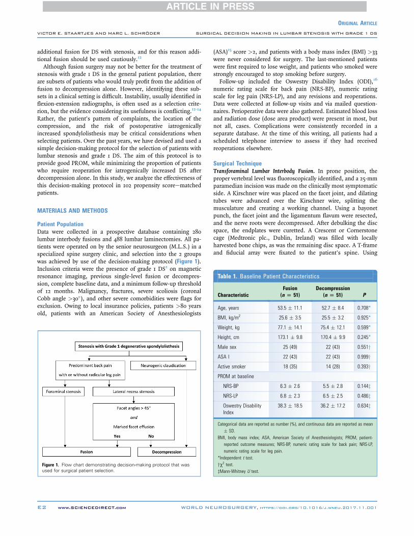
Table 1. Baseline Patient Characteristics
CharacteristicFusion(n [ 51)
Decompression(n [ 51) P
Age, years 53.5 � 11.1 52.7 � 8.4 0.708*2
ORIGINAL ARTICLE
VICTOR E. STAARTJES AND MARC L. SCHRÖDER SURGICAL DECISION MAKING IN LUMBAR STENOSIS WITH GRADE 1 DS
additional fusion for DS with stenosis, and for this reason addi-tional fusion should be used cautiously.12
Although fusion surgery may not be better for the treatment ofstenosis with grade 1 DS in the general patient population, thereare subsets of patients who would truly profit from the addition offusion to decompression alone. However, identifying these sub-sets in a clinical setting is difficult. Instability, usually identified inflexion-extension radiographs, is often used as a selection crite-rion, but the evidence considering its usefulness is conflicting.12-14
Rather, the patient’s pattern of complaints, the location of thecompression, and the risk of postoperative iatrogenicallyincreased spondylolisthesis may be critical considerations whenselecting patients. Over the past years, we have devised and used asimple decision-making protocol for the selection of patients withlumbar stenosis and grade 1 DS. The aim of this protocol is toprovide good PROM, while minimizing the proportion of patientswho require reoperation for iatrogenically increased DS afterdecompression alone. In this study, we analyze the effectiveness ofthis decision-making protocol in 102 propensity scoreematchedpatients.
MATERIALS AND METHODS
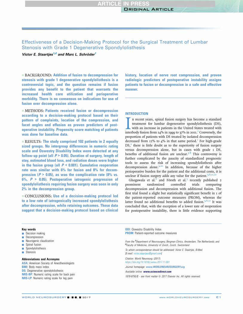
Patient PopulationData were collected in a prospective database containing 280lumbar interbody fusions and 488 lumbar laminectomies. All pa-tients were operated on by the senior neurosurgeon (M.L.S.) in aspecialized spine surgery clinic, and selection into the 2 groupswas achieved by use of the decision-making protocol (Figure 1).Inclusion criteria were the presence of grade 1 DS2 on magneticresonance imaging, previous single-level fusion or decompres-sion, complete baseline data, and a minimum follow-up thresholdof 12 months. Malignancy, fractures, severe scoliosis (coronalCobb angle >30�), and other severe comorbidities were flags forexclusion. Owing to local insurance policies, patients >80 yearsold, patients with an American Society of Anesthesiologists
Figure 1. Flow chart demonstrating decision-making protocol that wasused for surgical patient selection.
E2 www.SCIENCEDIRECT.com WORLD NE
(ASA)15 score >2, and patients with a body mass index (BMI) >33were never considered for surgery. The last-mentioned patientswere first required to lose weight, and patients who smoked werestrongly encouraged to stop smoking before surgery.Follow-up included the Oswestry Disability Index (ODI),16
numeric rating scale for back pain (NRS-BP), numeric ratingscale for leg pain (NRS-LP), and any revisions and reoperations.Data were collected at follow-up visits and via mailed question-naires. Perioperative data were also gathered. Estimated blood lossand radiation dose (dose area product) were present in most, butnot all, cases. Complications were consistently recorded in aseparate database. At the time of this writing, all patients had ascheduled telephone interview to assess if they had receivedreoperations elsewhere.
Surgical TechniqueTransforaminal Lumbar Interbody Fusion. In prone position, theproper vertebral level was fluoroscopically identified, and a 25-mmparamedian incision was made on the clinically most symptomaticside. A Kirschner wire was placed on the facet joint, and dilatingtubes were advanced over the Kirschner wire, splitting themusculature and creating a working channel. Using a bayonetpunch, the facet joint and the ligamentum flavum were resected,and the nerve roots were decompressed. After debulking the discspace, the endplates were curetted. A Crescent or Cornerstonecage (Medtronic plc., Dublin, Ireland) was filled with locallyharvested bone chips, as was the remaining disc space. A T-frameand fiducial array were fixated to the patient’s spine. Using
BMI, kg/m 25.6 � 3.5 25.5 � 3.2 0.925*
Weight, kg 77.1 � 14.1 75.4 � 12.1 0.599*
Height, cm 173.1 � 9.8 170.4 � 9.9 0.245*
Male sex 25 (49) 22 (43) 0.551yASA I 22 (43) 22 (43) 0.999yActive smoker 18 (35) 14 (28) 0.393yPROM at baseline
NRS-BP 6.3 � 2.6 5.5 � 2.8 0.144zNRS-LP 6.8 � 2.3 6.5 � 2.5 0.486zOswestry DisabilityIndex
38.3 � 18.5 36.2 � 17.2 0.634z
Categorical data are reported as number (%), and continuous data are reported as mean� SD.
BMI, body mass index; ASA, American Society of Anesthesiologists; PROM, patient-reported outcome measures; NRS-BP, numeric rating scale for back pain; NRS-LP,numeric rating scale for leg pain.
*Independent t test.yc2 test.zMann-Whitney U test.
UROSURGERY, https://doi.org/10.1016/j.wneu.2017.11.001
ORIGINAL ARTICLE
VICTOR E. STAARTJES AND MARC L. SCHRÖDER SURGICAL DECISION MAKING IN LUMBAR STENOSIS WITH GRADE 1 DS
fluoroscopy, the patient’s current spinal anatomy was matched tothe trajectories that were preplanned on computed tomographyimages. Kirschner wires were inserted percutaneously underguidance of the SpineAssist robot (Mazor Robotics Ltd., Caesarea,Israel) under fluoroscopic control. Pedicle screws were insertedover the wires. Using the Sextant system (Medtronic plc.),reduction was achieved, if necessary, and 2 curved rods wereinserted percutaneously.
Decompression. In knee-elbow position, a 50-mm midline incisionwas made after fluoroscopic identification of the proper vertebrallevel. Via a muscle-splitting approach, a mini-open retractor wasplaced. The interspinous ligament was cut, and the spinous pro-cess was partially resected. Bilateral partial hemilaminectomy wasthen performed. The interspinous ligament was deliberatelyresected to contralaterally undercut the hypertrophic ligamentum
Figure 2. Graphic representation of patient-reported outcome measuresduring the follow-up period. Error bars represent SEM. Change scoresrepresent mean difference between the values at baseline and last
WORLD NEUROSURGERY-: ---, - 2017
flavum and osteophytes. The facet joints were left untouchedwherever possible to preserve biomechanical integrity.17 If needed,only their hypertrophic medial part was partially resected. Thelateral recesses and foramina were further opened until thenerve roots appeared to be fully released. Discectomy wasperformed only in cases of significant nerve root compressionby a bulging disc at the index level.
StatisticsPatients who met inclusion and exclusion criteria were pooled,and 2 optimal groups were constructed using nearest-neighborpropensity scoreebased matching. This was achieved using theMatchIt18 code for R (R Foundation for Statistical Computing,Vienna, Austria; https://www.R-project.org/).19 Patients werematched for age; BMI; sex; ASA score; smoking status; andbaseline NRS-BP, NRS-LP, and ODI. Categorical data are
follow-up. ODI, Oswestry Disability Index; NRS, numeric rating scale;PROM, patient-reported outcome measures.
www.WORLDNEUROSURGERY.org E3
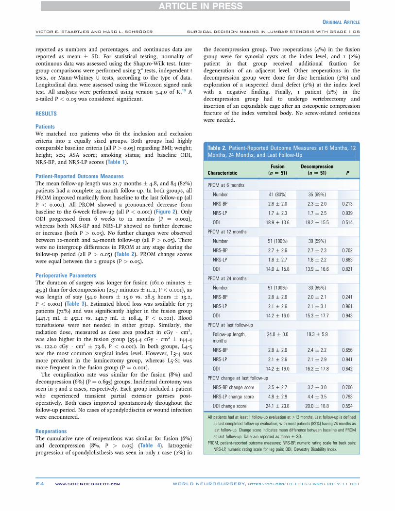
Table 2. Patient-Reported Outcome Measures at 6 Months, 12Months, 24 Months, and Last Follow-Up
CharacteristicFusion(n [ 51)
Decompression(n [ 51) P
PROM at 6 months
Number 41 (80%) 35 (69%)
NRS-BP 2.8 � 2.0 2.3 � 2.0 0.213
NRS-LP 1.7 � 2.3 1.7 � 2.5 0.939
ODI 18.9 � 13.6 18.2 � 15.5 0.514
PROM at 12 months
Number 51 (100%) 30 (59%)
NRS-BP 2.7 � 2.6 2.7 � 2.3 0.702
NRS-LP 1.8 � 2.7 1.6 � 2.2 0.663
ODI 14.0 � 15.8 13.9 � 16.6 0.821
PROM at 24 months
Number 51 (100%) 33 (65%)
NRS-BP 2.8 � 2.6 2.0 � 2.1 0.241
NRS-LP 2.1 � 2.6 2.1 � 3.1 0.961
ODI 14.2 � 16.0 15.3 � 17.7 0.943
PROM at last follow-up
Follow-up length,months
24.0 � 0.0 19.3 � 5.9
NRS-BP 2.8 � 2.6 2.4 � 2.2 0.656
NRS-LP 2.1 � 2.6 2.1 � 2.9 0.941
ODI 14.2 � 16.0 16.2 � 17.8 0.642
PROM change at last follow-up
NRS-BP change score 3.5 � 2.7 3.2 � 3.0 0.706
NRS-LP change score 4.8 � 2.9 4.4 � 3.5 0.793
ODI change score 24.1 � 20.8 20.0 � 18.8 0.594
All patients had at least 1 follow-up evaluation at �12 months. Last follow-up is definedas last completed follow-up evaluation, with most patients (82%) having 24 months aslast follow-up. Change score indicates mean difference between baseline and PROMat last follow-up. Data are reported as mean � SD.
PROM, patient-reported outcome measures; NRS-BP, numeric rating scale for back pain;NRS-LP, numeric rating scale for leg pain; ODI, Oswestry Disability Index.
ORIGINAL ARTICLE
VICTOR E. STAARTJES AND MARC L. SCHRÖDER SURGICAL DECISION MAKING IN LUMBAR STENOSIS WITH GRADE 1 DS
reported as numbers and percentages, and continuous data arereported as mean � SD. For statistical testing, normality ofcontinuous data was assessed using the Shapiro-Wilk test. Inter-group comparisons were performed using c2 tests, independent ttests, or Mann-Whitney U tests, according to the type of data.Longitudinal data were assessed using the Wilcoxon signed ranktest. All analyses were performed using version 3.4.0 of R.19 A2-tailed P < 0.05 was considered significant.
RESULTS
PatientsWe matched 102 patients who fit the inclusion and exclusioncriteria into 2 equally sized groups. Both groups had highlycomparable baseline criteria (all P > 0.05) regarding BMI; weight;height; sex; ASA score; smoking status; and baseline ODI,NRS-BP, and NRS-LP scores (Table 1).
Patient-Reported Outcome MeasuresThe mean follow-up length was 21.7 months � 4.8, and 84 (82%)patients had a complete 24-month follow-up. In both groups, allPROM improved markedly from baseline to the last follow-up (allP < 0.001). All PROM showed a pronounced decrease frombaseline to the 6-week follow-up (all P < 0.001) (Figure 2). OnlyODI progressed from 6 weeks to 12 months (P ¼ 0.002),whereas both NRS-BP and NRS-LP showed no further decreaseor increase (both P > 0.05). No further changes were observedbetween 12-month and 24-month follow-up (all P > 0.05). Therewere no intergroup differences in PROM at any stage during thefollow-up period (all P > 0.05) (Table 2). PROM change scoreswere equal between the 2 groups (P > 0.05).
Perioperative ParametersThe duration of surgery was longer for fusion (161.0 minutes �45.9) than for decompression (25.7 minutes � 11.2, P < 0.001), aswas length of stay (54.0 hours � 15.0 vs. 28.5 hours � 13.2,P < 0.001) (Table 3). Estimated blood loss was available for 73patients (72%) and was significantly higher in the fusion group(443.3 mL � 452.1 vs. 142.7 mL � 108.4, P < 0.001). Bloodtransfusions were not needed in either group. Similarly, theradiation dose, measured as dose area product in cGy $ cm2,was also higher in the fusion group (354.4 cGy $ cm2 � 144.4vs. 122.0 cGy $ cm2 � 73.6, P < 0.001). In both groups, L4-5was the most common surgical index level. However, L3-4 wasmore prevalent in the laminectomy group, whereas L5-S1 wasmore frequent in the fusion group (P ¼ 0.001).The complication rate was similar for the fusion (8%) and
decompression (6%) (P ¼ 0.695) groups. Incidental durotomy wasseen in 3 and 2 cases, respectively. Each group included 1 patientwho experienced transient partial extensor pareses post-operatively. Both cases improved spontaneously throughout thefollow-up period. No cases of spondylodiscitis or wound infectionwere encountered.
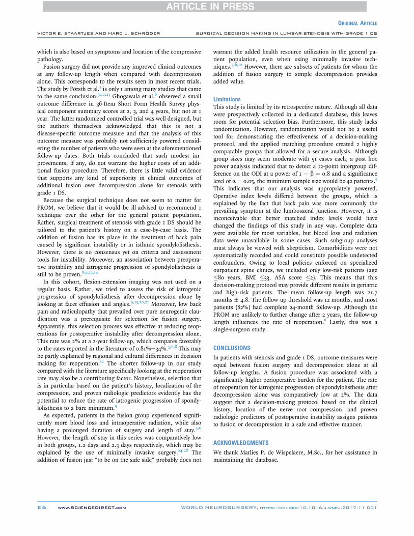
ReoperationsThe cumulative rate of reoperations was similar for fusion (6%)and decompression (8%, P > 0.05) (Table 4). Iatrogenicprogression of spondylolisthesis was seen in only 1 case (2%) in
E4 www.SCIENCEDIRECT.com WORLD NE
the decompression group. Two reoperations (4%) in the fusiongroup were for synovial cysts at the index level, and 1 (2%)patient in that group received additional fixation fordegeneration of an adjacent level. Other reoperations in thedecompression group were done for disc herniation (2%) andexploration of a suspected dural defect (2%) at the index levelwith a negative finding. Finally, 1 patient (2%) in thedecompression group had to undergo vertebrectomy andinsertion of an expandable cage after an osteopenic compressionfracture of the index vertebral body. No screw-related revisionswere needed.
UROSURGERY, https://doi.org/10.1016/j.wneu.2017.11.001
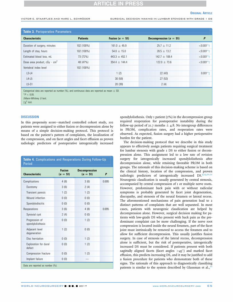
Table 3. Perioperative Parameters
Characteristic Patients Fusion (n [ 51) Decompression (n [ 51) P
Duration of surgery, minutes 102 (100%) 161.0 � 45.9 25.7 � 11.2 <0.001*yLength of stay, hours 102 (100%) 54.0 � 15.0 28.5 � 13.2 <0.001*yEstimated blood loss, mL 73 (72%) 443.3 � 452.1 142.7 � 108.4 <0.001*yDose area product, cGy $ cm2 48 (47%) 354.4 � 144.4 122.0 � 73.6 <0.001*yVertebral index level 102 (100%)
L3-L4 1 (2) 22 (43) 0.001*zL4-L5 30 (59) 27 (53)
L5-S1 20 (39) 2 (4)
Categorical data are reported as number (%), and continuous data are reported as mean � SD.*P < 0.05.yMann-Whitney U test.zc2 test.
ORIGINAL ARTICLE
VICTOR E. STAARTJES AND MARC L. SCHRÖDER SURGICAL DECISION MAKING IN LUMBAR STENOSIS WITH GRADE 1 DS
DISCUSSION
In this propensity scoreematched controlled cohort study, 102patients were assigned to either fusion or decompression alone bymeans of a simple decision-making protocol. This protocol isbased on the patient’s pattern of complaints, the localization ofthe compression, and on facet angles and facet effusion as provenradiologic predictors of postoperative iatrogenically increased
Table 4. Complications and Reoperations During Follow-UpPeriod
CharacteristicFusion(n [ 51)
Decompression(n [ 51) P
Complications 4 (8) 3 (6) 0.695
Durotomy 3 (6) 2 (4)
Transient paresis 1 (2) 1 (2)
Wound infection 0 (0) 0 (0)
Spondylodiscitis 0 (0) 0 (0)
Reoperations 3 (6) 4 (8) 0.695
Synovial cyst 2 (4) 0 (0)
Progression ofspondylolisthesis
0 (0) 1 (2)
Adjacent leveldegeneration
1 (2) 0 (0)
Disc herniation 0 (0) 1 (2)
Exploration for duraldefect
0 (0) 1 (2)
Compression fracture 0 (0) 1 (2)
Implant failure 0 (0) —
Data are reported as number (%).
WORLD NEUROSURGERY-: ---, - 2017
spondylolisthesis. Only 1 patient (2%) in the decompression grouprequired reoperation for postoperative instability during thefollow-up period of 21.7 months � 4.8. No intergroup differencesin PROM, complication rates, and reoperation rates wereobserved. As expected, fusion surgery had a higher perioperativeburden for the patient.The decision-making protocol that we describe in this study
appears to effectively assign patients requiring surgical treatmentfor lumbar stenosis with grade 1 DS to either fusion or decom-pression alone. This assignment led to a low rate of revisionsurgery for iatrogenically increased spondylolisthesis afterdecompression alone, while retaining favorable PROM in bothgroups. The rationale of this decision-making scheme is based onthe clinical history, location of the compression, and provenradiologic predictors of iatrogenically increased DS.9,13,20,21
Neurogenic claudication is usually generated by central stenosis,accompanied by central compression of 1 or multiple nerve roots.However, predominant back pain with or without radicularsymptoms is probably generated by facet joint degeneration,discopathy, and stenosis of the neural foramen or lateral recess.The aforementioned mechanisms of pain generation lead to 2distinct patterns of complaints that are well separated. In mostcases, patients with neurogenic claudication are helped bydecompression alone. However, surgical decision making for pa-tients with low-grade DS who present with back pain as the pre-dominant complaint can be more challenging. If the nerve rootcompression is located inside the neural foramen, part of the facetjoint must intrinsically be removed to access the foramen and toallow for sufficient decompression. This usually justifies fusionsurgery. In case of stenosis of the lateral recess, decompressionalone is sufficient, but the risk of postoperative, iatrogenicallyincreased DS must be considered. If patients present with bothsagittally aligned facets (facet angles >45�) and marked faceteffusion, this predicts increasing DS, and it may be justified to adda fusion procedure for patients who demonstrate both of thesesigns. The rationale of this approach to diagnostically classifyingpatients is similar to the system described by Glassman et al.,22
www.WORLDNEUROSURGERY.org E5
ORIGINAL ARTICLE
VICTOR E. STAARTJES AND MARC L. SCHRÖDER SURGICAL DECISION MAKING IN LUMBAR STENOSIS WITH GRADE 1 DS
which is also based on symptoms and location of the compressivepathology.Fusion surgery did not provide any improved clinical outcomes
at any follow-up length when compared with decompressionalone. This corresponds to the results seen in most recent trials.The study by Försth et al.5 is only 1 among many studies that cameto the same conclusion.3,12,23 Ghogawala et al.6 observed a smalloutcome difference in 36-Item Short Form Health Survey phys-ical component summary scores at 2, 3, and 4 years, but not at 1year. The latter randomized controlled trial was well designed, butthe authors themselves acknowledged that this is not adisease-specific outcome measure and that the analysis of thisoutcome measure was probably not sufficiently powered consid-ering the number of patients who were seen at the aforementionedfollow-up dates. Both trials concluded that such modest im-provements, if any, do not warrant the higher costs of an addi-tional fusion procedure. Therefore, there is little valid evidencethat supports any kind of superiority in clinical outcomes ofadditional fusion over decompression alone for stenosis withgrade 1 DS.Because the surgical technique does not seem to matter for
PROM, we believe that it would be ill-advised to recommend 1technique over the other for the general patient population.Rather, surgical treatment of stenosis with grade 1 DS should betailored to the patient’s history on a case-by-case basis. Theaddition of fusion has its place in the treatment of back paincaused by significant instability or in isthmic spondylolisthesis.However, there is no consensus yet on criteria and assessmenttools for instability. Moreover, an association between preopera-tive instability and iatrogenic progression of spondylolisthesis isstill to be proven.8,9,13,14
In this cohort, flexion-extension imaging was not used on aregular basis. Rather, we tried to assess the risk of iatrogenicprogression of spondylolisthesis after decompression alone bylooking at facet effusion and angles.9,13,20,21 Moreover, low backpain and radiculopathy that prevailed over pure neurogenic clau-dication was a prerequisite for selection for fusion surgery.Apparently, this selection process was effective at reducing reop-erations for postoperative instability after decompression alone.This rate was 2% at a 2-year follow-up, which compares favorablyto the rates reported in the literature of 0.81%e34%.5,6,8 This maybe partly explained by regional and cultural differences in decisionmaking for reoperation.12 The shorter follow-up in our studycompared with the literature specifically looking at the reoperationrate may also be a contributing factor. Nonetheless, selection thatis in particular based on the patient’s history, localization of thecompression, and proven radiologic predictors evidently has thepotential to reduce the rate of iatrogenic progression of spondy-lolisthesis to a bare minimum.9
As expected, patients in the fusion group experienced signifi-cantly more blood loss and intraoperative radiation, while alsohaving a prolonged duration of surgery and length of stay.3-6
However, the length of stay in this series was comparatively lowin both groups, 1.2 days and 2.3 days respectively, which may beexplained by the use of minimally invasive surgery.24-28 Theaddition of fusion just “to be on the safe side” probably does not
E6 www.SCIENCEDIRECT.com WORLD NE
warrant the added health resource utilization in the general pa-tient population, even when using minimally invasive tech-niques.5,6,12 However, there are subsets of patients for whom theaddition of fusion surgery to simple decompression providesadded value.
LimitationsThis study is limited by its retrospective nature. Although all datawere prospectively collected in a dedicated database, this leavesroom for potential selection bias. Furthermore, this study lacksrandomization. However, randomization would not be a usefultool for demonstrating the effectiveness of a decision-makingprotocol, and the applied matching procedure created 2 highlycomparable groups that allowed for a secure analysis. Althoughgroup sizes may seem moderate with 51 cases each, a post hocpower analysis indicated that to detect a 12-point intergroup dif-ference on the ODI at a power of 1 � b ¼ 0.8 and a significancelevel of a ¼ 0.05, the minimum sample size would be 42 patients.5
This indicates that our analysis was appropriately powered.Operative index levels differed between the groups, which isexplained by the fact that back pain was more commonly theprevailing symptom at the lumbosacral junction. However, it isinconceivable that better matched index levels would havechanged the findings of this study in any way. Complete datawere available for most variables, but blood loss and radiationdata were unavailable in some cases. Such subgroup analysesmust always be viewed with skepticism. Comorbidities were notsystematically recorded and could constitute possible undetectedconfounders. Owing to local policies enforced on specializedoutpatient spine clinics, we included only low-risk patients (age�80 years, BMI �33, ASA score �2). This means that thisdecision-making protocol may provide different results in geriatricand high-risk patients. The mean follow-up length was 21.7months � 4.8. The follow-up threshold was 12 months, and mostpatients (82%) had complete 24-month follow-up. Although thePROM are unlikely to further change after 2 years, the follow-uplength influences the rate of reoperation.6 Lastly, this was asingle-surgeon study.
CONCLUSIONS
In patients with stenosis and grade 1 DS, outcome measures wereequal between fusion surgery and decompression alone at allfollow-up lengths. A fusion procedure was associated with asignificantly higher perioperative burden for the patient. The rateof reoperation for iatrogenic progression of spondylolisthesis afterdecompression alone was comparatively low at 2%. The datasuggest that a decision-making protocol based on the clinicalhistory, location of the nerve root compression, and provenradiologic predictors of postoperative instability assigns patientsto fusion or decompression in a safe and effective manner.
ACKNOWLEDGMENTS
We thank Marlies P. de Wispelaere, M.Sc., for her assistance inmaintaining the database.
UROSURGERY, https://doi.org/10.1016/j.wneu.2017.11.001
ORIGINAL ARTICLE
VICTOR E. STAARTJES AND MARC L. SCHRÖDER SURGICAL DECISION MAKING IN LUMBAR STENOSIS WITH GRADE 1 DS
REFERENCES10. Sato S, Yagi M, Mach
Miyake A, et al. Reop
1. Kepler CK, Vaccaro AR, Hilibrand AS,Anderson DG, Rihn JA, Albert TJ, et al. Nationaltrends in the use of fusion techniques to treatdegenerative spondylolisthesis. Spine (Phila Pa1976). 2014;39:1584-1589.
2. Meyerding HW. Spondylolisthesis; surgical fusionof lumbosacral portion of spinal column andinterarticular facets; use of autogenous bonegrafts for relief of disabling backache. J Int CollSurg. 1956;26(5 Part 1):566-591.
3. Austevoll IM, Gjestad R, Brox JI, Solberg TK,Storheim K, Rekeland F, et al. The effectiveness ofdecompression alone compared with additionalfusion for lumbar spinal stenosis with degenera-tive spondylolisthesis: a pragmatic comparativenon-inferiority observational study from the Nor-wegian Registry for Spine Surgery. Eur Spine J.2017;26:404-413.
4. Chang W, Yuwen P, Zhu Y, Wei N, Feng C,Zhang Y, et al. Effectiveness of decompressionalone versus decompression plus fusion for lum-bar spinal stenosis: a systematic review and meta-analysis. Arch Orthop Trauma Surg. 2017;137:637-650.
5. Försth P, Ólafsson G, Carlsson T, Frost A,Borgström F, Fritzell P, et al. A randomized,controlled trial of fusion surgery for lumbar spinalstenosis. N Engl J Med. 2016;374:1413-1423.
6. Ghogawala Z, Dziura J, Butler WE, Dai F,Terrin N, Magge SN, et al. Laminectomy plusfusion versus laminectomy alone for lumbarspondylolisthesis. N Engl J Med. 2016;374:1424-1434.
7. Ghogawala Z, Resnick DK, Glassman SD,Dziura J, Shaffrey CI, Mummaneni PV. Random-ized controlled trials for degenerative lumbarspondylolisthesis: which patients benefit fromlumbar fusion? J Neurosurg Spine. 2016;26:260-266.
8. Guha D, Heary RF, Shamji MF. Iatrogenic spon-dylolisthesis following laminectomy for degener-ative lumbar stenosis: systematic review andcurrent concepts. Neurosurg Focus. 2015;39:E9.
9. Blumenthal C, Curran J, Benzel EC, Potter R,Magge SN, Harrington JF Jr, et al. Radiographicpredictors of delayed instability followingdecompression without fusion for degenerativegrade I lumbar spondylolisthesis. J Neurosurg Spine.2013;18:340-346.
WORLD NEUROSURGERY-: ---, - 2017
ida M, Yasuda A, Konomi T,eration rate and risk factors
of elective spinal surgery for degenerative spon-dylolisthesis: minimum 5-year follow-up. Spine J.2015;15:1536-1544.
11. Deyo RA, Mirza SK, Martin BI, Kreuter W,Goodman DC, Jarvik JG. Trends, major medicalcomplications, and charges associated with sur-gery for lumbar spinal stenosis in older adults.JAMA. 2010;303:1259-1265.
12. Peul WC, Moojen WA. Fusion for lumbar spinalstenosis—safeguard or superfluous surgicalimplant? N Engl J Med. 2016;374:1478-1479.
13. Hasegawa K, Kitahara K, Shimoda H, Ishii K,Ono M, Homma T, et al. Lumbar degenerativespondylolisthesis is not always unstable: clin-icobiomechanical evidence. Spine (Phila Pa 1976).2014;39:2127-2135.
14. Hu K, Feng D. Fusion surgery for lumbar spinalstenosis. N Engl J Med. 2016;375:598-599.
15. Saklad M. Grading of patients for surgical pro-cedures. Anesthesiology. 1941;2:281-284.
16. Fairbank JC, Couper J, Davies JB, O’Brien JP. TheOswestry low back pain disability questionnaire.Physiotherapy. 1980;66:271-273.
17. Detwiler PW, Spetzler CB, Taylor SB,Crawford NR, Porter RW, Sonntag VKH. Biome-chanical comparison of facet-sparing laminectomyand Christmas tree laminectomy. J Neurosurg Spine.2003;99:214-220.
18. Ho D, Imai K, King G, Stuart E. Matching asnonparametric preprocessing for reducing modeldependence in parametric causal inference. PolitAnal. 2007;15:199-236.
19. R Core Team. R: A Language and Environment forStatistical Computing. Vienna, Austria: R Foundationfor Statistical Computing; 2017.
20. Enyo Y, Yoshimura N, Yamada H, Hashizume H,Yoshida M. Radiographic natural course of lum-bar degenerative spondylolisthesis and its riskfactors related to the progression and onset in a15-year community-based cohort study: theMiyama study. J Orthop Sci. 2015;20:978-984.
21. Oishi Y, Murase M, Hayashi Y, Ogawa T,Hamawaki J. Smaller facet effusion in associationwith restabilization at the time of operation inJapanese patients with lumbar degenerativespondylolisthesis. J Neurosurg Spine. 2009;12:88-95.
22. Glassman SD, Carreon LY, Anderson PA,Resnick DK. A diagnostic classification for lum-bar spine registry development. Spine J. 2011;11:1108-1116.
23. Sigmundsson FG, Jönsson B, Strömqvist B.Outcome of decompression with and withoutfusion in spinal stenosis with degenerative spon-dylolisthesis in relation to preoperative painpattern: a register study of 1,624 patients. Spine J.2015;15:638-646.
24. Adogwa O, Parker SL, Bydon A, Cheng J,McGirt MJ. Comparative effectiveness of mini-mally invasive versus open transforaminal lumbarinterbody fusion: 2-year assessment of narcoticuse, return to work, disability, and quality of life.J Spinal Disord Tech. 2011;24:479-484.
25. Archavlis E, Carvi y Nievas M. Comparison ofminimally invasive fusion and instrumentationversus open surgery for severe stenotic spondylo-listhesis with high-grade facet joint osteoarthritis.Eur Spine J. 2013;22:1731-1740.
26. Goldstein CL, Phillips FM, Rampersaud YR.Comparative effectiveness and economic evalua-tions of open versus minimally invasive posterioror transforaminal lumbar interbody fusion: asystematic review. Spine (Phila Pa 1976). 2016;41(suppl 8):S74-89.
27. Mummaneni PV, Bisson EF, Kerezoudis P,Glassman S, Foley K, Slotkin JR, et al. Minimallyinvasive versus open fusion for grade I degenera-tive lumbar spondylolisthesis: analysis of theQuality Outcomes Database. Neurosurg Focus. 2017;43:E11.
28. Ntoukas V, Müller A. Minimally invasive approachversus traditional open approach for one levelposterior lumbar interbody fusion. Minim InvasiveNeurosurg. 2010;53:21-24.
Conflict of interest statement: M.L.S. is a consultant toMazor Robotics, Ltd. The other author has no conflicts toreport.
Received 11 October 2017; accepted 1 November 2017
Citation: World Neurosurg. (2017).https://doi.org/10.1016/j.wneu.2017.11.001
Journal homepage: www.WORLDNEUROSURGERY.org
Available online: www.sciencedirect.com
1878-8750/$ - see front matter ª 2017 Elsevier Inc. Allrights reserved.
www.WORLDNEUROSURGERY.org E7