Embed Size (px)
Citation preview

Citation: Gerhardt-Szep S, Schröder-Schichtel E, Zücker Q, Zahn T, Feierabend S, et al. (2016) Problems of Direct Composite Posterior Restorations: A Clinical Study. J Dent Probl Solut 3(1): 035-039. DOI: 10.17352/2394-8418.000032
Journal of Dental Problems and Solutions
035
IntroductionIn recent years, the increasing demand for aesthetically appealing,
naturally-coloured dental restoration options has given rise to a growth in the use of composites in the posterior dental area [1-5]. The declining acceptance of dental amalgam and the mercury problem also makes an alternative to amalgam necessary [6,7]. In a statement from the German Scientific Association for Operative Dentistry and European Federation of Conservative Dentistry, it is defined, that indications for the use of direct composite systems may vary according to specific circumstances [8]. Three different indications are named in this statement, including restorations of tooth structure and contour, shape changing restorations and combinations of these possibilities [8]. However it remains clear, that restorations in the posterior area, which are subject to high mechanical stresses, should always be performed using materials with high strength and good radiopacity properties. Many authors of comparable long-term studies referred to the use of hybrid composites, as only such materials demonstrated both superior restoration margin stability and much better physical properties, including adequate abrasion resistance and flexural strength, which maked them suitable replacements for amalgam [1,9-11]. To meet the requirements of good long-term clinical and aesthetically appealing therapy results using a composite filling, it is also essential to consistently adhere to processing parameters. Even the smallest deviations from the application recommendations can cause clinical failures [7,12-14].
In order to carry out clinical investigations of composite materials most researchers used different criteria (for example the
Research Article
Problems of Direct Composite Posterior Restorations: A Clinical Study
Susanne Gerhardt-Szep1*, Elke Schröder-Schichtel2, Quanita Zücker3, Tugba Zahn4, Stefanie Feierabend5 and Stefan Rüttermann1 1Department of Operative Dentistry, Carolinum Dental University-Institute GmbH, J.W. Goethe University, Frankfurt am Main, Germany2Bavarian Association Dental health e.V. (LAGZ), München, Germany3Dental office, Riedstadt, Germany4Department of Prosthetic Dentistry, Carolinum Dental University-Institute GmbH, J.W. Goethe University, Frankfurt am Main, Germany5Department of Restorative Dentistry and Periodontology, University Medical Center Freiburg, Germany
Dates: Received: 23 June, 2016; Accepted: 12 July, 2016; Published: 14 July, 2016
*Corresponding author: PD Dr. Susanne Gerhardt-Szep, MME, Department of Operative Dentistry, Carolinum Dental University-Institute GmbH, J.W. Goethe University, Theodor-Stern-Kai 7, Frankfurt am Main 60590, Germany, Tel: +49-69-6301-7505; E-mail:
www.peertechz.com
ISSN: 2394-8418
Keywords: Resin composites; Clinical trial; Modified FDI criteria; In vivo; In vitro
AbstractThe aim of this clinical study was to describe the quality of posterior composite restorations (n =
329) performed on a group of patients (n=219) during an observation period of three years at various intervals (6, 12, 18, 24 and 36 months) after application.
The parameters were assessed both In vivo and In vitro using clinical examinations, impressions and photography according to modified FDI criteria. For the statistical analysis of the results, the Wilcoxon test with a significance level of p = 0.05 was applied.
After three years, In vivo five from the seven parameters exhibited significant changes. Only “retention” and “approximal contact” remained unchanged. In vitro studied parameters “anatomical form”, “occlusal contour/wear” and “approximal contact” did not result in any significant changes, however “marginal adaptation”, “surface luster” and “overhangs” deteriorated significantly.
In summary, the results of this study show that composite posterior restorations were clinically acceptable in terms of specific parameters. However, unsatisfactory results have arisen in relation to the handling of composites, stated In vivo and In vitro especially in the reconstruction of the marginal adaptation, surface and overfilling.
traditional United States Public Health Service / USPHS also known as ‘Ryge criteria’) [15]. The 16 “FDI clinical criteria” for the evaluation of direct and indirect restorations were first published in 2007 and have since been applied by some investigators in clinical studies on resin composite restorations in posterior teeth [16]. The criteria were categorized into three groups: esthetic parameters (four criteria), functional parameters (six criteria) and biological parameters (six criteria). Each criterion can be expressed with five scores, three for acceptable and two for non-acceptable (one for reparable and one for replacement) situations. They are however not definitely fixed; modifications and/or alterations are possible [16]. Comparing the FDI- and the USPHS-criteria for the evaluation of restorations in deciduous teeth authors concluded that the newer FDI method was more sensitive for identifying differences in deciduous composite resin restorations [16]. Potential clinically assessed problems can thus be elicited by using them.
Therefore, the purpose of this study was to evaluate the clinical performance and potential problems of posterior composite restorations in a period of three years after application at different time intervals on the basis of modified FDI criteria. The primary outcome was defined using functional parameters in regard of the approximal contact, marginal adaptation, occlusal contour / wear, overhangs and retention. Secondary outcomes included esthetic parameters like the anatomical form and surface luster of the restoration.
Material and MethodsIn the context of this long-term study, posterior composite
restorations applied using an ultra-fine hybrid composite (Herculite XRV, Kerr, Karlsruhe, Germany) in the student courses of the clinical
Citation: Gerhardt-Szep S, Schröder-Schichtel E, Zücker Q, Zahn T, Feierabend S, et al. (2016) Problems of Direct Composite Posterior Restorations: A Clinical Study. J Dent Probl Solut 3(1): 035-039. DOI: 10.17352/2394-8418.000032
Gerhardt-Szep et al. (2016)
036
semesters at the Department of Operative Dentistry, Carolinum Dental University-Institute GmbH in Frankfurt / Main (Germany) were given follow-up examinations. The restorations were all applied after predefined clinical protocol (for example: 1. no bevelling of the preparation; 2. Consistent rubberdam application; 3. consistent metal matrix band application; 4. Total-Etch-Technique performed with Optibond FL (Kerr, Karlsruhe, Germany); 5. Incremental composite application technique in 2 mm layers each and 6. Surface contouring / finishing using carbide or fine diamond burs).
Two trained examiners carried out the rating and ranking procedures. Finally, in a case of disagreement between both, the less favorable rating was noted. The evaluation intervals of 6, 12, 18, 24 and 36 months after the application of the restorations were selected. The fillings were examined and photographed In vivo at these intervals. To enable a differentiated evaluation, all restorations were additionally assessed on the basis of replica models.
Patient group and examination intervalsPatient selected met the following criteria: 1. absence of pain;
2. Application of posterior composite restorations in the student courses of the department of operative dentistry in the last 36 months; 3. Application after predefined protocol; 4. age 18-70 years. A total of 229 patients were requested by letter to participate in the program. 219 patients attended the follow-up examinations during which it was possible to examine 329 composite posterior dental restorations. Not all of the included restorations were examined consistently with the specified intervals.
Clinical follow-up examinations The restorations were inspected and evaluated subjectively on
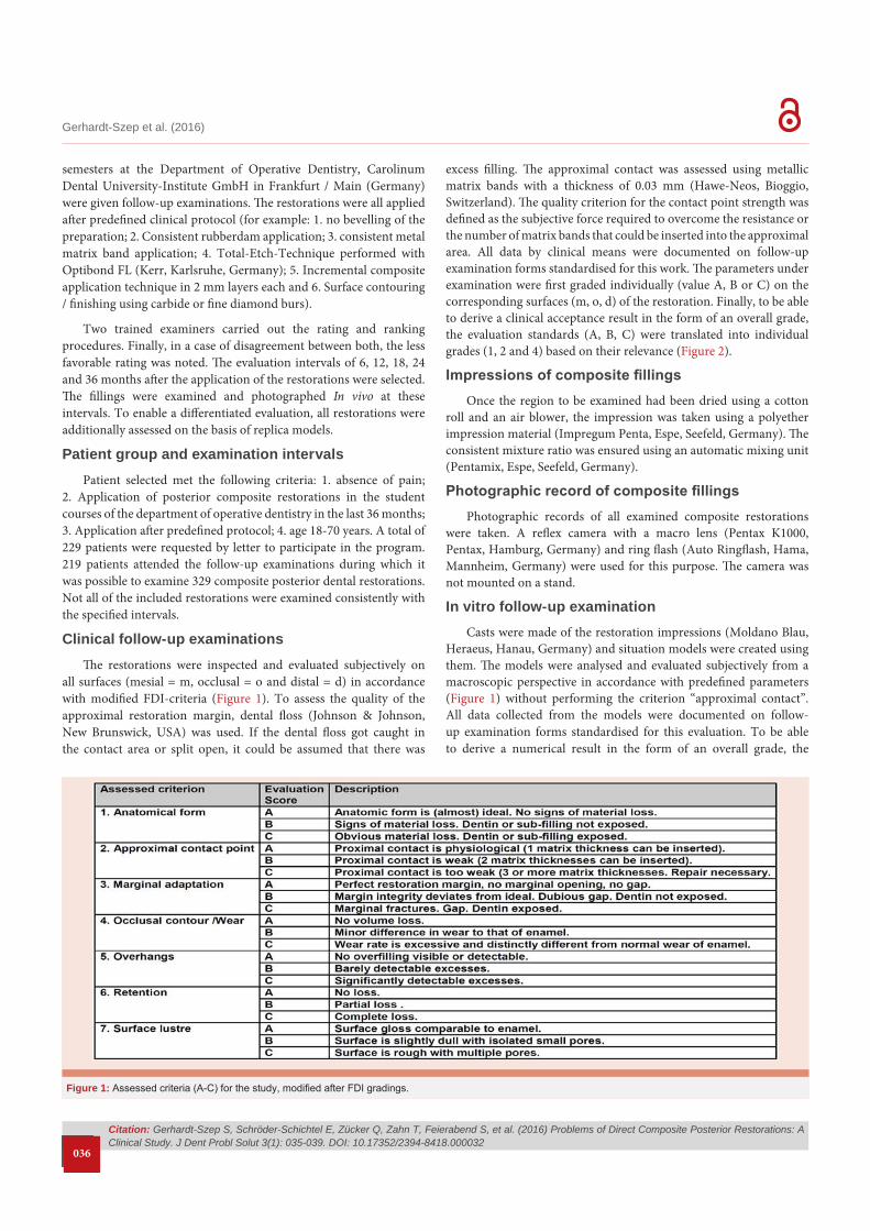
all surfaces (mesial = m, occlusal = o and distal = d) in accordance with modified FDI-criteria (Figure 1). To assess the quality of the approximal restoration margin, dental floss (Johnson & Johnson, New Brunswick, USA) was used. If the dental floss got caught in the contact area or split open, it could be assumed that there was
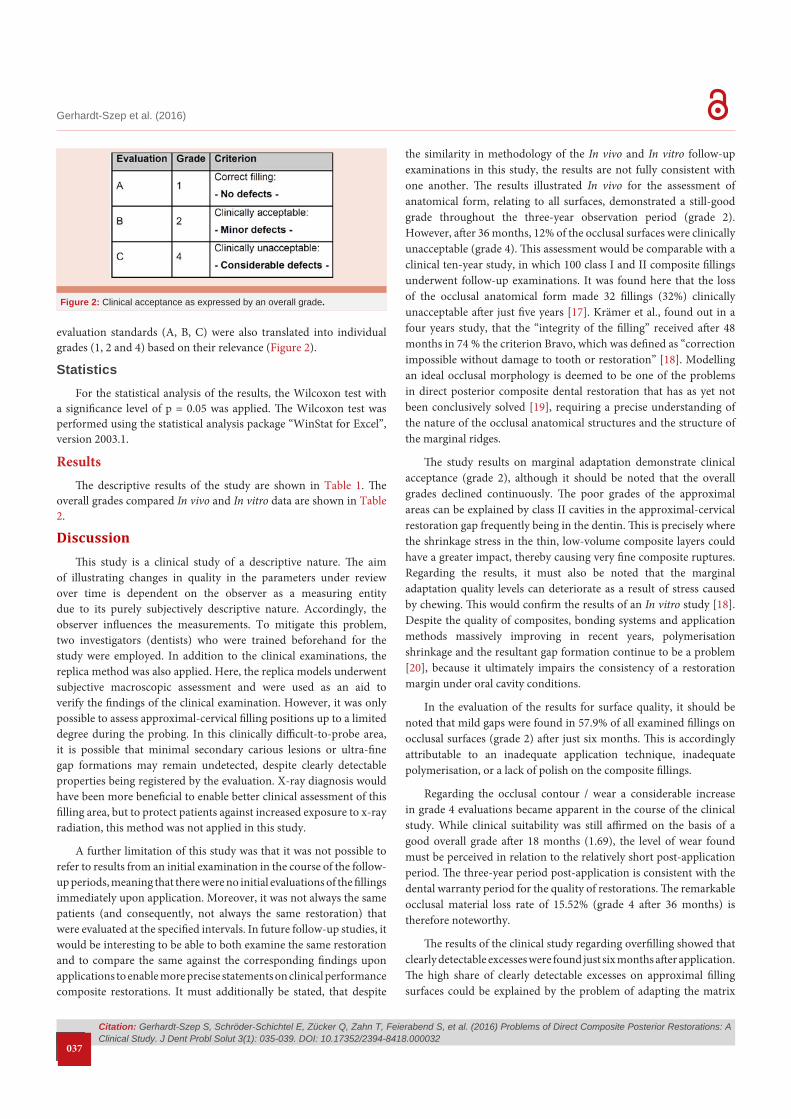
excess filling. The approximal contact was assessed using metallic matrix bands with a thickness of 0.03 mm (Hawe-Neos, Bioggio, Switzerland). The quality criterion for the contact point strength was defined as the subjective force required to overcome the resistance or the number of matrix bands that could be inserted into the approximal area. All data by clinical means were documented on follow-up examination forms standardised for this work. The parameters under examination were first graded individually (value A, B or C) on the corresponding surfaces (m, o, d) of the restoration. Finally, to be able to derive a clinical acceptance result in the form of an overall grade, the evaluation standards (A, B, C) were translated into individual grades (1, 2 and 4) based on their relevance (Figure 2).
Impressions of composite fillingsOnce the region to be examined had been dried using a cotton
roll and an air blower, the impression was taken using a polyether impression material (Impregum Penta, Espe, Seefeld, Germany). The consistent mixture ratio was ensured using an automatic mixing unit (Pentamix, Espe, Seefeld, Germany).
Photographic record of composite fillingsPhotographic records of all examined composite restorations
were taken. A reflex camera with a macro lens (Pentax K1000, Pentax, Hamburg, Germany) and ring flash (Auto Ringflash, Hama, Mannheim, Germany) were used for this purpose. The camera was not mounted on a stand.
In vitro follow-up examination Casts were made of the restoration impressions (Moldano Blau,
Heraeus, Hanau, Germany) and situation models were created using them. The models were analysed and evaluated subjectively from a macroscopic perspective in accordance with predefined parameters (Figure 1) without performing the criterion “approximal contact”. All data collected from the models were documented on follow-up examination forms standardised for this evaluation. To be able to derive a numerical result in the form of an overall grade, the
Figure 1: Assessed criteria (A-C) for the study, modified after FDI gradings.
Citation: Gerhardt-Szep S, Schröder-Schichtel E, Zücker Q, Zahn T, Feierabend S, et al. (2016) Problems of Direct Composite Posterior Restorations: A Clinical Study. J Dent Probl Solut 3(1): 035-039. DOI: 10.17352/2394-8418.000032
Gerhardt-Szep et al. (2016)
037
evaluation standards (A, B, C) were also translated into individual grades (1, 2 and 4) based on their relevance (Figure 2).
StatisticsFor the statistical analysis of the results, the Wilcoxon test with
a significance level of p = 0.05 was applied. The Wilcoxon test was performed using the statistical analysis package “WinStat for Excel”, version 2003.1.
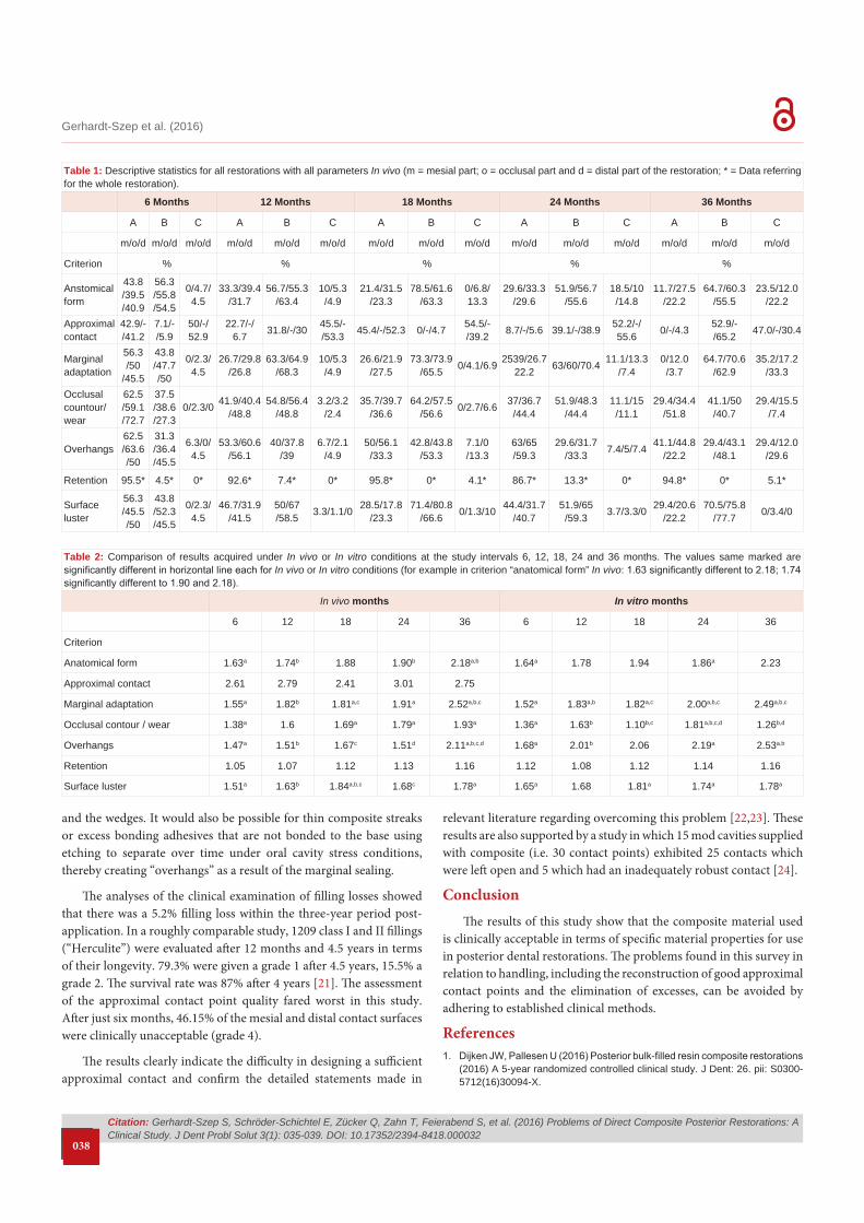
ResultsThe descriptive results of the study are shown in Table 1. The
overall grades compared In vivo and In vitro data are shown in Table 2.
DiscussionThis study is a clinical study of a descriptive nature. The aim
of illustrating changes in quality in the parameters under review over time is dependent on the observer as a measuring entity due to its purely subjectively descriptive nature. Accordingly, the observer influences the measurements. To mitigate this problem, two investigators (dentists) who were trained beforehand for the study were employed. In addition to the clinical examinations, the replica method was also applied. Here, the replica models underwent subjective macroscopic assessment and were used as an aid to verify the findings of the clinical examination. However, it was only possible to assess approximal-cervical filling positions up to a limited degree during the probing. In this clinically difficult-to-probe area, it is possible that minimal secondary carious lesions or ultra-fine gap formations may remain undetected, despite clearly detectable properties being registered by the evaluation. X-ray diagnosis would have been more beneficial to enable better clinical assessment of this filling area, but to protect patients against increased exposure to x-ray radiation, this method was not applied in this study.
A further limitation of this study was that it was not possible to refer to results from an initial examination in the course of the follow-up periods, meaning that there were no initial evaluations of the fillings immediately upon application. Moreover, it was not always the same patients (and consequently, not always the same restoration) that were evaluated at the specified intervals. In future follow-up studies, it would be interesting to be able to both examine the same restoration and to compare the same against the corresponding findings upon applications to enable more precise statements on clinical performance composite restorations. It must additionally be stated, that despite
the similarity in methodology of the In vivo and In vitro follow-up examinations in this study, the results are not fully consistent with one another. The results illustrated In vivo for the assessment of anatomical form, relating to all surfaces, demonstrated a still-good grade throughout the three-year observation period (grade 2). However, after 36 months, 12% of the occlusal surfaces were clinically unacceptable (grade 4). This assessment would be comparable with a clinical ten-year study, in which 100 class I and II composite fillings underwent follow-up examinations. It was found here that the loss of the occlusal anatomical form made 32 fillings (32%) clinically unacceptable after just five years [17]. Krämer et al., found out in a four years study, that the “integrity of the filling” received after 48 months in 74 % the criterion Bravo, which was defined as “correction impossible without damage to tooth or restoration” [18]. Modelling an ideal occlusal morphology is deemed to be one of the problems in direct posterior composite dental restoration that has as yet not been conclusively solved [19], requiring a precise understanding of the nature of the occlusal anatomical structures and the structure of the marginal ridges.
The study results on marginal adaptation demonstrate clinical acceptance (grade 2), although it should be noted that the overall grades declined continuously. The poor grades of the approximal areas can be explained by class II cavities in the approximal-cervical restoration gap frequently being in the dentin. This is precisely where the shrinkage stress in the thin, low-volume composite layers could have a greater impact, thereby causing very fine composite ruptures. Regarding the results, it must also be noted that the marginal adaptation quality levels can deteriorate as a result of stress caused by chewing. This would confirm the results of an In vitro study [18]. Despite the quality of composites, bonding systems and application methods massively improving in recent years, polymerisation shrinkage and the resultant gap formation continue to be a problem [20], because it ultimately impairs the consistency of a restoration margin under oral cavity conditions.
In the evaluation of the results for surface quality, it should be noted that mild gaps were found in 57.9% of all examined fillings on occlusal surfaces (grade 2) after just six months. This is accordingly attributable to an inadequate application technique, inadequate polymerisation, or a lack of polish on the composite fillings.
Regarding the occlusal contour / wear a considerable increase in grade 4 evaluations became apparent in the course of the clinical study. While clinical suitability was still affirmed on the basis of a good overall grade after 18 months (1.69), the level of wear found must be perceived in relation to the relatively short post-application period. The three-year period post-application is consistent with the dental warranty period for the quality of restorations. The remarkable occlusal material loss rate of 15.52% (grade 4 after 36 months) is therefore noteworthy.
The results of the clinical study regarding overfilling showed that clearly detectable excesses were found just six months after application. The high share of clearly detectable excesses on approximal filling surfaces could be explained by the problem of adapting the matrix
Figure 2: Clinical acceptance as expressed by an overall grade.
Citation: Gerhardt-Szep S, Schröder-Schichtel E, Zücker Q, Zahn T, Feierabend S, et al. (2016) Problems of Direct Composite Posterior Restorations: A Clinical Study. J Dent Probl Solut 3(1): 035-039. DOI: 10.17352/2394-8418.000032
Gerhardt-Szep et al. (2016)
038
and the wedges. It would also be possible for thin composite streaks or excess bonding adhesives that are not bonded to the base using etching to separate over time under oral cavity stress conditions, thereby creating “overhangs” as a result of the marginal sealing.
The analyses of the clinical examination of filling losses showed that there was a 5.2% filling loss within the three-year period post-application. In a roughly comparable study, 1209 class I and II fillings (“Herculite”) were evaluated after 12 months and 4.5 years in terms of their longevity. 79.3% were given a grade 1 after 4.5 years, 15.5% a grade 2. The survival rate was 87% after 4 years [21]. The assessment of the approximal contact point quality fared worst in this study. After just six months, 46.15% of the mesial and distal contact surfaces were clinically unacceptable (grade 4).
The results clearly indicate the difficulty in designing a sufficient approximal contact and confirm the detailed statements made in
relevant literature regarding overcoming this problem [22,23]. These results are also supported by a study in which 15 mod cavities supplied with composite (i.e. 30 contact points) exhibited 25 contacts which were left open and 5 which had an inadequately robust contact [24].
ConclusionThe results of this study show that the composite material used
is clinically acceptable in terms of specific material properties for use in posterior dental restorations. The problems found in this survey in relation to handling, including the reconstruction of good approximal contact points and the elimination of excesses, can be avoided by adhering to established clinical methods.
References1. Dijken JW, Pallesen U (2016) Posterior bulk-filled resin composite restorations
(2016) A 5-year randomized controlled clinical study. J Dent: 26. pii: S0300-5712(16)30094-X.
Table 1: Descriptive statistics for all restorations with all parameters In vivo (m = mesial part; o = occlusal part and d = distal part of the restoration; * = Data referring for the whole restoration).
6 Months 12 Months 18 Months 24 Months 36 Months
A B C A B C A B C A B C A B C
m/o/d m/o/d m/o/d m/o/d m/o/d m/o/d m/o/d m/o/d m/o/d m/o/d m/o/d m/o/d m/o/d m/o/d m/o/d
Criterion % % % % %
Anstomical form
43.8/39.5/40.9
56.3/55.8/54.5
0/4.7/4.5
33.3/39.4/31.7
56.7/55.3/63.4
10/5.3/4.9
21.4/31.5/23.3
78.5/61.6/63.3
0/6.8/13.3
29.6/33.3/29.6
51.9/56.7/55.6
18.5/10/14.8
11.7/27.5/22.2
64.7/60.3/55.5
23.5/12.0/22.2
Approximal contact
42.9/-/41.2
7.1/-/5.9
50/-/52.9
22.7/-/6.7 31.8/-/30 45.5/-
/53.3 45.4/-/52.3 0/-/4.7 54.5/-/39.2 8.7/-/5.6 39.1/-/38.9 52.2/-/
55.6 0/-/4.3 52.9/-/65.2 47.0/-/30.4
Marginal adaptation
56.3/50
/45.5
43.8/47.7/50
0/2.3/4.5
26.7/29.8/26.8
63.3/64.9/68.3
10/5.3/4.9
26.6/21.9/27.5
73.3/73.9/65.5 0/4.1/6.9 2539/26.7
22.2 63/60/70.4 11.1/13.3/7.4
0/12.0/3.7
64.7/70.6/62.9
35.2/17.2/33.3
Occlusal countour/wear
62.5/59.1/72.7
37.5/38.6/27.3
0/2.3/0 41.9/40.4/48.8
54.8/56.4/48.8
3.2/3.2/2.4
35.7/39.7/36.6
64.2/57.5/56.6 0/2.7/6.6 37/36.7
/44.451.9/48.3
/44.411.1/15/11.1
29.4/34.4/51.8
41.1/50/40.7
29.4/15.5/7.4
Overhangs62.5/63.6/50
31.3/36.4/45.5
6.3/0/4.5
53.3/60.6/56.1
40/37.8/39
6.7/2.1/4.9
50/56.1/33.3
42.8/43.8/53.3
7.1/0/13.3
63/65/59.3
29.6/31.7/33.3 7.4/5/7.4 41.1/44.8
/22.229.4/43.1
/48.129.4/12.0
/29.6
Retention 95.5* 4.5* 0* 92.6* 7.4* 0* 95.8* 0* 4.1* 86.7* 13.3* 0* 94.8* 0* 5.1*
Surface luster
56.3/45.5/50
43.8/52.3/45.5
0/2.3/4.5
46.7/31.9/41.5
50/67/58.5 3.3/1.1/0 28.5/17.8
/23.371.4/80.8
/66.6 0/1.3/10 44.4/31.7/40.7
51.9/65/59.3 3.7/3.3/0 29.4/20.6
/22.270.5/75.8
/77.7 0/3.4/0
Table 2: Comparison of results acquired under In vivo or In vitro conditions at the study intervals 6, 12, 18, 24 and 36 months. The values same marked are significantly different in horizontal line each for In vivo or In vitro conditions (for example in criterion “anatomical form” In vivo: 1.63 significantly different to 2.18; 1.74 significantly different to 1.90 and 2.18).
In vivo months In vitro months
6 12 18 24 36 6 12 18 24 36
Criterion
Anatomical form 1.63a 1.74b 1.88 1.90b 2.18a,b 1.64a 1.78 1.94 1.86a 2.23
Approximal contact 2.61 2.79 2.41 3.01 2.75
Marginal adaptation 1.55a 1.82b 1.81a,c 1.91a 2.52a,b,c 1.52a 1.83a,b 1.82a,c 2.00a,b,c 2.49a,b,c
Occlusal contour / wear 1.38a 1.6 1.69a 1.79a 1.93a 1.36a 1.63b 1.10b,c 1.81a,b,c,d 1.26b,d
Overhangs 1.47a 1.51b 1.67c 1.51d 2.11a,b,c,d 1.68a 2.01b 2.06 2.19a 2.53a,b
Retention 1.05 1.07 1.12 1.13 1.16 1.12 1.08 1.12 1.14 1.16
Surface luster 1.51a 1.63b 1.84a,b,c 1.68c 1.78a 1.65a 1.68 1.81a 1.74a 1.78a
Citation: Gerhardt-Szep S, Schröder-Schichtel E, Zücker Q, Zahn T, Feierabend S, et al. (2016) Problems of Direct Composite Posterior Restorations: A Clinical Study. J Dent Probl Solut 3(1): 035-039. DOI: 10.17352/2394-8418.000032
Gerhardt-Szep et al. (2016)
039
Copyright: © 2016 Gerhardt-Szep S, et al. This is an open-access article distributed under the terms of the Creative Commons Attribution License, which permits unrestricted use, distribution, and reproduction in any medium, provided the original author and source are credited.
2. Javaheri DS (2001) Placement technique for direct posterior composite restorations. Pract Proced Aesthet Dent 13: 195-200.
3. Krämer N, Reinelt C, Frankenberger R (2015) Ten-year Clinical Performance of Posterior Resin Composite Restorations. J Adhes Dent 17: 433-441.
4. Ferracane JL (2006) Is the wear of dental composites still a clinical concern? Is there still a need for In vitro wear simulating devices? Dent Mater 22: 689-692.
5. Opdam NJ, van de Sande FH, Bronkhorst E, Cenci MS, Bottenberg P et al. (2014) Longevity of posterior composite restorations: a systematic review and meta-analysis. J Dent Res 93: 943-949.
6. Colson DG (2012) A safe protocol for amalgam removal. J Environ Public Health 2012: 517391.
7. Rasines Alcaraz MG, Veitz-Keenan A, Sahrmann P, Schmidlin PR, Davis D, et al. (2014) Direct composite resin fillings versus amalgam fillings for permanent or adult posterior teeth. Cochrane Database Syst Rev 31: CD005620.
8. Hickel R, Heidemann D, Staehle HJ, Minnig P, Wilson NH (2004) German Scientific Association for Operative Dentistry; European Federation of Conservative Dentistry. Direct composite restorations: extended use in anterior and posterior situations. Clin Oral Investig 8: 43-44.
9. Gilmour AS, Evans P, Addy LD (2007) Attitudes of general dental practitioners in the UK to the use of composite materials in posterior teeth. Br Dent J 202: E32.
10. da Rosa Rodolpho PA, Cenci MS, Donassollo TA, Loguércio AD, Demarco FF (2006) A clinical evaluation of posterior composite restorations: 17-year findings. J Dent 34: 427-435.
11. Alexander G, Hopcraft MS, Tyas MJ, Wong RH (2014) Dentists’ restorative decision-making and implications for an ‘amalgamless’ profession. Part 1: a review. Aust Dent J 59: 408-419.
12. Deliperi S (2012) Functional and aesthetic guidelines for stress-reduced direct posterior composite restorations. Oper Dent 37: 425-431.
13. Giachetti L, Scaminaci Russo D, Bambi C, Grandini R (2006) A review of polymerization shrinkage stress: current techniques for posterior direct resin restorations. J Contemp Dent Pract 7: 79-88.
14. Peschke A, Blunck U, Roulet JF (2000) Influence of incorrect application of a water-based adhesive system on the marginal adaptation of Class V restorations. Am J Dent 13: 239-244.
15. Bayne SC, Schmalz G (2005) Reprinting the classic article on USPHS evaluation methods for measuring the clinical research performance of restorative materials. Clin Oral Investig 9: 209-214.
16. Hickel R, Peschke A, Tyas M, Mjör I, Bayne S et al. (2010) FDI World Dental Federation: clinical criteria for the evaluation of direct and indirect restorations-update and clinical examples. Clin Oral Investig 14: 349-366.
17. Raskin A. Michotte-Theall B, Vreven J, Wilson NH (1999) Clinical evaluation of a posterior composite - 10 year report. J Dent 27: 13-19.
18. Krämer N, Reinelt C, Richter G, Petschelt A, Frankenberger R (2009) Nanohybrid vs. fine hybrid composite in Class II cavities: clinical results and margin analysis after four years. Dent Mater 25: 750-759.
19. Martos J, Silveira LF, Ferrer-Luque CM, Gonzalez-López S (2010) Restoration of posterior teeth using occlusal matrix technique. Indian J Dent Res 21: 596-599.
20. Deliperi S, Bardwell DN (2002) An alternative method to reduce polymerization shrinkage in direct posterior composite restorations. J Am Dent Assoc 133: 1387-1398.
21. Geurtsen W, Schoeler U (1997) A 4-year retrospective clinical study of class I and class II composite restorations. J Dent 25: 229-232.
22. De Munck J, Mine A, Poitevin A, Van Ende A, Cardoso MV, et al. (2012) Meta-analytical review of parameters involved in dentin bonding. J Dent Res 91: 351-357.
23. Koczarsky MJ, Corredor AC (2002) Direct posterior composite restorations: simplified success through a systematic approach. Pract Proced Aesthet Dent 14: 87-94.
24. El-Bradawy W, Leung B, El-Mowafy O, Rubo JH, Rubo MH (2003) Evaluation of proximal contacts of posterior composite restorations with 4 placement techniques. Can Dent Assoc 69: 162-167.