Embed Size (px)
Citation preview

Edinburgh Research Explorer
The structure of the human RNase H2 complex defines keyinteraction interfaces relevant to enzyme function and humandisease
Citation for published version:Reijns, MAM, Bubeck, D, Gibson, LCD, Graham, SC, Baillie, GS, Jones, EY & Jackson, AP 2011, 'Thestructure of the human RNase H2 complex defines key interaction interfaces relevant to enzyme functionand human disease' Journal of Biological Chemistry, vol 286, no. 12, pp. 10530-9. DOI:10.1074/jbc.M110.177394
Digital Object Identifier (DOI):10.1074/jbc.M110.177394
Link:Link to publication record in Edinburgh Research Explorer
Document Version:Publisher's PDF, also known as Version of record
Published In:Journal of Biological Chemistry
Publisher Rights Statement:Copyright © 2011 by The American Society for Biochemistry and Molecular Biology, Inc.Author's Choice—Final version full access. Creative Commons Attribution Non-Commercial License applies toAuthor Choice Articles
General rightsCopyright for the publications made accessible via the Edinburgh Research Explorer is retained by the author(s)and / or other copyright owners and it is a condition of accessing these publications that users recognise andabide by the legal requirements associated with these rights.
Take down policyThe University of Edinburgh has made every reasonable effort to ensure that Edinburgh Research Explorercontent complies with UK legislation. If you believe that the public display of this file breaches copyright pleasecontact [email protected] providing details, and we will remove access to the work immediately andinvestigate your claim.
Download date: 20. Jun. 2018

JacksonBaillie, E. Yvonne Jones and Andrew P.C. D. Gibson, Stephen C. Graham, George S. Martin A. M. Reijns, Doryen Bubeck, Lucien and Human DiseaseInterfaces Relevant to Enzyme FunctionComplex Defines Key Interaction The Structure of the Human RNase H2Protein Structure and Folding:
doi: 10.1074/jbc.M110.177394 originally published online December 22, 20102011, 286:10530-10539.J. Biol. Chem.
10.1074/jbc.M110.177394Access the most updated version of this article at doi:
.JBC Affinity SitesFind articles, minireviews, Reflections and Classics on similar topics on the
Alerts:
When a correction for this article is posted•
When this article is cited•
to choose from all of JBC's e-mail alertsClick here
Supplemental material:
http://www.jbc.org/content/suppl/2011/01/03/M110.177394.DC1.html
http://www.jbc.org/content/286/12/10530.full.html#ref-list-1
This article cites 34 references, 11 of which can be accessed free at
by guest on August 8, 2013http://www.jbc.org/Downloaded from

The Structure of the Human RNase H2 Complex Defines KeyInteraction Interfaces Relevant to Enzyme Function andHuman Disease*□S
Received for publication, September 3, 2010, and in revised form, October 11, 2010 Published, JBC Papers in Press, December 22, 2010, DOI 10.1074/jbc.M110.177394
Martin A. M. Reijns‡1, Doryen Bubeck§1,2, Lucien C. D. Gibson¶, Stephen C. Graham§�3, George S. Baillie¶,E. Yvonne Jones§4, and Andrew P. Jackson‡
From the ‡Medical Research Council Human Genetics Unit, Institute of Genetics and Molecular Medicine, Western General Hospital,Edinburgh EH4 2XU, United Kingdom, the §Division of Structural Biology, Wellcome Trust Centre for Human Genetics, University ofOxford, Oxford OX3 7BN, United Kingdom, the ¶Institute of Neuroscience and Psychology, School of Life Sciences, College ofMedical, Veterinary and Life Sciences, University of Glasgow, Glasgow G12 8QQ, Scotland, United Kingdom, and the �CambridgeInstitute for Medical Research and Department of Clinical Biochemistry, University of Cambridge, Addenbrooke’s Hospital,Cambridge CB2 0XY, United Kingdom
Ribonuclease H2 (RNase H2) is the major nuclear enzymeinvolved in the degradation of RNA/DNA hybrids and removalof ribonucleotides misincorporated in genomic DNA. Muta-tions in each of the three RNase H2 subunits have been impli-cated in a human auto-inflammatory disorder, Aicardi-Gout-ieres Syndrome (AGS). To understand how mutations impacton RNase H2 function we determined the crystal structure ofthe human heterotrimer. In doing so, we correct several keyregions of the previously reported murine RNase H2 atomicmodel and provide biochemical validation for our structuralmodel. Our results provide new insights into how the subunitsare arranged to form an enzymatically active complex. In par-ticular, we establish that the RNASEH2A C terminus is a eu-karyotic adaptation for binding the two accessory subunits,with residues within it required for enzymatic activity. ThisC-terminal extension interacts with the RNASEH2C C termi-nus and both are necessary to form a stable, enzymatically ac-tive heterotrimer. Disease mutations cluster at this interfacebetween all three subunits, destabilizing the complex and/orimpairing enzyme activity. Altogether, we locate 25 out of 29residues mutated in AGS patients, establishing a firm basis forfuture investigations into disease pathogenesis and function ofthe RNase H2 enzyme.
RNA/DNA hybrids occur during fundamental cellular pro-cesses, such as DNA replication (1), transcription (2), and te-
lomere elongation (3). Ribonuclease H2 (RNase H2)5 is thepredominant nuclear enzyme to hydrolyze the RNA strand ofsuch RNA/DNA hybrids. In addition, RNase H2 is able tocleave the 5�-phosphodiester bond of one or more ribonucle-otides embedded in a DNA duplex (4, 5). As misincorporatedribonucleotides may be the most abundant non-canonicalnucleotides present in genomic DNA (6), their removal byfactors including RNase H2 is likely to be critical to maintaingenomic integrity.Eukaryotic RNase H2 has evolved markedly from its pro-
karyotic ancestry. In both archaea and bacteria, the RNase H2enzyme consists of a single protein, while eukaryotic RNaseH2 is a complex containing three protein subunits, all ofwhich are required for activity (5). The RNASEH2A subunitexhibits significant sequence homology, including the cata-lytic site residues, with the prokaryotic enzyme, whereas thetwo auxiliary subunits (RNASEH2B and RNASEH2C) have noknown prokaryotic equivalents. The precise role of the auxil-iary subunits is not fully defined; however they are thought toserve as a platform for assembly of the RNase H2 complexand to modulate its interaction with other proteins. Indeed,RNASEH2B binds PCNA (7), which is a scaffold and catalystfor DNA-editing enzymes during DNA replication and repair(8).Mutations in all three subunits of human RNase H2 cause
the autoinflammatory disorder Aicardi-Goutieres Syndrome(AGS) (9, 10). AGS is an early-onset progressive encephalopa-thy with similarities to congenital viral infections and showsimmunological features similar to those seen in the autoim-mune disease systemic lupus erythematosis (11). Inflamma-tion may be triggered by accumulation of nucleic acid byprod-ucts from recently replicated DNA (12), or by increasedcellular levels of endogenous retroelements (13). Elevated lev-els of these cytosolic nucleic acids are thought to be recog-nized by pattern recognition receptors, which normally act todetect viral infections (13).Recently, the structure of the murine RNase H2 trimer was
solved (14). Despite a high degree of sequence identity be-
* This research was funded in part by a Medical Research Council SeniorClinical Fellowship grant (to A. P. J.) and Cancer Research UK grant (toE. Y. J.). The work was also supported by the Wellcome Trust Core Award(Grant 075491/Z04).
The atomic coordinates and structure factors (codes 3P5J and 3P56) have beendeposited in the Protein Data Bank, Research Collaboratory for Structural Bioin-formatics, Rutgers University, New Brunswick, NJ (http://www.rcsb.org/).Author’s Choice—Final version full access.
□S The on-line version of this article (available at http://www.jbc.org) con-tains supplemental Tables S1–S3.
1 Both authors contributed equally to this work.2 Supported by EMBO, CRI, and SJC. To whom correspondence may be ad-
dressed. E-mail: [email protected] An 1851 Research Fellow.4 To whom correspondence may be addressed. E-mail: yvonne@
strubi.ox.ac.uk.
5 The abbreviations used are: RNase H2, ribonuclease H2; AGS, Aicardi-Gout-ieres Syndrome.
THE JOURNAL OF BIOLOGICAL CHEMISTRY VOL. 286, NO. 12, pp. 10530 –10539, March 25, 2011Author’s Choice © 2011 by The American Society for Biochemistry and Molecular Biology, Inc. Printed in the U.S.A.
10530 JOURNAL OF BIOLOGICAL CHEMISTRY VOLUME 286 • NUMBER 12 • MARCH 25, 2011 by guest on August 8, 2013http://www.jbc.org/Downloaded from

tween the mouse and human subunits, the authors only lo-cated three patient mutations in their structure, stating thatthe remaining mutations were in disordered loops. We there-fore determined the structure of the human RNase H2 trimerto enhance our understanding of how RNase H2 mutationscause disease. We report the crystal structure of humanRNase H2 at 4.1 Å resolution. We find that the core of thecatalytic domain adopts a canonical RNase H2 fold (15) simi-lar to murine RNASEH2A (14), however we report a differentarrangement of the RNASEH2B and RNASEH2C subunits. Inaddition, we define a more extensive interaction interface forthe C terminus of RNASEH2A that involves both auxiliarysubunits. Using biochemical assays, we demonstrate that theC termini of RNASEH2A and RNASEH2C form an integralcomponent of the heterotrimer architecture and are impor-tant for RNase H2 enzymatic function. Our human structurenow allows the mapping of 25 out of 29 AGS mutations, andshows that 7 of these cluster at the interface of theRNASEH2A and RNASEH2C C-terminal regions.
EXPERIMENTAL PROCEDURES
Cloning, Protein Expression, and Purification—A polycis-tronic construct based on pGEX6P1 was made to express thehuman RNase H2 trimer, allowing simultaneous expression ofGST-tagged RNASEH2B, and untagged RNASEH2A andRNASEH2C (supplemental Fig. S1A). Shine-Dalgarno (SD)sequences were introduced by PCR before RNASEH2A andRNASEH2C coding sequences. Similar polycistronic con-structs were used for all co-expression, with RNASEH2A,RNASEH2B, or RNASEH2C replaced by alternative codingsequences. Point mutations and internal deletions were intro-duced using the QuikChange method (Stratagene); C-termi-nal deletions were created with alternative reverse primers;chimeric Archaeoglobus fulgidus (Afu)/human RNASEH2Awas generated by overlap PCR. For a list of constructs gener-ated, see supplemental Table S1.Recombinant RNase H2 complexes were expressed in Ro-
setta-2 cells (Novagen). Cells were grown to an optical densityof 0.6 at 37 °C, cooled to room temperature, and induced with0.1 mM IPTG at 20 °C for 14–16 h. Cells were pelleted andlysed by sonication in a PBS buffer containing 0.2% Tween, 10mM MgCl2, Protease Inhibitor Mixture (Sigma, 50 �l pergram of cell pellet), 100 �g of DNase I and 100 �g of RNase A(Sigma). Cleared lysates were incubated with glutathione-Sepharose 4B beads (GE Healthcare Life Science) for 2 h at16 °C. GST-tagged and associated proteins were released fromthe beads by cleavage with PreScission protease (GE Health-care Life Sciences) for 14–16 h at 4 °C and further purified bysize-exclusion chromatography in 150 mM NaCl, 20 mM Tris-HCl pH 7.Crystallization, Data Collection, and Refinement—As hu-
man RNaseH2 containing full-length subunits did not yieldcrystals of sufficient quality, we improved x-ray diffraction bytruncating regions of predicted disorder and introducingpoint mutations. Our best diffraction data were obtained fromcrystals of a complex containing RNASEH2B residues 2–226,full-length RNASEH2C, and RNASEH2A with catalytic sitemutations D34A and D169A. Importantly, this complex
(without the point mutations) retains the same activity aswild-type RNase H2 (supplemental Fig. S1, B and C).
Crystals were grown by vapor diffusion at room tempera-ture in 96-well plates with 0.2 �l droplets at 4 °C. Equal vol-umes of pre-formed RNaseH2 complex (11 mg/ml in 150 mM
NaCl, 20 mM Tris, pH 7) and precipitant (20% PEG3350, 0.1 M
bis-Tris-propane pH 6.5, 0.2 M KNO3) were mixed. Crystalswere cryoprotected with reservoir solution plus 20% ethyleneglycol before cryocooling in liquid nitrogen.Diffraction data were collected at Diamond Light Source
(Harwell Science and Innovation Campus, UK) at beamlineI04. The data set was integrated and scaled using HKL2000(16). Pseudo-translational symmetry was detected from a na-tive Patterson map, and as a result MOLREP was used for themolecular replacement strategy (17). Coordinates depositedfor the murine RNase H2 (PDB ID: 3KIO) were initially usedas a search model for phasing, however limited refinementinto the human electron density resulted in poor geometryand statistics (supplemental Table S2). The deposited murinestructure was re-refined against deposited structure factorswith BUSTER-TNT (18), using non-crystallographic localstructure similarity restraints (19) and TLS constraints (whereeach chain corresponded to a TLS group; supplemental TableS3). As murine RNase H2 (PDB ID: 3KIO) was solved by ex-perimental phasing using selenomethionine derivates (14), wepreserved the location but not necessarily the sequence con-text of these methionines, including those introduced by site-directed mutagenesis (14).The re-refined murine RNase H2 coordinates were then
used as a molecular replacement model for the human struc-ture. To generate a human homology model as a startingpoint for refinement, point mutations were introduced usingCOOT into the murine coordinates. Atomic models for thesubunits were refined against the 4.1 Å human dataset as indi-vidual rigid bodies with PHENIX (20), followed by furthercycles of maximum-likelihood positional and B-factor refine-ment with BUSTER-TNT (18) using TLS and 2-fold NCS re-straints. The use of non-crystallographic local structure simi-larity restraints (19) plus strict stereochemical (18) andB-factor (21) restraints were coupled with highly conservativerebuilding to avoid over-fitting of the experimental data. Be-cause of steric clashes, RNASEH2A:143–166 were excludedfrom the refinement in one of the two heterotrimers. Theanalysis and figures presented were generated from the het-erotrimer best-resolved in electron density. Final refinementstatistics are reported in Table 1.Protein Co-precipitation—Recombinant complexes with
one subunit GST-tagged were expressed using polycistronicconstructs and purified by affinity chromatography (seeabove). Affinity-matrix-bound proteins before and after Pre-Scision Protease cleavage were separated by SDS-PAGE (12%)and visualized by Coomassie Brilliant Blue staining.RNase H2 Activity Assay—Enzyme activity assays were per-
formed using a fluorometric assay as previously described (9).Briefly, 10 �M of 3� fluorescein-labeled oligonucleotides(GATCTGAGCCTGGGaGCT or gatctgagcctgggagct; upper-case DNA, lowercase RNA) were annealed to a complemen-tary 5� DABCYL-labeled DNA oligonucleotide (Eurogentec)
Structure of Human RNase H2
MARCH 25, 2011 • VOLUME 286 • NUMBER 12 JOURNAL OF BIOLOGICAL CHEMISTRY 10531 by guest on August 8, 2013http://www.jbc.org/Downloaded from

in 60 mM KCl, 50 mM Tris-HCl, pH 8, by heating for 5 min at95 °C followed by slow cooling to room temperature. Reac-tions were performed in 100 �l of buffer (60 mM KCl, 50 mM
Tris-HCl, pH 8, 10 mM MgCl2, 0.01% BSA, 0.01% TritonX-100) with 250 nM substrate in 96-well flat-bottomed platesat 24 � 2 °C for 90 min. Fluorescence was read for 100 msusing a VICTOR2 1420 multilabel counter (Perkin Elmer),with a 480 nm excitation filter and a 535 nm emission filter.Reactions were carried out in triplicate with a range of differ-ent protein concentrations. A standard curve (relating fluo-rescence units to moles of converted substrate) was generatedusing fluorescein-labeled oligonucleotide annealed to DNAwithout DABCYL.Steady State Enzyme Kinetics—Experiments were per-
formed using the fluorometric assay and conditions describedabove, with 0.1 nM RNase H2 and substrate concentrationsranging from 0 to 1 �M for RNA18:DNA18 substrate or from 0to 0.5 �M for D14R1D3:D18 substrate. Initial velocities of sub-strate cleavage were determined and expressed as the rate ofthe converted substrate concentration (nM) over time (min).Substrate hydrolysis by RNase H2 follows Michaelis-Mentenkinetics. Kinetic parameters Km and Vmax were determinedusing non-linear regression in SigmaPlot 11.0 (Systat SoftwareInc.).ThermoFluor Assay—Thermal stability assays were per-
formed using the fluorescence-based thermal shift (Thermo-Fluor) method as previously described (22). Proteins werediluted in 50 �l volumes to 2 �M in 10 mM HEPES-KOH pH7.2, 100 mM potassium acetate, 2 mM magnesium acetate, and5� SYPRO Orange (Invitrogen). Assays were carried out in aBio-Rad iQ5 rtPCR Thermocycler, heating the samples from20 °C to 80 °C at 0.5 °C increments, holding each temperaturefor 30 s. Fluorescence intensity was measured in relative fluo-rescence units (RFU) using excitation/emission wavelengthsof 485 nm/575 nm. Thermal denaturation graphs were plot-
ted as a function of the gradient of protein unfolding [d(RFU)/dT] against the temperature gradient, with the melting tem-perature (Tm) defined as the temperature at the maximum ofd(RFU)/dT.Peptide Arrays—Peptides were generated by automatic syn-
thesis (23, 24) on chemically modified cellulose membranedisks derivatized with Fmoc-�-alanine using standard Fmoc(fluoren-9-ylmethoxycarbonyl) chemistry with the MultiPep(Intavis Bioanalytical Instruments AG) (23, 25). After synthe-sis, side chains were deprotected by incubating the disks in80% trifluoroacetic acid (TFA), 3% triisopropylsilane (TIPS),5% H2O, 12% dichloromethane for 1 h at room temperature.This solution was removed and the membrane disks were sol-ubilized in 88.5% TFA, 4% trifluoromethanesulfonic acid,2.5% TIPS, 5% H2O for 16 h at room temperature. Peptide-cellulose conjugates were precipitated with ice-cold tert-bu-tylmethylether, dried, and dissolved in DMSO. Peptide arrayswere created by spotting the peptide-cellulose conjugates oncoated microscope slides (CelluSpotsTM, Intavis BioanalyticalInstruments AG) in duplicate.Slides were blocked in TBST (50 mM Tris pH 7.5, 150
mM NaCl, 0.05% Tween-20) containing 5% BSA and thenoverlaid with TBST/0.5% BSA containing 0.4 �M recombi-nant His-RNASEH2BC. Control slides were overlaid withTBST/0.5% BSA only. Bound protein was detected withanti-His-HRP (Qiagen) and visualized by ECL (GE Health-care Life Sciences).Fluorescence Anisotropy—Peptide binding experiments
were performed at 20 °C in a Perkin Elmer LS55 Lumines-cence Spectrometer with excitation/emission wavelengths of480 nm/530 nm and slit widths of 15 nm/20 nm, respectively.The photomultiplier voltage used was 650 V with an integra-tion time of 5 s per measurement. A peptide corresponding tothe C terminus of RNASEH2A with an N-terminal fluoresceinplus a single glycine linker ([Fluorescein] GSSHRYFLERGLE-SATSL; Peptide Protein Research) was rehydrated in 150 mM
NaCl, 20 mM Tris, pH 8 and diluted in a 900 �l volume to afinal concentration of 20 nM. The RNASEH2BC heterodimer(54.4 �M) was titrated into a constantly stirred cuvette con-taining the fluorescein-labeled peptide. Anisotropy was mea-sured continuously, and the values recorded once measure-ments were unchanged for five consecutive readings.Dissociation constants (Kd) were calculated by fitting the ti-tration data (corrected for dilution) to a one-site bindingequilibrium model in Prism5 (GraphPad Software).
RESULTS
Crystal Structure of the Human RNase H2 Complex—Togain insight into how mutations in RNase H2 cause humandisease, we set out to solve the structure of the human RNaseH2 heterotrimer. Given significant sequence conservationbetween human and murine RNase H2 (86, 80, and 65%sequence identity for RNASEH2A, RNASEH2B, andRNASEH2C, respectively) the murine RNase H2 crystal struc-ture (PDB ID: 3KIO) (14) was used as a model for molecularreplacement to phase human RNase H2. However, limitedrefinement of this solution into the human RNase H2 electrondensity failed to give adequate statistics or geometry
TABLE 1X-ray data collection and refinement statistics for human RNase H2
Human RNaseH2
Data collectionSpace group C2Cell dimensionsa, b, c (Å) 212.2, 42.3, 186.9�, �, � (°) 90.0, 98.1, 90.0
Resolution (Å) 29.8-4.1 (4.4-4.1)aRmerge 0.18 (0.69)I/�(I) 5.6 (1.7)Completeness (%) 98.5 (98.7)Redundancy 3.9 (3.5)
RefinementNumber of reflections 13281 (2371)Rxpct
b 37.4 (28.7)Rfree 37.9 (28.0)Number of non-H atoms 7359RmsdBond lengths (Å) 0.008Bond angles (°) 0.96
Ramachandran plotFavored (%) 94.1Outliers (%) 0.5
Average B factors (Å2) 188.1a Values in parentheses are for highest-resolution shell.bRxpct � 100 � (�hkl ��Fobs� � �Fxpct��/�hkl �Fobs�), where �Fobs� and �Fxpct� are theobserved structure factor amplitude and the expectation of the model structurefactor amplitude, respectively (18).
Structure of Human RNase H2
10532 JOURNAL OF BIOLOGICAL CHEMISTRY VOLUME 286 • NUMBER 12 • MARCH 25, 2011 by guest on August 8, 2013http://www.jbc.org/Downloaded from

(supplemental Table S2). Re-analysis of the coordinates andstructure factors deposited in the Protein Data Bank (PDB)for murine RNase H2 established that the published mousemodel was not in agreement with the experimental electrondensity in a number of areas (mRNASEH2C residues 87–116and 136–144; mRNASEH2B residues 92–102, 146–159, and267–282; supplemental Fig. S2 and S3) (26). We thereforeused BUSTER-TNT to re-refine the deposited murine coordi-nates against the deposited structure factors. Our new inter-pretation of the murine model improved the refinement sta-tistics (supplemental Table S3) and produced a better visualfit to the density (supplemental Fig. S3). The two murinemodels are similar in their secondary structural elements butdiffer significantly in their connectivity and sequence assign-ment for all three RNase H2 subunits (see supplemental Fig.S3 and S4, and supplemental text for a detailed description ofthe differences observed).We then solved the structure of human RNase H2 to 4.1 Å
resolution by molecular replacement using coordinates forthis re-refined murine model. To avoid overfitting of the data,the human model was refined using tight geometric and B-factor restraints (21) resulting in refinement residuals Rexptand Rfree of 37.4 and 37.9%, respectively. The geometry of thefinal refinement was assessed using MOLPROBITY (27).94.1% of the residues were in the favored region of the Ram-achandran plot; 0.5% were outliers.As expected from the sequence conservation between mu-
rine and human RNase H2, we find their structures to be sim-ilar. Superposition of atomic models based on secondarystructure matching (28) resulted in a root mean square devia-tion of 1.01 Å for equivalent C� atom pairs. The largest differ-ence was found at (human) RNASEH2A residues 195–204.However, due to the low resolution of our human structure,this new conformation was not modeled and the �-helix wastruncated at residue 194.Human RNase H2 is a heterotrimeric complex in which the
core domains of the three subunits are linearly stacked (Fig.1A). The structure of the RNase H2 catalytic subunit is highlyconserved between prokaryotes and eukaryotes (14, 15). In-deed, we find that the core domain of human RNASEH2Aadopts a canonical RNase H2 fold. In eukaryotes, the se-quence of RNASEH2A extends beyond the catalytic core do-main to include nearly 50 additional residues. Whereas resi-dues 258–283 are not resolved in our human RNase H2structure, the extreme C terminus of RNASEH2A (residues284–299) forms an �/� motif that binds the RNASEH2BCheterodimer, and RNASEH2A residues 250–257 contributeto a �-sheet with RNASEH2C. The auxiliary RNASEH2B andRNASH2C subunits together with the C-terminal extensionof RNASEH2A adopt an interwoven triple �-barrel fold cen-tered on the C-terminal half of RNASEH2C. RNASEH2C resi-dues 118–126 and 128–134 form �-strands central to allthree �-barrels, whereas residues 143–160 form a kinked�-helix that interacts with secondary structure elements ofRNASEH2B and the C terminus of RNASEH2A.Analysis of the electrostatic surface potential of RNase H2
reveals two positively charged patches on the same face of themolecule (Fig. 1B). The first region corresponds to the active
site binding groove of the catalytic RNASEH2A subunit and issimilar to that observed for Archaeoglobus fulgidus (Afu)RNase HII (15). The second basic patch occurs in the vicinityof the central �-barrel formed by the RNASEH2B andRNASEH2C subunits, and may indicate an additional sub-strate binding site.The RNASEH2A C Terminus Is Necessary for an Enzymati-
cally Active Complex—In the crystal structure of humanRNase H2 the C terminus of RNASEH2A extends from the
FIGURE 1. Crystal structure of the heterotrimeric RNase H2 complex.A, ribbon diagram representation of human RNase H2 reveals a linear ar-rangement of subunits where the core of the catalytic domain (RNASEH2A;blue) is stacked on the interwoven auxiliary RNASEH2B (green) andRNASEH2C (red) subunits. The C terminus of RNASEH2A, residues 284 –299(blue arrow), extends from the core domain and bridges the other two sub-units. The first residue of the extension, RNASEH2A:284, and the last residueof the core domain, RNASEH2A:257, are indicated by gray and blue asterisks,respectively. The C terminus of RNASEH2C (red arrow) forms a kinked �-he-lix at the intersubunit interface. B, electrostatic surface of human RNase H2indicates two positively charged patches: one in the catalytic active sitegroove (gray arrow) and the second on the interwoven RNASEH2BC het-erodimer (black arrow). Electrostatic surface potential was calculated usingPDB2PQR (34) and colored by potential (�9kT/e to �9 kT/e; electropositivein blue and electronegative in red) on the solvent accessible surface usingthe APBS tool (35). These and all other molecular graphics were generatedusing PyMOL (DeLano Scientific LLC).
Structure of Human RNase H2
MARCH 25, 2011 • VOLUME 286 • NUMBER 12 JOURNAL OF BIOLOGICAL CHEMISTRY 10533 by guest on August 8, 2013http://www.jbc.org/Downloaded from

core catalytic domain and bridges the two auxiliary subunits(Fig. 2A). To assess the functional relevance of this interac-tion, we examined the effect of mutant and chimericRNASEH2A on complex assembly and enzyme activity. GST-tagged RNASEH2B was used to co-purify complexes byaffinity chromatography. When co-expressed, wild-typeRNASEH2A, RNASEH2B, and RNASEH2C subunits assemble
to form an enzymatically active complex (Fig. 2). In contrast,deletion of the RNASEH2A C-terminal extension (residues248–299; RNASEH2A�C) entirely abolished heterotrimerformation, resulting in isolation of the RNASEH2BC het-erodimer only (RNASEH2A�CBC; Fig. 2B). RNASEH2A�C
could be purified (supplemental Fig. S5), indicating that lackof assembly was not due to misfolding of the truncated pro-
Structure of Human RNase H2
10534 JOURNAL OF BIOLOGICAL CHEMISTRY VOLUME 286 • NUMBER 12 • MARCH 25, 2011 by guest on August 8, 2013http://www.jbc.org/Downloaded from

tein. Proteins purified after RNASEH2A�CBC expression didnot exhibit enzyme activity (data not shown). This is in agree-ment with the requirement for all three subunits for substratecleavage by eukaryotic RNase H2 (5, 7). However, in vivo as-sembly is not a pre-requisite for an enzymatically active com-plex, as we could reconstitute RNase H2 activity by mixingrecombinant RNASEH2A with separately purifiedRNASEH2BC in vitro (Fig. 2C). In contrast, mixing ofRNASEH2A�C with RNASEH2BC did not show activity abovebackground (Fig. 2C). We therefore conclude that theRNASEH2A C terminus is essential for the formation of anenzymatically active RNase H2 heterotrimer.The C Terminus of RNASEH2A Is a Eukaryotic Adaptation
Allowing Binding of Accessory Subunits—Prokaryotes whichlack the auxiliary RNASEH2B and RNASEH2C subunits alsolack the C-terminal extension of RNASEH2A present in eu-karyotes. It is therefore not surprising that Afu RNase HII isunable to bind the human RNASEH2BC heterodimer despitesharing 27% sequence identity with human RNASEH2A (Fig.2D). In contrast, a chimeric Afu RNase HII to which C-termi-nal residues 248–299 of human RNASEH2A were fused al-lowed efficient heterotrimer formation (Fig. 2D). This com-plex was able to cleave RNase H2 substrates (though withreduced activity compared with Afu RNase HII alone; Fig. 2E).Furthermore, murine RNASEH2A could bind humanRNASEH2BC (Fig. 2D), showing that residues essential forinteraction are conserved between the mouse and human cat-alytic subunits. This mouse/human chimeric RNase H2 wasfunctional and exhibited enzyme activity similar to both wild-type mouse and human RNase H2 (Fig. 2E).RNASEH2A Residues Critical for Interaction with the Acces-
sory Subunits—The extended C terminus of RNASH2A iscentral to the inter-subunit triple �-barrel architectureformed between all three RNase H2 subunits. As the chaintracing of this region (RNASEH2A:284–299) differs from thatpreviously reported for murine RNase H2 (14), we performedbiochemical and biophysical assays to corroborate our atomicmodel. In addition, these assays define specific residues in-volved in the interaction between the RNASEH2A C terminusand the accessory subunits.Firstly, to confirm binding of the C-terminal 17 residues of
RNASEH2A to the RNASEH2BC heterodimer, we assessed
the anisotropy of a fluorescein-labeled peptide (RNASEH2A:283–299) in the presence of increasing concentrations ofRNASEH2BC heterodimer (Fig. 2F). The peptide bound theheterodimer both with high affinity and specificity (Kd �123 � 10 nM), and the addition of purified full-lengthRNASEH2A successfully competed for RNASEH2BC bindingwhile the RNASEH2A�C C-terminal truncation mutant hadno effect (data not shown).Secondly, peptide arrays were used to examine the
interaction between the RNASEH2A C terminus and theRNASEH2BC heterodimer in more detail. Use of such peptidearrays has proved an accurate method for the mapping of pro-tein-protein interactions (29, 30). RNASEH2A peptides cover-ing residues Ile-253–Ala-259 and Tyr-287–Leu-299 were suf-ficient to bind RNASEH2BC (Fig. 2G and supplemental Fig.S6A). Furthermore, systematic mutational analysis of a pep-tide corresponding to RNASEH2A:275–299 identified resi-dues Tyr-287, Phe-288, Arg-291, Leu-293, and Ala-296 asessential for binding (Fig. 2G). This is in agreement with ourstructural model, which shows direct involvement for the sidechains of these amino acids in inter-subunit interactions. Allof these residues are conserved between human and mouse(Fig. 2H), consistent with the formation of a functional com-plex between mouse RNASEH2A and human RNASEH2BC.Significantly, substitution of one of these residues (Arg-291)has been found to cause AGS (10).Furthermore, though unresolved in our crystal structure,
peptide array analysis indicates that RNASEH2A residuesGly-264—Thr-270 are also involved in RNASEH2BC binding.We show that Arg-266 and Lys-267 are essential for binding,and mutagenesis of these two residues (R266A/K267A) inrecombinant RNase H2 leads to reduced enzyme activity,without affecting complex stability (supplemental Fig. S6). Inconclusion, our data demonstrate that the RNASEH2A C ter-minus has a key role in enzyme function, being critical bothfor complex formation and enzymatic activity.RNASEH2C as a Platform for Complex Assembly—The hu-
man RNase H2 structure shows that the C terminus ofRNASEH2C (Arg-118—Gln-160) forms an integral compo-nent of the heterotrimer architecture. Residues in the C-ter-minal kinked helix (RNASEH2C:143–160) contact bothRNASEH2A and RNASEH2B, while �-strands formed by
FIGURE 2. The C terminus of RNASEH2A is essential and sufficient for its interaction with RNASEH2B and -C. A, surface representation of the interfacebetween human RNASEH2B (green), RNASEH2C (red) and the C terminus of RNASEHA:284 –299 (blue ribbon). Amino acids found to be essential for bindingin the peptide array (Tyr-287, Phe-288, Arg-291, Leu-293, Ala-296) are shown as sticks. B, Coomassie Blue stained SDS-PAGE gels show purification of GST-tagged RNASEH2B (GST-B) and associated proteins. Schematics illustrate subunit compositions of expressed complexes. Complexes composed of full-length subunits (RNASEH2ABC or RNASEH2BC) or with RNASEH2A truncated after residue 247 (RNASEH2A�CBC) were bound to glutathione-Sepharose(bound) and released by cleavage with Precision Protease (cleaved). C, reconstitution of enzyme activity after mixing of RNASEH2A and RNASEH2BC in vitro.RNA18:DNA18 substrate was cleaved with increasing concentrations of purified proteins. � BC (dashed lines) indicates addition of RNASEH2BC heterodimer;RNASEH2Acat and RNASEH2AcatBC indicate D34A and D169A point mutations in RNASEH2A (catalytically inactive); RNASEH2A�C indicates truncatedRNASEH2A:1–247; mock indicates background activity from the cleaved fraction of a GST-only purification. D, affinity purification after co-expression of hu-man GST-RNASEH2B and RNASEH2C with either murine RNASEH2A (RNASEH2mmA-hsBC), A. fulgidus RNaseHII (RNASEH2Afu-hsBC) or Afu RNaseHII fused toresidues 248 –299 of human RNASEH2A (RNASEH2Afu-hsAC-BC). E, cleavage of D14R1D3:D18 substrate by chimeric RNase H2 complexes described under D,as well as wild-type human RNase H2 (RNASEH2ABC), murine RNase H2 (mmRNASEH2ABC) and A. fulgidus RNaseHII (Afu RNASEH2). F, fluorescence anisot-ropy of a fluorescein-labeled peptide (RNASEH2A:283–299) as a function of increasing concentrations of RNASEH2BC. G, peptide array corresponding toRNASEH2A residues 275–299 probed with His-tagged RNASEH2BC and anti-His-HRP. Alanine point mutations are printed in red; RNASEH2A residues essen-tial for binding are indicated in bold; “antibody only” shows background binding of anti-His-HRP to the array. H, sequence alignment for human (Hs) andMus musculus (Mm) RNASEH2A with RNase HII from Archeoglobus fulgidus (Afu_rnhB). Secondary structure elements for human RNASEH2A are shown inblue (arrows for �-strands; loops for �-helices). The vertical black arrow marks where RNASEH2A�C was truncated; dark blue boxes highlight peptides suffi-cient for binding RNASEH2BC; dark blue arrowheads indicate residues essential for binding in the peptide arrays described above; * mark residues mutatedin AGS. This and all other sequence alignments were generated using ALINE (36).
Structure of Human RNase H2
MARCH 25, 2011 • VOLUME 286 • NUMBER 12 JOURNAL OF BIOLOGICAL CHEMISTRY 10535 by guest on August 8, 2013http://www.jbc.org/Downloaded from
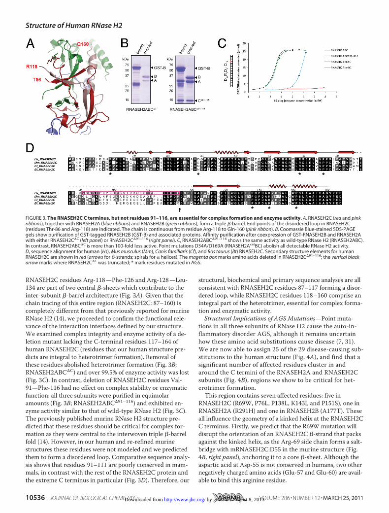
RNASEH2C residues Arg-118—Phe-126 and Arg-128—Leu-134 are part of two central �-sheets which contribute to theinter-subunit �-barrel architecture (Fig. 3A). Given that thechain tracing of this entire region (RNASEH2C: 87–160) iscompletely different from that previously reported for murineRNase H2 (14), we proceeded to confirm the functional rele-vance of the interaction interfaces defined by our structure.We examined complex integrity and enzyme activity of a de-letion mutant lacking the C-terminal residues 117–164 ofhuman RNASEH2C (residues that our human structure pre-dicts are integral to heterotrimer formation). Removal ofthese residues abolished heterotrimer formation (Fig. 3B;RNASEH2ABC�C) and over 99.5% of enzyme activity was lost(Fig. 3C). In contrast, deletion of RNASEH2C residues Val-91—Phe-116 had no effect on complex stability or enzymaticfunction: all three subunits were purified in equimolaramounts (Fig. 3B; RNASEH2ABC�91–116) and exhibited en-zyme activity similar to that of wild-type RNase H2 (Fig. 3C).The previously published murine RNase H2 structure pre-dicted that these residues should be critical for complex for-mation as they were central to the interwoven triple �-barrelfold (14). However, in our human and re-refined murinestructures these residues were not modeled and we predictedthem to form a disordered loop. Comparative sequence analy-sis shows that residues 91–111 are poorly conserved in mam-mals, in contrast with the rest of the RNASEH2C protein andthe extreme C terminus in particular (Fig. 3D). Therefore, our
structural, biochemical and primary sequence analyses are allconsistent with RNASEH2C residues 87–117 forming a disor-dered loop, while RNASEH2C residues 118–160 comprise anintegral part of the heterotrimer, essential for complex forma-tion and enzymatic activity.Structural Implications of AGS Mutations—Point muta-
tions in all three subunits of RNase H2 cause the auto-in-flammatory disorder AGS, although it remains uncertainhow these amino acid substitutions cause disease (7, 31).We are now able to assign 25 of the 29 disease-causing sub-stitutions to the human structure (Fig. 4A), and find that asignificant number of affected residues cluster in andaround the C termini of the RNASEH2A and RNASEH2Csubunits (Fig. 4B), regions we show to be critical for het-erotrimer formation.This region contains seven affected residues: five in
RNASEH2C (R69W, P76L, P138L, K143I, and P151S), one inRNASEH2A (R291H) and one in RNASEH2B (A177T). Theseall influence the geometry of a kinked helix at the RNASEH2CC terminus. Firstly, we predict that the R69Wmutation willdisrupt the orientation of an RNASEH2C �-strand that packsagainst the kinked helix, as the Arg-69 side chain forms a salt-bridge with mRNASEH2C:D55 in the murine structure (Fig.4B, right panel), anchoring it to a core �-sheet. Although theaspartic acid at Asp-55 is not conserved in humans, two othernegatively charged amino acids (Glu-57 and Glu-60) are avail-able to bind this arginine residue.
FIGURE 3. The RNASEH2C C terminus, but not residues 91–116, are essential for complex formation and enzyme activity. A, RNASEH2C (red and pinkribbons), together with RNASEH2A (blue ribbons) and RNASEH2B (green ribbons), form a triple �-barrel. End points of the disordered loop in RNASEH2C(residues Thr-86 and Arg-118) are indicated. The chain is continuous from residue Arg-118 to Gln-160 (pink ribbon). B, Coomassie Blue-stained SDS-PAGEgels show purification of GST-tagged RNASEH2B (GST-B) and associated proteins. Affinity purification after coexpression of GST-RNASEH2B and RNASEH2Awith either RNASEH2C�C (left panel) or RNASEH2C�91–116 (right panel). C, RNASEH2ABC�91–116 shows the same activity as wild-type RNase H2 (RNASEH2ABC).In contrast, RNASEH2ABC�C is more than 100-fold less active. Point mutations D34A/D169A (RNASEH2AcatBC) abolish all detectable RNase H2 activity.D, sequence alignment for human (Hs), Mus musculus (Mm), Canis familiaris (Cf), and Bos taurus (Bt) RNASEH2C. Secondary structure elements for humanRNASEH2C are shown in red (arrows for �-strands; spirals for � helices). The magenta box marks amino acids deleted in RNASEH2C�91–116; the vertical blackarrow marks where RNASEH2C�C was truncated; * mark residues mutated in AGS.
Structure of Human RNase H2
10536 JOURNAL OF BIOLOGICAL CHEMISTRY VOLUME 286 • NUMBER 12 • MARCH 25, 2011 by guest on August 8, 2013http://www.jbc.org/Downloaded from
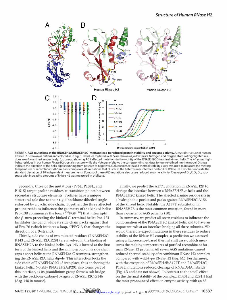
Secondly, three of the mutations (P76L, P138L, andP151S) target proline residues at transition points betweensecondary structure elements. Prolines have a uniquestructural role due to their rigid backbone dihedral angleenforced by a cyclic side chain. Together, the three affectedproline residues influence the geometry of the kinked helix:Pro-138 commences the loop (138PGP140) that interruptsthe �-turn preceding the kinked C-terminal helix; Pro-151facilitates the bend, while its side chain packs against thatof Pro-76 (which initiates a loop, 76PPG78, that changes thedirection of a �-strand).
Thirdly, side chains of two mutated residues (RNASEH2C:K143 and RNASEH2A:R291) are involved in the binding ofRNASEH2A to the kinked helix. Lys-143 is located at the firstturn of the kinked helix and the amino group of its side chaincaps a short helix at the RNASEH2A C terminus, strengthen-ing the RNASEH2A helix dipole. This interaction locks theside chain of RNASEH2C:K143 into place, thus anchoring thekinked helix. Notably RNASEH2A:R291 also forms part ofthis interface, as its guanidinium group forms a salt bridgewith the backbone carbonyl oxygen of RNASEH2C:G146(Arg-148 in mouse).
Finally, we predict the A177T mutation in RNASEH2B todisrupt the interface between a RNASEH2B �-helix and theRNASEH2C kinked helix. The affected alanine residue sits ina hydrophobic pocket and packs against RNASEH2C:A156of the kinked helix. Notably, the A177T substitution inRNASEH2B is the most common mutation, found in morethan a quarter of AGS patients (10).In summary, we predict all seven residues to influence the
conformation of the RNASEH2C kinked helix and to have animportant role at an interface bridging all three subunits. Wewould therefore expect mutations in these residues to reducestability of the RNase H2 complex; a prediction we assessedusing a fluorescence-based thermal shift assay, which mea-sures the melting temperatures of purified recombinant hu-man RNase H2 proteins. All seven AGS mutations causedreduced thermal stability of recombinant RNase H2 complexcompared with wild-type RNase H2 (Fig. 4C). Furthermore,with the exception of RNASEH2B:A177T and RNASEH2C:P138L, mutations reduced cleavage of RNA/DNA hybrids(Fig. 4D and data not shown). In contrast to the small effecton the thermal stability of the complex, K143I and R291H hadthe most pronounced effect on enzyme activity, with an 85
FIGURE 4. AGS mutations at the RNASEH2A/RNASEH2C interface lead to reduced protein stability and enzyme activity. A, crystal structure of humanRNase H2 is shown as ribbons and colored as in Fig. 1. Residues mutated in AGS are shown as yellow sticks. Nitrogen and oxygen atoms of highlighted resi-dues are blue and red, respectively. B, close-up showing AGS affected mutations in the vicinity of the RNASEH2C C-terminal kinked helix. The left panel high-lights residues in our human RNase H2 crystal structure while the right panel shows the corresponding residues for our re-refined murine model. (Arrowsindicate the direction of the helix dipole running from positive to negative). C, fluorescence-based thermal stability assay was used to measure the meltingtemperatures of recombinant AGS mutant complexes. All mutations that cluster at the heterotrimer interface destabilize RNase H2. Error bars indicate thestandard deviation of 10 independent measurements. D, most of these AGS mutations also cause reduced enzyme activity. Cleavage of D14R1D3:D18 sub-strate with increasing amounts of RNase H2 was measured in triplicate.
Structure of Human RNase H2
MARCH 25, 2011 • VOLUME 286 • NUMBER 12 JOURNAL OF BIOLOGICAL CHEMISTRY 10537 by guest on August 8, 2013http://www.jbc.org/Downloaded from

and 94% reduction in specific activity respectively (Fig. 4D).This is particularly striking given their distance from the cata-lytic center and predicted substrate binding groove. Measure-ments of enzyme kinetics for RNase H2 with K143I andR291H mutations (Table 2) indicate that their enzyme activi-ties are decreased due to reduced turnover (lower kcat) andnot reduced substrate affinity (no change in Km). In conclu-sion, all these mutations either disrupt complex stabilityand/or in vitro enzymatic activity. Atomic coordinates havebeen deposited at the RCSB Protein Data bank under IDcodes 3P56 for human RNase H2 and 3P5J for the re-refinedmurine RNase H2.
DISCUSSION
Our structure of the human RNase H2 complex identifiesnovel structural elements of the RNASEH2BC heterodimerand catalytic RNASEH2A subunit relevant to enzyme func-tion and disease. We establish that the C-terminal extensionof RNASEH2A spans the two auxiliary subunits and is neces-sary for the formation of an enzymatically active complex.The C terminus of RNASEH2C also contributes to the uniqueinterwoven architecture of the heterotrimer, by forming ahelix that bridges the RNASEH2A and RNASEH2B subunits.Analysis of human disease mutations that cluster in this re-gion demonstrates the importance of this interface for com-plex stability and enzymatic activity.Role of the RNASEH2A C terminus in Enzymatic Activity—
Our data establish that the RNASEH2A C-terminal tail isrequired for enzymatic activity. A unique adaptation of eu-karyotic RNase H2 enzymes, this C-terminal tail permits en-zyme activity only when accessory subunits are bound. Strik-ingly, substitution of specific residues in this C-terminalextension (R291H, K266A/R267A) impair enzyme activity,with minimal or no effect on complex stability (Fig. 4 andsupplemental Fig. S6). Furthermore, mutation of a directlyinteracting residue of the RNASEH2C subunit (K143I) has thesame effect. Notably, all these residues are distant from thecatalytic center. These residues may therefore influence en-zyme activity by forming an additional substrate-binding sitedistinct from the proposed RNASEH2A binding groove (14,15). Alternatively, they could influence RNase H2 quaternarystructure to alter enzymatic activity.Spatial and temporal regulation of DNA replication and
repair pathways is essential for genome stability in eukaryoticcells. Intriguingly, the RNASEH2BC heterodimer can exist as
a stable subcomplex in vitro (Fig. 2) (7). As the RNASEH2Aprotein is present at much lower levels than the other sub-units,6 this catalytic subunit might bind only when and whereenzyme activity is required.AGS Mutations Cause RNase H2 Dysfunction—We are able
to map the majority of AGS mutations onto the human RNaseH2 structure (86%). This provides a significant advance inunderstanding the structural context of these mutations inhuman disease. It is evident that some mutations directly af-fect enzyme catalysis (7, 9, 31, 32). It has also been establishedthat other AGS mutations do not dramatically alter enzymaticactivity in vitro (7, 31) and our findings are consistent withthese studies. However, we find that mutations with no orlimited effects on in vitro enzyme activity (RNASEH2B:A177T, and RNASEH2C:R69W, P76L, and P138L) signifi-cantly destabilize the RNase H2 complex, as measured by areduction in thermal stability of recombinant RNase H2.Though such instability does not necessarily have a direct invivo correlation, these mutations may lead to diminishedRNase H2 levels and impaired cellular activity. Hence, ourdata are consistent with the notion that human mutationscause AGS through reduced enzymatic activity, in an analo-gous manner to disease mutations in TREX1 (33). Future ex-amination of other human mutations may well provide addi-tional insight into the biology of RNase H2. For instance,mutations in surface residues (e.g. RNASEH2A:T240M,RNASEH2B: K162T and V185G, and RNASEH2C:R13H)could impair interactions with nucleic acid substrates or otherproteins.In summary, our findings provide important structural in-
sight into the requirements for RNase H2 complex integrityand regulation of enzyme activity, relevant to the cellularprocess of RNA/DNA hybrid degradation and the pathogene-sis of systemic autoimmune disease. Additionally, the struc-ture of the RNase H2 complex provides a framework for fu-ture investigation of RNase H2 function and its regulation inthe eukaryotic cell.
Acknowledgments—We thank J. Ren and K. Harlos (Oxford) for as-sistance in crystallographic data collection and refinement, N. M.Devine (INP, Glasgow) for help with the peptide arrays, E. Black-burn (Edinburgh Biophysical Characterization Facility; use of thisfacility was supported by The Wellcome Trust, the Scottish Univer-sity Life Sciences Alliance and the BBSRC), and S. Corless (IGMM)for technical assistance; and T. Walter, E. Seiradake, B. Janssen, N.Hastie, D. Stuart, and members of the Jackson Laboratory for usefuldiscussions. We thank the ESRF and Diamond Light Source for syn-chrotron access.
REFERENCES1. Machida, Y., Okazaki, T., and Okazaki, R. (1977) Proc. Natl. Acad. Sci.
U.S.A. 74, 2776–27792. Li, X., and Manley, J. L. (2005) Cell 122, 365–3783. Forstemann, K., and Lingner, J. (2005) EMBO Rep. 6, 361–3664. Eder, P. S., and Walder, J. A. (1991) J. Biol. Chem. 266, 6472–64795. Jeong, H. S., Backlund, P. S., Chen, H. C., Karavanov, A. A., and Crouch,
6 M. A. M. Reijns and A. P. Jackson, unpublished data.
TABLE 2Kinetic parameters for human RNase H2 with and without AGSmutations
Km kcat kcat/Km
nM min�1 min�1nM�1
D14R1D3:DNA18Wild type 28 � 2.7a 24 � 0.7 0.9A:R291H 27 � 1.8 9.5 � 0.2 0.3C:K143I 30 � 3.4 21 � 0.8 0.7
RNA18:DNA18Wild type 143 � 1.7 209 � 0.8 1.5A:R291H 144 � 4.5 59 � 0.6 0.4C:K143I 158 � 3.4 126 � 0.9 0.8
a Values indicate standard errors.
Structure of Human RNase H2
10538 JOURNAL OF BIOLOGICAL CHEMISTRY VOLUME 286 • NUMBER 12 • MARCH 25, 2011 by guest on August 8, 2013http://www.jbc.org/Downloaded from

R. J. (2004) Nucleic Acids Res. 32, 407–4146. Nick McElhinny, S. A., Watts, B. E., Kumar, D., Watt, D. L., Lundstrom,
E. B., Burgers, P. M., Johansson, E., Chabes, A., and Kunkel, T. A. (2010)Proc. Natl. Acad. Sci. U.S.A. 107, 4949–4954
7. Chon, H., Vassilev, A., DePamphilis, M., Zhao, Y., Zhang, J., Burgers,P. M., Crouch, R. J., and Cerritelli, S. M. (2009) Nucleic Acids Res. 37,96–110
8. Maga, G., and Hubscher, U. (2003) J. Cell Sci. 116, 3051–30609. Crow, Y., Leitch, A., Hayward, B., Garner, A., Parmar, R., Griffith, E., Ali,
M., Semple, C., Aicardi, J., Babul-Hirji, R., Baumann, C., Baxter, P., Ber-tini, E., Chandler, K., Chitayat, D., Cau, D., Dery, C., Fazzi, E., Goizet, C.,King, M. D., Klepper, J., Lacombe, D., Lanzi, G., Lyall, H., Martínez-Frías, M., Mathieu, M., McKeown, C., Monier, A., Oade, Y., Quarrell, O.,Rittey, C., Rogers, R., Sanchis, A., Stephenson, J., Tacke, U., Till, M., Tol-mie, J., Tomlin, P., Voit, T., Weschke, B., Woods, C. G., Lebon, P., Bon-thron, D. T., Ponting, C. P., and Jackson, A. P. (2006) Nat. Genet. 38,910–916
10. Rice, G., Patrick, T., Parmar, R., Taylor, C. F., Aeby, A., Aicardi, J., Ar-tuch, R., Montalto, S. A., Bacino, C. A., Barroso, B., Baxter, P., Benko,W. S., Bergmann, C., Bertini, E., Biancheri, R., Blair, E. M., Blau, N., Bon-thron, D. T., Briggs, T., Brueton, L. A., Brunner, H. G., Burke, C. J., Carr,I. M., Carvalho, D. R., Chandler, K. E., Christen, H. J., Corry, P. C.,Cowan, F. M., Cox, H., D’Arrigo, S., Dean, J., De Laet, C., De Praeter, C.,Dery, C., Ferrie, C. D., Flintoff, K., Frints, S. G., Garcia-Cazorla, A.,Gener, B., Goizet, C., Goutieres, F., Green, A. J., Guet, A., Hamel, B. C.,Hayward, B. E., Heiberg, A., Hennekam, R. C., Husson, M., Jackson,A. P., Jayatunga, R., Jiang, Y. H., Kant, S. G., Kao, A., King, M. D., Kings-ton, H. M., Klepper, J., van der Knaap, M. S., Kornberg, A. J., Kotzot, D.,Kratzer, W., Lacombe, D., Lagae, L., Landrieu, P. G., Lanzi, G., Leitch,A., Lim, M. J., Livingston, J. H., Lourenco, C. M., Lyall, E. G., Lynch,S. A., Lyons, M. J., Marom, D., McClure, J. P., McWilliam, R., Melancon,S. B., Mewasingh, L. D., Moutard, M. L., Nischal, K. K., Ostergaard, J. R.,Prendiville, J., Rasmussen, M., Rogers, R. C., Roland, D., Rosser, E. M.,Rostasy, K., Roubertie, A., Sanchis, A., Schiffmann, R., Scholl-Burgi, S.,Seal, S., Shalev, S. A., Corcoles, C. S., Sinha, G. P., Soler, D., Spiegel, R.,Stephenson, J. B., Tacke, U., Tan, T. Y., Till, M., Tolmie, J. L., Tomlin, P.,Vagnarelli, F., Valente, E. M., Van Coster, R. N., Van der Aa, N., Vander-ver, A., Vles, J. S., Voit, T., Wassmer, E., Weschke, B., Whiteford, M. L.,Willemsen, M. A., Zankl, A., Zuberi, S. M., Orcesi, S., Fazzi, E., Lebon,P., and Crow, Y. J. (2007) Am. J. Hum. Genet. 81, 713–725
11. Ramantani, G., Kohlhase, J., Hertzberg, C., Innes, A. M., Engel, K., Hun-ger, S., Borozdin, W., Mah, J. K., Ungerath, K., Walkenhorst, H., Rich-ardt, H. H., Buckard, J., Bevot, A., Siegel, C., von Stulpnagel, C., Ikono-midou, C., Thomas, K., Proud, V., Niemann, F., Wieczorek, D., Hausler,M., Niggemann, P., Baltaci, V., Conrad, K., Lebon, P., and Lee-Kirsch,M. A. (2010) Arthritis Rheum. 62, 1469–1477
12. Yang, Y. G., Lindahl, T., and Barnes, D. E. (2007) Cell 131, 873–88613. Stetson, D. B., Ko, J. S., Heidmann, T., and Medzhitov, R. (2008) Cell
134, 587–59814. Shaban, N. M., Harvey, S., Perrino, F. W., and Hollis, T. (2010) J. Biol.
Chem. 285, 3617–362415. Chapados, B. R., Chai, Q., Hosfield, D. J., Qiu, J., Shen, B., and Tainer,
J. A. (2001) J. Mol. Biol. 307, 541–55616. Otwinowski, Z., and Minor, W. (1997)Macromolecular Crystallography,
Part A, Academic Press, New York17. Vagin, A., and Teplyakov, A. (2010) Acta Crystallogr. D. Biol. Crystal-
logr. 66, 22–2518. Blanc, E., Roversi, P., Vonrhein, C., Flensburg, C., Lea, S. M., and Bri-
cogne, G. (2004) Acta Crystallogr. D. Biol. Crystallogr. 60, 2210–222119. Smart, O., Brandl, M., Flensburg, C., Keller, P., Paciorek, W., Vonrhein,
C., Womack, T., and Bricogne, G. (2008) Annual Meeting of the ACA,137, Knoxville, TN
20. Adams, P. D., Afonine, P. V., Bunkoczi, G., Chen, V. B., Davis, I. W.,Echols, N., Headd, J. J., Hung, L. W., Kapral, G. J., Grosse-Kunstleve,R. W., McCoy, A. J., Moriarty, N. W., Oeffner, R., Read, R. J., Richard-son, D. C., Richardson, J. S., Terwilliger, T. C., and Zwart, P. H. (2010)Acta Crystallogr. D. Biol. Crystallogr. 66, 213–221
21. Tronrud, D. E. (1996) J. Appl. Crystallogr. 29, 100–10422. Nettleship, J. E., Brown, J., Groves, M. R., and Geerlof, A. (2008)Meth-
ods Mol. Biol. 426, 299–31823. Winkler, D. F., Hilpert, K., Brandt, O., and Hancock, R. E. (2009)Meth-
ods Mol. Biol. 570, 157–17424. Dikmans, A., Beutling, U., Schmeisser, E., Thiele, S., and Frank, R. (2006)
Qsar Combinatorial Science 25, 1069–108025. Frank, R. (2002) J. Immunol. Methods 267, 13–2626. Kleywegt, G. J., Harris, M. R., Zou, J. Y., Taylor, T. C., Wahlby, A., and
Jones, T. A. (2004) Acta Crystallogr. D. Biol. Crystallogr. 60, 2240–224927. Davis, I. W., Leaver-Fay, A., Chen, V. B., Block, J. N., Kapral, G. J., Wang,
X., Murray, L. W., Arendall, W. B., 3rd, Snoeyink, J., Richardson, J. S.,and Richardson, D. C. (2007) Nucleic Acids Res. 35,W375–383
28. Krissinel, E., and Henrick, K. (2004) Acta Crystallogr. D. Biol. Crystal-logr. 60, 2256–2268
29. Li, X., MacLeod, R., Dunlop, A. J., Edwards, H. V., Advant, N., Gibson,L. C., Devine, N. M., Brown, K. M., Adams, D. R., Houslay, M. D., andBaillie, G. S. (2009) FEBS Lett. 583, 3310–3316
30. Meng, D., Lynch, M. J., Huston, E., Beyermann, M., Eichhorst, J., Adams,D. R., Klussmann, E., Houslay, M. D., and Baillie, G. S. (2009) J. Biol.Chem. 284, 11425–11435
31. Perrino, F. W., Harvey, S., Shaban, N. M., and Hollis, T. (2009) J. Mol.Med. 87, 25–30
32. Rohman, M. S., Koga, Y., Takano, K., Chon, H., Crouch, R. J., and Ka-naya, S. (2008) FEBS J. 275, 4836–4849
33. Crow, Y., Hayward, B. E., Parmar, R., Robins, P., Leitch, A., Ali, M.,Black, D. N., van Bokhoven, H., Brunner, H., Hamel, B., Corry, P.,Cowan, F., Frints, S., Klepper, J., Livingston, J., Lynch, S., Massey, R.,Meritet, J., Michaud, J., Ponsot, G., Voit, T., Lebon, P., Bonthron, D.,Jackson, A., Barnes, D., and Lindahl, T. (2006) Nat. Genet. 38, 917–920
34. Dolinsky, T. J., Nielsen, J. E., McCammon, J. A., and Baker, N. A. (2004)Nucleic Acids Res. 32,W665–667
35. Baker, N. A., Sept, D., Joseph, S., Holst, M. J., and McCammon, J. A.(2001) Proc. Natl. Acad. Sci. U.S.A. 98, 10037–10041
36. Bond, C. S., and Schuttelkopf, A. W. (2009) Acta Crystallogr. D. Biol.Crystallogr. 65, 510–512
Structure of Human RNase H2
MARCH 25, 2011 • VOLUME 286 • NUMBER 12 JOURNAL OF BIOLOGICAL CHEMISTRY 10539 by guest on August 8, 2013http://www.jbc.org/Downloaded from


![Edinburgh Research Explorer - COnnecting REpositories · Edinburgh Research Explorer ... demonstrated good inter-rater reliability [14,15]. Moreover, we ... Participants were community-resident,](https://img.dokumen.tips/doc/110x75/5e85a48b4a019226407e6438/edinburgh-research-explorer-connecting-repositories-edinburgh-research-explorer.jpg)