Embed Size (px)
Citation preview
Edinburgh Research Explorer
Pangenome and phylogenomic analysis of the pathogenicactinobacterium Rhodococcus equi
Citation for published version:Anastasi, E, MacArthur, I, Scortti, M, Alvarez, S, Giguère, S & Vazquez-Boland, J 2016, 'Pangenome andphylogenomic analysis of the pathogenic actinobacterium Rhodococcus equi' Genome Biology andEvolution, vol 8, no. 10, pp. 3140-3148. DOI: 10.1093/gbe/evw222
Digital Object Identifier (DOI):10.1093/gbe/evw222
Link:Link to publication record in Edinburgh Research Explorer
Document Version:Publisher's PDF, also known as Version of record
Published In:Genome Biology and Evolution
Publisher Rights Statement:© The Author(s) 2016. Published by Oxford University Press on behalf of the Society for Molecular Biology andEvolution.This is an Open Access article distributed under the terms of the Creative Commons Attribution Non-CommercialLicense (http://creativecommons.org/licenses/by-nc/4.0/), which permits non-commercial re-use, distribution, andreproduction in any medium, provided the original work is properly cited. For commercial re-use, please [email protected]
General rightsCopyright for the publications made accessible via the Edinburgh Research Explorer is retained by the author(s)and / or other copyright owners and it is a condition of accessing these publications that users recognise andabide by the legal requirements associated with these rights.
Take down policyThe University of Edinburgh has made every reasonable effort to ensure that Edinburgh Research Explorercontent complies with UK legislation. If you believe that the public display of this file breaches copyright pleasecontact [email protected] providing details, and we will remove access to the work immediately andinvestigate your claim.
Download date: 25. Jun. 2018

Pangenome and Phylogenomic Analysis of the Pathogenic
Actinobacterium Rhodococcus equi
Elisa Anastasi1,y, Iain MacArthur1,y, Mariela Scortti1,2, Sonsiray Alvarez1, Steeve Giguere3, andJose A. V�azquez-Boland1,2,4,*1Division of Infection and Immunity, The Roslin Institute, University of Edinburgh, Edinburgh, United Kingdom2Edinburgh Medical School (Biomedical Sciences), University of Edinburgh, Edinburgh, United Kingdom3Department of Large Animal Medicine, University of Georgia, Georgia, USA4Grupo de Patogenomica Bacteriana, Universidad de Leon, Leon, Spain
yThese authors contributed equally to the study.
*Corresponding author: E-mail: [email protected].
Accepted: September 2, 2016
Data deposition: This project has been deposited at GenBank under accessions LWTX00000000, LWIC00000000, LWBN00000000,
LWHS00000000, LWHT00000000, LWHU00000000, LWHV00000000, LWHW00000000, LWHX00000000, LWHY00000000,
LWHZ00000000, LWIA00000000, LWIB00000000, LWTO00000000, LXFI00000000, LXFH00000000, LXFG00000000, LWHR00000000,
LWTP00000000, LWTY00000000, LWTQ00000000, LWTR00000000, LWTS00000000, LWTT00000000, LWTU00000000, LWTV00000000,
and LWTW00000000.
Abstract
Wereportacomparativestudyof29representativegenomesof theanimalpathogenRhodococcusequi. Theanalyses showedthatR.
equi is genetically homogeneous and clonal, with a large core genome accounting for&80% of an isolates’ gene content. An open
pangenome, even distribution of accessory genes among the isolates, and absence of significant core–genome recombination,
indicated that gene gain/loss is a main driver of R. equi genome evolution. Traits previously predicted to be important in R. equi
physiology, virulence and niche adaptation were part of the core genome. This included the lack of a phosphoenolpyruvate:carbo-
hydrate transport system (PTS), unique among the rhodococci except for the closely related Rhodococcus defluvii, reflecting selective
PTSgene loss in theR.equi–R.defluvii sublineage.Thought tobeasaccharolytic, rbsCBandglcPnon-PTSsugarpermeasehomologues
were identified in the core genome and, albeit inefficiently, R. equi utilized their putative substrates, ribose and (irregularly) glucose.
Therewasnocorrelationbetween R. equiwhole-genome phylogenyandhostorgeographical source,withevidenceofglobal spread
of genomovars. The distribution of host-associated virulence plasmid types was consistent with the exchange of the plasmids (and
corresponding host shifts) across the R. equi population, and human infection being zoonotically acquired. Phylogenomic analyses
demonstrated thatR.equioccupiesacentralposition in theRhodococcusphylogeny,not supporting the recentlyproposedtransferof
the species to a new genus.
Key words: Rhodococcus equi, pangenome analysis, comparative genomics, genome diversity and evolution, phylogenomics,
Corynebacteriales, Actinobacteria.
Introduction
The soil-dwelling actinobacterium Rhodococcus equi is the
causative agent of a purulent bronchopneumonic disease
that affects foals in equine farms worldwide. In addition to
horses, R. equi can also infect other animal species and is
associated with severe opportunistic infections in immuno-
compromised people (Prescott 1991; von Bargen and Haas
2009; Vazquez-Boland et al. 2013). We previously reported
the complete genome sequence of an equine isolate of R. equi
(strain 103S). This work provided key information about the
genome structure of the pathogen and the mechanisms of
rhodococcal niche-adaptive genome plasticity and virulence
evolution (Letek et al. 2010). Here we present the first com-
prehensive comparative genomic analysis of R. equi, involving
GBE
� The Author(s) 2016. Published by Oxford University Press on behalf of the Society for Molecular Biology and Evolution.
This is an Open Access article distributed under the terms of the Creative Commons Attribution Non-Commercial License (http://creativecommons.org/licenses/by-nc/4.0/), which permits
non-commercial re-use, distribution, and reproduction in any medium, provided the original work is properly cited. For commercial re-use, please contact [email protected]
3140 Genome Biol. Evol. 8(10):3140–3148. doi:10.1093/gbe/evw222 Advance Access publication September 16, 2016
at Edinburgh U
niversity on Novem
ber 1, 2016http://gbe.oxfordjournals.org/
Dow
nloaded from

multiple isolates from different sources. Our new study
provides insight into the core features, diversity, popula-
tion structure and genome evolution of R. equi. It also
clarifies the phylogenetic position of the species, repeat-
edly questioned based on equivocal 16S rDNA and numer-
ical phenetic studies (Goodfellow et al. 1998; Gurtler et al.
2004; Jones and Goodfellow 2012; Jones et al. 2013b),
unambiguously confirming R. equi is a bona fide member
of the genus Rhodococcus.
Materials and Methods
Bacteria
The isolates sequenced in this study (supplementary table S1,
Supplementary Material online) were selected to include at
least two representatives from each of the seven major R.
equi genogroups defined by AseI PFGE genotyping
(Vazquez-Boland et al. 2008 and our unpublished data) plus
the type strain of the species, DSM 20307T (= ATCC
6939T=ATCC 25729T=NBRC 101255T). Isolates of different
animal sources (equine, bovine, porcine, ovine, human), geo-
graphical origin (13 countries) and host-associated virulence
plasmid type carriage (pVAPA, pVAPB, pVAPN) (Takai et al.
2000; Letek et al. 2008; Valero-Rello et al. 2015)were analyzed.
Genome Sequencing and Analysis
Rhodococcus equi DNA was isolated from exponential cul-
tures in BHI (OD600 & 1.0) using the GenEluteTM kit
(Sigma–Aldrich). Shotgun 101-bp pair-end DNA sequencing
was performed at Beijing Genomics Institute (BGI, China)
using TruSeq DNA PCR-Free Sample library preparation kit
on Illumina HiSeq 2000 instruments. Strains 2274 to 2288
(supplementary table S1, Supplementary Material online)
were sequenced at the genomics facility of the University of
Georgia (USA) as previously described (Anastasi et al. 2015).
Adaptors and low quality reads were trimmed using Scythe
(https://github.com/vsbuffalo/scythe) and Sickle (https://
github.com/najoshi/sickle), respectively, and assembled using
SPAdes (Bankevich et al. 2012). Annotation was performed
using Prokka V1.11 (Seemann 2014) and the complete 103S
genome (Letek et al. 2010) as a reference. Pangenome
analyses were performed using Get_Homologues V2.0
(Contreras-Moreira and Vinuesa 2013) with OrthoMCL clus-
tering algorithm and 70% sequence identity–75% coverage
as minimum BLASTp homology cutoff. Functional annotation
was performed using BLASTKOALA (Kanehisa, et al. 2016)
and the prokaryotes KEGG GENES search database.
Genome Diversity and Phylogenomic Analyses
Average nucleotide identity (ANI) was calculated using
JSpecies (Richter and Rossello-Mora 2009) with MUMmer
alignment (ANIm) as described in Goris et al. (2007) (settings
-X 150, -q -1, -F F, -e 1e-15, -a 2). Rhodococcus equi whole-
genome Maximum Likelihood (ML) phylogenetic reconstruc-
tion was performed with RealPhy (Bertels et al. 2014) using
RAxML (Stamatakis 2014) for tree construction with the gen-
eral time-reversible (GTR) model of nucleotide evolution and
gamma distributed rate variation. The Corynebacteriales ML
tree was constructed from alignments of concatenated con-
served protein products using PhyloPhlan (Segata et al. 2013).
Trees were graphed using FigTree (http://tree.bio.ed.ac.uk/
software/figtree/).
Results and Discussion
Rhodococcus equi Is Genetically Homogeneous
Twenty-seven de novo determined R. equi whole-genome
shotgun assemblies, the available draft genome of ATCC
33707, and the complete 103S genome (Letek, et al. 2010)
were analyzed (supplementary table S1, Supplementary
Material online). The average CDS number was 4,933
(range 4,525–5,325), similar to the gene content of the man-
ually annotated 5.04-Mbp 103S genome (4,598) (Letek, et al.
2010). Mean G + C content was 68.77%, also similar to that
previously determined for 103S (68.82%). The mean ANI
value was 99.13% (range 98.86–99.28%), well above the
consensus 95–96% threshold for prokaryotic species demar-
cation (Goris et al. 2007; Richter and Rossello-Mora 2009; Kim
et al. 2014). This corresponded to 100% 16S rDNA sequence
identity (1,519 nt) across all the isolates.
In comparison, the ANI values with members of the two
other main Rhodococcus lines of descent as defined based
on 16 rDNA phylogenies (McMinn, et al. 2000; Jones and
Goodfellow 2012), that is, the “erythropolis” clade (R. erythro-
polis, R. jostii, R. opacus and R. fascians included in the analysis)
and the “rhodochrous” clade” (Rhodococcus rhodochrous,
Rhodococcus rhodnii, Rhodococcus ruber, and Rhodococcus
pyridinivorans included in the analysis), were 72.27–74.58%
and 68.55–75.15%, respectively. The ANI with the recently
described R. equi close relative, Rhodococcus defluvii (strain
Ca11T) (Kampfer et al. 2014), was 82.96%. This corresponded
to 16S rDNA identity values of 96–98% and 95–97% for rep-
resentatives of the “erythropolis” and “rhodochrous” clades,
respectively, and 99% for R. defluvii.
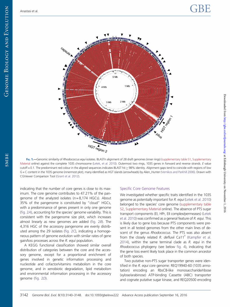
The above data correlate with a strong degree of genome
similarity and synteny conservation in BLASTn alignments
(fig. 1), indicating that R. equi is a genetically homogeneous
species.
Rhodococcus equi Core and Pangenome
The core genome shared by all 29 R. equi strains comprises
3,858 homologous gene clusters (HGC) (fig. 2A), equivalent
to 81.5% of the 103S genome or 78.2% of the average gene
content of the analyzed isolates, reflecting a low degree of
intraspecies genomic variability. A core genome size estima-
tion plot starts plateauing at about 25–27 genomes (fig. 2B),
Comparative Genomics and Phylogenomics of Rhodococcus equi GBE
Genome Biol. Evol. 8(10):3140–3148. doi:10.1093/gbe/evw222 Advance Access publication September 16, 2016 3141
at Edinburgh U
niversity on Novem
ber 1, 2016http://gbe.oxfordjournals.org/
Dow
nloaded from
indicating that the number of core genes is close to its max-
imum. The core genome contributes to 47.21% of the pan-
genome of the analyzed isolates (n = 8,174 HGCs). About
35% of the pangenome is constituted by “cloud” HGCs,
with a predominance of genes present in only one genome
(fig. 2A), accounting for the species’ genome variability. This is
consistent with the pangenome size plot, which increases
almost linearly as new genomes are added (fig. 2B). The
4,316 HGC of the accessory pangenome are evenly distrib-
uted among the 29 isolates (fig. 2C), indicating a homoge-
neous pattern of genome evolution with similar rates of gene
gain/loss processes across the R. equi population.
A KEGG functional classification showed similar overall
distribution of categories between the core and the acces-
sory genome, except for a proportional enrichment of
genes involved in genetic information processing and
nucleotide and cofactor/vitamins metabolism in the core
genome, and in xenobiotic degradation, lipid metabolism
and environmental information processing in the accessory
genome (fig. 2D).
Specific Core Genome Features
We investigated whether specific traits identified in the 103S
genome as potentially important for R. equi (Letek et al. 2010)
belonged to the species’ core genome (supplementary table
S2, Supplementary Material online). The absence of PTS sugar
transport components (EI, HPr, EII complex/permeases) (Letek
et al. 2010) was confirmed as a general feature of R. equi. This
is likely due to gene loss because PTS components were pre-
sent in all tested genomes from the other main lines of de-
scent of the genus Rhodococcus. The PTS was also absent
from the closely related R. defluvii Ca11T (Kampfer et al.
2014), within the same terminal clade as R. equi in the
Rhodococcus phylogeny (see below fig. 4), indicating that
the gene loss event likely took place in the common ancestor
of both species.
Two putative non-PTS sugar transporter genes were iden-
tified in the R. equi core genome: REQ19940-60 (103S anno-
tation) encoding an RbsCB-like monosaccharide/ribose
(xylose/arabinose) ATP-binding Cassette (ABC) transporter
and cognate putative sugar kinase, and REQ20500 encoding
FIG. 1.—Genomic similarity of Rhodococcus equi isolates. BLASTn alignment of 28 draft genomes (inner rings) (supplementary table S1, Supplementary
Material online) against the complete 103S chromosome (Letek, et al. 2010). Outermost two rings, 103S genes in forward and reverse strands. E value
cutoff = 0.1. The predominant red colour in the aligned sequences indicates BLAST hit� 98% identity. Alignment gaps tend to coincide with regions of low
G + C content in the 103S genome (innermost plot), many identified as HGT islands (arrowheads) by Alien_Hunter (Vernikos and Parkhill 2006). Drawn with
CGViewer Comparison Tool (Grant et al. 2012).
Anastasi et al. GBE
3142 Genome Biol. Evol. 8(10):3140–3148. doi:10.1093/gbe/evw222 Advance Access publication September 16, 2016
at Edinburgh U
niversity on Novem
ber 1, 2016http://gbe.oxfordjournals.org/
Dow
nloaded from

a Major Facilitator Superfamily (MFS) permease similar to the
Streptomyces coelicolor glucose transporter GlcP (van Wezel
et al. 2005) (supplementary table S2, Supplementary Material
online). Phenotype MicroArray (PMA) carbon source utilization
tests (Bochner 2009) showed positive reactions for D-ribose,
2-deoxy-D-ribose, D-xylose (and its C0-2 carbon epimer L-lyxose),
and D/L-arabinose (supplementary fig. S1A, Supplementary
Material online). To exclude false positives due to abiotic
dye reduction, growth curves were also performed in a chem-
ically defined medium (mREMM, see supplementary fig. S1,
Supplementary Material online, for details) using as a control
L-lactate, a main carbon source for R. equi (Letek et al. 2010).
Here only D-ribose consistently promoted R. equi growth, al-
though after a protracted lag phase and to a lesser extent than
L-lactate (supplementary fig. S1B, Supplementary Material
online). In some experiments, delayed, weak growth was
also observed with a-D-glucose (supplementary fig. S1B,
Supplementary Material online). Thus, while thought to be
unable to metabolize carbohydrates (Letek et al. 2010),
R. equi might utilize some sugars, albeit less efficiently than
L-lactate and other preferred carbon sources (i.e., acetate and
in general short- and long-chain monocarboxylates and fatty
acids [Letek et al. 2010 and our unpublished observations]).
Virtually, all 103S loci potentially involved in tolerance to
desiccation and oxidative stress, and thus important for R. equi
survival in dry soil and transmission by aerosolized dust
(Muscatello et al. 2007; Vazquez-Boland et al. 2013), were
also found to be part of the core genome (supplementary
table S2, Supplementary Material online). The same applies
to the intrinsic resistome identified in 103S (9/10 b-lacta-
mases, 5/5 aminoglycoside phosphotransferases and 4/4
multidrug efflux systems were conserved in all strains) (sup-
plementary table S2, Supplementary Material online). Indeed,
in vitro resistance to a number of antimicrobials, particularly
FIG. 2.—Rhodococcus equi core- and pangenome. (A) Pangenome distribution into strict core (present in 100% of isolates), soft-core (95% of isolates),
cloud (�2 genomes, cutoff defined as the class next to most populated noncore HGC) and shell (rest of HGCs). (B) Size estimation of core genome (left) and
pangenome (right) by sequential sampling of n genomes in 10 random combinations using Tettelin exponential decay function fit (orthology threshold
�50% for C and S) (Tettelin et al. 2005). Analyses in (A) and (B) performed with Get_Homologues (Contreras-Moreira and Vinuesa 2013). (C) Distribution of
accessory genes in R. equi isolates. The (manually curated) complete 103S genome (Letek et al. 2010) was subjected to automated annotation as a control;
the lower number of accessory genes in the manually annotated 103S sequence (n=667) suggests that the gene content is overestimated in the draft
genome sequences. (D) KEGG categories of core and accessory genome HGCs. Only 15.6% of the accessory genes could be categorized versus 45.2% for
the core genome, indicating that the accessory genome is a source of functional innovation in R. equi.
Comparative Genomics and Phylogenomics of Rhodococcus equi GBE
Genome Biol. Evol. 8(10):3140–3148. doi:10.1093/gbe/evw222 Advance Access publication September 16, 2016 3143
at Edinburgh U
niversity on Novem
ber 1, 2016http://gbe.oxfordjournals.org/
Dow
nloaded from

b-lactams and quinolones, has been observed in 103S (Letek
et al. 2010) and reported in the literature for R. equi
(Nordmann and Ronco 1992; Mascellino et al. 1994;
Soriano et al. 1998; Makrai et al. 2000; Jacks et al. 2003;
Jones and Goodfellow 2012).
All putative virulence-associated loci found in 103S, includ-
ing those identified as HGT islands, that is, mce2, srt1, srt2 and
the pilus and capsule biosynthesis determinants (Letek et al.
2010), also belonged to the R. equi core genome (supplemen-
tary table S2 and fig. S2, Supplementary Material online). Two
large HGT regions previously identified in 103S, likely gener-
ated by multiple horizontal gene acquisitions (Letek, et al.
2010), were also at least partially conserved in all isolates
(fig. 1 and supplementary fig. S3, Supplementary Material
online). Since these genomic islands are all at the same chro-
mosomal location in the genomes analyzed, the correspond-
ing HGT events clearly occurred before R. equi diversification
into sublineages (see below). The maintenance of a foreign
DNA signature indicates a relatively recent acquisition, consis-
tent with an evolutionarily young species.
Rhodococcus equi Core Genome Diversity andPopulation Structure
The species’ phylogeny was reconstructed by analysis of single
nucleotide polymorphisms in alignments of the draft genomes
to the 103S reference genome. All R. equi isolates branched
radially at a short distance (&0.001–0.002 substitutions per
FIG. 3.—Rhodococcus equi core–genome phylogeny. ML trees inferred using RealPhy (Bertels et al. 2014). Nodes indicate bootstrap support from 500
replicates. Scale bars indicate substitutions per site. (A) Unrooted tree with R. equi subclades (a–f) highlighted in different colours. (B) Unrooted tree as in (A)
including the genome of the closely related species R. defluvii Ca11T (GenBank assembly accession GCA_000738775.1) to illustrate the tight clustering of R.
equi strains (see also supplementary fig. S6, Supplementary Material online). (C) Same tree as in (B) rooted with R. defluvii Ca11T. Tips show strain name,
source of isolation (host, geographic origin) and plasmid type (confirmed by sequence analysis: A, equine pVAPA; B, porcine pVAPB; N, ruminant pVAPN; –,
no plasmid; a detailed comparative analysis of the virulence plasmid genomes will be reported elsewhere). Arrowheads indicate the reference genome strain
103S (Letek et al. 2010) and the type strain of R. equi (DSM 20307T). Rhodococcus equi isolates are split into two major lineages, I and II.
Anastasi et al. GBE
3144 Genome Biol. Evol. 8(10):3140–3148. doi:10.1093/gbe/evw222 Advance Access publication September 16, 2016
at Edinburgh U
niversity on Novem
ber 1, 2016http://gbe.oxfordjournals.org/
Dow
nloaded from

site between nodes of the major species’ sublineages), denot-
ing strong intraspecies genetic relatedness (fig. 3). The high
degree of relatedness is most evident in a genomic ML tree
including R. defluvii Ca11T (fig. 3B and C), a species most
closely related to R. equi according to16S rDNA phylogenies
(Kampfer et al. 2014) and whole genome comparisons (see
above and supplementary fig. S7, Supplementary Material
online). A recombination analysis showed no evidence of sig-
nificant core–genome exchanges between strains (supple-
mentary fig. S4, Supplementary Material online).
Comparison of a parsimony tree based on a gene presence/
absence matrix (supplementary fig. S5, Supplementary
Material online) and the ML core–genome tree (fig. 3C)
showed similar relationships between strains, indicating that
the different R. equi sublineages tend to be associated with a
similar accessory proteome composition. Overall, the above
data is consistent with a clonal diversification pattern and a
recent evolutionary origin for R. equi.
There was no obvious association between core–genome
phylotypes and host source, whereas the latter was clearly
linked with the host-associated plasmid type (fig. 3C). No cor-
relation between genomic types and the geographical origin
of the isolates was observed. This is illustrated by the equine
strains DSM20307T and PAM1271 or the bovine strains
PAM1354 and PAM1557, which essentially share the same
core and accessory genome while originating from Sweden
and Canada, or Ireland and Japan, respectively (fig. 3C and
supplementary fig. S5, Supplementary Material online).
Rhodococcus Phylogenomics
In a whole-genome phylogeny, the genus Rhodococcus ap-
pears as a distinct, well-defined monophyletic grouping of the
Corynebacteriales (fig. 4 and supplementary fig. S6,
Supplementary Material online). Rhodococcus equi isolates
are clustered together in a Rhodococcus subclade (no. 3 or
“equi” subclade) that contains two sister sublineages, one
comprising R. equi and R. defluvii Ca11T, confirming their
close relatedness (Kampfer et al. 2014), and the other,
Rhodococcus triatomae BKS15-14 and an unclassified isolate
(fig. 4 and supplementary fig. S6, Supplementary Material
online). Two other Rhodococcus subclades correspond to
the 16S rDNA monophyletic groupings “rhodochrous” (sub-
clade 1, with two sublineages: one encompassing R. ruber,
another the type species of the genus, R. rhodochrous, and
Rhodococcus pyridinivorans) and “erythropolis” (subclade 2,
also with two sublineages: one with R. opacus, R. jostii,
Rhodococcus imtechensis and Rhodococcus wratislaviensis,
the other comprising R. erythropolis and Rhodococcus qing-
shengii). Of note, subclades 2 (“erythropolis/jostii-opacus”)
and 3 (“equi”) are sister lineages of a main Rhodococcus sub-
division at the top of the genus tree (fig. 4 and supplementary
fig. S6, Supplementary Material online). Supplementary figure
S7, Supplementary Material online, illustrates the genomic
relatedness between R. equi and representative members of
Rhodococcus subclades 1, 2 and 3 in pairwise DNA sequence
alignments.
Rhodococcus rhodnii LMG 5362 and R. fascians isolates
define respectively two novel, more distantly related
Rhodococcus subclades (nos. 4 and 5), the latter (“fascians”)
branching off at an early bifurcation in the genus phylogeny
(fig. 4).
Rhodococcus and Nocardia form two clearly differentiated
clades under a common node in the intermediate branchings
of the Corynebacteriales (fig. 4 and supplementary fig. S6,
Supplementary Material online). Both genera belong to a
well-supported phyletic line that also comprises
Smaragdicoccus niigatensis DSM44881T, classified in the
Nocardiaceae (as is Rhodococcus), as well as Mycobacterium
spp. and Amycolicicoccus subflavus (Hoyosella subflava)
DQS3-9A1T, classified in the Mycobacteriaceae (Ludwig
et al. 2012). Another major Corynebacteriales phylogenomic
subdivision is formed by members of the genera Tsukamurella,
of the monogeneric Tsukamurellaceae, and Gordonia and
Williamsia, in some taxonomies classified within the
Nocardiaceae (Ludwig et al. 2012). The phylogenomic data
therefore indicate that the Nocardiaceae taxon is polyphyletic
and call for a reclassification of the genera Rhodococcus,
Nocardia and Smaragdicoccus into a same
(Mycobacteriaceae) family together with Amycolicicoccus
(Hoyosella) and Mycobacterium.
Conclusions
Our whole-genome comparative analyses show that R. equi is
largely monomorphic, not supporting the commonly held
view that R. equi is heterogeneous (McMinn et al. 2000;
Jones and Goodfellow 2012; Jones et al. 2013b) and its iso-
lates phylogenetically very diverse (Gurtler et al. 2004). The
tendency of the core–genome sublineages to associate with a
specific composition of the accessory genome and the lack of
significant core–genome recombination indicate that R. equi
evolution is primarily clonal. Although the accessory genome
represents a relatively small fraction of an isolates’ gene con-
tent (&20%), R. equi possesses an open pangenome that
constitutes the basis of its genomic variability. The coincidence
of the gaps in the genomic alignments with HGT islands in the
complete 103S genome sequence indicates that lateral ge-
netic exchanges have played a key role in the shaping of the
R. equi accessory genome.
Our analyses show no evidence of phylogeographic corre-
lation but instead of ample global circulation of genomotypes,
probably linked to international livestock trade. The distribu-
tion of the host-associated virulence plasmid types in the
R. equi phylogeny is consistent with the dynamic conjugal
exchange of the plasmids across the R. equi population
(Tripathi et al. 2012; Valero-Rello et al. 2015) and their key
role in animal host tropism (Vazquez-Boland et al. 2013;
Comparative Genomics and Phylogenomics of Rhodococcus equi GBE
Genome Biol. Evol. 8(10):3140–3148. doi:10.1093/gbe/evw222 Advance Access publication September 16, 2016 3145
at Edinburgh U
niversity on Novem
ber 1, 2016http://gbe.oxfordjournals.org/
Dow
nloaded from

Valero-Rello et al. 2015). Strains sharing the same core and
accessory genomotype and virulence plasmid type were asso-
ciated with both the corresponding adapted animal host and
people (e.g., pVAPB-carrying 1413 and 1533 isolates, pVAPN-
carrying 1354 and 1557 isolates) (fig. 3C and supplementary
fig. S5, Supplementary Material online), strongly supporting
that R. equi infection is zoonotically transmitted to humans
(Ocampo-Sosa et al. 2007; Vazquez-Boland et al. 2013).
Further illustrating the remarkable uniformity of R. equi,
virtually all major determinants predicted in 103S to be impor-
tant for the species’ biology, virulence and niche adaptation
(Letek et al. 2010) were part of the core genome. This includes
the absence of a PTS and other specific metabolic traits such
as the �thiC thiamin auxotrophic mutation or lactate utiliza-
tion via a lutABC operon (Letek et al. 2010). These features
may represent an adaptation to, and competitive advantage
within the main saprophytic habitats of R. equi, manure-rich
soil and the intestine (Muscatello, et al. 2007; Vazquez-
Boland, et al. 2013), where microbially derived thiamine,
and lactate and short-chain fatty acids produced by carbohy-
drate-fermenting microbiota, are presumably abundant.
Finally, our phylogenomic analyses resolve the lingering
problem of R. equi taxonomy (Goodfellow et al. 1998;
McMinn et al. 2000; Gurtler et al. 2004; Jones and
FIG. 4.—Whole-genome Corynebacteriales phylogeny. Constructed with PhyloPhlAn (Segata et al. 2013) using the genomes listed in supplementary
table S3, Supplementary Material online. Streptomyces albus NBRC 1304T was used as outgroup for tree rooting. Type strains are indicated by a T. All clades
in the tree have been collapsed except the Rhodococcus equi–R. defluvii sublineage of Rhodococcus suclade 3. All nodes are strongly supported; see
supplementary figure S7, Supplementary Material online, for a detailed tree with bootstrap values. Rhodococcus genus is in red, numbers designate major
subclades (with letter suffix for sublineages). In blue, the genome of the type strain of R. rhodnii NRRL B-16535T (GenBank assembly accession
GCA_000720375.1) probably represents a case of strain mix-up or sequence mislabelling.
Anastasi et al. GBE
3146 Genome Biol. Evol. 8(10):3140–3148. doi:10.1093/gbe/evw222 Advance Access publication September 16, 2016
at Edinburgh U
niversity on Novem
ber 1, 2016http://gbe.oxfordjournals.org/
Dow
nloaded from

Goodfellow 2012; Ludwig et al 2012). It is evident from our
data that R. equi is not at the periphery or outwith the genus
Rhodococcus, closer to the Nocardia, as previously claimed
(Goodfellow et al. 1998; McMinn et al. 2000; Jones et al.
2013b), but deeply embedded in the rhodococcal phylogeny.
Indeed, the “equi-defluvii-triatomae” subclade (no. 3) forms
with its sister “erythropolis/jostii-opacus” subclade (no. 2) a
major monophyletic subdivision central to the genus
Rhodococcus (fig. 4). In complete genome comparisons, R.
equi 103S shows the same degree of pairwise homology to
R. erythropolis PR4 and R. jostii RHA1 as these two subclade 2
members between themselves (Letek et al. 2010; Vazquez-
Boland et al. 2013). This means that the recent proposal of
transferring R. equi to a new genus “Prescotella”, with
“Prescotella equi” as its sole species (Jones et al. 2013a,
2013b), would only be justified if new genera were also cre-
ated for each R. erythropolis and R. jostii. Such an atomization
of the genus Rhodococcus is unwarranted, because the rho-
dococci form, in the Corynebacteriales phylogenomic tree (see
supplementary fig. S6, Supplementary Material online), a dis-
tinct monophyletic grouping equivalent in rank and diversity
to other well-established genera, such as Corynebacterium,
Gordonia or Mycobacterium.
Supplementary Material
Supplementary figures S1–S7 and tables S1–S3 are available
at Genome Biology and Evolution online (http://www.gbe.
oxfordjournals.org/).
Acknowledgments
We are greatly indebted to N. Fujita, National Institute of
Technology and Evaluation (NITE), Japan, for making available
the draft genome sequence of R. rhodochrous NBRC16069T.
We also thank D. Lewis for her contribution in establishing our
labaratory’s global R. equi isolate collection, and B. Contreras-
Moreira for help with Get_Homologues software. This work
was supported by the Horserace Betting Levy Board (grant
nos. vet/prj/712 and vet/prj753; to J.V.-B.) and core BBSRC
funding from the Roslin Institute (BB/J004227/1). E.A. was
supported by a BBSRC-funded Zoetis-sponsored CASE PhD
studentship from the Centre for Infectious Diseases of the
University of Edinburgh.
Literature CitedAnastasi E, et al. 2015. Novel transferable erm(46) determinant responsible
for emerging macrolide resistance in Rhodococcus equi. J Antimicrob
Chemother. 70:3184–3190.
Bankevich A, et al. 2012. SPAdes: a new genome assembly algorithm and
its applications to single-cell sequencing. J Comp Biol. 19:455–477.
Bertels F, Silander OK, Pachkov M, Rainey PB, van Nimwegen E. 2014.
Automated reconstruction of whole-genome phylogenies from short-
sequence reads. Mol Biol Evol. 31:1077–1088.
Bochner BR. 2009. Global phenotypic characterization of bacteria. FEMS
Microbiol Rev. 33:191–205.
Contreras-Moreira B, Vinuesa P. 2013. GET_HOMOLOGUES, a versatile
software package for scalable and robust microbial pangenome anal-
ysis. Appl Environ Microbiol. 79:7696–7701.
Goodfellow M, Alderson G, Chun J. 1998. Rhodococcal systematics: prob-
lems and developments. Antonie Van Leeuwenhoek 74:3–20.
Goris J, et al. 2007. DNA-DNA hybridization values and their relationship
to whole-genome sequence similarities. Int J Syst Evol Microbiol.
57:81–91.
Grant JR, Arantes AS, Stothard P. 2012. Comparing thousands of circular
genomes using the CGView Comparison Tool. BMC Genomics
13:202.
Gurtler V, Mayall BC, Seviour R. 2004. Can whole genome analysis refine
the taxonomy of the genus Rhodococcus FEMS Microbiol Rev.
28:377–403.
Jacks SS, Giguere S, Nguyen A. 2003. In vitro susceptibilities of
Rhodococcus equi and other common equine pathogens to azithro-
mycin, clarithromycin, and 20 other antimicrobials. Antimicrob Agents
Chemother. 47:1742–1745.
Jones AL, Goodfellow M. 2012. Genus IV. Rhodococcus. In: Goodfellow,
M., editors. Bergey’s manual of systematic bacteriology, Vol. 5, The
Actinobacteria. New York: Springer. pp. 437–464.
Jones AL, Sutcliffe IC, Goodfellow M. 2013a. Proposal to replace the ille-
gitimate genus name Prescottia Jones et al. 2013 with the genus name
Prescottella gen. nov. and to replace the illegitimate combination
Prescottia equi Jones et al. 2013 with Prescottella equi comb. nov.
Antonie Van Leeuwenhoek 103:1405–1407.
Jones AL, Sutcliffe IC, Goodfellow M. 2013b. Prescottia equi gen. nov.,
comb. nov.: a new home for an old pathogen. Antonie Van
Leeuwenhoek 103:655–671.
Kampfer P, Dott W, Martin K, Glaeser SP. 2014. Rhodococcus defluvii sp.
nov., isolated from wastewater of a bioreactor and formal proposal to
reclassify [Corynebacterium hoagii] and Rhodococcus equi as
Rhodococcus hoagii comb. nov. Int J Syst Evol Microbiol. 64:755–761.
Kanehisa M, Sato Y, Morishima K. 2016. BlastKOALA and GhostKOALA:
KEGG Tools for Functional Characterization of Genome and
Metagenome Sequences. J Mol Biol. 428:726–731.
Kim M, Oh HS, Park SC, Chun J. 2014. Towards a taxonomic coherence
between average nucleotide identity and 16S rRNA gene sequence
similarity for species demarcation of prokaryotes. Int J Syst Evol
Microbiol. 64:346–351.
Letek M, et al. 2008. Evolution of the Rhodococcus equi vap pathogenicity
island seen through comparison of host-associated vapA and vapB
virulence plasmids. J Bacteriol. 190:5797–5805.
Letek M, et al. 2010. The genome of a pathogenic Rhodococcus: cooptive
virulence underpinned by key gene acquisitions. PLoS Genet.
6:e1001145.
Ludwig W, et al. 2012. Road map of the phylum Actinobacteria. In:
Goodfellow M, et al., editors. Bergey’s Manual of Systematic
Bacteriology, Vol. 5, The Actinobacteria. New York: Springer. pp.
1–28.
Makrai L, et al. 2000. Characterisation of Rhodococcus equi strains isolated
from foals and from immunocompromised human patients. Acta Vet
Hung. 48:253–259.
Mascellino MT, Iona E, Ponzo R, Mastroianni CM, Delia S. 1994. Infections
due to Rhodococcus equi in three HIV-infected patients: microbiolog-
ical findings and antibiotic susceptibility. Int J Clin Pharmacol Res.
14:157–163.
McMinn EJ, Alderson G, Dodson HI, Goodfellow M, Ward AC. 2000.
Genomic and phenomic differentiation of Rhodococcus equi and re-
lated strains. Antonie Van Leeuwenhoek 78:331–340.
Muscatello G, et al. 2007. Rhodococcus equi infection in foals: the science
of ‘rattles’. Equine Vet J. 39:470–478.
Nordmann P, Ronco E. 1992. In-vitro antimicrobial susceptibility of
Rhodococcus equi. J Antimicrob Chemother. 29:383–393.
Comparative Genomics and Phylogenomics of Rhodococcus equi GBE
Genome Biol. Evol. 8(10):3140–3148. doi:10.1093/gbe/evw222 Advance Access publication September 16, 2016 3147
at Edinburgh U
niversity on Novem
ber 1, 2016http://gbe.oxfordjournals.org/
Dow
nloaded from

Ocampo-Sosa AA, et al. 2007. Molecular epidemiology of Rhodococcus
equi based on traA, vapA, and vapB virulence plasmid markers. J Infect
Dis. 196:763–769.
Prescott JF. 1991. Rhodococcus equi: an animal and human pathogen.
Clin Microbiol Rev. 4:20–34.
Richter M, Rossello-Mora R. 2009. Shifting the genomic gold standard for
the prokaryotic species definition. Proc Natl Acad Sci U S A.
106:19126–19131.
Seemann T. 2014. Prokka: rapid prokaryotic genome annotation.
Bioinformatics 30:2068–2069.
Segata N, Bornigen D, Morgan XC, Huttenhower C. 2013. PhyloPhlAn is a
new method for improved phylogenetic and taxonomic placement of
microbes. Nat Commun. 4:2304.
Soriano F, Fernandez-Roblas R, Calvo R, Garcia-Calvo G. 1998. In vitro
susceptibilities of aerobic and facultative non-spore-forming gram-
positive bacilli to HMR 3647 (RU 66647) and 14 other antimicrobials.
Antimicrob Agents Chemother. 42:1028–1033.
Stamatakis A. 2014. RAxML version 8: a tool for phylogenetic analysis and
post-analysis of large phylogenies. Bioinformatics 30:1312–1313.
Takai H, Hines SA, Sekizaki T, Nicholson VM, Alperin DA, Osaki M,
Takamatsu D, Nakamura M, Suzuki K, Ogino N, Kakuda T, Dan H,
Prescott J.F. 2000. DNA sequence and comparison of virulence
plasmids from Rhodococcus equi ATCC 33701 and 103. Infect.
Immun. 68:6840–6847.
Tettelin H, et al. 2005. Genome analysis of multiple pathogenic isolates of
Streptococcus agalactiae: implications for the microbial “pan-
genome”. Proc Natl Acad Sci U S A. 102:13950–13955.
Tripathi VN, Harding WC, Willingham-Lane JM, Hondalus MK. 2012.
Conjugal transfer of a virulence plasmid in the opportunistic intracel-
lular actinomycete Rhodococcus equi. J Bacteriol. 194:6790–6801.
Valero-Rello A, et al. 2015. An invertron-like linear plasmid mediates in-
tracellular survival and virulence in bovine isolates of Rhodococcus
equi. Infect Immun. 83:2725–2737.
van Wezel GP, et al. 2005. GlcP constitutes the major glucose uptake
system of Streptomyces coelicolor A3(2). Mol Microbiol. 55:624–636.
Vazquez-Boland JA, et al. 2008. Epidemiology and evolution of R. equi:
new insights from molecular studies. In Proceedings of the 4th
Havemeyer Workshop on Rhodococcus equi, p. 74. Edinburgh, UK.
Vazquez-Boland JA, et al. 2013. Rhodococcus equi: the many facets of a
pathogenic actinomycete. Vet Microbiol. 167:9–33.
Vernikos GS, Parkhill J. 2006. Interpolated variable order motifs for iden-
tification of horizontally acquired DNA: revisiting the Salmonella path-
ogenicity islands. Bioinformatics 22:2196–2203.
von Bargen K, Haas A. 2009. Molecular and infection biology of the horse
pathogen Rhodococcus equi. FEMS Microbiol Rev. 33:870–891.
Associate editor: Howard Ochman
Anastasi et al. GBE
3148 Genome Biol. Evol. 8(10):3140–3148. doi:10.1093/gbe/evw222 Advance Access publication September 16, 2016
at Edinburgh U
niversity on Novem
ber 1, 2016http://gbe.oxfordjournals.org/
Dow
nloaded from


![Edinburgh Research Explorer - COnnecting REpositories · Edinburgh Research Explorer ... demonstrated good inter-rater reliability [14,15]. Moreover, we ... Participants were community-resident,](https://img.dokumen.tips/doc/110x75/5e85a48b4a019226407e6438/edinburgh-research-explorer-connecting-repositories-edinburgh-research-explorer.jpg)