Embed Size (px)
Citation preview

Edinburgh Research Explorer
Pregnancy in Obese Mice Protects Selectively against VisceralAdiposity and Is Associated with Increased Adipocyte EstrogenSignalling
Citation for published version:Pedroni, SMA, Turban, S, Kipari, T, Dunbar, DR, McInnes, K, Saunders, PTK, Morton, NM & Norman, JE2014, 'Pregnancy in Obese Mice Protects Selectively against Visceral Adiposity and Is Associated withIncreased Adipocyte Estrogen Signalling' PLoS ONE, vol. 9, no. 4, pp. e94680. DOI:10.1371/journal.pone.0094680
Digital Object Identifier (DOI):10.1371/journal.pone.0094680
Link:Link to publication record in Edinburgh Research Explorer
Document Version:Publisher's PDF, also known as Version of record
Published In:PLoS ONE
Publisher Rights Statement:Copyright: © 2014 Pedroni et al. This is an open-access article distributed under the terms of the CreativeCommons Attribution License, which permits unrestricted use, distribution, and reproduction in any medium,provided the original author and source are credited.
General rightsCopyright for the publications made accessible via the Edinburgh Research Explorer is retained by the author(s)and / or other copyright owners and it is a condition of accessing these publications that users recognise andabide by the legal requirements associated with these rights.
Take down policyThe University of Edinburgh has made every reasonable effort to ensure that Edinburgh Research Explorercontent complies with UK legislation. If you believe that the public display of this file breaches copyright pleasecontact [email protected] providing details, and we will remove access to the work immediately andinvestigate your claim.
Download date: 25. May. 2019
Pregnancy in Obese Mice Protects Selectively againstVisceral Adiposity and Is Associated with IncreasedAdipocyte Estrogen SignallingSilvia M. A. Pedroni1,2, Sophie Turban3, Tiina Kipari3, Donald R. Dunbar3, Kerry McInnes3,
Philippa T. K. Saunders2, Nicholas M. Morton4, Jane E. Norman1,2*
1 Tommy’s Centre for Maternal and Fetal Health, The University of Edinburgh, Queen’s Medical Research Institute, Edinburgh, United Kingdom, 2 MRC Centre for
Reproductive Health, The University of Edinburgh, Queen’s Medical Research Institute, Edinburgh, United Kingdom, 3 Endocrinology Unit, University/BHF Centre for
Cardiovascular Science, The University of Edinburgh, Queen’s Medical Research Institute, Edinburgh, United Kingdom, 4 Molecular Metabolism Group, University/BHF
Centre for Cardiovascular Science, The University of Edinburgh, Queen’s Medical Research Institute, Edinburgh, United Kingdom
Abstract
Maternal obesity is linked with increased adverse pregnancy outcomes for both mother and child. The metabolic impact ofexcessive fat within the context of pregnancy is not fully understood. We used a mouse model of high fat (HF) feeding toinduce maternal obesity to identify adipose tissue-mediated mechanisms driving metabolic dysfunction in pregnant andnon-pregnant obese mice. As expected, chronic HF-feeding for 12 weeks preceding pregnancy increased peripheral(subcutaneous) and visceral (mesenteric) fat mass. However, unexpectedly at late gestation (E18.5) HF-fed mice exhibited aremarkable normalization of visceral but not peripheral adiposity, with a 53% reduction in non-pregnant visceral fat massexpressed as a proportion of body weight (P,0.001). In contrast, in control animals, pregnancy had no effect on visceral fatmass proportion. Obesity exaggerated glucose intolerance at mid-pregnancy (E14.5). However by E18.5, there were nodifferences, in glucose tolerance between obese and control mice. Transcriptomic analysis of visceral fat from HF-fed damsat E18.5 revealed reduced expression of genes involved in de novo lipogenesis (diacylglycerol O-acyltransferase 2 - Dgat2)and inflammation (chemokine C-C motif ligand 2 - Ccl2) and upregulation of estrogen receptor a (ERa) compared to HF nonpregnant. Attenuation of adipose inflammation was functionally confirmed by a 45% reduction of CD11b+CD11c+ adiposetissue macrophages (expressed as a proportion of all stromal vascular fraction cells) in HF pregnant compared to HF nonpregnant animals (P,0.001). An ERa selective agonist suppressed both de novo lipogenesis and expression of lipogenicgenes in adipocytes in vitro. These data show that, in a HF model of maternal obesity, late gestation is associated withamelioration of visceral fat hypertrophy, inflammation and glucose intolerance, and suggest that these effects are mediatedin part by elevated visceral adipocyte ERa signaling.
Citation: Pedroni SMA, Turban S, Kipari T, Dunbar DR, McInnes K, et al. (2014) Pregnancy in Obese Mice Protects Selectively against Visceral Adiposity and IsAssociated with Increased Adipocyte Estrogen Signalling. PLoS ONE 9(4): e94680. doi:10.1371/journal.pone.0094680
Editor: Marcia B. Aguila, State University of Rio de Janeiro, Biomedical Center, Institute of Biology, Brazil
Received December 14, 2013; Accepted March 17, 2014; Published April 14, 2014
Copyright: � 2014 Pedroni et al. This is an open-access article distributed under the terms of the Creative Commons Attribution License, which permitsunrestricted use, distribution, and reproduction in any medium, provided the original author and source are credited.
Funding: This study was funded by Tommy’s the Baby Charity [http://www.tommys.org/]. The funders had no role in study design, data collection and analysis,decision to publish, or preparation of the manuscript.
Competing Interests: The authors have declared that no competing interests exist.
* E-mail: [email protected]
Introduction
Obesity is an important risk factor for adverse pregnancy
outcomes for the mother, (eg preeclampsia, gestational diabetes,
caesarean section) and the baby (eg miscarriage, stillbirth and
macrosomia) [1]. Pregnancy itself alters maternal metabolism to
accomodate the increasing metabolic need of the fetus. During the
first trimester, pregnancy is characterized by increased deposition
of fat and normal or improved glucose/insulin homeostasis,
followed by increased adipose tissue inflammation, adipocytokine
production and adipose tissue insulin resistance in the third
trimester [2–9]. Adipose tissue insulin resistance may, via elevation
of plasma fatty acids, further contribute to the observed increase in
hepatic glucose production (HGP) and hepatic insulin resistance
[4,10,11]. Given the known association between hyperglycaemia,
hyperinsulinaemia and adverse pregnancy outcome shown in the
Hyperglycemia and Adverse Pregnancy Outcomes (HAPO) study
and others [12,13], it is likely that metabolic dysfunction is causal
in the link between maternal obesity and pregnancy complications.
Although many effect of pregnancy on maternal metabolism have
been defined, the interactions between obesity and pregnancy on
adipose tissue function, which plays an essential role on
metabolism, are incompletely understood. Improvements in
understanding the impact of pregnancy on adipose tissue
metabolism in obese pregnant women could aid the development
of therapies to reduce the risk of adverse pregnancy outcome in
obesity. In order to address this issue, we performed a detailed
phenotyping study using a high fat (HF) fed murine model of
obesity, together with a transcriptomic and an in vitro functional
analysis of visceral fat to identify adipose tissue-mediated
mechanisms driving metabolic dysfunction in obese and lean
pregnancy.
PLOS ONE | www.plosone.org 1 April 2014 | Volume 9 | Issue 4 | e94680
Methods
AnimalsAll experiments were approved by The University of Edinburgh
ethics committee and conducted according to the U.K. Animals
(Scientific Procedures) Act 1986. Five week old female mice
(C57Bl/6J) obtained from different litters and with different
mothers, were fed with either a high fat diet (Research Diets
D12331: 58%kcal fat, 25.5%kcal sucrose and 16.4%kcal protein)
(HF) or a low fat control diet (Research Diets D12328: 10.5%kcal
fat, 73%kcal corn starch and 16.4%kcal protein) (control) for 12
weeks before being mated with males who had been fed with chow
diet. The day on which a plug was detected was considered day
0.5 of pregnancy. Pregnant females were weighed on gestational
days 10.5, 14.5 and 18.5 (E10.5, 14.5 and 18.5), non pregnant
mice were also weighed. Mice were sedated prior to sacrifice in the
morning, and blood was collected by aortic puncture, in sedated
mice, with a 28 G needle into an EDTA-coated 1 ml tube.
Subcutaneous (around the thigh) and visceral (gonadal and
mesenteric) fat were recovered and weighed, visible lymph nodes
were dissected from fat tissue and samples snap frozen in liquid
nitrogen. In a parallel group of mice, a glucose tolerance test was
performed at E14.5 and E18.5 on fasted mice (see below).
Oral glucose tolerance test (GTT)After fasting for 6 , 2 mg/g body weight D-glucose (25% v/w)
was administered by oral gavage. Blood samples were collected by
tail bleeding at 0, 15, 30, 60 and 120-minute intervals after the
administration of glucose.
ELISAPlasma leptin (90030-Crystal chem, Downers Grove, USA;
assay range 0.2–12.8 ng/mL), high molecular weight adiponectin
(47-ADPMS-E01-Alpco, Salem, USA; assay range 0.125–8.0 ng/
mL), estradiol (ES180S-100-CalBiotech, Spring Valley, USA;
assay range 3–300 pg/mL) [14] and insulin (90080-Crystal chem,
Downers Grove, USA; Wide Range Assay: 0.1–12.8 ng/mL) were
measured by ELISA using the kits denoted here.
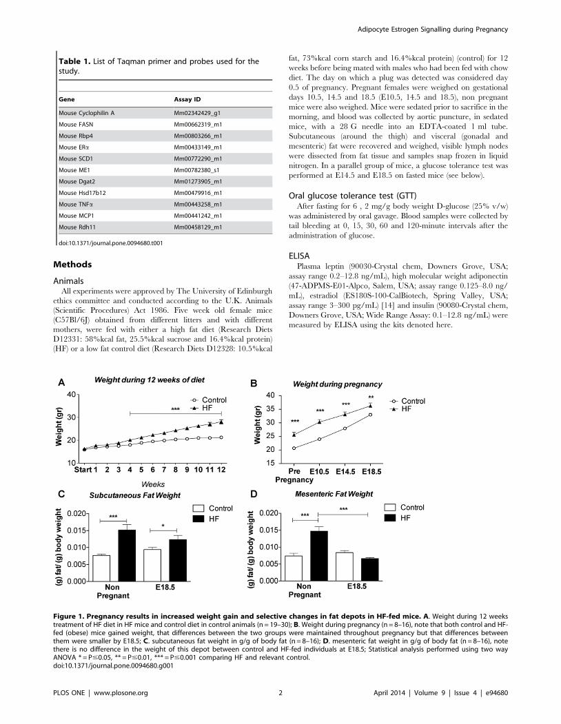
Table 1. List of Taqman primer and probes used for thestudy.
Gene Assay ID
Mouse Cyclophilin A Mm02342429_g1
Mouse FASN Mm00662319_m1
Mouse Rbp4 Mm00803266_m1
Mouse ERa Mm00433149_m1
Mouse SCD1 Mm00772290_m1
Mouse ME1 Mm00782380_s1
Mouse Dgat2 Mm01273905_m1
Mouse Hsd17b12 Mm00479916_m1
Mouse TNFa Mm00443258_m1
Mouse MCP1 Mm00441242_m1
Mouse Rdh11 Mm00458129_m1
doi:10.1371/journal.pone.0094680.t001
Figure 1. Pregnancy results in increased weight gain and selective changes in fat depots in HF-fed mice. A. Weight during 12 weekstreatment of HF diet in HF mice and control diet in control animals (n = 19–30); B. Weight during pregnancy (n = 8–16), note that both control and HF-fed (obese) mice gained weight, that differences between the two groups were maintained throughout pregnancy but that differences betweenthem were smaller by E18.5; C. subcutaneous fat weight in g/g of body fat (n = 8–16); D. mesenteric fat weight in g/g of body fat (n = 8–16), notethere is no difference in the weight of this depot between control and HF-fed individuals at E18.5; Statistical analysis performed using two wayANOVA * = P#0.05, ** = P#0.01, *** = P#0.001 comparing HF and relevant control.doi:10.1371/journal.pone.0094680.g001
Adipocyte Estrogen Signalling during Pregnancy
PLOS ONE | www.plosone.org 2 April 2014 | Volume 9 | Issue 4 | e94680
Microarray analysisVisceral adipose tissue RNA was prepared using Qiagen
RNeasy Mini kits (QIAGEN, Crawley-West Sussex, U.K.). RNA
integrity was calculated using a RNA 6000 Nano chip on an
Agilent 2100 bioanalyzer (Agilent Technologies, Palo Alto, CA).
Microarray analysis was performed at the ARK Genomics Facility
(Roslin Institute, Edinburgh, U.K.) on samples with a RIN of .
8.5. Array analysis was performed using standard protocols.
Briefly, samples were hybridized to the Affymetrix Mouse Genome
430-2.0 GeneChip which recognises 39,000 transcripts. Array data
were extracted through the GeneChip Operating Software
(GCOS), and CEL files were imported into Bioconductor and
normalized by robust multi-array average in the ‘‘Affy’’ module.
We used the Limma and Rank products (RankProd) packages to
perform statistical analysis. Spotfire DecisionSite (http://spotfire.
tibco.com/) was used to plot gene expression data. We performed
pathway analysis using DAVID (http://david.abcc.ncifcrf.gov/),
Webgestalt (http://bioinfo.vanderbilt.edu/webgestalt/) and Me-
tacore (from GeneGo Inc., St. Joseph, MI, USA) tools for genes
with Rank Product P-value of ,0.05, expression level .100 and
fold change 61.5. Microarray data were deposited in the Gene
Expression Omnibus (GEO) with accession number GSE48811.
Quantitative RT-PCR600 ng of total RNA, from visceral adipose tissue, was reverse
transcribed using the cDNA Synthesis Kit (4368813-Applied
Biosystems, Carlsbad, US). We analysed levels of gene-specific
mRNA using an ABI 7900HT (Applied Biosystems, Hill, U.K.)
with inventoried probes and primer sets included in Table 1
(Applied Biosystems, Hill, U.K.) and expressed values normalized
against cyclophilin A mRNA levels.
Western blottingMesenteric adipose tissue was homogenized in ice-cold lysis
buffer (50 mmol/L Tris, pH 7.4, 0.27 mol/L sucrose, 1 mmol/L
sodium orthovanadate, pH 10, 1 mmol/L EDTA, 1 mmol/L
EGTA, 10 mmol/L sodium b-glycerophosphate, 50 mmol/L
NaF, 5 mmol/L sodium pyrophosphate, 1% [w/v] Triton X-
100, 0.1% [v/v] 2-mercaptoethanol and one tablet of complete
TM protease inhibitor (Roche, Burgess Hill, U.K.). Thereafter,
50 mg of protein was run on 4–12% Bis-Tris gels for Western
blotting. To measure plasma concentrations of Rbp4 we
denatured 1 ml of plasma in standard loading buffer. Protein
signals were visualized using enhanced chemiluminescence (Pierce
Biotechnology, Rockford, IL) by exposure to Amersham Hyper-
filmTH ECL film (Amersham). We used primary antibodies raised
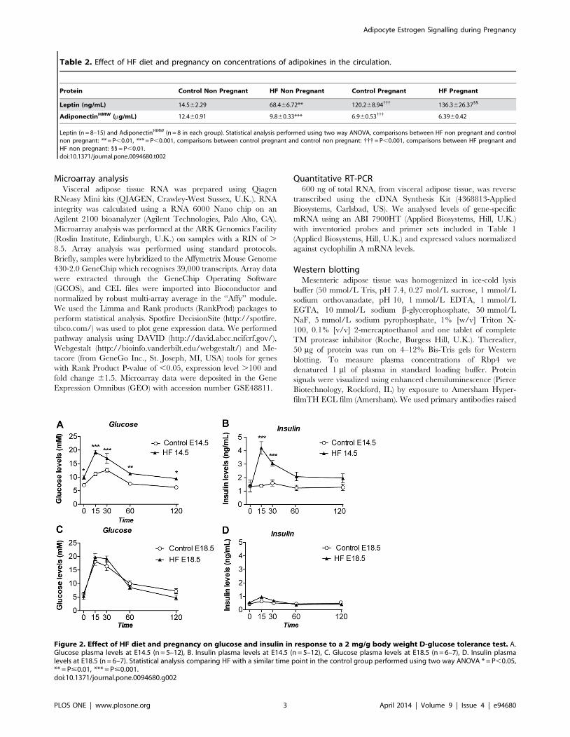
Table 2. Effect of HF diet and pregnancy on concentrations of adipokines in the circulation.
Protein Control Non Pregnant HF Non Pregnant Control Pregnant HF Pregnant
Leptin (ng/mL) 14.562.29 68.466.72** 120.268.94{{{ 136.3626.3711
AdiponectinHMW (mg/mL) 12.460.91 9.860.33*** 6.960.53{{{ 6.3960.42
Leptin (n = 8–15) and AdiponectinHMW (n = 8 in each group). Statistical analysis performed using two way ANOVA, comparisons between HF non pregnant and controlnon pregnant: ** = P,0.01, *** = P,0.001, comparisons between control pregnant and control non pregnant: {{{= P,0.001, comparisons between HF pregnant andHF non pregnant: 11 = P,0.01.doi:10.1371/journal.pone.0094680.t002
Figure 2. Effect of HF diet and pregnancy on glucose and insulin in response to a 2 mg/g body weight D-glucose tolerance test. A.Glucose plasma levels at E14.5 (n = 5–12), B. Insulin plasma levels at E14.5 (n = 5–12), C. Glucose plasma levels at E18.5 (n = 6–7), D. Insulin plasmalevels at E18.5 (n = 6–7). Statistical analysis comparing HF with a similar time point in the control group performed using two way ANOVA * = P,0.05,** = P#0.01, *** = P#0.001.doi:10.1371/journal.pone.0094680.g002
Adipocyte Estrogen Signalling during Pregnancy
PLOS ONE | www.plosone.org 3 April 2014 | Volume 9 | Issue 4 | e94680

against ERa (8644S Cell Signaling Technologies, U.K), b-tubulin
(2128S Cell Signaling Technologies, U.K.) and Rbp4 (A0040-
Dako, U.K.) and visualized the bands by adding horseradish
peroxidase anti-rabbit secondary antibody (Cell Signaling Tech-
nology).
Adipose tissue fractionationGonadal fat was digested in Krebs–Ringer solution with 2 mg/
mL collagenase type I (Worthington Biochemicals, NJ, USA) at
37uC for 1 h, filtered through 250-micron size exclusion mesh and
centrifuged at 600xg for 10 minutes to separate adipocytes from
stromal vascular cells (SVCs). Erythrocytes were lysed by re-
suspending the SVCs in 1 mL of erythrocyte lysis buffer (Sigma
Aldrich, Dorset, U.K.) for 5 min. Cells were then collected by
centrifugation and re-suspended in flow cytometry buffer.
Flow cytometry of pro-inflammatory macrophages inadipose tissue
For flow cytometry, 56105 of gonadal fat stromal vascular cells
were pre-incubated in 100 mL PBS with 1 mg/mL FcR block (BD
Biosciences, Oxford, U.K.) and then incubated with 0.2 mg each of
rat anti-mouse– CD11b FITC, and hamster anti-mouse-CD11c
PE (Caltag, Invitrogen, Paisley, U.K.) in PBS with 10% mouse
serum (Sigma Aldrich, Dorset, U.K.) for 30 min at 4uC in the
dark. Cells were sorted using a FACScalibur (BD Biosciences) flow
cytometer and analyzed using FlowJo 8.0 software (Treestar Inc.,
Ashland, OR).
Figure 3. Array analysis of mesenteric fat depots from control and HF-fed obese mice using Spotfire Decision Site. A. Log (base 2)ratios of gene expression intensities in pregnant and non-pregnant mice on the high fat and control diets. The y-axis shows the comparison of datafrom pregnant and non-pregnant mice regardless of diet. The x-axis compares high fat and control diets regardless of pregnancy status. Red spotsrepresent genes that are significantly differentially expressed in pregnant mice on HF compared to control diet. Genes expressed below the arbitrarythreshold (100) throughout the experiment were removed for clarity. Several genes of interest with higher expression in pregnant mice and on a highfat diet are marked and labelled. B. and C. Log (base 2) gene expression intensities in (B) Pregnant mice and (C) Non-pregnant mice on the high fatand control diets. For each group (pregnant or non pregnant), the y-axis shows the log2 expression in HF mice and the x-axis shows log2 expressionin control mice. Red spots represent genes that are significantly differentially expressed in high fat diet compared to control diet for each pregnancystatus. Several genes of interest discussed in the text are marked and labelled on graphs. Genes expressed below the arbitrary threshold (100)throughout the experiment were removed for clarity.doi:10.1371/journal.pone.0094680.g003
Adipocyte Estrogen Signalling during Pregnancy
PLOS ONE | www.plosone.org 4 April 2014 | Volume 9 | Issue 4 | e94680
Female clonal adipocyte Chub-S7 cell lineAn aliquot of immortalized primary human preadipocytes
(subsequently termed the Chub-S7 cell line), a gift from Dr
Christian Darimont (Nestle Research Centre, Lausanne, Switzer-
land) was differentiated according to published protocols [15].
After 17 days of differentiation, fully differentiated Chub-S7 cells
were treated with the estrogen receptor alpha agonist 4,49,40-(4-
Propyl-[1H]-pyrazole-1,3,5-triyl) trisphenol (PPT) (1426-Tocris,
Bristol, U.K.) in DMEM media (D6046-Sigma-Aldrich). A vehicle
DMSO control was included. Samples were incubated for 6 hours
in a cell culture incubator at 37uC with 5%CO2 in 6 wells plates.
Messenger RNA levels were measured in extracted total RNA as
described above.
Measurement of de novo lipogenesis in isolated primarymurine adipocytes
We quantified lipogenesis in isolated fat cells as previously
described [16]. Briefly, isolated adipocytes from gonadal adipose
tissue from groups of four non pregnant control female mice were
pooled and incubated for 12 hours with vehicle or 10 nM PPT in
a Krebs phosphate buffer. Following the incubation of adipocytes,
the cells were washed twice with 10 volumes of Krebs phosphate
prior to starting incubation with 3H Glucose to a final
concentration of 0.5 mCi/ml with or without the presence of
10 nM insulin. Typical Media/Cells ratio was 5/1 by volume.
Cells were lysed by adding.
1 ml/10 ml experimental medium of 6 M H2SO4 and vortex-
ing. Organic scintillant (POPOP+PPO in toluene; Fluka 327123)
was added (2 volumes/1 volumes of media), without mixing, and
then left for at least 2 hours without agitation to allow the lipids to
diffuse into the organic layer before counts were measured in a
scintillation counter (Tri-Carb 2100 TR Liquid Scinillation
Analyser, Packard). Lipid were extracted using a solution
composed by 4v Isopropanol, 1v Heptane and 1v Sulfuric acid
1N. We calculated relative lipogenesis by determining radioactive
glucose incorporation into total lipid content.
Statistical analysisData are presented as the mean 6 standard error of the mean
(S.E.M). Statistical analysis was performed using SigmaStat 3.5
and graphs were created using Graph Pad (GraphPad Prism 4).
Unless otherwise stated two-way ANOVA (Holm-Sidak, pairwise
multiple comparison) was performed with pregnancy and diet
status (HF or control) as independent variables. A P value of ,
0.05 was considered to show a statistically significant difference
between the groups.
Results
Normalization of visceral fat mass at late gestation in HFmice
High fat (HF) feeding for 12 weeks resulted in a 30% increase in
body weight in female mice (Figure 1A) which was in agreement
with previous studies from our laboratory using this model [17].
Although at time of conception mice on the control diet weighed
less than their HF counterparts they gained more weight (as a
proportion of body fat) than HF in pregnancy (60% vs 42% from
conception to E18.5) and there was a convergence in weight by the
end of pregnancy (Figure 1B). Despite this convergence, the HF
remained heavier than controls at E18.5. In both pregnant and
non-pregnant groups, HF-feeding resulted in a significant increase
in subcutaneous fat mass (as a fraction of total body weight) that
was largely maintained throughout pregnancy (Figure 1C). In
contrast, there was a significant and selective net reduction (a 2
53% reduction) in mesenteric fat (as a fraction of total body
weight) mass in HF-fed pregnant mice leading to no observable
differences in mesenteric fat mass between HF and control mice by
E18.5 (Figure 1D).
HF pregnant mice exhibit a normalised adipokine profileat late-stage pregnancy
To determine whether the reduction in visceral fat mass found
in HF pregnant mice was associated with an improvement in
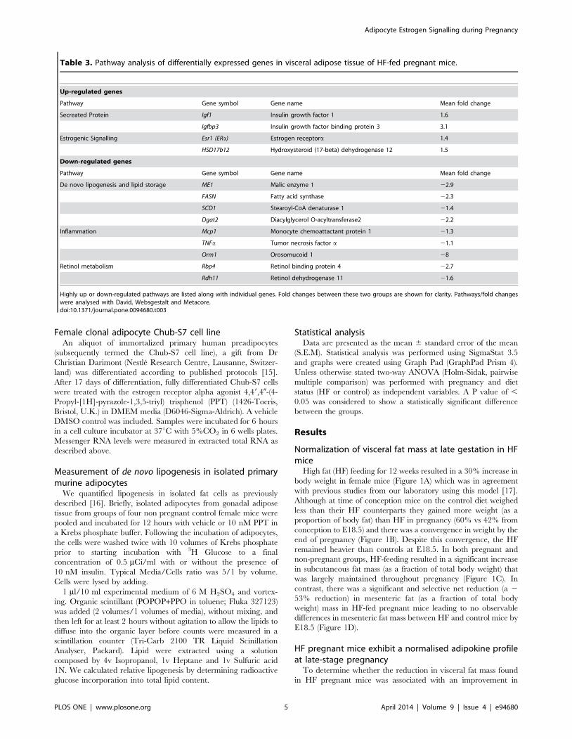
Table 3. Pathway analysis of differentially expressed genes in visceral adipose tissue of HF-fed pregnant mice.
Up-regulated genes
Pathway Gene symbol Gene name Mean fold change
Secreated Protein Igf1 Insulin growth factor 1 1.6
Igfbp3 Insulin growth factor binding protein 3 3.1
Estrogenic Signalling Esr1 (ERa) Estrogen receptora 1.4
HSD17b12 Hydroxysteroid (17-beta) dehydrogenase 12 1.5
Down-regulated genes
Pathway Gene symbol Gene name Mean fold change
De novo lipogenesis and lipid storage ME1 Malic enzyme 1 22.9
FASN Fatty acid synthase 22.3
SCD1 Stearoyl-CoA denaturase 1 21.4
Dgat2 Diacylglycerol O-acyltransferase2 22.2
Inflammation Mcp1 Monocyte chemoattactant protein 1 21.3
TNFa Tumor necrosis factor a 21.1
Orm1 Orosomucoid 1 28
Retinol metabolism Rbp4 Retinol binding protein 4 22.7
Rdh11 Retinol dehydrogenase 11 21.6
Highly up or down-regulated pathways are listed along with individual genes. Fold changes between these two groups are shown for clarity. Pathways/fold changeswere analysed with David, Websgestalt and Metacore.doi:10.1371/journal.pone.0094680.t003
Adipocyte Estrogen Signalling during Pregnancy
PLOS ONE | www.plosone.org 5 April 2014 | Volume 9 | Issue 4 | e94680
adipokine secretion and metabolic profile, we measured plasma
levels of leptin and adiponectinHMW (Table 2). As expected, prior
to pregnancy, HF mice had greater plasma leptin and lower
adiponectinHMW levels than control mice (Table 2). During
pregnancy, circulating leptin increased in both lean and HF
pregnant animals, perhaps reflecting leptin production from other
organs, such as placenta [18] (Table 2). Notably, during
pregnancy, plasma adiponectin decreased in control diet-fed
pregnant mice, but not in HF mice compared to non pregnant
animals so that by the end of pregnancy there were no differences
in adiponectinHMW levels between the HF and control groups
(Table 2).
Metabolic dysfunction converges with that of leanpregnant mice in late gestation HF mice
HF-fed pregnant mice displayed pronounced glucose intoler-
ance and apparent insulin resistance compared with control diet-
fed pregnant mice at E14.5 (Figure 2A and 2B). However, at
E18.5, control diet-fed mice showed a significant worsening of
glucose tolerance whereas HF-fed mice showed no further
deterioration in glucose tolerance compared to E14.5, leading to
an unexpected convergence in the glucose homeostasis profile of
the two groups (Figure 2C and 2D). Remarkably, the convergence
in glucose tolerance was accompanied by a correction of the
hyperinsulinaemia exhibited by the HF pregnant mice at mid
gestation (compare Figure 2B and 2D) suggesting if anything an
enhancement in whole body insulin sensitivity in the HF-fed E18.5
compared to the HF-fed E14.5 stages. There were no differences
in fasting glucose/insulin ratio comparing control and HF-fed
pregnant mice either at E14.5 (ratios of 7.7561.1 and 7.561.2
respectively) or at E18.5 (ratios of 15.165.1 and 15.962.7
respectively).
Transcriptomic analysis reveals gene pathways linked toreduced visceral adiposity and inflammation in HFpregnant mice
Microarray analysis was performed to understand the molecular
mechanisms associated with altered visceral fat mass in HF
pregnant mice. We used Spotfire DecisionSite to show the
distribution of the differentially expressed genes in our control
and HF, both non pregnant and E18.5 (Figure 3). We aimed to
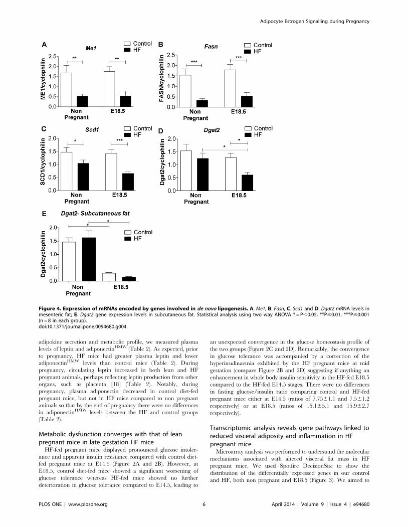
Figure 4. Expression of mRNAs encoded by genes involved in de novo lipogenesis. A. Me1, B. Fasn, C. Scd1 and D. Dgat2 mRNA levels inmesenteric fat; E. Dgat2 gene expression levels in subcutaneous fat. Statistical analysis using two way ANOVA * = P,0.05, **P#0.01, ***P#0.001(n = 8 in each group).doi:10.1371/journal.pone.0094680.g004
Adipocyte Estrogen Signalling during Pregnancy
PLOS ONE | www.plosone.org 6 April 2014 | Volume 9 | Issue 4 | e94680
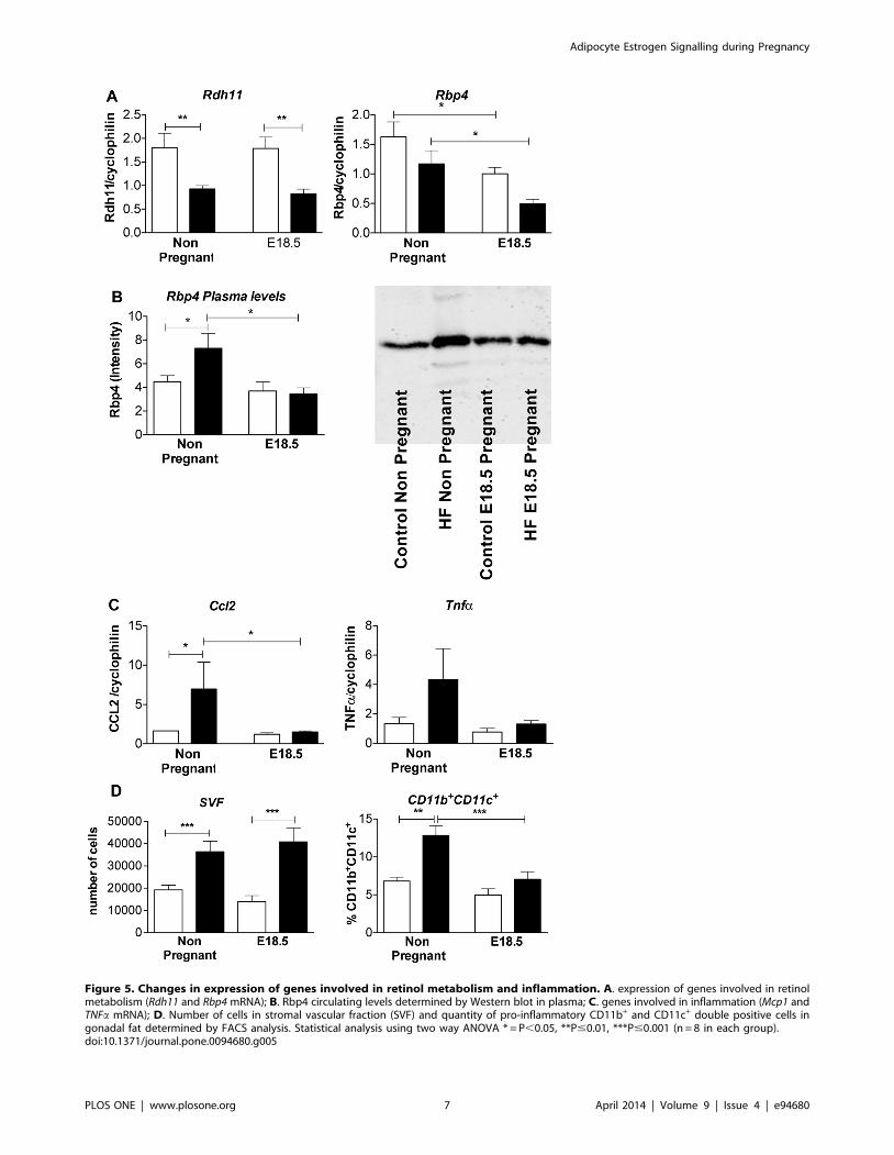
Figure 5. Changes in expression of genes involved in retinol metabolism and inflammation. A. expression of genes involved in retinolmetabolism (Rdh11 and Rbp4 mRNA); B. Rbp4 circulating levels determined by Western blot in plasma; C. genes involved in inflammation (Mcp1 andTNFa mRNA); D. Number of cells in stromal vascular fraction (SVF) and quantity of pro-inflammatory CD11b+ and CD11c+ double positive cells ingonadal fat determined by FACS analysis. Statistical analysis using two way ANOVA * = P,0.05, **P#0.01, ***P#0.001 (n = 8 in each group).doi:10.1371/journal.pone.0094680.g005
Adipocyte Estrogen Signalling during Pregnancy
PLOS ONE | www.plosone.org 7 April 2014 | Volume 9 | Issue 4 | e94680
identify specific genes altered by the interaction between diet and
pregnancy.
Particular genes of interest are marked and labelled on Figure 3
and summarized in Table 3.
Bioinformatics pathway analysis (Table 3) suggested that
expression of genes involved in de novo lipogenesis and lipid
storage (Me1, Fasn, Scd1 and Dgat2), inflammation (Ccl2, Tnfa) and
retinol metabolism (Rbp4, Rdh11) were down-regulated in visceral
adipose tissue of HF-fed pregnant, compared with HF non
pregnant mice. In contrast, there was an increased expression of
genes involved in estrogen biosynthesis/action (Esr1, Hsd17b12)
(Table 3).
We performed qRT-PCR to validate the array findings. We
found that genes involved in de novo lipogenesis (Me1, Fasn and
Scd1) were down regulated by HF feeding independently of
pregnancy in visceral fat (Figure 4A–C). However Dgat2, which
plays a key role in triglyceride storage, was selectively decreased in
visceral fat of HF-fed pregnant mice compared to both non-
pregnant HF-fed animals and control diet-fed pregnant mice
(Figure 4.D). This effect was in addition to the effects of pregnancy
itself in decreasing Dgat2 mRNA levels in both subcutaneous and
mesenteric fat (Figure 4E).
Rdh11 mRNA was down regulated in HF mice independently of
pregnancy (Figure 5A). In contrast, mesenteric fat Rbp4 mRNA
level was down regulated by pregnancy but not HF feeding
(Figure 5A). A decline in Rbp4 plasma levels in pregnancy was
limited to HF mice, with increased plasma Rbp4 levels in HF non-
pregnant mice compared to non pregnant controls (Figure 5B).
Ccl2 mRNA was up regulated in mesenteric fat of HF compared
with control non pregnant mice (Figure 5A). A similar trend was
observed with Tnfa. Pregnancy suppressed Ccl2 mRNA levels in
visceral fat of HF pregnant mice, leading to convergence of Ccl2
and Tnfa mRNA expression in HF and control animals by E18.5
(Figure 5C). Consistent with reduced adipose inflammatory
burden, pro-inflammatory CD11b+/CD11c+ macrophage density
in gonadal fat was increased with HF in non-pregnant mice but
converged with, and was comparable to, density in control diet-fed
pregnant mice by E18.5 (Figure 5D).
Reduced visceral adiposity in HF pregnancy is associatedwith increased adipocyte estrogens signalling
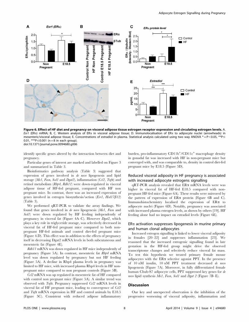
qRT-PCR analysis revealed that ERa mRNA levels were was
higher in visceral fat of HF-fed E18.5 compared with non-
pregnant HF-fed mice (Figure 6A). These results were mirrored by
the pattern of expression of ERa protein (Figure 6B and C).
Immunohistochemistry localised the expression of ERa to
adipocyte nuclei (Figure 6D). Notably, pregnancy was associated
with increased plasma estrogen levels, as shown by others [19]. HF
feeding alone had no impact on estradiol levels (Figure 6E).
ERa activation suppresses lipogenesis in murine primaryand human clonal adipocytes
Increased estrogen signalling is linked to lower visceral adiposity
in females [20–22] and suppresses inflammation [23]. We
reasoned that the increased estrogenic signalling found in late
gestation in the HF-fed group might drive the observed
transcriptome changes and selectively reduce visceral fat mass.
To test this hypothesis we treated primary female mouse
adipocytes with the ERa selective agonist PPT. In the presence
of 10 nM insulin, 10 nM PPT treatment decreased de novo
lipogenesis (Figure 7A). Moreover, in fully differentiated female
human Chub-S7 adipocyte cells, PPT suppressed key genes for de
novo lipid synthesis Me1, Fasn, Scd1 and Dgat 2 (Figure 7B–E).
Discussion
Our key and unexpected observation is the inhibition of the
progressive worsening of visceral adiposity, inflammation and
Figure 6. Effect of HF diet and pregnancy on visceral adipose tissue estrogen receptor expression and circulating estrogen levels. A.Esr1 (ERa) mRNA; B, C. Western analysis of ERa in visceral adipose tissue; D. Immunolocalisation of ERa to adipocyte nuclei (arrowheads) inmesenteric/visceral adipose tissue; E. Concentrations of estradiol in plasma. Statistical analysis calculated using two way ANOVA * = P,0.05, **P#0.01, ***P#0.001 (n = 8 in each group).doi:10.1371/journal.pone.0094680.g006
Adipocyte Estrogen Signalling during Pregnancy
PLOS ONE | www.plosone.org 8 April 2014 | Volume 9 | Issue 4 | e94680

metabolic dysfunction observed during normal pregnancy when
mice are fed a HF diet prior to and throughout gestation. The
protection and reversal of visceral adiposity happens at a late stage
in pregnancy and occurs despite an exaggerated deterioration in
metabolic function at mid-gestation in HF-fed mice. We suggest
that reduced visceral adiposity might be mediated, in part, through
direct actions of estrogen to suppress de novo lipogenesis.
Our results showed that prior to pregnancy, HF animals display
greater mesenteric fat weight and glucose intolerance than control
mice. At E14.5 (mid gestation), impaired glucose tolerance in HF
compared with control animals is maintained. However, by late
gestation (E18.5), glucose tolerance has not worsened further in
the HF-fed group and indeed insulin levels are reduced, suggesting
an increase in whole body insulin sensitivity or an improvement in
pancreatic beta cell function. Further, visceral fat mass, pro-
inflammatory cytokine gene expression and proinflammatory
adipose tissue macrophage density are reduced in HF-fed mice
at E18.5. The pregnancy related reduction in visceral fat mass
observed in parallel with maintained subcutaneous fat expansion
in HF mice contrasts with the observed increase in adipose tissue
expansion in pregnant rats treated with a HF diet [29,30] and with
the increased general adiposity observed in a mouse model treated
with a more moderate high fat diet than the one used in the study
reported here [24]. Although our HF mice continued to gain
weight in pregnancy, and weighed more than the control mice at
term, their pregnancy weight gain as a proportion of body weight
was lower than that for control mice in pregnancy. We believe that
the convergence in body weight observed in HF-fed and control
mice cannot be due to differences in fetal weight, given that King
et al (using the mouse model presented here) showed minimal
differences in birth weight in female offspring of HF fed mice
(limited to 6% in female fetuses, with no effect observed in the
birth weight of male fetuses) [17]. Furthermore, we believe that the
convergence in maternal weight by E18.5 is not due to calorie
restriction in HF mice because previously published work by King
et al has shown that energy intake is similar in control and HF
pregnant mice [17].
Increased estrogen signalling through ERa drives an improved
metabolic phenotype and a favourable fat distribution in rodents
and humans in males and non-pregnant females [21,22]. Our
results reveal a novel role for a selective increase in estrogen
signalling through increased ERa expression in visceral adipocytes
of pregnant animals given HF diet. These changes in ERa occur
during a period of pregnancy characterised by increased
Figure 7. Effect of the ERa-selective agonist PPT on de novo lipogenesis of primary adipocytes and expression levels of relevantgenes in fully differentiated Chub-S7. A. effect of ERa activation mediated by treatment of primary adipocytes with 10 nM PPT, on de novolipogenesis in presence of 10 nM insulin. Effect of different concentration of PPT treatment (1 nM, 10 nM, 0.1 mM and 1 mM) on gene expression of B.Me1, C. FASN, D. SCD1 and E. Dgat2 (n = 6 in each groups). Normality of data distribution was confirmed using the Kolmogorv-Smirnov test. Statisticalanalysis was performed using the Kruskal-Wallis Test with Dunns as a post hoc test: comparison of pairs of columns* = P,0.05, ** = P,0.01 and*** = P,0.001.doi:10.1371/journal.pone.0094680.g007
Adipocyte Estrogen Signalling during Pregnancy
PLOS ONE | www.plosone.org 9 April 2014 | Volume 9 | Issue 4 | e94680

circulating concentrations of estradiol. Our transcriptomic and
functional studies in Chub-S7 cells and primary adipocytes, in
which PPT treatment (acting through ERa) inhibits de novo
lipogenesis, are supported by similar finding in PPT-treated 3T3-
L1 adipocytes [25]. Our data are also consistent with the effect of
estradiol treatment to decrease lipogenesis and triglyceride storage
[26], and with the phenotype of increased lipogenesis in ERa2/2
mice [27–28]. Thus we hypothesize that estrogen plays a
fundamental role in suppression of lipogenic pathways in visceral
adipose tissue of HF pregnant mice. The driver to increased ERaexpression is unclear, but a likely possibility is increased local
synthesis of E2, which upregulates the ERa receptor.
Our microarray analysis also identifies reduced adipose tissue
inflammation in HF pregnant mice. In particular, we observed a
pregnancy related decrease in the production of visceral fat Ccl2
mRNA and in the numbers of adipose tissue pro-inflammatory
macrophages in HF-fed but not control diet-fed pregnant animals.
This attenuation in inflammation in HF animals may also be
driven by increased estrogen action on adipocytes leading to
reduced macrophage accumulation into adipose tissue (possibly via
reduced chemokine release), given that ovariectomized mice show
increased adipose tissue Ccl2 and Tnfa mRNA production which is
reversed by estradiol administration [29]. As in our mouse model,
a pregnancy-induced attenuation in circulating inflammatory
markers has also been shown in a longitudinal study of 240 obese
pregnant women [30], although data from our own group shows
that an excess of circulating and placentally derived pro-
inflamatory cytokines is still present in obese women at term
[31]. In parallel with the decrease in adipose tissue mRNA
expression of Ccl2 and Tnfa in our HF mice as pregnancy
advanced, we also observed a decreased presence of pro-
inflammatory adipose tissue macrophages.
We found retinol metabolism, in particular Rbp4, to be
decreased by pregnancy in HF-fed mice. Rpb4 is secreted by
adipocytes and induces insulin resistance by reducing PI3K
signaling in muscle and increasing gluconeogenesis in liver [32].
Thus, in HF-fed mice, reduced Rbp4 levels in pregnancy could
contribute to the alleviation of worsened glucose homeostasis
observed in HF-fed compared to control diet-fed mice by E18.5. It
is also known that ERa signaling increases Rpb4 expression in
3T3-L1 adipocytes, supporting the central role for estrogen in our
phenotype [33]. Leptin levels were increased by pregnancy,
despite the decrease visceral adipose tissue and unchanged
subcutaneous fat weight, suggesting an alternative source of
production, possibly the placenta [18]. Higher leptin levels per
unit fat mass may also reflect increased production by adipocytes
with increased ERa that are also exposed to a higher concentra-
tion of estradiol in pregnancy [34,35].
In summary, we hypothesize that increased ERa expression
contributes to a novel pregnancy-related and fat depot-specific
reduction in visceral adiposity and inflammation that counteracts
the progressive metabolic perturbations associated with pregnan-
cy. Given the positive association between visceral fat mass and
exaggerated cardiovascular risk, the elucidation of the molecular
pathway by which ERa suppresses visceral fat accumulation – and
the interplay of ERa with the hormonal milieu of pregnancy,
might provide insight into therapeutic strategies against metabolic
disease [36]. These results provide further support for the rationale
for interventions early in pregnancy (or prior to pregnancy) to
improve pregnancy outcome.
Acknowledgments
We thank Jon Henderson, Lynn Ramage, Jean Wade, Graham Harold,
Frances Collins and Mike Millar and staff of the Histology and Imaging
Core Facility for expert technical assistance. We thank Mandy Drake and
Vicky King for sharing materials with us. This study was funded by
Tommy’s the Baby Charity [?http://www.tommys.org/]. Donald Dunbar
was supported by a BHF Centre of Research Excellence Award to the
University of Edinburgh. Jane Norman is guarantor for this work.
Author Contributions
Conceived and designed the experiments: SMAP NMM JEN. Performed
the experiments: SMAP ST TK DRD KM. Analyzed the data: SMAP
PTKS NMM JEN. Wrote the paper: SMAP PTKS NMM JEN.
References
1. Norman JE, Reynolds RM (2011) The consequences of obesity and excess
weight gain in pregnancy. Proc Nutr Soc. 70(4):450–6.
2. Barbour LA, McCurdy CE, Hernandez TL, Kirwan JP, Catalano PM, et al.
(2007) Cellular mechanisms for insulin resistance in normal pregnancy and
gestational diabetes. Diabetes Care. 30 Suppl 2:S112–9.
3. Catalano PM, Tyzbir ED, Wolfe RR, Calles J, Roman NM, et al Carbohydrate
metabolism during pregnancy in control subjects and women with gestational
diabetes. (1993) Am J Physiol 264(1 Pt 1):E60–7.
4. Catalano PM, Huston L, Amini SB, Kalhan SC (1999) Longitudinal changes in
glucose metabolism during pregnancy in obese women with normal glucose
tolerance and gestational diabetes mellitus. Am J Obstet Gynecol. 180(4):903–
16.
5. Zhang L, Sugiyama T, Murabayashi N, Umekawa T, Ma N, et al. (2011) The
inflammatory changes of adipose tissue in late pregnant mice. J Mol Endocrinol.
47(2):157–65.
6. de Castro J, Sevillano J, Marciniak J, Rodriguez R, Gonzalez-Martin C, et al.
(2011) Implication of low level inflammation in the insulin resistance of adipose
tissue at late pregnancy. Endocrinology. 152(11):4094–105.
7. Resi V, Basu S, Haghiac M, Presley L, Minium J, et al. (2012) Molecular
inflammation and adipose tissue matrix remodeling precede physiological
adaptations to pregnancy. Am J Physiol Endocrinol Metab. 303(7):E832–40.
8. Locher LF, Meyer N, Weber EM, Rehage J, Meyer U, et al. (2011) Hormone-
sensitive lipase protein expression and extent of phosphorylation in subcutaneous
and retroperitoneal adipose tissues in the periparturient dairy cow. J Dairy Sci.
94(9):4514–23.
9. Fernandes FS, Sardinha FL, Badia-Villanueva M, Carulla P, Herrera E, et al.
(2012) Dietary lipids during early pregnancy differently influence adipose tissue
metabolism and fatty acid composition in pregnant rats with repercussions on
pup’s development. Prostaglandins Leukot Essent Fatty Acids. 86(4–5):167–74.
10. Friedman JE, Ishizuka T, Shao J, Huston L, Highman T, et al. (1999) Impaired
glucose transport and insulin receptor tyrosine phosphorylation in skeletal
muscle from obese women with gestational diabetes. Diabetes. 48(9):1807–14.
11. Catalano PM, Nizielski SE, Shao J, Preston L, Qiao L, et al. (2002)
Downregulated IRS-1 and PPARgamma in obese women with gestational
diabetes: relationship to FFA during pregnancy. Am J Physiol Endocrinol
Metab. 282(3):E522–33.
12. Kjerulff LE, Sanchez-Ramos L, Duffy D (2011) Pregnancy outcomes in women
with polycystic ovary syndrome: a metaanalysis. Am J Obstet Gynecol.
204(6):558 e1–6.
13. Metzger BE, Lowe LP, Dyer AR, Trimble ER, Chaovarindr U, et al. (2008)
Hyperglycemia and adverse pregnancy outcomes. N Engl J Med. 358(19):1991–
2002.
14. Haisenleder DJ, Schoenfelder AH, Marcinko ES, Geddis LM, Marshall JC
(2011) Estimation of estradiol in mouse serum samples: evaluation of commercial
estradiol immunoassays. Endocrinology. 152(11):4443–7.
15. Darimont C, Zbinden I, Avanti O, Leone-Vautravers P, Giusti V, et al. (2003)
Reconstitution of telomerase activity combined with HPV-E7 expression allow
human preadipocytes to preserve their differentiation capacity after immortal-
ization. Cell Death Differ. 10(9):1025–31.
16. Kaaman M, Sparks LM, van Harmelen V, Smith SR, Sjolin E, et al. (2007)
Strong association between mitochondrial DNA copy number and lipogenesis in
human white adipose tissue. Diabetologia. 50(12):2526–33.
17. King V, Dakin RS, Liu L, Hadoke PW, Walker BR, et al. (2013) Maternal
obesity has little effect on the immediate offspring but impacts on the next
generation. Endocrinology. 154(7):2514–24.
18. Hoggard N, Hunter L, Duncan JS, Williams LM, Trayhurn P, et al. (1997)
Leptin and leptin receptor mRNA and protein expression in the murine fetus
and placenta. Proc Natl Acad Sci U S A. 30;94(20):11073–8.
Adipocyte Estrogen Signalling during Pregnancy
PLOS ONE | www.plosone.org 10 April 2014 | Volume 9 | Issue 4 | e94680
19. McCormack JT, Greenwald GS (1974) Progesterone and oestradiol-17beta
concentrations in the peripheral plasma during pregnancy in the mouse. JEndocrinol. 62(1):101–7.
20. Turgeon JL, Carr MC, Maki PM, Mendelsohn ME, Wise PM (2006) Complex
actions of sex steroids in adipose tissue, the cardiovascular system, and brain:Insights from basic science and clinical studies. Endocr Rev. 27(6):575–605.
21. Heine PA, Taylor JA, Iwamoto GA, Lubahn DB, Cooke PS (2000) Increasedadipose tissue in male and female estrogen receptor-alpha knockout mice. Proc
Natl Acad Sci U S A. 97(23):12729–34.
22. Okura T, Koda M, Ando F, Niino N, Ohta S, et al. (2003) Association ofpolymorphisms in the estrogen receptor alpha gene with body fat distribution.
Int J Obes Relat Metab Disord. 27(9):1020–7.23. Ribas V, Nguyen MT, Henstridge DC, Nguyen AK, Beaven SW, et al. (2010)
Impaired oxidative metabolism and inflammation are associated with insulinresistance in ERalpha-deficient mice. Am J Physiol Endocrinol Metab.
298(2):E304–19.
24. Jones HN, Woollett LA, Barbour N, Prasad PD, Powell TL, et al. (2009) High-fat diet before and during pregnancy causes marked up-regulation of placental
nutrient transport and fetal overgrowth in C57/BL6 mice. FASEB J. 23(1):271–8.
25. Homma H, Kurachi H, Nishio Y, Takeda T, Yamamoto T, et al. (2000)
Estrogen suppresses transcription of lipoprotein lipase gene. Existence of aunique estrogen response element on the lipoprotein lipase promoter. J Biol
Chem. 275(15):11404–11.26. Gao H, Bryzgalova G, Hedman E, Khan A, Efendic S, et al. (2006) Long-term
administration of estradiol decreases expression of hepatic lipogenic genes andimproves insulin sensitivity in ob/ob mice: a possible mechanism is through
direct regulation of signal transducer and activator of transcription 3. Mol
Endocrinol. 20(6):1287–99.27. Bryzgalova G, Gao H, Ahren B, Zierath JR, Galuska D, et al. (2006) Evidence
that oestrogen receptor-alpha plays an important role in the regulation of
glucose homeostasis in mice: insulin sensitivity in the liver. Diabetologia.
49(3):588–97.
28. Bryzgalova G, Lundholm L, Portwood N, Gustafsson JA, Khan A, et al. (2008)
Mechanisms of antidiabetogenic and body weight-lowering effects of estrogen in
high-fat diet-fed mice. Am J Physiol Endocrinol Metab. 295(4):E904–12.
29. Stubbins RE, Najjar K, Holcomb VB, Hong J, Nunez NP (2012) Oestrogen
alters adipocyte biology and protects female mice from adipocyte inflammation
and insulin resistance. Diabetes Obes Metab. 14(1):58–66.
30. Friis CM, Paasche Roland MC, Godang K, Ueland T, Tanbo T, et al. (2013)
Adiposity-related inflammation: effects of pregnancy. Obesity (Silver Spring).
21(1):E124–30.
31. Roberts KA, Riley SC, Reynolds RM, Barr S, Evans M, et al. (2011) Placental
structure and inflammation in pregnancies associated with obesity. Placenta.
32(3):247–54.
32. Yang Q, Graham TE, Mody N, Preitner F, Peroni OD, et al. (2005) Serum
retinol binding protein 4 contributes to insulin resistance in obesity and type 2
diabetes. Nature. 436(7049):356–62.
33. Jung US, Jeong KJ, Kang JK, Yi K, Shin JH, et al. (2013) Effects of estrogen
receptor alpha and beta on the expression of visfatin and retinol-binding protein
4 in 3T3-L1 adipocytes. Int J Mol Med. 32(3):723–8.
34. Piermaria J, Console G, Perello M, Moreno G, Gaillard RC, et al. (2003) Impact
of estradiol on parametrial adipose tissue function: evidence for establishment of
a new set point of leptin sensitivity in control of energy metabolism in female rat.
Endocrine. 20(3):239–45.
35. Yi KW, Shin JH, Seo HS, Lee JK, Oh MJ, et al. (2008) Role of estrogen
receptor-alpha and -beta in regulating leptin expression in 3T3-L1 adipocytes.
Obesity. 16(11):2393–9.
36. Hamdy O, Porramatikul S, Al-Ozairi E (2006) Metabolic obesity: the paradox
between visceral and subcutaneous fat. Curr Diabetes Rev. 2(4):367–73.
Adipocyte Estrogen Signalling during Pregnancy
PLOS ONE | www.plosone.org 11 April 2014 | Volume 9 | Issue 4 | e94680