Embed Size (px)
Citation preview

Research ArticleEco-Friendly, Green Packaging Materials fromAkaganeite and Hematite Nanoparticle-Reinforced ChitosanNanocomposite Films
G. T. D. Chandrakumara,1,2 D. M. S. N. Dissanayake,1,2 M. M. M. G. P. G. Mantilaka ,1,2
R. T. De Silva,1 H. M. T. G. A. Pitawala,2,3 and K. M. Nalin de Silva 1,4
1Sri Lanka Institute of Nanotechnology (SLINTEC), Nanotechnology and Science Park, Mahenwatte, Pitipana,Homagama, Sri Lanka2Postgraduate Institute of Science, University of Peradeniya, Peradeniya, Sri Lanka3Department of Geology, Faculty of Science, University of Peradeniya, Peradeniya, Sri Lanka4Department of Chemistry, University of Colombo, Colombo, Sri Lanka
Correspondence should be addressed to M. M. M. G. P. G. Mantilaka; [email protected]
Received 8 September 2019; Revised 18 November 2019; Accepted 3 December 2019; Published 23 December 2019
Academic Editor: Francesco Marotti de Sciarra
Copyright © 2019 G. T. D. Chandrakumara et al. This is an open access article distributed under the Creative Commons AttributionLicense, which permits unrestricted use, distribution, and reproduction in any medium, provided the original work isproperly cited.
In this study, chitosan nanocomposite thin films were successfully fabricated by incorporating hematite nanoparticles (HNPs) andakaganeite nanoparticles (ANPs) as reinforcing fillers using the solution casting method. HNPs and ANPs were synthesized via aurea-assisted synthesis route using naturally occurring ferruginous laterites. Scanning electronmicroscopic (SEM) images indicatedthe spherical to subhexagonal morphology of the HNPs and rice-like morphology of the ANPs. X-ray diffractograms indicate thecrystalline structure of iron oxides as hematite and akaganeite. Tensile tests were carried out to evaluate the mechanical propertiesof the nanocomposite films where maximum tensile stress of the chitosan/HNP composites was improved as high as 35.7% whilechitosan/ANP composites indicated 43.5%. Thermal decomposition curves obtained by thermogravimetric analysis (TGA) indicatethat the thermal stability of the nanocomposites has improved remarkably compared to neat chitosan films. Furthermore, thesenanocomposites exhibited excellent UV barrier properties as identified by UV-visible spectrometry. Fourier-transform infrared(FTIR) spectroscopic results are evident in the presence of Fe-O bond in the wavenumber around 480-500 cm-1, and the resultalso indicated that the nanofillers interact with the chitosan matrix via hydrogen bonding, which enhanced the physicalproperties of the nanocomposites. Incorporation of iron oxide nanoparticle varieties into chitosan has led to improvements ofcertain physical and chemical properties, which make chitosan a promising material for packaging applications.
1. Introduction
Over several decades, various biodegradable materials havegained considerable attention as alternatives for petrochemical-based packaging materials [1]. Production of biopolymericpackaging films provides a number of economic and envi-ronmental benefits. Chitosan (CH) is one of the key naturallyderived green polymers used in the synthesis of packagingmaterials [2–4]. It is the linear polymer of β-(1-4)-2-aceta-mido-2-deoxy-D-glucopyranose synthesized by deacetyla-tion of chitin, the second most abundant polysaccharide in
nature. Chitin is extracted by the exoskeleton of crustaceanssuch as shrimps and crab shells [5–7]. Chitosan exhibitssuperior properties such as nontoxicity, biodegradability,biocompatibility, antiviral, and antimicrobial properties[8–12]. This unique biopolymer is widely applied in variousfields including agriculture [13], food packaging [14–16],textiles [10], bioengineering, and medicine [17, 18].
Although CH can easily be formulated into thin films, itspoor mechanical and thermal properties limit the suitabilityfor packaging applications. Since the properties of the pack-aging material directly affect the quality and the shelf life of
HindawiJournal of NanomaterialsVolume 2019, Article ID 1049142, 11 pageshttps://doi.org/10.1155/2019/1049142

the product, suitable modifications are required. Severalstrategies have been tested and discussed in the literatureto overcome the above-mentioned drawbacks. Recentreviews discussed the chemical modifications such as cross-linking of biopolymer chains by crosslinking agents viaintra- or intermolecular linkages which enhanced themechanical and barrier properties of the biopolymers. More-over, the excess amount of the crosslinking agents can act asplasticizer agents [19]. Biopolymers such as starch, cellulose,alginate, pectin, glucose, and some proteins including gelatinhave been blended with chitosan to improve the quality offood [16, 20].
Recent studies proposed various methods to enhance theproperties by reinforcing numerous inorganic materials intothe chitosan polymer matrix. Reinforcing nanomaterialssuch as nanohydroxyapatite [18, 21, 22], nano-MgO [6],nanoclays including montmorillonite [1, 23], and halloysitenanotubes [24, 25] had been incorporated, and numerousphysicochemical properties of the packaging materials havebeen enhanced. Moreover, incorporation of graphene oxideand carbon nanotubes has successfully increased themechanical properties of the films [3, 16]. Chitosan has beentested for antimicrobial studies due to its inherent antibacte-rial properties against both gram-positive and gram-negativebacteria and fungi. Reinforcement of Ag nanoparticles in asmall percentage inactivated E. coli compared with the neatchitosan [26]. Ag nanoparticles have the ability to enter themicrobial cells and induce the damage of the cell membraneand the reactions that damage the DNA of the cells [27]. Fur-thermore, incorporation of both zinc oxide (ZnO) nanoparti-cles and silver (Ag) nanoparticles into chitosan to formCH/Ag/ZnO blend had prevented the growth of microbialpathogens than CH/Ag and CH/ZnO films [26]. Strongerantibacterial properties against S. aureus than E. coli wereexhibited by different kinds of graphene oxide- (GO-) chito-san nanohybrids (pristine powder, sphericle, and nanofibri-lar network structures) [28]. Yadav et al. discussed asynthesizing route of cellulose/iron oxide nanocomposite[29] which enhanced the properties significantly. Chitosan-Fe-Al-Mn oxyhydroxide composites have been developedby Chaudhary et al. targeting adsorption applications [30].To the best of our knowledge, there is no attempt that hasbeen taken to incorporate iron oxide-based nanofillers intothe chitosan structure targeting active packaging applica-tions. But in conventional production, these nanomaterialsare pricey, require a lot of time and effort, and most of allare not industrially viable. Nevertheless, the nanomaterialsused in this study are extracted directly from an earth mate-rial called laterites. As stated in the method developed by theauthors previously [31], these nanoparticles are cost-effectiveand industrially viable.
In this work, we reinforced CH polymer films by hema-tite nanoparticles (HNPs) and akaganeite nanoparticles(ANPs) by developing polymer nanocomposites and inves-tigated their properties in relation to advanced green pack-aging applications. Hematite and akaganeite nanoparticleswere synthesized using naturally occurring widely distrib-uted ferruginous laterites [31, 32], and these nanoparticleswere incorporated into CH by solution casting. The mor-
phological, thermal, chemical, optical, and mechanicalproperties of synthesized nanocomposites were investigatedusing SEM, XRD, TGA, FTIR, UV-Vis-IR spectrometer,and tensile tester. The materials, chemicals, and methodsused in this synthesis process are cost-effective, facile, andeasily industrialized. Also, the iron oxide/hydroxide nano-materials that were used to reinforce the chitosan matrixare nontoxic and environmentally friendly [33, 34]. Herein,eco-friendly polymer nanocomposites with enhanced prop-erties are introduced, which could be considered as promis-ing alternatives for petroleum-derived materials in futureactive packaging activities.
2. Experimental
2.1. Materials. Chitosan flakes from shrimp shells (molecularweight ~50000Da based on viscosity; ≥75% of deacetylation),acetic acid (≥99.8%), sodium hydroxide (≥98.0%), hydro-chloric acid (≥39wt. %), and urea (99%) were purchasedfrom Sigma-Aldrich and were used without further purifica-tion. Laterite rock samples were collected from Homagama,Sri Lanka.
2.2. Urea-Assisted Synthesis of ANPs and HNPs UsingFerruginous Laterite. Extraction of Fe3+ ions from lateritesto the acid solution was performed by mixing 10 g of lateritepowder (250μm particle size), 20ml of 5M HCl solution,and 20ml of distilled water under reflux condition for 5 h at100°C as in [31]. The supernatant was separated by filtrationand treated with 5 g of urea under reflux conditions at 90°Cfor 5 h. The resultant precipitate was separated by centrifuga-tion, washed by 3 sediment-decant cycles, and oven-dried toget ANPs. One portion of the dry precipitate was calcined at650°C for 2 h in a muffle furnace to produce HNPs.
2.3. Fabrication of Chitosan/HNP and Chitosan/ANPComposite Films. 1 g of chitosan flakes was dissolved in100ml of 1% (w/v) acetic acid solution by stirring at500 rpm for 12 h. The chitosan solution was filtered by180μm sieve to remove undissolved particles. In order toprepare chitosan/HNP composites with 0.25% and 1%(w/w%) filler compositions, a predetermined amount ofHNPs was added to the chitosan solution under the vigorousstirring at 1000 rpm. The homogeneous dispersions obtainedafter 24 h magnetic stirring followed by 10-minute sonicationwere poured into 8 cm size Petri dishes and vacuum dried for3 days at 50°C to evaporate the solvent. Remaining acetic acidin the resultant nanocomposite films was neutralized by 5mlof 1M NaOH solution and rinsed with distilled water. Com-posite films were peeled off from the Petri dishes and placedin a desiccator for further testing. A similar procedure wasfollowed to synthesize chitosan/ANP composites where thevarious amounts of ANPs were added to the chitosan solu-tion instead of HNPs.
2.4. Characterization of HNPs and Composite Films
2.4.1. Morphological Analysis. Synthesized ANPs, HNPs, andthe fracture surfaces of the composite films were examined by
2 Journal of Nanomaterials

a field emission scanning electron microscope (FE-SEM)(Hitachi SU6600, Japan).
2.4.2. X-Ray Diffraction (XRD) Analysis. The crystallinity ofthe nanoparticles and the composite films were analyzedusing diffraction patterns obtained from an X-ray diffrac-tometer (Siemens D5000, Germany) working with an X-raytube operated at 40 kV voltage and 40mA current whichemits Cu-Kα radiation. Data was collected at diffractionangle (2θ) values between 6° and 70° with step size of 2θ =0:1° at room temperature. The diffraction patterns were ana-lyzed using the XPowder12 software package with the aid ofthe ICDD PDF-2 database.
2.4.3. Chemical Properties. Fourier-transform infrared(FTIR) spectra were analyzed using a spectrometer (BrukerVERTEX 80, USA) based on the Attenuated Total Reflection(ATR) technique within 500-4000 cm-1 with 32 scans permeasurement and 4 cm-1 resolution to identify the interfacialinteractions of the fabricated films.
2.4.4. Thermal Properties. Thermal behaviours of the com-posite samples between 25 and 800°C were determined usingthermogravimetric analysis (TA Instruments SDT Q600,USA) with a ramp of 10°C/min in a nitrogen environment.
2.4.5. UV Shielding Properties. The UV absorbance of theproduced films was examined over the range of 200-800 cm-1 using a UV-Vis-IR spectrophotometer (ShimadzuUV-3600, Japan).
2.4.6. Mechanical Properties. The average thicknesses of thetest specimens were determined using a digital micrometerscrew gauge (Mitutoyo, Japan) prior to the tensile tests. Thinfilm specimens with the dimensions of 8mm × 70mm weremounted on a cardboard window for ease of handling andfixed between the upper and lower clamps of the test rig withthe gauge length of 35mm. The maximum tensile strength(σ), elastic modulus (E), and elongation at break (ε) wereperformed as stated by ASTM D882-02 [35] using a tensiletester (Instron, USA) with a load cell of 500 kN and a strainrate of 5mm/min. Six replicate thin film stripes were testedfor each composition, and the mean values were reported.
2.4.7. Statistical Methods. One-way analysis of variance(ANOVA) was conducted by Tukey’s post hoc test usingcommercially available software (IBM SPSS Statistics, version21) to investigate the significant difference between the meanvalues of σ, E, and ε with respect to the HNP and ANP con-centration. The mean difference was considered significant atthe 0.05 level.
3. Results and Discussion
3.1. Morphologies of Nanomaterials. Laterite samples col-lected from the similar locality indicated that the composi-tion consists of iron-bearing minerals, namely, goethite(FeO(OH)) and hematite (Fe2O3). The ferruginous natureof the laterite was a great advantage to use it as the ironsource for the synthesis of nanoparticles. The iron extractionprocess from powdered laterite by hydrochloric acid solution
4.00 𝜇m
(a)
500 nm
(b)
1.00 𝜇m
(c)
500 nm
(d)
Figure 1: Scanning electron micrographs of nanofillers: (a, b) ANPs and (c, d) HNPs.
3Journal of Nanomaterials
and the urea-assisted nanoparticle synthesis method werediscussed elsewhere [31]. Due to the low cost and noncom-plexity, the above method was applied to produce hematitenanoparticles (HNPs) by calcination of akaganeite nanopar-ticles (ANPs).
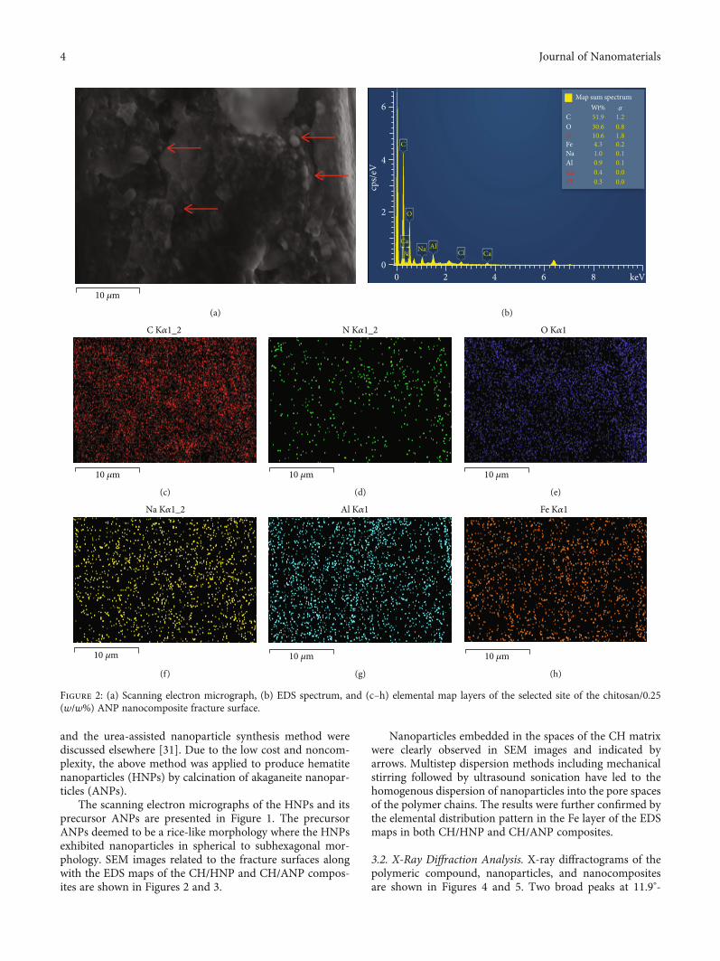
The scanning electron micrographs of the HNPs and itsprecursor ANPs are presented in Figure 1. The precursorANPs deemed to be a rice-like morphology where the HNPsexhibited nanoparticles in spherical to subhexagonal mor-phology. SEM images related to the fracture surfaces alongwith the EDS maps of the CH/HNP and CH/ANP compos-ites are shown in Figures 2 and 3.
Nanoparticles embedded in the spaces of the CH matrixwere clearly observed in SEM images and indicated byarrows. Multistep dispersion methods including mechanicalstirring followed by ultrasound sonication have led to thehomogenous dispersion of nanoparticles into the pore spacesof the polymer chains. The results were further confirmed bythe elemental distribution pattern in the Fe layer of the EDSmaps in both CH/HNP and CH/ANP composites.
3.2. X-Ray Diffraction Analysis. X-ray diffractograms of thepolymeric compound, nanoparticles, and nanocompositesare shown in Figures 4 and 5. Two broad peaks at 11.9°-
10 𝜇m
(a)
6
CaClAlNa
Ca
O
C
51.9 1.20.830.6
10.6 1.80.24.3
1.0 0.10.10.00.00.3
0.40.9
Ca
N
CWt%
Map sum spectrum𝜎
O
FeNaAl
Cl
8 keV4200
2
4
cps/
eV
6
(b)
10 𝜇m
C K𝛼1_2
(c)
10 𝜇m
N K𝛼1_2
(d)
10 𝜇m
O K𝛼1
(e)
10 𝜇m
Na K𝛼1_2
(f)
10 𝜇m
Al K𝛼1
(g)
10 𝜇m
Fe K𝛼1
(h)
Figure 2: (a) Scanning electron micrograph, (b) EDS spectrum, and (c–h) elemental map layers of the selected site of the chitosan/0.25(w/w%) ANP nanocomposite fracture surface.
4 Journal of Nanomaterials

17.5° and 20°-26° appeared in the diffractogram of chitosan(Figure 6) due to the amorphous nature of the polymericcompound. For HNPs (Figure 6), major diffraction peakswere found at 24.2°, 33.1°, 35.6°, 40.8°, 49.4°, and 54.0° relatedto (012), (104), (110), (113), (024), and (116) crystal planes,respectively. This crystallographic pattern is inherent inthe hematite (Fe2O3) crystal structure (JCPDS card no.0469-72). The resultant polymer nanocomposite (Figure 6)chitosan/HNPs exhibit two diffraction peaks at 33.1° and35.6° related to hematite which confirms the reinforcementof HNPs into the CH.
Diffractogram related to ANPs exhibits peaks at 11.9°,16.8°, 26.9°, 34.2°, 35.3°, 39.4°, 46.7°, 52.4°, and 56.2° relatedto 110, 200, 310, 400, 211, 301, 411, 600, and 521 crystalplanes (Figure 7) and confirms the crystal structure of theakaganeite (FeO·OH) mineral (JCPDS card no. 0157-13).
3.3. Chemical Properties. The FTIR spectra of chitosan andchitosan/iron nanocomposites are shown in Figure 8. Thedominant bands appearing in the neat chitosan are due tothe vibrations of functional groups such as symmetricalstretching vibrations of N-H at 3356 cm-1, stretching of
5 𝜇m
(a)
6
AlNaN
O
C
57.0 0.80.6
4.831.8
0.21.24.2
1.2 0.10.01.1
N
CWt%
Map sum spectrum𝜎
OFe
NaAl
8 keV4200
2
4
cps/
eV
6
(b)
C K𝛼1_2
5 𝜇m
(c)
N K𝛼1_2
5 𝜇m
(d)
O K𝛼1
5 𝜇m
(e)
Na K𝛼1_2
5 𝜇m
(f)
Al K𝛼1
5 𝜇m
(g)
Fe K𝛼1
5 𝜇m
(h)
Figure 3: (a) Scanning electron micrograph, (b) EDX spectrum, and (c–h) elemental map layers of the fracture surface of chitosan/0.25(w/w%) HNP nanocomposite.
5Journal of Nanomaterials

200 300 400 500 600 7000
2
4
6
8
10
12
Abs
orba
nce
Wavelength (nm)Chitosan/0.25 (w/w%) HNPsChitosan/1 (w/w%) ANPs
Chitosan/0.25 (w/w%) ANPsChitosan/1 (w/w%) HNPs
Chitosan
Figure 4: UV-Vis-IR spectra of the chitosan and nanocomposite films.
HNPsANPs
0.0 0.2 0.4 0.6 0.8 1.0
32
36
40
44
48
52
56
60
Max
imum
tens
ile st
ress
(MPa
)
NP composition (w/w%)
(a)
HNPsANPs
0.0 0.2 0.4 0.6 0.8 1.00.5
1.0
1.5
2.0
2.5
3.0
3.5
Elas
tic m
odul
us (G
Pa)
NP composition (w/w%)
(b)
0.0 0.2 0.4 0.6 0.8 1.0
4
6
8
10
12
Elon
gatio
n at
bre
ak (%
)
NP composition (w/w%)
HNPsANPs
(c)
0 2 4 6 8 10 120
10
20
30
40
50
60
Tens
ile st
ress
(MPa
)
Tensile strain (%)
ChitosanChitosan/0.25 (w/w%) HNPsChitosan/1 (w/w%) HNPsChitosan/0.25 (w/w%) ANPsChitosan/1 (w/w%) ANPs
(d)
Figure 5: Plots of tensile properties, (a) maximum tensile stress, (b) elastic modulus, and (c) elongation at break, versus NP compositions and(d) stress-strain curves of polymer nanocomposites.
6 Journal of Nanomaterials

-OH bonds corresponding to the strong and broadbandcentered at 3288 cm-1 [5], asymmetric stretching vibrationsof C-H bonds at 2920 cm-1, and stretching of C-H at2874 cm-1. Furthermore, vibrations of carbonyl bonds ofthe secondary amide group at 1647 cm-1, C=O vibrations inthe protonated amine group at 1566 cm-1, CH bending ofmethylene at 1420 cm-1, and a methyl group at 1377 cm-1 also
indicate the characteristic peaks of chitosan. The peak at1315 cm-1 is corresponding to the –CH3 stretching vibrationsof tertiary amide. The sharp peak centered at 1151 cm-1 isrelated to the asymmetric vibrations of CO. Moreover, thepeak at 895 cm-1 indicates the wagging motions of the sac-charide structure. The above vibrational patterns related tothe bonds in the chitosan structure are frequently discussedin previous research works [36].
Each and every ATR-IR spectra of chitosan iron oxidenanocomposite comprises the above dominant peaks whichdenote the conserved chitosan structure after the productionof nanocomposite films. As shown in Figure 8 inset 1, thepeak corresponding to the C=O vibrations of the protonatedamine group (1566 cm-1) has been shifted towards the lowerwavenumbers (1560 cm-1 with 0.25% HNPs, 0.25% ANPs,and 1% ANPs as well as 1558 cm-1 with 1% HNPs) byincorporating nanoparticles into the polymer. Additionally,the vibration band at 3288 cm-1 relevant to -OH bondstretching of chitosan indicates a wavenumber shift towardsthe lower direction. The value has been decreased as3260 cm-1, 3283 cm-1, 3281 cm-1, and 3275 cm-1 by reinforc-ing 0.25% HNPs, 1% HNPs, 0.25% ANPs, and 1% ANPs,respectively. This can be attributed to the possible interac-tions of iron nanoparticles along with the hydroxyl groupsand amine groups of the chitosan skeleton through hydro-gen bonding [6].
3.4. Thermal Properties. TGA curves for HNP- and ANP-reinforced chitosan composites are shown in Figure 9. Amultistep decomposition pattern with 3 mass losses [6] wasobserved for all compositions. The mass loss near the 100°Cregion may be inherent in the removal of absorbed waterand remaining solvents used during the casting step of thecomposite films. The rate of the mass loss increased whenincreasing the temperature from 250°C to 300°C which gavethe next mass loss and decreased further when increasingthe temperature giving third mass loss.
The second mass loss can be attributed to the degradationof amine and –CH2OH groups of the polymer. The thirdmass loss may be due to the decomposition of the glucos-amine group of chitosan. 50% mass loss (WL) of chitosanwas found at 325°C (Figure 9). At the same temperature con-ditions, the addition of 0.25 and 1 (w/w%) HNPs reduced thepercentage mass loss into 26.74% and 16.89% while loadingthe same amount of ANPs decreased the mass loss into48.42% and 38.03%, respectively. This improved thermal sta-bility of nanocomposite films compared to that of neat chito-san is due to the impregnated hematite and akaganeitenanoparticles which have high heat resistivity. Furthermore,the particles act as a barrier to removal of the decomposedpolymer matrix.
3.5. UV Shielding Properties. UV shielding ability is animportant property of active packaging. Especially in light-sensitive foods, the photodegradation process can be under-gone upon radiation during storage time. Figure 4 indicatesthe UV-Vis-IR absorbance spectra related to the neat CHand its nanocomposites. Absorbance measurements wereconducted at different locations of the thin film samples,
10 15 20 25 30 35 40 45 50 55 60 65
(110)
2 theta (degrees)
Chitosan
(020)
(116)(024)(113)
(110)(104)
HNPs
(012)
(110)
Inte
nsity
(a.u
.)
(104)
Chitosan/ HNPs
Figure 6: X-ray diffractogramsof chitosan,HNPs, and chitosan/HNPnanocomposites.
10 15 20 25 30 35 40 45 50 55 60 65 70
(110)(020)
2 theta (degrees)
Chitosan
(521)(600)(301)
(211)(400)(200)
(110)
Inte
nsity
(a.u
.)
ANPs
(310)
(411)
Chitosan/ANPs
Figure 7: X-ray diffractograms of chitosan, ANPs, and chitosan/ANPnanocomposites.
7Journal of Nanomaterials

and similar spectrum patterns were obtained. That could bedue to the homogeneous dispersion of iron nanoparticleswithin the polymer. Nanocomposite samples were moreeffective than the pristine polymer in blockage of harmfulUV radiation. The higher light absorption of wavelengthscorresponding to the UV region by nanocomposites could
be inherent in the reinforcement of HNPs and ANPs intothe pores in the CH structure.
The UV shielding properties are higher in the lower wave-length UV (400nm) and middle wavelength UV (500nm).Compared to other chitosan composites, this material is acompetent UV shielding material [6, 37, 38].
4000 3500 3000 2500 2000 1500 1000 500
1675 1650 1625 1600 1575 1550 1525
15581560
1560
1566
1560
Inset 2
3600 3500 3400 3300 3200 3100 3000
Inset 1 3288
32833281
32753260
Tran
smitt
ance
Wavenumber (cm–1)
Chitosan/0.25 (w/w%) HNPsChitosan/1 (w/w%) ANPsChitosan/0.25 (w/w%) ANPs
Chitosan/1 (w/w%) HNPsChitosan
Figure 8: Fourier-transform infrared spectra of chitosan and nanocomposites.
100 200 300 400 500 600 700 80020
40
60
80
100
Wei
ght (
%)
Temperature (°C)
38.03% WL
48.42% WL
50.0% WL
16.89% WL
26.74% WL
Chitosan/0.25 (w/w%) HNPs Chitosan/1 (w/w%) ANPsChitosan/0.25 (w/w%) ANPs
Chitosan/1 (w/w%) HNPs
Chitosan
Figure 9: Thermogravimetric curves related to chitosan and nanocomposites.
8 Journal of Nanomaterials

3.6. Mechanical Properties. Mechanical properties, namely,ultimate tensile stress (σ), elastic modulus (E), and elonga-tion at break (ε), versus filler compositions and typicalstress-strain curves of the nanocomposites are depicted inFigures 5(a)–5(d).
Statistical results obtained by one-way analysis of vari-ance (ANOVA) along with Tukey’s post hoc test indicatedthat there is a statistical significance (P < 0:05) betweenthe mean values of σ among 0 and 0.25 (w/w%) HNPs, 0and 0.25 (w/w%) ANPs, and 0 and 1 (w/w%) ANPs. Differ-ences between the mean σ related to 0 and 1 (w/w%) HNPsand 0.25 and 1 (w/w%) HNPs as well as 0.25 and 1 (w/w%)ANPs are not significant. Optimum average σ values of thenanocomposites are deemed to be 50:2 ± 6:2 MPa and43:6 ± 4:4 MPa with the addition of 0.25 (w/w%) HNPsand 1 (w/w%) HNP loadings, respectively, while 53:1 ±2:3 MPa and 50:4 ± 10:3 MPa at 0.25 (w/w%) ANPs and1 (w/w%) ANP loadings, respectively.
The differences between mean values of E are statisticallysignificant in various compositions except for the differencebetween 0.25 and 1 (w/w%) ANP contents. E was improvedby 187% and 106% by adding 0.25 (w/w%) and 1 (w/w%)HNPs while 191% and 167% by 0.25 (w/w%) and 1 (w/w%)ANP reinforcement to the polymer.
The ε value of the neat chitosan without nanofillersindicates 4:5 ± 0:5 GPa which was increased by 104%and 55.5% by adding 0.25 (w/w%) and 1 (w/w%) ANPs.In contrast, nanocomposites with 0.25 (w/w%) HNPs indi-cated 4.4% decrease of mean ε value with respect to thecontrol sample. Differences between the mean ε of HNPcompositions are not statistically significant. The differencebetween the ε values corresponding to each ANP compo-sition is statistically significant (P < 0:05), other than themean ε values between 0 (w/w%) and 1 (w/w%) ANPs.In that condition, the mean ε value was similar to the neatchitosan.
The addition of both nanofillers individually into thechitosan matrix has improved the tensile properties. Theimprovements in σ and E could be due to the interactionsof hydroxyl and amine groups in chitosan with iron nanopar-ticles occurring via hydrogen bonding. This is furtherexplained in Chemical Properties. Similar interactionshad led to improvements in mechanical properties byincorporating different fillers such as nanoclay and MgO,which had facilitated the effective stress transfer from thematrix to the filler as a result of superior filler-matrix interac-tion [6, 24, 39]. Plots related to σ and E of both compositetypes followed similar patterns where there is an increasingtrend from 0 to 0.25 (w/w%) and decreasing trend after thatuntil 1 (w/w%). All parameters related to tensile propertiesindicated optimum values at 0.25 (w/w%) compositions inboth HNP and ANP filler loadings. However, further load-ing of both nanofillers to the polymer has led to the decre-ment of the properties which indicate the saturation ofreinforcing ability. When the filler loading is higher thanthe optimum level, the particles tend to agglomerate by cre-ating high-stress concentrations. In consequence, the mate-rial gets ruptured easily through the weak planes under anexternal force.
These results indicate that the nanometer scale ironoxide nanoparticles can be used as an effective filler materialin the production of chitosan nanocomposites. Further-more, various properties of the polymer nanocompositecan be tuned by varying the nanofiller loading. Also, thismaterial is in good agreement with the previously reportedmaterials that the chitosan matrix is reinforced by variousmaterials like MgO nanoparticles, carbon nanotubes, andcellulose [6, 40, 41].
4. Conclusions
Chitosan/iron nanocomposite films were successfully pre-pared by incorporating either hematite nanoparticles orakaganeite nanoparticles using the solution casting method.Nanoparticles were synthesized using a novel iron source;laterites and HNPs yielded to be spherical to subhexagonalmorphology with 45 nm average size, while ANPs yieldedto be rice-like morphology. XRD results confirm thecrystalline phases of HNPs and ANPs as hematite and aka-ganeite, respectively. Tensile tests revealed the improve-ments of mechanical properties of the samples wheremaximum tensile stress of the chitosan/HNP compositeswas improved by 35.7% and 17.8% by incorporating 0.25and 1 (w/w%), respectively, while chitosan/ANP compos-ites indicated 43.5% and 36.2% enhancement with 0.25%and 1% (w/w%) reinforcement. Chemical properties evalu-ated by FTIR explained possible interfacial interactions ofiron nanoparticles along with hydroxyl and amine groupsin the chitosan skeleton. The TGA results indicate theenhancement of thermal properties due to thermal resistiv-ity of the filler counterparts in composite films. Further-more, the UV blocking ability of the nanocomposites wassignificant than unmodified chitosan films. The incorpora-tion of a small amount of two nanofiller types into chitosanhas led to the nanocomposite with enhanced physicochem-ical properties which can be considered as promising mate-rials for packaging applications in the future.
Data Availability
The analytical data used to support the findings of this studyare available from the corresponding author upon request.
Conflicts of Interest
The authors declare that they have no conflicts of interest.
Authors’ Contributions
D.M.S.N. Dissanayake and G.T.D. Chandrakumara contrib-uted to the manuscript equally and co-first authors.
Acknowledgments
This research was funded by the National Research Councilof Sri Lanka, grant number 16-123. The authors would liketo acknowledge all the staff members of the Sri Lanka Insti-tute of Nanotechnology for the support and guide in instru-mentation and analytical facilities.
9Journal of Nanomaterials

References
[1] N. Bumbudsanpharoke and S. Ko, “Nanoclays in food andbeverage packaging,” Journal of Nanomaterials, vol. 2019,Article ID 8927167, 13 pages, 2019.
[2] A. Giannakas, C. Salmas, A. Leontiou, D. Tsimogiannis,A. Oreopoulou, and J. Braouhli, “Novel LDPE/chitosan rose-mary and Melissa extract nanostructured active packagingfilms,” Nanomaterials, vol. 9, no. 8, p. 1105, 2019.
[3] I. S. Fahim, N. Marei, H. G. Salem, and W. Mamdouh, “Effectof graphene and fullerene nanofillers on controlling the poresize and physicochemical properties of chitosan nanocompos-ite mesoporous membranes,” Journal of Nanomaterials,vol. 2015, Article ID 979561, 10 pages, 2015.
[4] S. H. Othman, N. R. A. Kechik, R. A. Shapi’i, R. A. Talib, andI. S. M. A. Tawakkal, “Water sorption and mechanical proper-ties of starch/chitosan nanoparticle films,” Journal of Nanoma-terials, vol. 2019, Article ID 3843949, 12 pages, 2019.
[5] E. A. Takara, J. Marchese, and N. A. Ochoa, “NaOH treatmentof chitosan films: impact on macromolecular structure andfilm properties,” Carbohydrate Polymers, vol. 132, pp. 25–30,2015.
[6] R. T. De Silva, M. M. M. G. P. G. Mantilaka, S. P. Ratnayake,G. A. J. Amaratunga, and K. M. N. de Silva, “Nano-MgOreinforced chitosan nanocomposites for high performancepackaging applications with improved mechanical, thermaland barrier properties,” Carbohydrate Polymers, vol. 157,pp. 739–747, 2017.
[7] H. Ehrlich, D. Janussen, P. Simon et al., “Nanostructural Orga-nization of Naturally Occurring Composites—Part II: Silica-Chitin-Based Biocomposites,” Journal of Nanomaterials,vol. 2008, Article ID 670235, 8 pages, 2008.
[8] S. Y. Lee, H. E. Shim, J. E. Yang, Y. J. Choi, and J. Jeon,“Continuous flow removal of anionic dyes in water bychitosan-functionalized Iron oxide nanoparticles incorpo-rated in a dextran gel column,” Nanomaterials, vol. 9,no. 8, p. 1164, 2019.
[9] A. Mukheem, S. Shahabuddin, N. Akbar et al., “Boron nitridedoped polyhydroxyalkanoate/chitosan nanocomposite for anti-bacterial and biological applications,” Nanomaterials, vol. 9,no. 4, p. 645, 2019.
[10] I. O. Silva, R. Ladchumananandasivam, J. H. O. Nascimentoet al., “Multifunctional chitosan/gold nanoparticles coatingsfor biomedical textiles,” Nanomaterials, vol. 9, no. 8, p. 1064,2019.
[11] R. Nisticò, A. Bianco Prevot, G. Magnacca, L. Canone,S. García-Ballesteros, and A. Arques, “Sustainable magneticmaterials (from chitosan and municipal biowaste) for theremoval of diclofenac from water,” Nanomaterials, vol. 9,no. 8, p. 1091, 2019.
[12] Y. Zhang, Y. Shi, B. Yan et al., “Flocculant-assisted synthesis ofgraphene-like carbon nanosheets for oxygen reduction reac-tion and supercapacitor,” Nanomaterials, vol. 9, no. 8,p. 1135, 2019.
[13] A. El Hadrami, L. R. Adam, I. El Hadrami, and F. Daayf,“Chitosan in plant protection,” Marine Drugs, vol. 8, no. 4,pp. 968–987, 2010.
[14] M. Z. Elsabee and E. S. Abdou, “Chitosan based edible filmsand coatings: a review,” Materials Science and Engineering: C,vol. 33, no. 4, pp. 1819–1841, 2013.
[15] S. Shankar and J. W. Rhim, “Polymer nanocomposites for foodpackaging applications,” in Functional and Physical Properties
of Polymer Nanocomposites, A. Dasari and J. Njuguna, Eds.,Wiley, 2016.
[16] H. Wang, J. Qian, and F. Ding, “Emerging chitosan-basedfilms for food packaging applications,” Journal of Agriculturaland Food Chemistry, vol. 66, no. 2, pp. 395–413, 2018.
[17] R. M. Jin, N. Sultana, S. Baba, S. Hamdan, and A. F. Ismail,“Porous PCL/chitosan and nHA/PCL/chitosan scaffolds fortissue engineering applications: fabrication and evaluation,”Journal of Nanomaterials, vol. 2015, Article ID 357372, 8pages, 2015.
[18] M. A. Nazeer, E. Yilgor, and I. Yilgor, “Intercalated chitosan/-hydroxyapatite nanocomposites : promising materials forbone tissue engineering applications,” Carbohydrate Polymers,vol. 175, pp. 38–46, 2017.
[19] F. Garavand, M. Rouhi, S. H. Razavi, I. Cacciotti, andR. Mohammadi, “Improving the integrity of natural biopoly-mer films used in food packaging by crosslinking approach:A review,” International Journal of Biological Macromolecules,vol. 104, Part A, pp. 687–707, 2017.
[20] S. M. Noorbakhsh-Soltani, M. M. Zerafat, and S. Sabbaghi,“A comparative study of gelatin and starch-based nano-composite films modified by nano-cellulose and chitosanfor food packaging applications,” Carbohydrate Polymers,vol. 189, pp. 48–55, 2018.
[21] L. Pighinelli and M. Kucharska, “Chitosan-hydroxyapatitecomposites,” Carbohydrate Polymers, vol. 93, no. 1, pp. 256–262, 2013.
[22] R. De Silva, P. Pasbakhsh, A. J. Qureshi, A. G. Gibson, andK. L. Goh, “Stress transfer and fracture in nanostructuredparticulate-reinforced chitosan biopolymer composites :influence of interfacial shear stress and particle slender-ness,” Composite Interfaces, vol. 21, no. 9, pp. 807–818,2015.
[23] S. I. Hong, J. H. Lee, H. J. Bae et al., “Effect of shear rate onstructural, mechanical, and barrier properties of chitosan/-montmorillonite nanocomposite film,” Journal of AppliedPolymer Science, vol. 119, no. 5, pp. 2742–2749, 2011.
[24] R. T. De Silva, P. Pasbakhsh, K. L. Goh, S. P. Chai, andH. Ismail, “Physico-chemical characterisation of chitosan/hal-loysite composite membranes,” Polymer Testing, vol. 32, no. 2,pp. 265–271, 2013.
[25] B. Huang, M. Liu, and C. Zhou, “Chitosan composite hydro-gels reinforced with natural clay nanotubes,” CarbohydratePolymers, vol. 175, pp. 689–698, 2017.
[26] M. Hosseinnejad and S. M. Jafari, “Evaluation of differentfactors affecting antimicrobial properties of chitosan,” Interna-tional Journal of Biological Macromolecules, vol. 85, pp. 467–475, 2016.
[27] M. Hoseinnejad, S. M. Jafari, and I. Katouzian, “inorganic andmetal nanoparticles and their antimicrobial activity in foodpackaging applications,” Critical Reviews in Microbiology,vol. 44, no. 2, pp. 161–181, 2017.
[28] H. J. Majidi, A. Babaei, Z. A. Bafrani, D. Shahrampour,E. Zabihi, and S. M. Jafari, “Investigating the best strategy todiminish the toxicity and enhance the antibacterial activity ofgraphene oxide by chitosan addition,” Carbohydrate Polymers,vol. 225, p. 115220, 2019.
[29] M. Yadav, S. Mun, J. Hyun, and J. Kim, “Synthesis andcharacterization of iron oxide/cellulose nanocomposite film,”International Journal of Biological Macromolecules, vol. 74,pp. 142–149, 2015.
10 Journal of Nanomaterials

[30] M. Chaudhary, S. Rawat, N. Jain, A. Bhatnagar, and A. Maiti,“Chitosan-Fe-Al-Mn metal oxyhydroxides composite ashighly efficient fluoride scavenger for aqueous medium,”Carbohydrate Polymers, vol. 216, pp. 140–148, 2019.
[31] D. M. S. N. Dissanayake, M. M. M. G. P. G. Mantilaka, T. C.Palihawadana et al., “Facile and low-cost synthesis of purehematite (α-Fe2O3) nanoparticles from naturally occurringlaterites and their superior adsorption capability towardsacid-dyes,” RSC Advances, vol. 9, no. 37, pp. 21249–21257,2019.
[32] T. D. Pham, H. H. Nguyen, N. V. Nguyen et al., “Adsorptiveremoval of copper by using surfactant modified laterite soil,”Journal of Chemistry, vol. 2017, Article ID 1986071, 10 pages,2017.
[33] K. Rajendran, S. Sen, G. Suja, S. L. Senthil, and T. V. Kumar,“Evaluation of cytotoxicity of hematite nanoparticles in bacte-ria and human cell lines,” Colloids and Surfaces B: Biointer-faces, vol. 157, pp. 101–109, 2017.
[34] M. I. Mohamed, M. K. A. Mohammad, H. R. Abdul Razak,K. Abdul Razak, and W. M. Md Saad, “Nanotoxic profilingof novel iron oxide nanoparticles functionalized with perchlo-ric acid and SiPEG as a radiographic contrast medium,”BioMed Research International, vol. 2015, Article ID 183525,7 pages, 2015.
[35] ASTM 882-46T, Standard Test Method for Tensile Properties ofThin Plastic Sheeting 1, American National Standards Insti-tute, 2002.
[36] R. N. Wijesena, N. Tissera, Y. Y. Kannangara, Y. Lin, G. A. J.Amaratunga, and K. M. N. De Silva, “A method for top downpreparation of chitosan nanoparticles and nanofibers,” Carbo-hydrate Polymers, vol. 117, pp. 731–738, 2015.
[37] L. Cano, E. Pollet, L. Avérous, and A. Tercjak, “Effect of TiO2nanoparticles on the properties of thermoplastic chitosan-based nano-biocomposites obtained by mechanical kneading,”Composites Part A: Applied Science and Manufacturing,vol. 93, pp. 33–40, 2017.
[38] Y.-S. Huang, S.-H. Yu, Y.-R. Sheu, and K.-S. Huang, “Prepara-tion and thermal and anti-UV properties of chitosan/micacopolymer,” Journal of Nanomaterials, vol. 2010, Article ID513798, 6 pages, 2010.
[39] I. Malagurski, S. Levic, A. Nesic, M. Mitric, V. Pavlovic, andS. Dimitrijevic-brankovic, “Mineralized agar-based nanocom-posite films : potential food packaging materials with antimi-crobial properties,” Carbohydrate Polymers, vol. 175, pp. 55–62, 2017.
[40] A. Khan, R. A. Khan, S. Salmieri et al., “Mechanical and barrierproperties of nanocrystalline cellulose reinforced chitosanbased nanocomposite films,” Carbohydrate Polymers, vol. 90,no. 4, pp. 1601–1608, 2012.
[41] S.-F. Wang, L. Shen, W. D. Zhang, and Y. J. Tong, “Prepara-tion and mechanical properties of chitosan/carbon nanotubescomposites,” Biomacromolecules, vol. 6, no. 6, pp. 3067–3072,2005.
11Journal of Nanomaterials

CorrosionInternational Journal of
Hindawiwww.hindawi.com Volume 2018
Advances in
Materials Science and EngineeringHindawiwww.hindawi.com Volume 2018
Hindawiwww.hindawi.com Volume 2018
Journal of
Chemistry
Analytical ChemistryInternational Journal of
Hindawiwww.hindawi.com Volume 2018
Scienti�caHindawiwww.hindawi.com Volume 2018
Polymer ScienceInternational Journal of
Hindawiwww.hindawi.com Volume 2018
Hindawiwww.hindawi.com Volume 2018
Advances in Condensed Matter Physics
Hindawiwww.hindawi.com Volume 2018
International Journal of
BiomaterialsHindawiwww.hindawi.com
Journal ofEngineeringVolume 2018
Applied ChemistryJournal of
Hindawiwww.hindawi.com Volume 2018
NanotechnologyHindawiwww.hindawi.com Volume 2018
Journal of
Hindawiwww.hindawi.com Volume 2018
High Energy PhysicsAdvances in
Hindawi Publishing Corporation http://www.hindawi.com Volume 2013Hindawiwww.hindawi.com
The Scientific World Journal
Volume 2018
TribologyAdvances in
Hindawiwww.hindawi.com Volume 2018
Hindawiwww.hindawi.com Volume 2018
ChemistryAdvances in
Hindawiwww.hindawi.com Volume 2018
Advances inPhysical Chemistry
Hindawiwww.hindawi.com Volume 2018
BioMed Research InternationalMaterials
Journal of
Hindawiwww.hindawi.com Volume 2018
Na
nom
ate
ria
ls
Hindawiwww.hindawi.com Volume 2018
Journal ofNanomaterials
Submit your manuscripts atwww.hindawi.com