Embed Size (px)
Citation preview

JOURNAL OF CLINICAL MICROBIOLOGY, Dec. 1978, p. 740-7470095-1137/78/0008-0740$02.00/0Copyright © 1978 American Society for Microbiology
Vol. 8, No. 6
Printed in U.S.A.
Early Detection and Identification of Trichophytonverrucosum
JULIUS KANE* AND CATHERINE SMITKALaboratory Services Branch, Ontario Ministry of Health, Toronto, Ontario M5W 1R5
Received for publication 10 September 1978
A new medium for the early detection and identification of Trichophytonverrucosum has been formulated. The key ingredients of the medium are 4%casein and 0.5% yeast extract. T. verrucosum is recognized by its early hydrolysisof casein and very slow growth. Microconidia were produced by 19 out of 35isolates (54%), and macroconidia were produced by 8 out of 35 isolates (23%). Allisolates formed chains of chlamydospores at 37°C, and 24 out of 35 isolates formedchains at 28°C. Nutritional requirements of all 35 strains of T. verrucosum wereconfirmed. The medium was evaluated by isolating 570 suspected T. verrucosumfrom skin scrapings. The early detection of hydrolysis, formation of characteristicchains of chlamydospores, and restricted slow growth of this dermatophytedifferentiate it from T. schoenleinii.
Trichophyton verrucosum is the commoncause ofringworm in cattle in Ontario. Infectionsoccur most frequently during late winter andearly spring and present a hazard to those per-sons caring for cattle.
In this laboratory, human isolates of T. ver-rucosum have been grown most frequently fromthe trunk, hands, scalp, and face. However, otherareas of the body, such as the feet, legs, groin,anus, fingernails, and ears, have also been in-fected.
T. verrucosum grows poorly on peptone dex-trose agar (Sabouraud), and sometimes thegrowth is hardly noticeable even after 4 weeksof incubation. The slow growth of this zoophilicfungus has presented a serious problem in itsisolation and identification. In the past, identi-fication has rested largely upon gross morphol-ogy (glabrous), lack of sporulation, capacity togrow well at 37°C (4), and a nutritional require-ment for thiamine and inositol. This nondescriptappearance is shared by T. schoenleinii, but itdoes not have the nutritional requirement. Witheither organism, the lack of a characteristic ap-pearance makes detection in primary culturesproblematic. Following the introduction ofbromocresol purple (BCP) milk dextrose agar inthe identification of T. rubrum, T. mentagro-phytes (1), and T. megninii (3), studies weremade to determine the value of this medium inthe identification of other dermatophytes. T.verrucosum produced a rapidly clearing zone ofhydrolysis around the primary growth. This wasemphasized by the BCP indicator and was foundto be very useful in indicating the presence of
this fungus. Frequently the hydrolysis becameapparent before any growth was visible. Rosen-thal and Sokolsky (5) noted the hydrolysis ofcasein by T. verrucosum while studying the en-zymatic activities of pathogenic fungi.The purpose of this study is to report the
efficacy of BCP casein yeast extract agar me-dium in the isolation and identification of T.verrucosum.
MATERIALS AND METHODSCultures. Thirty-five recent human primary iso-
lates from skin scrapings and hair submitted by localphysicians to our diagnostic laboratory were used inthe experimental study. As our controls, we used 19cultures of other dermatophytes to compare the de-gree of casein hydrolysis and rate of growth on thenew medium with T. verrucosum. Nutritional require-ments for inositol and thiamine were determined forall 35 isolates of T. verrucosum. Ail cultures werechecked for purity (1). Skin scrapings and hair frompatients living in rural areas, or when T. verrucosumwas otherwise suspected, have been cultured on thenew medium since 1972.
Media. Media used in this study were composed ofthe following substances: (i) BCP 0.25% yeast extractbase with 0.5, 1.0, 2.0, and 4.0% casein. (il) BCP 4%casein base with 0.1, 0.3, 0.5, and 0.7% yeast extract.(iii) BCP with 4% casein and 0.5% yeast extract agar(BCPCYA). (iv) BCPCYA with 0.5 mg of cyclohexi-mide per ml, 50 gg of chloramphenicol per ml, and 20gg of gentamicin per ml (BCPCYA-CCG). (v)BCPCYA with 2% dextrose. (vi) BCPCYA with 0.25,0.5, and 0.7% Lab-Lemco beef extract powder (Oxoidcode L29). (vii) Peptone dextrose CCG agar (Sabour-aud) (1).
Procedures. Thirty-five isolates of T. verrucosum
740
on May 16, 2020 by guest
http://jcm.asm
.org/D
ownloaded from
T. VERRUCOSUM DETECTION AND IDENTIFICATION 741
were selected for testing. The cultures were inoculatedon BCP yeast extract base slants with various concen-trations of casein, and the tubes were incubated at280C in order to determine the optimal concentrationof casein for the growth of T. verrucosum. In the samemanner, the isolates were tested on BCP casein basewith various concentrations of yeast extract. Once theoptimal concentrations of both substances necessaryfor the earliest detection of the fungus were deter-mined, the effects of dextrose and beef extract on thedegree of growth, hydrolysis, and sporulation of T.verrucosum were observed. The completely formu-lated medium was then tested with all isolates at 280Cas well as at 370C.
Fifteen cultures of other dermatophytes, as well asfour isolates of T. schoenleinii, were inoculated ontoBCPCYA for comparison with T. verrucosum andincubated at 28°C. Ail cultures were checked on adaily basis for 3 weeks.
RESULTSThe results of comparing the growth of 35
isolates of T. verrucosum on various concentra-tions of casein is shown in Table 1. The bestgrowth was obtained with 2 to 4% casein. Theaddition of 0.3 to 0.5% yeast extract to caseinbase improved the growth of 7 of the 20 culturestested. Neither the addition of 2% dextrose norbeef extract to the medium stimulated thegrowth of T. verrucosum as compared to sugar-and beef extract-free media.
Hydrolysis by all 35 isolates of T. verrucosumwas distinct at 280C as well as at 37°C (Table2). Most isolates showed hydrolysis as early as48 h at both incubation temperatures. Figure 1shows the hydrolysis of a primary isolate after 6days of incubation at 280C. The colony shown isvery limited, and its diameter did not outgrowthe original inoculum. Findings of the micro-scopic examination of the tiny colonies producedin 10 days are shown in Fig. 2 and 3. Microco-nidia were produced by 19 of 35 isolates (54%),and macroconidia were produced by 8 of 35organisms (23%) at 280C. Twenty-four isolatesformed chains of flattened chlamydospores at28°C, and all 35 isolates produced these chainsat 370C.
Ail of the 19 other dermatophytes used ascontrols for T. verrucosum growth on the newmedium hydrolyzed casein in 3 to 5 days at280C. The aerial mycelium became pronouncedduring this short time, and its diameter exceededthe original inoculum several times (Fig. 4-7).The final formula for the BCPCYA medium
used for the early detection of T. verrucosum is:casein (powdered skim milk), 40 g; yeast extractpowder, 5 g; agar, 15 g; BCP (1.6% in alcohol), 1ml; distilled water, 1,000 ml; cycloheximide, 0.5mg/ml; chloramphenicol, 50,g/ml; gentamicin,20 ,ug/ml.
TABLE 1. Growth of 35 isolates of T. verrucosum onyeast extract base with various concentrations of
casein in 2 weeks at 28°CNo. of isolates in indicated concn of casein
Degree of (:growth'
0.5 1 2 4- il 8 0 0Tr 6 7 8 7
+ and ++ 18 20 25 26+++ 0 0 2 2
a, No growth; Tr, trace; +, 2 to 3 mm; ++, 4 to 5mm; +++, 6 to 7 mm.
TABLE 2. Response of 35 isolates of T. verrucosumon BCPCYA medium after 2 weeks of incubation
No. of isolates at:Response
280C 370C
Hydrolysis 35Microconidia 19Macroconidia 8Chlamydospores 24 35
The yeast extract powder is dissolved in 50 mlof the distilled water and filter sterilized. Therest of the ingredients are combined and heatedto dissolve them. The pH is adjusted to 7.0, andthe medium is autoclaved for 10 min at 15 lb/in2pressure. The antibiotics and yeast extract arethen added to the medium, which is immediatelydispensed into tubes (153 by 30 mm) and slanted.
DISCUSSIONThe hydrolysis of casein by T. verrucosum is
a dependable characteristic which is producedat the onset of metabolism and is clearly shownon BCPCYA. The development of the aerialmycelium of T. verrucosum is restricted andslow, and therefore clearly offset by the zone ofhydrolysis. The early detection of hydrolysis-compensates for this phenomenon and enablesone to suspect the presence of the dermatophytein only a few days. The microscopic examinationof the tiny colonies discloses typical chains offlattened chlamydospores that are characteristicof T. verrucosum. The production of chains ofchlamydospores on BCPCYA is more rapid at37°C and frequently observed in 2 to 3 days.The production of micro- and macroconidiafrom the described medium is also helpful in theidentification of T. verrucosum. On rare occa-sions pleomorphic isolates are seen (4 out of 570primary isolates), and the requirements for thia-mine and inositol must be determined. A recentpleomorphic isolate, F6243/78, showed hydrol-ysis and restricted whitish growth, but did notshow any chlamydospores. Three consecutive
VOL. 8, 1978
on May 16, 2020 by guest
http://jcm.asm
.org/D
ownloaded from

742 KANE AND SMITKA
FIG. 1. Hydrolysis of casein by primary growth of T. verrucosum on BCPCYA in 6 days at 28°C. (A) Singlecolonies. (B) Group of colonies.
subcultures on BCPCYA resulted in the produc-tion of chains of chlamydospores. A nutritionaltest confirmed T. verrucosum.
Previous studies (1) have shown that dextroseretards the growth of T. rubrum in a casein base.By following the findings of Georg (2) with yeastextract, dextrose was replaced by 0.5% yeast
extract for the primary isolation of T. verru-cosum.Other dermatophytes are able to hydrolyze
casein in BCPCYA; however, compared to T.verrucosum, their aerial mycelium does not re-main restricted to the center but rather coversthe hydrolyzed portion of the medium as well.
J. CLIN. MICROBIOL.
on May 16, 2020 by guest
http://jcm.asm
.org/D
ownloaded from

T. VERRUCOSUM DETECTION AND IDENTIFICATION 743
FIG. 2. Microconidia and macroconidia produced by T. verrucosum on casein yeast extract agar in 10days at 28°C (phase contrast, x400).
The growth of T. verrucosum on the new me- examination differentiates it easily from T. ver-dium is unique and characteristic when com- rucosum. Some bacteria may also hydrolyze ca-pared with that of other dermatophytes. sein, but these colonies are soft compared to T.The growth of a frequent contaminant, Sco- verrucosum.
pulariopsis brevicaulis, is often restricted due The comparison of growth of T. verrucosumto CCG present in BCPCYA. This fungus hy- and T. schoenleinii is shown in Fig. 7. T. schoen-drolyzes casein in the medium, but microscopic leinii produces spreading growth on the
VOL. 8, 1978
on May 16, 2020 by guest
http://jcm.asm
.org/D
ownloaded from

744 KANE AND SMITKA
À*
".s
^* #'. ettt
* . QZ4\Nraj¼-
e.
*04%.
r.
C:)<'b .
fz
i: 9
1.
's~~~~~~~~~
FIG. 3. Characteristic chlamydospores produced by T. verrucosum on casein yeast extract agar (x400).
BCPCYA in contrast to the restricted colonygrowth of T. verrucosum. Also microscopic ex-amination of T. schoenleinii reveals neither thepresence of chains ofchlamydospores nor typicalmacroconidia.The experimental results with 35 confirmed
strains of T. verrucosum encouraged adoption ofBCPCYA medium for use in our diagnostic
work. Ail skin scrapings and hair of patientsfrom rural areas and where T. verrucosum isotherwise suspected are set up on this mediumin addition to peptone dextrose antibiotic agar(1). From 1972, when the use of BCPCYA me-dium began, until 1977, 570 isolates of T. verru-cosum were detected and identified. Only 4 ofthese isolates required the nutritional tests de-
J. CLIN. MICROBIOL.
e *,r.s
4M
on May 16, 2020 by guest
http://jcm.asm
.org/D
ownloaded from
T. VERRUCOSUM DETECTION AND IDENTIFICATION 745
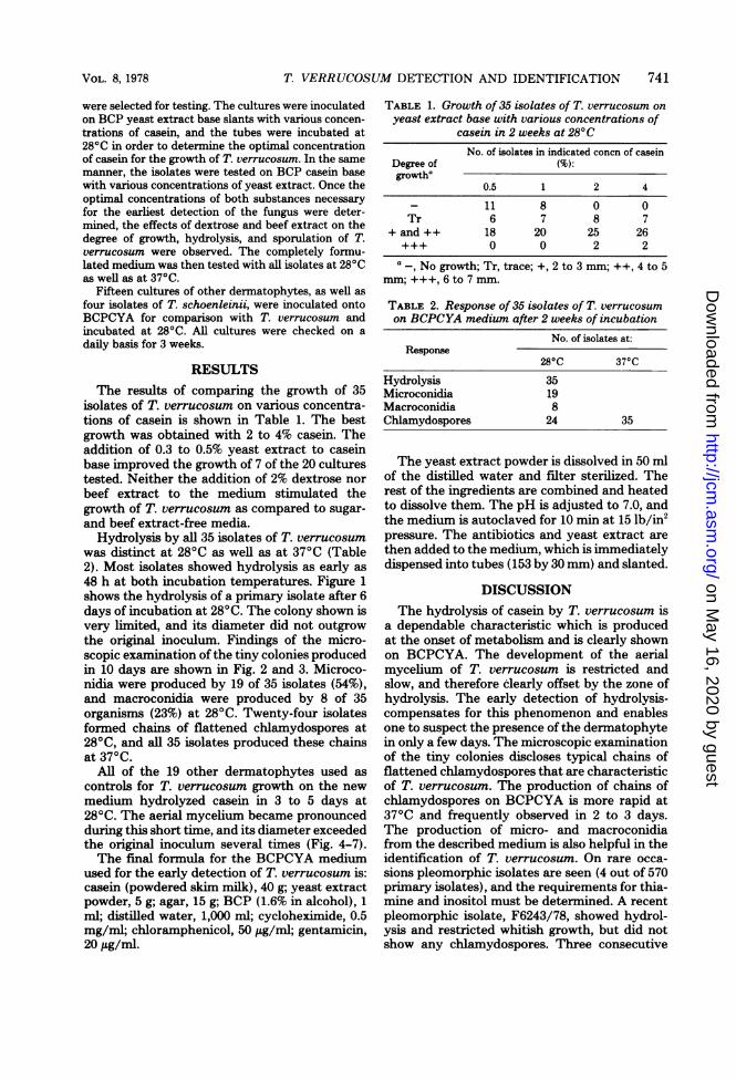
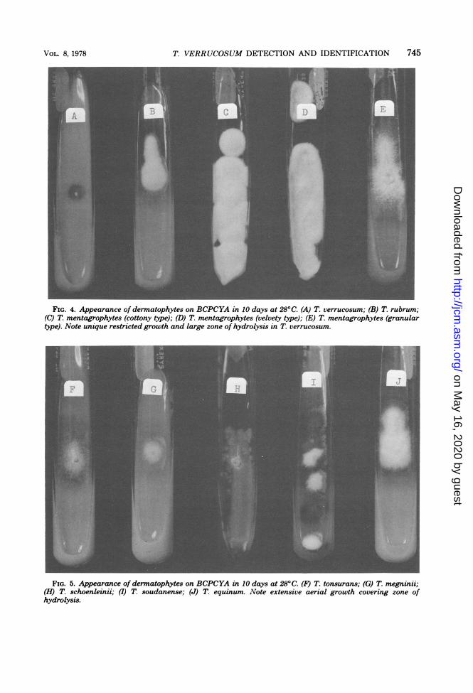
FIG. 4. Appearance of dermatophytes on BCPCYA in 10 days at 280C. (A) T. verrucosum; (B) T. rubrum;(C) T. mentagrophytes (cottony type); (D) T. mentagrophytes (velvety type); (E) T. mentagrophytes (granulartype). Note unique restricted growth and large zone of hydrolysis in T. verrucosum.
FIG. 5. Appearance of dermatophytes on BCPCYA in 10 days at 280C. (F) T. tonsurans; (G) T. megninii;(H) T. schoenleinii; (I) T. soudanense; (J) T. equinum. Note extensive aerial growth covering zone ofhydrolysis.
VOL. 8, 1978
on May 16, 2020 by guest
http://jcm.asm
.org/D
ownloaded from

746 KANE AND SMITKA
FIG. 6. Appearance of dermatophytes on BCPCYA in 10 days at 28°C. (K) Microsporum canis; (L) M.gypseum; (M) M. audouinii; (N) M. nanum; (O) Epidermophyton floccosum. Note spreading aerial myceliumin all five dermatophytes.
A.m,
..:..
FIG. 7. Comparison ofgrowth of T. verrucosum (A) with T. schoenleinii (B, C, D, and E). Note the uniquerestricted growth and large zone of hydrolysis of T. verrucosum.
J. CLIN. MICROBIOL.
on May 16, 2020 by guest
http://jcm.asm
.org/D
ownloaded from

T. VERRUCOSUM DETECTION AND IDENTIFICATION 747
scribed by Georg (2). The remainder were iden-tified as T. verrucosum by the production ofearly hydrolysis, very restricted growth, and for-mation of chains of chlamydospores from pri-mary isolates at 28°C or subsequent incubationat 37°C for 2 to 3 days. The majority of scrapingsfrom which T. verrucosum was isolated hadmycelium present in the hydroxide mount. Inmost cases the presumptive diagnosis of thephysician was stated as cattle ringworm. Asmentioned previously, almost all isolates of T.verrucosum come from rural areas where mostof the population comes into contact with cattle.A small number of T. verrucosum isolates were
identified from employees of meat-packing com-
panies. In the identification of T. verrucosum,the following criteria are considered: source ofspecimen, clinical diagnosis, geographic area, hy-drolysis of casein, slow, confmed growth, chla-mydospore production in typical chains, andenhanced formation of typical macroconidia onthis new medium. We wish to emphasize that
the adoption of BCPCYA in diagnostic workalleviates the fear of overlooking this importantdermatophyte.
ACKNOWLEDGMENTS
We thank Wilhelm Van der Kolk, Ministry of Health, forthe photography and Margaret Kwok of the Media Depart-ment for the preparation of all media.
LITERATURE CITED1. Fischer, J. B., and J. Kane. 1971. The detection of
contamination in Trichophyton rubrum and Trichophy-ton mentagrophytes. Mycopathol. Mycol. Apple. 43:169-180.
2. Georg, L. K. 1950. The relation of nutrition to the growthand morphology of Trichophyton faviforme. Mycologia42:683-692.
3. Kane, J., and J. B. Fischer. 1975. Occurrence of Tricho-phyton megninii in Ontario. Identification with a simplecultural procedure. J. Clin. Microbiol. 2:111-114.
4. Rebell, G., and D. Taplin. 1970. Dermatophytes: theirrecognition and identification, rev. ed. University ofMiami Press, Coral Gables, Fla.
5. Rosenthal, S. A., and H. Sokolsky. 1965. Enzymaticstudies with pathogenic fungi. Dermatol. Int. 4:72-79.
VOL. 8, 1978
on May 16, 2020 by guest
http://jcm.asm
.org/D
ownloaded from