Embed Size (px)
Citation preview
PREVALENCE OF Trichophyton, Microsporum AND Epidermophyton SPECIES
CAUSING Tinea capitis IN CHILDREN AGED 3-14 YEARS IN MATHARE
INFORMAL SETTLEMENT, NAIROBI, KENYA
MOTO JEDIDAH NDUNGE (B. Ed. Sc.)
Reg. No. I56/21427/2010
A THESIS SUBMITTED IN PARTIAL FULFILMENT OF THE REQUIREMENT
FOR THE AWARD OF DEGREE OF MASTER OF SCIENCE (MICROBIOLOGY) IN
THE SCHOOL OF PURE AND APPLIED SCIENCES OF KENYATTA UNIVERSITY.
October 2014

ii
DECLARATION
This is my original work and has not been presented for a degree or any other award in
any other university or any other institution of higher learning.
Signature Date
Name: Moto Jedidah Ndunge
Department of Microbiology
APPROVAL BY SUPERVISORS
This thesis has been submitted for examination with our approval as University
supervisors.
Dr. John M. Maingi
Department of Microbiology,
Kenyatta University.
Signature Date
Dr. Anthony Kebira
Department of Microbiology,
Kenyatta University.
Signature Date

iii
DEDICATION
To the Lord my God, I give the sacrifice of thanks giving. To my beloved husband Mr.
Patrick Mulandi, I thank you for your love, moral support, constant encouragement and
financial assistance to make this dream come true of pursuing a higher education. To our
children Mark Musyoka and Blessing Mutheu who in your tender age needed your
mother’s love and care and yet very patiently waited for the entire months that I was
away from home.
GOD BLESS YOU.

iv
ACKNOWLEDGEMENTS
I acknowledge with gratitude the tireless efforts and support made by the following: - The
lecturers of Department of Plant and Microbial Sciences, Kenyatta University for their
advisory contributions in various ways.
In a very special way, I thank my supervisors Dr. John Maingi and Dr. Anthony Kebira
both of the Department of Microbiology, with whose patience and guidance this work has
been successful. I am specially indebted to my family; loving husband Patrick Mulandi,
children Mark Musyoka and Blessing Mutheu who sacrificed a lot to ensure that I got all
that I needed for this study. This project was fully sponsored by them.
I cannot forget to thank Daniel Ng’ang’a a laboratory technician, research laboratory
Department of Plant and Microbial Sciences of Kenyatta University, who patiently and
diligently accorded me assistance whenever I required. Over and above all, I thank the
Almighty God for all his care and favors for me throughout the study.
v
TABLE OF CONTENTS
Declaration……………………………………………………………………….............ii
Dedication………………………………………………………………………..............iii
Acknowledgements…………………………………………………………...................iv
Table of contents……………………………………………………………….................v
List of figures …………………………………………………………………................ix
List of tables……………………………………………………………………................x
List of plates……………………………………………………………………..............xi
Abbreviations and acronyms……………………………………………......................xii
Abstract…………………………………………………………………………...........xiii
CHAPTER ONE………………………………………………………………................1
INTRODUCTION……………………………………………………………….............1
1.1: Background information…………………………………………………...................1
1.2 Statement of the problem and justification……………………………………………6
1.3 Research questions………………………………………………………….................8
1.4 Research hypothesis…………………………………………………………………...8
1.5 Study objectives……………………………………………………………….............8
1.5.1 General objectives…………………………………………………………...............8
1.5.2 Specific objectives……………………………………………………………..........9
CHAPTER TWO……………………………………………………………………….10
LITERATURE REVIEW………………………………………………………………10
2.1 Etiology of Tinea capitis……………………………………………………………..10
2.2 Clinical manifestation………………………………………………………………..11
vi
2.3 Transmission of Tinea capitis…………………………………………………….....12
2.4 Epidemiology of Tinea capitis………………………………………………….......15
2.5 Diagnosis of Tinea capitis……………………………………………………….....19
2.5.1 Wood’s lamp examination……………………………………………….................20
2.5.2 Direct microscopy………………………………………………………….............21
2.5.3 Mycological culture……………………………………………………..................21
2.6 Identification of dermatological agents causing Tinea capitis………………………..21
2.7 Standard therapy……………………………………………………………...........25
CHAPTER THREE…………………………………………………………………….27
MATERIALS AND METHODS...……………………………………………………..27
3.1 Study area and population………………………………………………....................28
3.2 Structural questionnaire……………………………………………………………...29
3.3 Study design…………………………………………………….................................29
3.3.1 Inclusion criteria…………………………………………………………………...30
3.3.2 Exclusion criteria…………………………………………………………………..30
3.4 Sample size determination………………………………………………...................30
3.5 Specimen collection techniques……………………………………………………...31
3.6 Examination of the specimen……………………………………………...................31
3.6.1 Direct microscopy………………………………………………………….............31
3.6.2 Mycological culture……………………………………………………..................32
3.6.3 Screening for Trichophyton species………………………………..........................32
3.7 Data analysis…………………………………………………………….................33
3.8 Ethical consideration………………………………………………………………....33
vii
CHAPTER FOUR………………………………………………………………………34
RESULTS………………………………………………………………………………..34
4.1 Demographic profile……………………………………………………………...34
4.1.1 Prevalence of Tinea capitis infection in children aged 3-14 in Mathare
informal settlement……………………………....................................................34
4.2 Multiple infections with dermatological agents causing Tinea capitis among the
study subjects in Mathare informal settlement......................................................39
4.3 Significant predisposing factors for Tinea capitis infections in children aged
3-14 in Mathare informal settlement…………………………..............................40
4.3.1 Socio-economic status of the children and prevalence of Tinea capitis………...40
4.3.1.1 Employment status of the father and prevalence of Tinea capitis in children
aged 3-14 in Mathare informal settlement…………...........................................40
4.3.1.2 Employment status of the mother and prevalence of Tinea capitis in
children aged 3-14 years in Mathare informal settlement……………………...42
4.3.1.3 Approximate monthly income levels of the family and prevalence of
Tinea capitis in children aged 3-14years in Mathare informal settlement………42
4.3.2 Number of children in a family and prevalence of Tinea capitis in children aged
3-14 years in Mathare informal settlement……………………………………...43
4.3.3. Respondents’ source of information and prevalence of Tinea capitis in
children aged 3-14 years in Mathare informal settllement………......................43
4.3.4 Knowledge of the children on ways of transmission of Tinea capitis and its
prevalence among the study subjects……...........................................................44
viii
4.3.5 Relationship between sharing of combs and towels with the prevalence of
Tinea capitis in children aged 3-14 years in Mathare informal settlement………..44
4.3.6 Frequency of hair shaving and prevalence of Tinea capitis in children aged
3-14 years in Mathare informal settlement……………………………………….45
4.3.7 Place of hair shaving and prevalence of Tinea capitis in children aged 3-14
years in Mathare informal settlement …………………………………………...45
4.3.8 Level of knowledge on ways of transmission of Tinea capitis and its prevalence
among the study subjects.......................................................................................46
4.4 Dermatological agents causing Tinea capitis in children aged 3-14 years in
Mathare informal settlement………………………………………………………46
CHAPTER FIVE …........................................................................................................52
DISCUSSION, CONCLUSIONS AND RECOMMENDATIONS..............................52
5.1 Discussion…………………………………………………………………………...52
5.1.1 Etiological agents of Tinea capitis............................................................................52
5.1.2 Prevalence of dermatological agents causing Tinea capitis in children aged
3-14years..................................................................................................................54
5.1.3 Relationship between prevalence of Tinea capitis and various risk factors............57
5.2 Conclusions................................................................................................................59
5.3 Recommendations.......................................................................................................59
REFERENCES.................................................................................................................61
APPENDICES..................................................................................................................73
ix
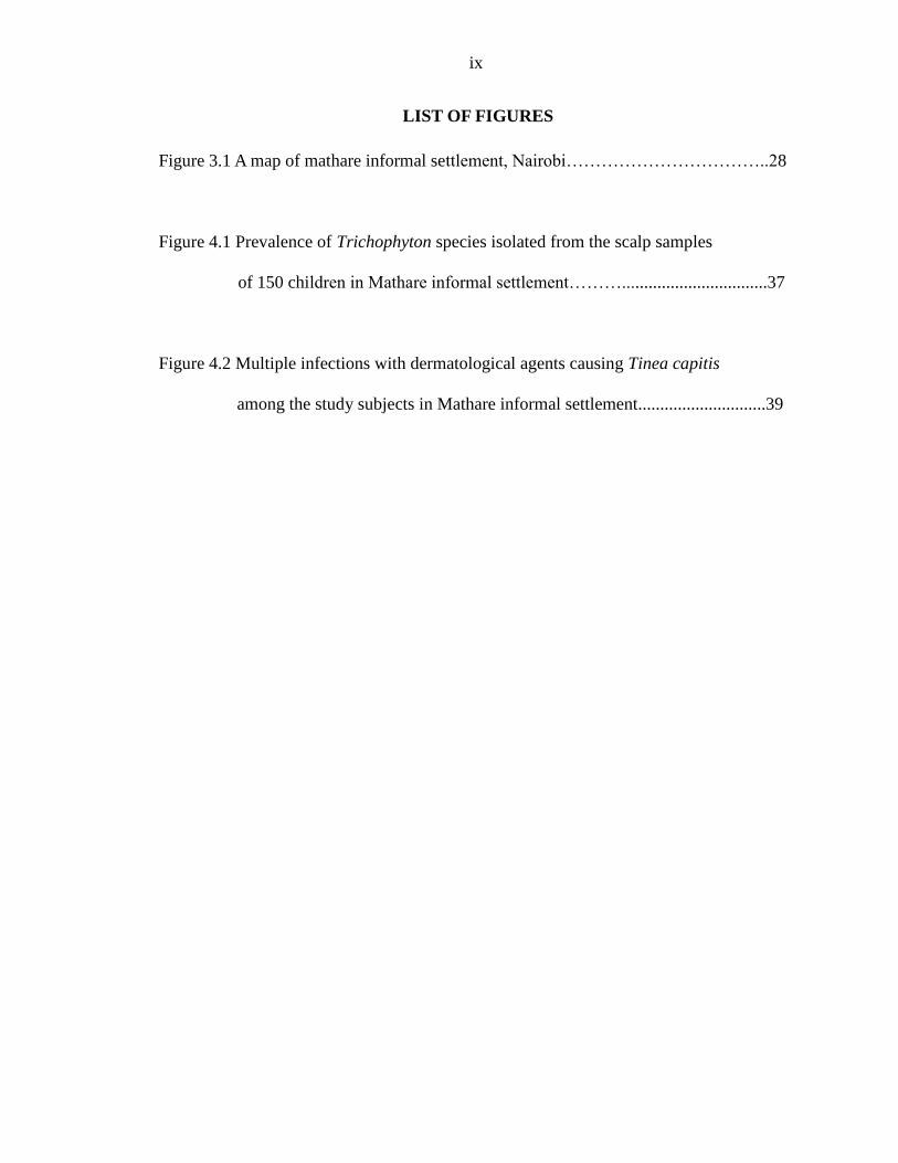
LIST OF FIGURES
Figure 3.1 A map of mathare informal settlement, Nairobi……………………………..28
Figure 4.1 Prevalence of Trichophyton species isolated from the scalp samples
of 150 children in Mathare informal settlement……….................................37
Figure 4.2 Multiple infections with dermatological agents causing Tinea capitis
among the study subjects in Mathare informal settlement.............................39
x
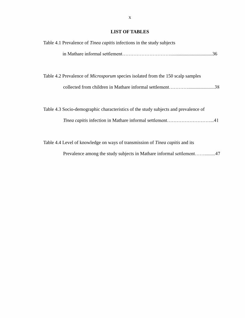
LIST OF TABLES
Table 4.1 Prevalence of Tinea capitis infections in the study subjects
in Mathare informal settlement…………………………...................................36
Table 4.2 Prevalence of Microsporum species isolated from the 150 scalp samples
collected from children in Mathare informal settlement…………......................38
Table 4.3 Socio-demographic characteristics of the study subjects and prevalence of
Tinea capitis infection in Mathare informal settlement………………………...41
Table 4.4 Level of knowledge on ways of transmission of Tinea capitis and its
Prevalence among the study subjects in Mathare informal settlement…….........47
xi
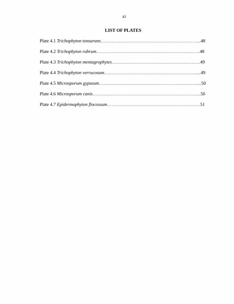
LIST OF PLATES
Plate 4.1 Trichophyton tonsurans………………………………………………………...48
Plate 4.2 Trichophyton rubrum…………………………………………………………..48
Plate 4.3 Trichophyton mentagrophytes………………………………………………….49
Plate 4.4 Trichophyton verrucosum……………………………………………………....49
Plate 4.5 Microsporum gypseum……………………………………………………….....50
Plate 4.6 Microsporum canis……………………………………………………………..50
Plate 4.7 Epidermophyton flocossum…………………………………………………….51
xii
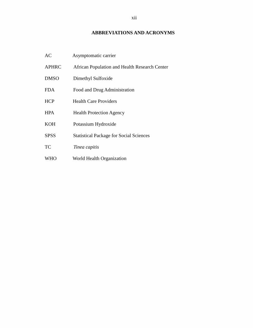
ABBREVIATIONS AND ACRONYMS
AC Asymptomatic carrier
APHRC African Population and Health Research Center
DMSO Dimethyl Sulfoxide
FDA Food and Drug Administration
HCP Health Care Providers
HPA Health Protection Agency
KOH Potassium Hydroxide
SPSS Statistical Package for Social Sciences
TC Tinea capitis
WHO World Health Organization
xiii
ABSTRACT Tinea capitis is a worldwide public health problem that affects children below 15 years of
age and requires identification of the specific causative fungal agent. The hair and skin of
the scalp are associated with symptoms and signs of inflammation and hair loss. Poor
hygiene, low standards of living, sharing of hair devices or garment, climate conditions
and overcrowding are some of the predisposing factors contributing to frequent
transmission of the infection. Several previous studies have concentrated on symptomatic
cases of Tinea capitis infection with limited studies in Kenya. However, no such study
has been done in Mathare informal settlement despite the existence of predisposing
factors such as low standards of living of the people in the area. This study therefore
aimed at determining the prevalence of Tinea capitis infection and its significant risk
factors in Mathare informal settlement in both symptomatic and asymptomatic cases. The
study also aimed at determining the prevalence of Trichophyton, Epidermophyton and
Microsporum species causing Tinea capitis infection among school going children in
Mathare informal settlement, Nairobi. A total of 150 children were systematically and
randomly sampled from five public primary schools in Mathare informal settlement. Skin
scrapings specimens were collected and inoculated on potato dextrose agar. Fungal
cultural characteristics were observed macroscopically (pigmentation formation),
microscopically (microconidia or macroconidia formation) and Trichophyton species
differentiated by use of biochemical tests. In addition, a structural questionnaire was
administered to consenting children’s guardians and socio-demographic data collected. In
a total of one hundred and fifty (150) children aged between 3-14 years consisting of 89
(59.3 %) males and 61 (40.7 %) females, 123 (82 %) were infected with Tinea capitis.
The dermatophytes consisted of 61.3 % Trichophyton, 13.3 % Microsporum and 7.3 %
Epidermophyton with infections occurring either singly (56 %), duo (38 %) or tripple
coinfections (6 %). Males were most affected with socio-economic factors such as
employment status of the parents and monthly income levels of the family significantly
influencing infections (p<0.001). Other factors that significantly influenced the infection
include; knowledge on ways of transmission of Tinea capitis (p<0.001), sharing of combs
and towels (p<0.001), place of hair shaving (p = 0.037) and frequency of hair shaving (p
= 0.02). The prevalence of the infection was higher in lower age groups than the upper
age group of 12-14 years. These findings suggest that prevalence of Tinea capitis
infection in the informal settlement of Nairobi is high. There is therefore a need to
improve personal and community hygiene including the economic status of people living
in the informal settlement.
1
CHAPTER ONE
INTRODUCTION
1.1 Background
Tinea capitis is a disease caused by cutaneous infection of the scalp, eye lashes and eye
brows. Several synonyms are used to describe the infection, including ringworm of the
scalp and Tinea tonsurans. It is caused by dermatophyte species of genera Trichophyton
and Microsporum. The most important causative agents are species which cause an
endothrix infection, such as Trichophyton gourvilli, Trichophyton soudanense,
Trichophyton tonsurans, Trichophyton violaceum and Trichophyton yaoundei and species
that cause an ectothrix infection such as Microsporum audouinii, Microsporum canis and
Microsporum gypseum (Ngwogu et al., 2007). Infections with Epidermophyton
floccosum, the only species of epidermophyton that is pathogenic on man, usually occur
on the skin of the torso, limbs, soles of feet or palms of hands and nails, but rarely does it
invade the hair (Summerbell et al., 2007). However, a recent study from Brazil suggests
that E. floccosum may be a possible etiological agent of Tinea capitis (Cerqueira et al.,
2005).
It is predominantly a disease of preadolescent children. It accounts for up to 92.5 % of
dermatophytoses in children younger than 10 years. Tinea capitis also occurs in adults,
although this is less common (Silverberg et al., 2002; Abdel-Rahman et al., 2005). Tinea
capitis is considered to be almost exclusive to children and rarely occurs after puberty,
probably due to changes in the pH of the scalp and an increase in fatty acids serving a
protective role. Consequently, most cases occurring in adults involve women with
2
hormonal disorders resulting in carryover of Tinea capitis from childhood or in patients
with severe immunodepression due to leukemia, lymphoma, or treatment with
immunosuppressant drugs (Rebollo et al., 2008).
Dermatophytes are considered as group of closely related filamentous keratinophylic
fungi belonging to the genera Trichophyton, Epidermophyton and Microsporum .While
the genus Epidermophyton is represented by a single species (E. floccosum), the genera
Microsporum and Trichophyton are complex and consist of many species (Liu et al.,
2000). Worldwide, they are among the most common infectious agents for human (Yehia
et al., 2010) and prevalence of infections caused by them has dramatically risen to such a
level in the last decades that skin mycoses now affect more than 20–25 % of the world's
population, which make them one of the most frequent forms of infections (Neji et al.,
2008). The distribution of dermatophytes varies among different countries and exhibits
geographical and seasonal variations depending on several factors, including life style,
type of the population, migration of people and climatic conditions (Asticcioli et al.,
2008).
The primary host of Epidermophyton floccosum, is man (Larone, 2002). E. floccosum is
widespread in most countries of the world, accounting for 5 % of all dermatophytes
isolated (Summerbell et al., 2007). It is an anthropophilic dermatophyte that is
transmitted between individuals by contact, particularly in community swimming pool
areas, common showers, and gym facilities (Summerbell et al., 2007).
3
Tinea capitis infections are classified into three major groups: anthropophilic, zoophilic,
and geophilic (H P A, 2007). The anthropophilic infections are parasitic on humans and
usually form larger hyphae and spores inside the hair shaft while in zoophilic infections,
they are smaller with spores outside the hair shaft. However, in geophilic infections, they
are identified by location (H P A, 2007). In immunocompetent individuals, anthropophilic
species cause mild lesions with minimal inflammation, but geophilic and zoophilic
species may result in extensive lesions secondary to inflammation, leading to abscesses
and pustules (Krajewska-Kulak et al., 2003).
Tinea capitis is further divided in terms of invasion of the hair shaft; endothrix and
ectothrix. In endothrix, the hair shaft is filled with hyphae and spores. Some causes of
endothrix infection are Trichopthton tonsurans and Trichophyton schoenleinii species
(Sarabi, 2008). In the ectothrix types, the hyphae and spores cover the outside of the hair,
which results in the destruction of the cuticle. All of the Microsporum species and
Trichophyton verrucosum are involved. Microsporum infections (Microsporum canis)
cause a "gray patch" Tinea capitis (Kao, 2006). A very rare and severe form of Tinea
capitis infection is favus, primarily caused by T. schoenleinii. This infection results in a
honeycomb-type destruction of the hair follicle, giving the hair a yellowish color (Kao,
2006). Severe scaling of the scalp may result, and due to cuticle breakage, the hair may
become brittle which is often seen on the patient (Habif, 2004).
Tinea capitis occurs predominantly in rural or sub urban areas and some of the factors
associated with this increased frequency include poor personal hygiene, overcrowding,
and low socioeconomic level (Rebollo et al., 2008). These organisms are usually found as
4
fomites on items such as combs, hats, pillows, and theater seats, where the spores can live
for long periods of time, contributing to spread of the disease (Arenas, 2002). Therefore,
the prevalence rates of Tinea capitis in a particular area depends upon the environmental
conditions, personal hygiene and individual susceptibility (Seema et al., 2011).
The isolation of different species of dermatophytes also varies from one ecological niche
to another, depending on their primary natural habitat (Seema et al., 2011). Contact in
schools is probably the most important independent factor affecting the rapid spread of
Tinea capitis. Infection in children of school age is usually followed by infection of
younger siblings. In addition, progressive migration and climate change, social and
economic conditions that affect skin exposure to fungal pathogens, and therapeutic
methods are also important (Szepietowski and Baran, 2005). Transmission is increased
with decreased personal hygiene and low socioeconomic status. Asymptomatic carriers
(AC) are common, making Tinea capitis difficult to eradicate (Kawachi et al., 2010).
Anthropophilic dermatophytes such as T. tonsurans, T. violaceum, and M. audouinii have
been associated with high rates of asymptomatic carriers (Ginter-Hanselmeyer et al.,
2007). These organisms generally produce mild signs of infection. Asymptomatic carriers
at home or school are potentially important sources of disease transmission (Ginter-
Hanslmeyer et al., 2007). Other studies have determined that carrier rate may increase to
as high as 44 % for the siblings of index cases (Chen and Friedlander, 2001). AC should
therefore be detected and treated. Increased surveillance in schools would be helpful in
prevention of the disease (Ginter-Hanselmayer et al., 2007). Previous studies have
indicated; race, socio economic conditions, cultural patterns and public health measures
5
as some of the predisposing factors to the infections (Ayanbimpe et al., 2003, Anosike et
al., 2005, Bassiri and Khaksar, 2006). The emergence of T. tonsurans infection in
developed countries has been attributed to low socioeconomic status, crowded living
conditions, and the sharing of combs (Fuller et al., 2003).
Tinea capitis disease has been recognized as an important public health problem in the
United States with 13 % of school children, especially those of African-American
descent, testing positive for the dermatophytes (Ghannoum et al., 2003). Tinea capitis has
decreased in developed countries, while it presents a high prevalence in developing
countries (Caputo et al., 2001).
In Kenya, limited studies have been done on Tinea capitis infection. In a study done in
school going children in Kibera informal settlement, Nairobi, the prevalence of
dermatophyte infections was found to be 11.2 % with Tinea capitis infection being the
most prevalent (Chepchirchir et al., 2009). In the study T. violecium was the most
common with 72.9 % dermatophyte species isolated.
In other studies done in a rural school in Kisumu and an urban school in Eldoret in
Kenya, the prevalence of Tinea capitis infection was found to be 10.1 % and 33.3 %
respectively (Schmeller et al., 1997, Ayaya et al., 2001). Trichophyton tonsurans was the
most prevalent (77.8 %) in Eldoret urban school (Ayaya et al., 2001) while in Kisumu
rural school, Microsporum audouinii was the most prevalent with 62 % of the
dermatophyte species isolated (Schmeller et al., 1997). Presence of healthy asymptomatic
carriers is one of the greatest challenges in eradication of dermatophytosis and therefore
6
asymptomatic carriers should be identified and treated in order to prevent transmission of
the infection.
1.2 Statement of the problem and justification
Tinea capitis causes hair loss, scaling, erythema, and impetigo-like lesions. It is the most
common dermatophyte infection found in children under the age of 12 years (Sarabi,
2008). Most of dermatophyte infections of the hair (Tinea captis) are caused by species of
genera Trichophyton and Microsporum leading to itching and hair loss. These fungi are
highly adapted to nonliving keratinized tissues of the hair (Kao, 2006). The source of
infection may be humans, animals, or soil since various dermatophyte species have both
ecologic and geographic differences in their occurrence.
Although no specific preventive measures like vaccine exist, dermatophyte infection can
be prevented by observing simple general hygiene measures. Epidemiological studies of
Tinea capitis have however demonstrated that poor hygiene, low standards of living,
climate conditions and overcrowding are interrelated and all contribute to frequent
transmission of the infection. Tinea capitis is a cause of concern because of its contagious
nature and its cause of social stigma in children.
Contact among children is more frequent between the school ages of 4-14 years than in
early childhood and therefore this age group is at greater risk of contracting infectious
diseases (Fathi and Al-Samarai, 2000). For this reason conducting school surveys is the
best way of measuring the magnitude of the problem in order to enable designing of
7
programmes to meet the needs of those mostly infected by Tinea capitis and also develop
appropriate preventive and control measures.
There is inadequate information on the causative agent of Tinea capitis in Kenya (Ayaya
et al., 2001). Previous studies carried out in Kenyan primary school children in Eldoret,
Kisumu and Kibera informal settlement considered only symptomatic cases of
dermatophytosis. However, no such studies have been carried out in Mathare informal
settlement despite the availability of predisposing factors of the infection such as low
socio-economic levels of the people living in the area. Existence asymptomatic carriers
among children may lead to high rates transmission of the infection making it difficult to
be eradicated.
Identification of dermatological agents causing Tinea capitis in both symptomatic and
asymptomatic individuals would therefore help in determining the actual prevalence rates
of the infection in an area in order to reduce the risk of transmission of the infection. The
causative agents of Tinea capitis infection in asymptomatic carriers can therefore be
identified and treated to reduce its prevalence rates among the children. Isolation and
identification of specific causative agent of Tinea capitis would help in the right choice of
chemotherapy hence reducing the prevalence of the infection among children.

8
1.3 Research questions
i) Are Trichophyton, Microsporum and Epidermophyton species present in all the hair
samples collected from children in Mathare informal settlement, Nairobi?
ii) Is there variation in prevalence of Trichophyton, Microsporum and Epidermophyton
species isolated from the specimen collected?
iii) Is there a relationship between the prevalence of Tinea capitis and the socio-economic
background, age and gender of the children?
1.4 Research Hypotheses
i) Trichophyton, Microsporum and Epidermophyton species are not present in most of the
hair samples collected from children in Mathare informal settlement, Nairobi.
ii) There is no significant difference between the prevalence of the isolates.
iii) There is no significant relationship between the socio-economic background, age and
gender and the prevalence of Tinea capitis in children aged between 3-14 years in
Mathare informal settlement, Nairobi.
1.5 Objectives of the study
1.5.1 General objective
The prevalence of Trichophyton, Microsporum and Epidermophyton species causing
Tinea capitis infection among children aged between 3-14 years in Mathare informal
settlement, Nairobi.
9
1.5.2 Specific objectives
i) To determine the prevalence of Tinea infection.
ii) To determine the relationship between socio-economic back ground, age, and gender
of children and the prevalence rates of Tinea capitis in Mathare informal settlement
dwellers.
iii) To isolate and identify dermatological agents causing Tinea capitis infection from hair
samples collected from children in the age bracket of 3-14 years in Mathare informal
settlement.
10
CHAPTER TWO
LITERATURE REVIEW
2.1 Etiology of Tinea capitis
The predominance of specific pathogens causing Tinea capitis varies with geography,
environments, climates, occupations, ethnic groups and life styles. The dermatophytes
that cause Tinea capitis can invade other parts of the body such as the nails and the body,
but rarely the feet or groins. Children or adults who have neither signs nor symptoms of
infection, but from whose scalps causative fungi can be grown are described as “carriers”
(H P A, 2007).
The disease is caused by species of genera Trichophyton and Microsporum (Emele and
Oyeka, 2008). The most important causative agents are species, which cause an endothrix
infection, such as Trichophyton gourvilli, Trichophyton soudanense, Trichophyton
tonsurans, Trichophyton violaceum and Trichophyton yaoundei and species that cause an
ectothrix infection such as Microsporum audouinii, Microsporum canis and Microsporum
gypseum (Ngwogu and Otokunefor, 2007). Ecto-endothrix invasion of the hair is often
associated with M. audouinii, M. canis, M. distortum, M. ferrugineum, M. gypseum, M.
nanum, and T. verrucosum. Some of these cause fluorescence under Wood light (Rebollo
et al., 2008).
The organism that commonly causes Tinea capitis in the Western world is Trichophyton
tonsurans while there is inadequate information on the actual causative agent in Kenya
(Ayaya et al., 2001). In poor African countries, the most common causative organisms are
11
Trichophyton soudanense and Microsporum audouinii (Havlickova et al., 2008).
Infection by M. audouinii is of historical interest in many parts of the world because it
was responsible for epidemics in Europe in the 19th century before arriving in the
Americas and then finally almost disappearing 50 years ago (Arenas, 2008).
2.2 Clinical Manifestations
TC has three clinical forms: TC superficial (non-inflammatory), TC profunda
(inflammatory) and TC favosa (favus). Inflammatory TC presents with painful,
inflammatory, indurated, and postulated mass that can be accompanied by regional
lymphadenopathy (Aktas et al., 2009). Massive follicular destruction and big nodules
presenting with pustule and sinus tractions can rarely occur. This acute inflammatory
nodule is called kerion celci which occur as a result of intense hypersensitivity reaction to
dermatophyte infections (Aktas et al., 2009). If the zoophilic dermatophytes are the
causative agent, pustules and deep indurations can occur (Corting, 2009).
The clinical presentation of TC is determined by the form of invasion of the hair by the
pathogenic fungi (ectothrix or endothrix), the size of the inoculum, and the immune status
of the host (Rebollo et al., 2008). A wide variety of presentations have been described,
ranging from asymptomatic carriers, diffuse scaling similar to seborrheic dermatitis, areas
of alopecia without inflammation, alopecia with black dots, and if the causative agent is
zoophilic or geophilic, a variable inflammatory response is triggered in the host and is
clinically manifested as folliculitis or kerion (Rebollo et al., 2008) . It is also common to
12
encounter enlargement of the auricular and posterior occipital lymph nodes, and this can
be the primary manifestation of the disease (Elewski, 2000).
Genetic immunological predispositions and also genetic differences of keratins affect the
ability of a fungus to attach to the stratum corneum (Joshi et al., 2011). Dermatophytes
have the ability to form molecular attachments to keratin and use it as a source of
nutrients allowing them to colonize keratinized tissues, including the stratum corneum of
the epidermis, hair, nails (Kemal et al., 2013). Resistance factors to the colonization of
fungi are composed of UV light, variation in temperature and moisture, and fungistatic
fatty acids and sphingosines produced by keratinocytes (Kemal et al., 2013).
2.3 Transmission of Tinea capitis
The etiologic agents originate from different sources based on host preference and natural
habitat. The natural reservoir of dermatophytes can be humans (anthropophilic
dermatophytes), animals (zoophilic dermatophytes), or soil (geophilic dermatophytes)
(Adamski and Batura-Gabryel, 2007).
Transmission requires contact with intact or detached hair. Human-to–human
transmission usually requires close contact with infected subject or person because
dermatophytes are of low infectivity and virulence. In most cases transmission takes
place within families or in situations involving direct contact with detached hair; for
example in barber shops.
13
The source of infection of zoophilic dermatophytes in children and adults are mostly
domestic animals – cats, dogs, hamsters, guinea pigs, rabbits or even some birds. Farmers
also often suffer from dermatomycoses transmitted from breeding cattle, pigs, sheep,
horses and goats (Adamski and Batura-Gabryel 2007). Infection with geophilic
dermatophytes usually happens as a result of contact with soil and it is common among
people who cultivate the soil (gardeners, farmers) (Kalinowska, 2012). The disease more
often affects males than females working without protective gloves and unsuitable
hygiene is conductive for transmission of pathogen. Infection through direct contact with
ill people occurs rather rarely (Kalinowska, 2012).
Occasionally, dermatophytes infection may become chronic and wide spread. This
progression has been related to both host and organism factors (Fathi and Al-Samarai,
2000). Approximately half of these patients have underlying diseases affecting their
immune response or are receiving treatments which compromise T-lymphocyte function.
On the basis of the type of hair invasion, dermatophytes are also classified as endothrix,
ectothrix or favus. In endothrix infection the fungus grows completely within the hair
shaft, the hyphae are converted to arthroconidia (spores) within the hair while the cuticle
surface of the hair remains intact (Fuller et al., 2003).
In ectothrix infection hair invasion develops in a manner similar to endothrix except that
the hyphae destroy the hair cuticle and grow around the exterior of the hair shaft.
Arthroconidia may develop both within and outside the hair shaft. Elongated hyphae,
parallel to the long axis of the hair, persist within the hair. Favus is a rare type of TC
14
characterized by typical honey-colored, cup-shaped, follicular crusts called scutula
(Brajesh and Mahadeva, 2013). Ectothrix anthropophilic infections potentially spread
rapidly whereas endothrix and favic infections are less contagious (Rebollo, et al., 2008).
Fungal conidia are shed in the air, and may remain viable for long periods on combs,
brushes, blankets and telephones (Habif et al., 2005).
These dermatophytes can be transmitted from person to person and through fomites
(Panasitti et al., 2006). The clinical presentation of the disease varies depending on the
etiological agent and type of hair invasion, the level of host resistance and the degree of
inflammatory host response (Liu et al., 2000). Asymptomatic carriage (AC) seems to be
organism specific. Anthropophilic dermatophytes such as T. tonsurans, T. violaceum, and
M. audouinii have been associated with high rates of AC (Ginter-Hanselmeyer et al.,
2007). These organisms generally produce mild signs of infection. Asymptomatic carriers
at home or school are potentially important sources of disease transmission (Ginter-
Hanslmeyer et al., 2007). Other studies have determined that carrier rate may increase to
as high as 44 % for the siblings of index cases (Chen and Friedlander, 2001).
Dermatophytes are keratinophilic fungi, which parasitize on corneous structures, such as
stratum corneum, hair or nails (Kalinowska et al., 2012). Of great importance may also
be some specific anatomic regions of the skin, greatly facilitating the colonization by
fungi. Scalp hair can therefore arrest arthrospores spread by air. Similarly, spores are
arrested in the hyponychium under or in the interdigital spaces, or in the folds of the skin
where additionally occlusion helps them to develop (Dworacka-Kaszak, 2004). The

15
spores are particularly resistant to environmental conditions, such as variable temperature
and drying (Hryncewicz-Gwozdz et al., 2005). In addition to the progressive migration
and climate change, social and economic conditions that affect skin exposure to fungal
pathogens, and therapeutic methods are also important (Szepietowski and Baran, 2005).
Transmission is increased with decreased personal hygiene and low socioeconomic
status. Asymptomatic carriers are common, making TC difficult to eradicate (Kawachi et
al., 2010).
2.4 Epidemiology of Tinea capitis
The epidemiology of TC has changed with the advent of griseofulvin and the sensitivity
of M. audouinii to this antifungal medication (Elewski, 2000). Since the 1970’s, there has
been a progressive spread of infections caused by Trichophyton tonsurans through inner
city areas of much of the USA and more recently in the UK and other European cities. By
contrast Trichophyton schoenleinii, which causes a characteristic scalp infection, favus, is
becoming less common, partly because it’s striking clinical appearances and the tendency
to scar are recognized even in remote communities. Patients with favus, or their parents,
are more likely to present for treatment (H P A, 2007).
Trichophyton tonsurans arrived in the Americas with the Spanish conquistadores and
currently, in Mexico, this organism accounts for between 15 % and 28 % of cases
(Arenas, 2002). In the United States of America, it is the predominant causative organism
of TC (98 %), whereas the dermatophyte Microsporum canis is more common in some
parts of Europe, Arab countries, Iran, Brazil, Mexico, and the Dominican Republic

16
(Arenas, 2002). The Countries with the highest incidence are Italy and other
Mediterranean countries, although other nearby countries such as Austria, Hungary,
Germany, and Poland also has high incidences (Ginter-Hanselmayer et al., 2007). The
increase in anthropophilic dermatophytes is due to Trichophyton tonsurans, mainly in the
United Kingdom and to Trichophyton soudanense and Microsporum audouinii in France
(Panasiti et al., 2007).
The commonest cause of this infection in the UK is Microsporum canis. Its geographic
range is, however, worldwide as it is spread from cats or dogs. In many parts of the UK,
Microsporum canis infections are infrequent but still the commonest forms of TC in those
locations. Microsporum canis infections are also seen in children who do not have a
history of exposure to cats or dogs (H P A, 2007). The likely explanation is that they have
acquired the infection from a contaminated environment. In addition, other
anthropophilic fungi such as T. violaceum, T. soudanense and M. audouinii are seen in
cities. A dramatic increase in T. tonsurans infections has been reported in the USA
(Nelson et al., 2003). Additionally, T. tonsurans had become the most common cause and
today more than 95 % of Tinea capitis cases are caused by T. tonsurans (Foster et al.,
2004).
In the USA T. tonsurans is also the most frequent isolate; it appears to be common in
urban populations, particularly black American children, than in other cultural or ethnic
groups. Little is known about the risk factors for anthropophilic infection (HPA, 2007).
Previous studies have indicated; race, socio economic conditions, cultural patterns and

17
public health measures as some of the predisposing factors to the infections (Ayanbimpe
et al., 2003, Anosike et al., 2005, Bassiri and Khaksar, 2006). The emergence of T.
tonsurans infection in developed countries has been attributed to low socioeconomic
status, crowded living conditions, and the sharing of combs (Fuller et al., 2003).
In a recent US survey, TC was found in 6.6 % of the population (Fungal Research Trust,
2011). However an infection range from 0 % to 19.4 % is more common in deprived
areas and black children, suggesting a global prevalence of 200 million cases (Fungal
Research Trust, 2011). In Germany, before World War II, Microsporum audouinii and
Epidermophyton floccosum occupied the top of the list of causative organisms by
frequency, but from the 1950s onwards T. rubrum (80 % to 90 %) has been the
predominant dermatophyte at all sites apart from the head (Seebacher et al., 2008).
In Africa, however, TC continues to be an important public health problem, where it has
been reported to affect 10 % to 30 % of school-aged children (Sidat et al., 2007). Hair
infection (Tinea capitis) is most common among children, often resulting in bald patches
with psychological consequences. Although TC, like other dermatophytoses, is of public
health importance, it is not a notifiable disease and as a result, the actual prevalence
figures are unknown in many endemic areas (Ayaya et al., 2001; Ameh and Okolo, 2004;
Anosike et al., 2005).
In the Netherlands, Sweden, and Belgium, there have been increases in M. canis TC, but
there are also increases of anthropophilic TC caused by T. violaceum, T. soudanense, and

18
T. tonsurans, which is a reflection of immigration patterns, particularly from East Africa
(Kolivras et al., 2003; Hallgren et al., 2004). The most common species responsible for
TC in Australia and New Zealand are M. canis and T. Tonsurans (Ameen, 2010).
However, since the early 1990s, T. soudanense, T. violaceum, and M. audouinii TC have
been increasingly reported in children who have immigrated from East Africa, in
particular, with evidence for transmission of these agents to local populations (Mc
Pherson et al., 2008).
In Nigeria, the head is affected in 13.7 % of cases and the most common causative
organisms are Trichophyton soudanense (30.6 %), Microsporum ferrugineum (7.7 %),
and Microsporum audouinii (7.7 %), with cases involving Trichophyton tonsurans
occurring less frequently (Ayanbimpe et al., 2008). In Mozambique, the prevalence of TC
is 9.6 %, due mainly to Microsporum audouinii, Trichophyton violaceum, and
Trichophyton mentagrophytes (Sidat et al., 2007). There is limited data on TC infections
in Kenya. However, for those that have evaluated the prevalence of TC among school
going children, have shown 33.3 % infection rates with the prevalence of T. tonsurans
being 77.8 % and 4 % for T. rubrum (Ayaya et al., 2001).
The distribution of dermatophyte infections and their causative agents varies with
geographical region. It is influenced by a wide range of factors, such as type of
population, climatic factors, lifestyle, migration of people, cultural practices and
socioeconomic conditions (Havlickova et al., 2008; Ameen, 2010). Some dermatophyte
species appear to be homogeneously distributed worldwide whereas others show a
19
geographic restriction (Havlickova et al., 2008). In recent decades, an ever-growing
etiological role of some anthropophilic dermatophytes has become evident all over the
world (Jankowska-Konsur et al., 2011).
2.5 Diagnosis of Tinea capitis
Apart from cultural characteristics for identification of dermatophytes, clinically TC
agents such as M. audouinii and M. canis, can mimic impetigo and pediculosis or
psoriasis and seborrhea, respectively. However, for impetigo, the pain is generally more
severe and individual hairs do not appear to be broken. In psoriasis, the scales on the
scalp are thicker, but the hair is not broken off (Johnson and Nunley, 2000). Alopecia
areata also causes hair loss and may mimic T. tonsurans infections, but does not cause
scaling of the scalp (Sarabi, 2008). TC is diagnosed by several methods.
A Wood's lamp examination may show hairs that turn blue-green. A potassium hydroxide
test on the hair or scalp may show fungi under the microscope. A fungal culture of the
hair or scalp may show what type of fungus is causing the infection (Mounsey and Reed,
2009). Clinical diagnosis can also be applied. However, dependence on the clinical
diagnosis of TC is unreliable and has a low specificity even though certain signs such as
lymphadenopathy are useful predictors of the infection. For this reason, wherever
possible, the diagnosis should be confirmed by appropriate laboratory tests.
20
2.5.1 Wood’s lamp examination
The Wood's lamp produces invisible long-wave ultraviolet light (340-450 nm wave
length). Filtered ultraviolet (Wood’s) light elicits a green fluorescence from some
dermatophyte fungi, mainly Microsporum species, in hair infections. Exposure to Wood’s
light is a useful screening procedure for taking specimens from Microsporum infections
(H P A, 2007). The first use of Wood's lamp for the detection of TC was based on the fact
that some dermatophyte species produce characteristic fluorescence under UV light. The
chemical responsible for the fluorescence is pteridine. Wood's lamp is helpful in the
diagnosis and treatment of an individual patient as well as for mass screening and control
of epidemics in schools. It can also be helpful in assessing the length and response to
treatment; the end point being emergence of non-fluorescent hair. Dermatophytes that
cause fluorescence are generally members of the Microsporum genus. However, the
absence of fluorescence does not necessarily rule out TC as most Trichophyton species,
with the exception of T. schoenleinii, are non-fluorescent (Gupta and Singhi, 2004).
Wood's light fluorescence is helpful but not diagnostic as it is only positive if the
responsible organism fluoresces and fluorescence is sometimes seen for other reasons.
The diagnosis of TC should be confirmed by microscopy and culture of skin scrapings
and hair pulled out by the roots (Higgins et al., 2000). However, with TC infections that
are caused by the Trichophyton species, the fungal spores form on the inside of the hair
shaft (endothrix), and there is no fluorescence (Fuller et al., 2003). Therefore, the Wood's
light examination is not a reliable method for diagnosing TC caused by the Trichophyton
species because this species does not fluoresce.
21
2.5.2 Direct microscopy
Microscopic examination and/or fungal culture should be used to confirm the clinical
diagnosis of TC because of the extended nature of most treatment regimens (Ali et al.,
2007). Microscopic examination consists of scraping the scales of the lesions onto a slide
and viewing the sample, which is prepared with a 20 % potassium hydroxide (KOH)
solution, under the microscope to look for the presence of hyphae (Chen and Friedlander,
2001). This test may be difficult to interpret or may be falsely negative with early or
inflammatory lesions. Therefore, the final diagnosis of TC should be made by culture.
2.5.3 Mycological culture
Culture documentation of the infection is a crucial component to treatment of TC
(Roberts and Friedlander, 2005). Plucked hair fragments and skin scrapings are placed
directly in culture media. The most commonly used media is Sabourand’s agar.
Chlorampenicol and Cycloheximide are used to inhibit bacteria and saprobic fungi.
Cultures are incubated at 25ºC for 3-4 weeks and if T. verrucosum, T. violaceum or T.
soudanense are suspected, they are incubated for 6 weeks. Fungal identification is based
on macroscopic (pigmentation formation) and microscopic morphology (macroconidia or
microconidia formation).
2.6 Identification of dermatological agents causing Tinea capitis
Like a number of fungi, dermatophytes may exhibit two phases in their life cycle: the
anamorph state (imperfect or asexual phase), which is isolated in the laboratory; and the
teleomorph state (perfect or sexual phase) (Enany et al., 2013). Not all of the teleomorph
22
dermatophyte species have been identified (Mukherjee et al, 2011). Anamorphic states of
dermatophytes include genera Epidermophyton, Microsporum and Trichophyton and
belong to the class Hyphomycetes and phylum Deuteromycota (Enany et al., 2013).
Teleomorphic states include majority of geophilic and zoophilic species of Microsporum
and Trichophyton. They are classified in the teleomorphic genus Arthroderma, order
Onygenales, phylum Ascomycota, and are usually found in their anamorphic state
(Molina, 2011).
There are 3 major genera of Dermatophytes. These are Epidermophyton, Microsporum
and Trichophyton. Epidermophyton are characterised by large thin-walled, multicellular,
club-shaped and clustered bunches of macroconidia (Ayorinde et al., 2013). No
Microconidia are produced. However, Microsporum produces both microconidia and
macroconidia (Ayorinde et al., 2013). Macroconidia are multiseptate, with echinulations
on the cell wall. The thickness of the cell wall and shape varies depending on the species
(Simpanya, 2000). Trichophyton produces smooth walled macroconidia. Macroconidia
are thin walled and cigar-shaped (Centre for Food Security and Public Health, 2005).
Microsporum canis grow on culture media to form white cotton radiated colony, golden
yellow on reverse (Marques et al., 2005). Macroconidia are fusoid, thick and rough-
walled with curved apex with greater than 6 cells (Mcdonald, 2000). Epidermophyton
flocossum grow with khaki pigmentation on front and yellow brown reverse (Ellis et al.,
2007). The macroconidia are similar to those of Microsporum except that they are smooth
thin walled, club shaped and they occur in clusters and appear to be directly growing
23
from the hyphae (Mcdonald, 2000) but with no microconidia.
The cultural morphhology and microscopic chararacteristics for Trichophyton species
include; numerous smooth walled and clavate to pyriform microconidia. Macroconidia
are less distinctive and often absent in this genus (Mcdolnald, 2000). Trichophyton
tonsurans show dark-brown pigmentation with reddish brown to mahogany reverse after
21 days. In microscopy it reveals numerous microconidia of varying sizes and shape
which appear to be formed at right angle to the hypha (Ellis et al., 2007).
Trichophyton mentagrophytes produce a flat, white to cream and a powdery appearance
in potato dextrose agar with a pinkish brown reverse. The microscopy show single-celled
spherical microconidia and spiral hyphae. T. rubrum produce white to cream flat colony
with yellow-brown reverse after 21 days. On microscopy, numerous pyriform
microconidia are observed (Ellis et al., 2007). T. mentagrophytes is urease positive after 7
days while T. rubrum is urease negative after 7 days (Mcdolnald, 2000). The Colony of T.
verrucosum grows slowly in potato dextrose agar. It is small, button or disc-shaped, white
to cream-coloured, with a suede-like to velvety surface, a raised centre, and flat periphery
with a yellow reverse. On microscopy, it is observed to produce clavate to pyriform
microconidia borne singly along the hyphae (Ellis et al., 2007).
Traditionally, most commercially available identification systems of dermatophytes are
based on physiological (growth temperature), nutritional (sugar assimilation and/or
fermentation, enzyme production profiles) and morphological characteristics (Elsayed et
24
al., 2010). Tinea capitis is generally identified by the presence of branching hyphae and
spores on KOH microscopy (Ayorinde et al., 2013). If hyphae and spores are not
visualized, Wood's lamp examination can be performed. If KOH microscopy and Wood's
lamp examinations are negative, fungal culture may be considered when Tinea capitis is
strongly suspected (Barry and Hainer, 2003).
Laboratory diagnosis is routinely performed by direct microscopic examination of a
clinical specimen followed by sample culture in specific agars (Côbo et al., 2010). This
combination of techniques is time-consuming and notoriously low in sensitivity (Uchida
et al., 2009). Furthermore, sample cultures on agar have a high risk of contamination by
non-dermatophytic moulds and yeasts (Arabtzis et al., 2007). The final diagnosis must be
made based on isolation of the organism from affected tissues and visualization of tissue
invasion by organisms with compatible morphology (Arabtzis et al., 2007).
PCR-based techniques used in dermatophytes diagnosis, are highly specific and sensitive
methodologies but demand well-equipped laboratories, expensive reagents, laboratory
personnel expertise and protocol standardization, so, actually, molecular techniques for
dermatophytes diagnosis are far from being routinely used and need more studies before
implementation (Garg et al., 2009). Some associations with other techniques, such as
histology, are proposed to increase diagnosis sensitivity (Karimzadegan-Nia et al., 2007)
but this would bring more difficulties in the routine, demanding professionals,
equipments and more time (Côbo et al., 2010).
25
2.7 Standard therapy
Griseofulvin was first approved by the U.S. Food and Drug Administration (FDA) for
systemic treatment of TC in 1958. Before then, the only available treatments were
shaving the head, applying mercury/sulfur to the scalp, or resorting to high-fat diets
(Möhrenschlager et al., 2005). However, griseofulvin quickly became the mainstay of
treatment and the use of terbinafine and itraconazole in patients allergic to griseofulvin
were also successful (Trivino-Duran et al., 2005). In a meta-analysis study, Fleece et al
(2004) showed terbinafine treatment for up to four weeks to be as effective in treating
Trichophyton spp. as 8 weeks of griseofulvin treatment. Griseofulvin is a mitotic inhibitor
and interferes with nucleic acid, protein, and cell wall synthesis of replicating
dermatophyte cells (Brendan, 2012). There is also evidence that griseofulvin has an anti-
inflammatory effect, which is unique among the systemic antifungal agents (Brendan,
2012). A recent extensive review quoted mycologic cure rates of 80 % to 95 % and
effective therapy rates of 88 % to 100 % for griseofulvin (Gupta and Cooper, 2008).
In 2007, terbinafine became the second FDA-approved drug to treat TC in children. The
drug is an allyl-amine whose antifungal effect is due to inhibition of squa-lene epoxidase
(Brendan, 2012). However the responses of Microsporum species to terbinafine are
generally slower than those of Trichophyton and in some patients there is treatment
failure (Caceres-Rios et al., 2000). However higher doses of terbinafine, more than 6
mg/kg per day, appear to produce good responses (Devliotou-Panagiotidou and
Koussidou-Eremondi, 2004).
26
Itraconazole has both fungistatic and fungicidal activity, depending on its tissue
concentration, unlike other azoles, its principal mechanism of action is fungistatic,
through depletion of ergosterol in the cell membrane, leading to alteration of membrane
permeability (Rebollo et al., 2008). It is highly lipophilic and keratinophilic, and it
persists in the stratum corneum for 3 to 4 weeks after suspension of treatment, allowing it
to be used in pulses of 1 week separated by periods of 2 weeks without treatment
(Higgins et al., 2000).
Currently, the recommended therapy for TC is a 6 to 8 week course of oral griseofulvin.
Griseofulvin has a long-standing history of safety and efficacy when used to treat fungal
scalp infections in children (Bennet et al., 2000). Griseofulvin is effective when treating
Microsporum, Epidermophyton, and Trichophyton (Chan and Friedlander, 2004). When
compared to other treatments in Trichophyton infections, griseofulvin and terbinafine are
equally effective, but griseofulvin is most effective against Microsporum infections
(Fuller et al., 2001). Despite the existence of antifungal agents effective on
dermatophytes, there is need to search for alternatives (Tra Bi et al., 2005). The relatively
high cost and constraints due to the length of the modern treatment curb the control of the
dermatomycoses in developing countries (Tra Bi et al., 2005). Resource-poor people
from remote areas still use traditional medicine for the treatment of various diseases of
microbial and non microbial origin (Kone et al., 2002).

27
CHAPTER THREE
MATERIALS AND METHODS
3.1 Study area and population
The study was carried out in Mathare valley informal settlement in Nairobi. Mathare
valley is situated five kilometers northeast of Nairobi’s city center. As one of the largest
slums in East Africa and the oldest in Nairobi, Mathare is divided into different villages.
The area is comprised of 13 villages: Mashimoni, Mabatini, Village No. 10, Village 2,
Kosovo, 3A, 3B, 3C, 4A, 4B, Gitathuru, Kiamutisya, and Kwa Kariuki (Crow and Odaba,
2010). The 2009 Kenyan Census reported 80,309 residents in the 13 Mathare villages.
The settlement sits within a valley of the Mathare and Gitathuru. Mathare Valley is
enclosed by Pangani on the West. On the north, it is enclosed by the police depot,
Mathare primary school, and Mathare Mental Hospital. Juja Road borders Mathare on the
south, separating it from Eastleigh, an estate dominated by Somali immigrants and
entrepreneurs. To the east, it borders Huruma estate (Fig. 3.1). It stretches out for about 3
kilometres along side Juja road (APHRC, 2002). Water quality and reliability is
inconsistent, with frequent contamination from vandalized pipes and 90 % of residents
lacking access to in-home piped water. The sanitary infrastructure in Mathare is equally
bad and in many cases worse than the water. The sewerage pipe system is in total
disrepair, and there is limited or no solid waste management.
28
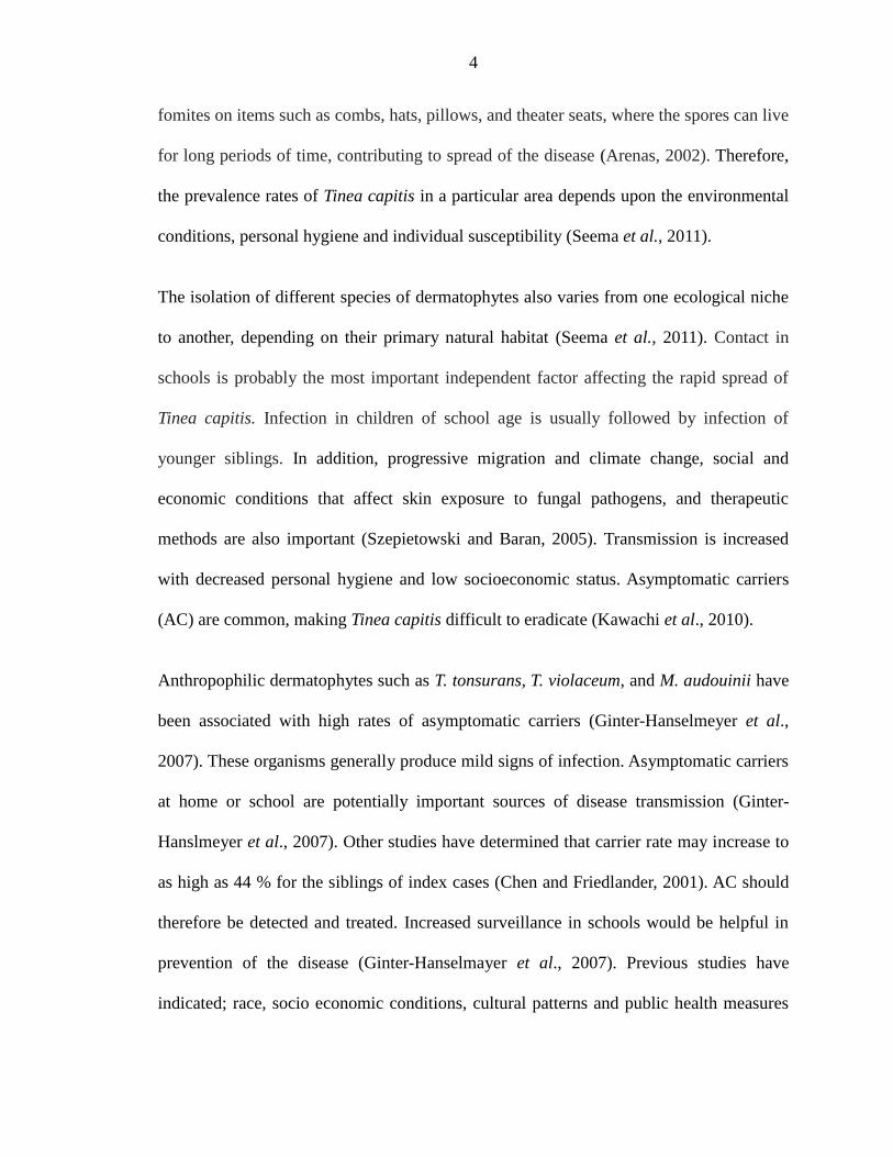
Figure 3.1: A map showing Mathare informal settlement, Nairobi (www.
openstreetmap.com).
Mathare
3A
3C
Mashimoni
Kwa
Kariuki
Kiamutisya
N
29
3.2 Structured questionnaires
A structured questionnaire (Appendix 1) was administered among the children’s
guardians and socio-economical and demographic data collected. The questionnaires
were administered to the parents/ guardians of the children who were selected for the
study after the collection of the specimen. The questionnaires were divided into two parts.
The first section was on socio demographic data of the study subjects while the second
part was on knowledge on hygienic practices in relation to TC infection. The
questionnaires were personally administered to the children’s parents/guardians by the
researcher. All the five public primary schools in Mathare informal settlement were
involved in the study. The schools involved include; Mathare 4A, Mathare north primary
school, Kiboro primary school, St. Teresa’s and Muthaiga primary school.
3.3 Study design
This was a cross-sectional study. The study subjects were randomly selected from the five
public primary schools in Mathare informal settlement. Systematic random sampling
method was used to select the subjects. The desired sample size (n) was 150 while the
study population (p) was 2250. The interval of study subjects’ selection was given by p /n
(2250/150) which was 15. Every 15th
pupil was therefore selected for the study. Sampling
from each school was done considering the total pupil population: Mathare 4A 477,
Mathare north 480, Kiboro primary 410, St.Teresa’s 420 and Muthaiga 463. The average
population in each school was given by study population/number of schools (2250/5)
which was 450. Therefore, thirty pupils (450/5) were randomly selected from each of the
five public primary schools. The children were grouped in age groups of year 3-5, 6-8, 9-
30
11 and 12-14. Samples were collected and questionnaires were personally administered
by the researcher to the subjects through their guardians/ parents. Inclusion and exclusion
criteria were used in this study.
3.3.1 Inclusion criteria
All pupils aged between 3-14 years in Mathare informal settlement who were present in
school at the period of study, whose parents consented as well as those who assented to
participate in the study were included for the study. This included the children without
clinical symptoms (asymptomatic) of TC and the ones with clinical symptoms
(symptomatic).
3.3.2 Exclusion criteria
Children whose parents did not consent to their children’s participation in the study were
excluded from the study.
3.4 Sample size determination
Sample size was calculated from the sample size formula:-
n= Z2 p (1-p)/d
2 (Daniel, 1999), and a confidence level of 95 %
where, n = sample size,
Z = Z statistic for a level of confidence,
P = expected prevalence or proportion,
d = precision
The prevalence of dermatophytosis in school going children in Kibera informal
settlement is 11.2 % (Chepchirchir et al., 2009).
31
Therefore, p = 0.11
d = 0.05(half of p).
z = 1.96 (For the level of confidence of 95 %, which is conventional, Z value is 1.96).
Therefore,
n = 1.962*0.11(1-0.11)/0.05
2 = 150.437 = 150.
3.5 Specimen collection
Scalp skin scrapping/hair stubs were collected from school going children in the selected
primary schools. The scalp was first sterilized with 70 % alcohol and skin scrapped by
sterile surgical blades and hair stubs collected into dry Petri dishes. The samples were
then wrapped with sterile parafilm and transported to Kenyatta University laboratory for
analysis.
3.6 Examination of the specimen
3.6.1 Direct microscopy
Some of the samples were inoculated onto the media while the other treated with 10 %
Potassium hydroxide (KOH) + 36 % dimethyl sulfoxide (DMSO) without vigorous
squashing of the specimen on the slide. The slide was gently heated and viewed under
light microscope (Robbert and Pihet, 2008). Hair samples were examined within 30
minutes to allow softening and digestion of the specimen (HPA, 2007). Slides were
evaluated for the presence of fungal elements under a microscope (x100, x200 and x400)
magnification. The presence of fungal hyphae and/or spores within (endothrix) and/or
around (ectothrix) hair shafts was considered to be a positive test.
32
3.6.2 Mycological culture
Plucked hair fragments and skin scrapings were placed directly on culture media. Potato
dextrose agar was used as culture media. 0.1g/L Chloramphenicol and 0.1g/L
cycloheximide were added to inhibit bacterial and saprobic fungal contamination. The
cultures were incubated at 25oC to ensure that the media does not dry up and the growth
was examined regularly for 3–4 weeks. Fungal identification was carried out based on
macroscopic (growth characteristics, pigment formation) as well as microscopic
morphology (formation of macro conidia and micro conidia or other typical elements).
Identification was carried out by the gross colony morphology and microscopically by
lacto phenol cotton blue mounts and slide cultures (Roberts and Friedlander, 2005). The
identification was done according to mycological identification of dermatophytes by
Mcdonald et al. (2000) and Ellis et al. (2007).
3.6.3 Screening for Trichophyton and Microsporum species
The cultures were macroscopically screened twice a week for signs of fungal growth.
Change in colony pigmentation on both reverse and front of the plate was also recorded.
Additionally biochemical tests were performed. This included urease activity (Betty et
al., 2007) in order to differentiate between the members of Trichophyton species.
3.7 Data analysis
All the field data were collected and stored in Microsoft Excel Software package. The
scalp scrapings /hair stubs data and questionnaire data were then exported to SPSS 16 for
33
analysis. Chi- square test was used for comparing prevalence of the infection by sex, age
and to determine significant predisposing factors of Tinea capitis infection. P < 0.05 was
considered significant. The results were then presented in descriptive statistics using
frequency tables, cross tabulation and bar charts.
3.8 Ethical consideration
Authority to carry out research was sought from Kenyatta University Graduate School.
Kenyatta University Ethics Review Committee gave the approval. National commission
for science, technology and innovation gave the permit. Consenting parents and children
signed an informed consent form to allow participation of their children in the study.
34
CHAPTER FOUR
RESULTS
4.1 Demographic profile
A total of 150 children aged 3-14 years from the five public primary schools in Mathare
informal settlement were examined for Tinea capitis infection. Both symptomatic and
asymptomatic cases were considered whereby the asymptomatic and symptomatic cases
picked at random were 48 (32 %) and 102 (68 %) respectively. All the 102 (68 %)
symptomatic cases were positive for Tinea capitis infection. Of the 48(32 %)
asymptomatic cases only 21 (43.75 %) were positive for Tinea capitis infection. In the
other 27 (56.52 %) samples no dermatological agent was isolated. The study population
comprised of 89 (59.3 %) males and 61 (40.7 %) females. The mean average age was 8.5
+ 1.86. The number of children selected at random comprised of 47 children between the
ages of 3-5, 72 children between the ages of 6-8, 20 children between the ages of 9-11
and 11 children between the ages of 12-14. The overall prevalence of the three genera of
dermatological agents causing Tinea capitis infections was 61.3 %, 13.3 % and 7.3 % for
Trichophyton, Microsporum and Epidermophyton, respectively (Table 4.1).
4.1.1 Prevalence of Tinea capitis infection in children aged 3-14 years in Mathare
informal settlement
Of the 150 children examined, 123 (82 %) were infected with either one of three
dermatophytes. These were; Trichophyton (61.3 %), Microsporum (13.3 %) and
Epidermophyton (7.3 %). There was a significant difference between the prevalence of
the three dermatological agents isolated (χ2
= 6.602, df = 1, p = 0.026). According to sex,
males were significantly infected compared to females (45.3 % versus 36.7 %, χ² = 7.142,
35
df = 1, p = 0.009). The prevalence of Trichophyton species was 62.9 % in males versus
59.0 % in females. There was a significant difference in the prevalence of Trichophyton
species between sexes (χ² = 5.673, df =1, p = 0.020). The prevalence of Microsporum
species was 35.3 % in males versus 22.9 % in females. The prevalence of Microsporum
species differed significantly between sexes (χ² = 7.503), df = 1, p = 0.006).
By age the levels of infection were different in the age groups 6-8 years (χ² = 4.236, df =
1, p = 0.049), for Trichophyton in the age category 9-11 years (χ² = 4.738, df = 1, P =
0.031) and Microsporum in the age category 6-8 years (χ² = 5.438, df = 1, P = 0.021)
(Table 4.1). The highest infection prevalence was observed in males. In the age category
12-14 years there was a significant difference between males and females. Males had
higher prevalence of 66.7 % compared to 33.3 % for females. (χ² = 5.438. df = 1, P =
0.020).

36
Table 4.1 Prevalence of Tinea capitis infections among the study subjects in
Mathare informal settlement
Age Sex Number Trichophyton Microsporum Epidermophyton overall
(years) examined(N) species species species 3-5 Male 25 22(91.6)* 1(4.2) 0(0) 23(95.8)
Female 22 15(65.2) 4(17.4) 1(4.3) 20(87.0)
Total 47 37(78.7) 5(10.6) 1(2.1) 43(91.4)
6-8 Male 44 26(61.9) 3(7.1) 5(11.9) 34(81.0)
Female 28 14(46.7) 6(20.0) 2(6.7) 22(73.3)
Total 72 40(55.6) 9(12.5)* 7(9.7) 56(77.8)*
9-11 Male 12 8(57.1) 4(42.9) 0(0) 12(100)*
Female 8 4(60.0) 1(10.0) 2(20) 7(90)
Total 20 10(58.8)* 4(23.5) 2(11.8) 16(94.10)
12-14 Male 8 4(66.7) 0(0) 1(16.7) 5(83.3)
Female 3 1(33.3) 2(66.7) 0(0) 3(100)
Total 11 5(55.6) 2(22.2) 1(11.1) 8(88.9)
All Male 89 56(62.9)* 7(7.9)* 5(5.6) 68(76.4)*
Female 61 36(59.0) 13(21.3) 6(9.8) 55(90.2)
Total 150 92(61.3) 20(13.3) 11(7.3) 123(82.0)
*5% or less significant difference in prevalence of dermatological agents causing Tinea
capitis
37
The highest prevalence of Microsporum species infection was found in the age category
9-11 years followed by age group 6-8 years. In the age category 9-11 years, males had
significantly higher infections than females (χ² = 4.591, df = 1, P = 0.032) (Table 4.1).
Among the Trichophyton species, Trichophyton tonsurans (33.3 %) was the most
prevalent followed by Trichophyton mentagrophytes (10.7 %) and Trichophyton
verrucossum and Trichophyton rubrum as the least (8.0 %) (Figure 4.1).
Figure 4.1 Prevalence of Trichophyton species isolated from the scalp samples of the 150
children of Mathare informal settlement
Key: N; Total number of samples collected (150), n; frequency of Trichophyton species
identified, percentage (prevalence) = (n/N)* 100.

38
Of the 150 hair and scalp scrapings examined from the children, 11 (7.3 %) were positive
for Epidermophyton infections with children of the age group 6-8 years and males most
affected (χ² = 0.948, df = 1, p = 0.330). Only a few children in the age group 12-14 and 3-
5 years were found positive for Epidermophyton species (Table 4.1). The only
Epidermophyton species isolated from the hair samples was Epidermophyton floccosum.
The most prevalent species of Microsporum was Microsporum gypseum (7.3 %) followed
by Microsporum canis (6.0 %) (Table 4.2).
Table 4.2 Prevalence of Microsporum species isolated from the 150 samples collected
from children in Mathare informal settlement
Microsporum species Frequency (n) Percentage (n/N)*100
Microsporum canis 9 6.0
Microsporum gypseum 11 7.3
Total 20 13.3
Key: N; Total number of samples collected (150), n; Frequency of dermatophytes
isolated.
39
4.2 Multiple infections with dermatological agents causing Tinea capitis among the
study subjects in Mathare informal settlement
Some dermatological agents occurred singly whereas others occurred in a multiple
fashion (Figure 4.2). Among 56 % who had only one infection, Trichophyton occurred
singly at the highest rate of 30 % followed by Microsporum at 22 % while the least single
infection was by Epidermophyton (4 %).
The dermatological agents also co-occurred at the rate of 38 % of all the cases observed.
Trichophyton and Microsporum species co-occurred most at the rate of 30 %, followed by
Trichophyton and Epidermophyton combination at the rate of 6 % and the least occurring
combination was Microsporum species with Epidermophyton species at the rate of 2 %.
Triple infections with all the three dermatological agents observed in this study occurred
at a very small rate of 6 % (Figure 4.2).
Figure 4.2: Multiple infections with dermatological agents causing Tinea capitis among
the study subjects in Mathare informal settlement
40
4.3 Significant pre-disposing factors for Tinea capitis infections in children aged 3-14
years in Mathare informal settlement
The demographic characteristics of the study subjects included; the socio economic status
of the family, number of children in a family and practice of personal hygiene (Table 4.3).
Practice of personal hygiene among the children was demonstrated by frequency and
place of hair shaving, sharing of personal effects such as combs and towels and
knowledge on ways of transmission of Tinea capitis.
4.3.1 Social economic status of the children
The socio economic status of the children included the employment status of the parents
and the monthly income of the family. Most of children came from families whose both
parents were unemployed and earned less than 5,000 Kenya shillings per month (Table
4.3).
4.3.1.1 Employment status of the father and prevalence of Tinea capitis in children
aged 3-14 years in Mathare informal settlement
In a total of 34 children whose parents were employed, 7 (20.6 %) had Tinea capitis
infections while among self-employed, unemployed and retired the percentages of
children infected were 92.6 %, 96.9 % and 95.8 % respectively (Table 4.3). The highest
prevalence was therefore observed among children whose parents were unemployed
63/65 (96.9 %). The employment status of the father was found to have a significant
influence on the prevalence rates of Tinea capitis among the study subjects (χ² = 102.287,
df = 3, P < 0.001).
41
Table 4.3 socio-demographic characteristics of the study subjects and prevalence of
Tinea capitis infection in Mathare informal settlement No. of Frequency with % of Pvalue
Participants Tinea infection (n) participants
(N) Infected (n/N) %
Father’s employment status
Employed 34 7 20.6
self-employed 27 27 92.6
unemployed 65 65 96.9 p < 0.001
retired 24 24 95.5
Mother’s employment status
Employed 23 9 39.1
Self-employed 27 19 70.1
Unemployed 67 66 98.5 p < 0.001
retired 33 28 84.8
Family Monthly income
Below 5,000 82 76 92.7
5,000-10,000 30 26 86.7
10,000-15,000 21 11 52.4 p < 0.001
15,000-20,000 12 4 33.3
Above 20,000 5 0 0
Number of children in a family
1-4 42 1 2
5-8 50 11 22 p = 0.210
>8 68 52 76.5
Respondents’ source information
Teachers 54 2 3 .7
Friends 22 15 68.2
HCP 24 17 70.8 p = 0.727
Parents 27 19 70.4
Others 23 12 52.2
Knowledge on ways of transmission
Have knowledege 21 12 57.1
Have no knowledge 129 114 88.4 p < 0.001
Sharing of combs and towels
Yes 92 85 92.4
No 58 38 65.5 p < 0.001
Frequency of hair shaving
Weekly 30 19 63.3
After two weeks 42 36 85.7 p = 0.02
Monthly 78 68 87.2
Place of hair shaving
Home 47 33 70.2
Barber shop 68 65 95 p = 0.037
Both home and barber shop 35 25 71.4
42
4.3.1.2 Employment status of the mother and prevalence of Tinea capitis in children
aged 3-14 years in Mathare informal settlement
Of the 150 children examined, 9/23 (39.1 %) children whose mothers were employed
were positive for Tinea infection (Table 4.3). Of the 27 children whose mothers were self-
employed, 19 (70.1 %) were positive for Tinea capitis while 28 out of 33 (84.8 %) were
observed in children whose mothers were retired. However the highest prevalence of
Tinea capitis (66/67) was observed in children whose mothers were unemployed. The
employment status of the mother was found to be a significant risk factor to Tinea capitis
infections in the children (χ² = 52.056, df = 3, P < 0.001).
4.3.1.3 Approximate monthly income levels of the family in Kenya shillings (Ksh)
and prevalence of Tinea capitis in children aged 3-14 years in Mathare informal
settlement
Monthly income levels of the family had a significant influence on the prevalence rates of
Tinea capitis in children (χ² = 237.170, df = 4, p < 0.001) (Table 4.3). The greatest
percentage of the families 82 (54.7 %) reported to have an income of less than Ksh 5,000
per month, 30 (20 %) earn an income of more than Ksh 5,000 but less than Ksh 10,000
per month while 21 (14 %) earn an income of between Ksh 10,000 and 15,000 per month.
Only 12 (8.0 %) of the families earn more than Ksh 15,000 but less than Ksh 20,000
while very few 5 (3.3 %) of the families gets an income of more than Ksh 20,000 per
month. However the highest prevalence (92.7 %) was observed in children whose family
income levels was below Ksh 5,000.
43
4.3.2 Number of children in a family and prevalence of Tinea capitis in children
aged 3-14 years in Mathare informal settlement
The largest number of children who were positive for Tinea capitis (76.5 %) came from
families with more than eight children (Table 4.3). The other 22 % and 2 % were from
families with a total number of children between 5-8 and 1-4 respectively. However the
number of children in a family was not a significant risk factor to the infection among the
study subjects (χ² = 3.123, df = 2, P = 0.210).
4.3.3 Respondents’ source of information on personal hygyiene and prevalence of
Tinea capitis in children aged 3-14 in Mathare informal settlement
The study showed that 36 % (54/150) of the respondents aged between 6 and 14 obtain
information on personal hygiene from their teachers with majority 78 % being within the
age category of 12-14 years (Table 4.3). Of the 54 respondents whose source of
information was teachers, only 2 (3.7 %) were infected with Tinea capitis. There was no
significant difference between teachers as source of information and age group of
children with 78% of those whose source of information were teachers being those aged
between 12-14 years mostly in standard 6-8 (χ² = 7.934, df = 3, P = 0.47).
Other sources of information were friends (14.7 %), Health care providers (HCP) (16 %)
and parents (18 %). There was no significant difference between parents as source of
information and sex of children with 64 % of those whose source of information were
parents being girls (χ² = 2.965; df = 1; p = 0.85). Other sources of information accounted
for 15.3 %. The source of information had no significant influence on the prevalence
rates of Tinea capitis among the study subjects (χ² = 2.049, df = 4, P = 0.727). However
44
the highest prevalence of Tinea capitis (70.8 %) was observed in children whose source
of information was HCP.
4.3.4 Knowledge of the children on ways of transmission of Tinea capitis and its
prevalence among the study subjects in Mathare informal settlement
With regard to transmission of Tinea capitis, the majority 129 (86 %) had no knowledge
on the ways in which they can be infected with the ring worms of the scalp and only 21
(14%) had knowledge on how they can be infected (Table 4.3). Out of the 129 children
who had no knowledge on the ways of transmission of Tinea capitis, 114 (88.4 %) were
infected with Tinea capitis. Only 12 (57.1 %) out of 21 who reported to have knowledge
on the ways of transmission of Tinea capitis were infected.
Children in the age group 12-14 accounted for 84 % of the respondents with knowledge
on ways of transmission of Tinea capitis, the other 16 % were from age group 9-11. There
was no significant difference between the knowledge on transmission of Tinea capitis and
sex of children (χ² = 0.477, df = 1, p = 0.490). The prevalence of Tinea capitis was
significantly high in children who had no knowledge on ways of transmission of the
infection (χ² = 21.820, df = 1, P < 0.001).
4.3.5 Relationship between sharing of combs and towels with the prevalence of Tinea
capitis in children aged 3-14 years in Mathare informal settlement
This study shows that 92 (61.3 %) of the respondents shared combs and bathing towels
while 58 (38.7 %) did not share. Among those who shared combs and towels, 85 (92.4 %)
were infected with Tinea capitis while 38 (65.5 %) were among those who did not share
45
(Table 4.3). Sharing of combs and towels was observed to be a significant risk factor to
Tinea capitis infection among the study subjects (χ² = 18.187, df = 1, P < 0.001).
4.3.6 Frequency of hair shaving and prevalence of Tinea capitis in children aged 3-14
years in Mathare informal settlement
Out of the 150 children, only 30 (20 %) of them had their hair shaved weekly, 42 (28 %)
had their hair shaved after two weeks and 78 (52 %) had their hair shaved monthly (Table
4.3). Out of 30 children who shaved their hair weekly, only 19 (63.3 %) were found to be
Tinea infected. Of the 42 who shaved their hair after two weeks, 36 (85.7 %) were
infected. The highest infection 68/78 (87.2 %) was observed in children whose hair was
shaved monthly. The frequency of hair shaving was found to be a significant risk factor to
Tinea capitis infection among the study subjects (χ² = 9.876, df = 3, P = 0.020).
4.3.7 Place of hair shaving and prevalence of Tinea capitis in children aged 3-14
years in Mathare informal settlement
Out of the 150 children whose samples were examined, the majority 68 (45.3 %) had
their hair shaved in barber shops, 47 (31.3 %) had their hair shaved at home while 35
(23.4 %) had their hair shaving done at both home and barber shops (Table 4.3).
However, the highest prevalence of Tinea capitis was observed in children whose place
of hair shaving was barber shop (92.4 %), followed by those who shaved their hair at
both barber shop and home (71.4 %). The lowest prevalence was observed in children
who did their hair shaving at home (70.2 %). The place of hair shaving was found to have
significant influence on the prevalence rates of the infection (χ² = 6.604, df = 2, P =
0.037).

46
4.3.8 Level of knowledge of the children on ways of transmission of Tinea capitis and
its prevalence among the study subjects in Mathare informal settlement
The findings show that, of the 21 children who reported to have knowledge on ways of
transmission 6 (28.7 %) mentioned at least 3 correct methods of transmission of Tinea
capitis. These were ranked highly knowledgeable (Table 4.4). About 13 (61.9 %)
mentioned at least two correct ways of transmission and were ranked as average while
only 2 (9.4 %) mentioned only one correct answer and therefore were ranked as low.
Table 4.4: Level of knowledge of the children on ways of transmission of Tinea
capitis
Level of knowledge Frequency Percentage
High 6 28.7
Average 13 61.9
Low 2 9.4
Total 21 100
4.4 Dermatological agents causing Tinea capitis in children aged 3-14 years in
Mathare informal settlement
The three genera of dermatological agents; Microsporum, Trichophyton and
Epidermophyton were isolated and identified from the hair samples collected from the
150 children (Plate 4.1 - 4.7). Based on the colony morphology and microscopic
observations, seven different species were identified. Trichophyton tonsurans (Plate 4.1)

47
grew on potato dextrose agar to produce dark-brown pigmentation with reddish brown/
mahogany reverse after 14 days. In microscopy it revealed numerous microconidia of
varying sizes and shapes which appeared to be formed at right angle to the hypha.
Trichophyton rubrum (Plate 4.2) produced white flat velvet colony with yellow-brown
reverse after 14 days. On microscopy, numerous small pyriform microconidia were
observed. Trichophyton mentagrophytes (Plate 4.3) grew to produce flat, white to cream
and a powdery appearance with a pinkish brown reverse and single-celled spherical
microconidia under microscope on the tenth day of growth. Trichopyton mentagrophytes
was urease positive after 7 days.
The Colony of Trichophyton verrucosum (Plate 4.4) was identified by its slow growth in
potato dextrose agar. The colony appeared small, button or disc-shaped, cream-coloured,
with a suede-like to velvety surface, a raised centre, and flat periphery with a yellow
reverse on the twenty first day of growth. On microscopy, it was observed to produce
clavate to pyriform microconidia borne singly along the hyphae. Microsporum gypseum
(Plate 4.5) was identified by production of a colony which was powdery with tawny buff
surface and yellow brown on reverse side of the medium. On microscopy, abundant, thin
and smooth walled macroconidia with 4-6 septa were observed.
Microsporum canis (Plate 4.6) produced flat, white colored with a dense cottony surface
and golden yellow on reverse side of the medium after 10 days of growth. Under
microscope, spindle shaped, rough and thick walled macroconidia were observed.
48
Epidermophyton floccosum (Plate 4.7) grew with khaki pigmentation on front and yellow
brown reverse. On microscopy, large thin-walled, multicellular, club-shaped and
clustered bunches of macro conidia were observed.
a
b c a
c b
Microconidia Hyphae
Plate 4.2: Trichophyton rubrum, a; microscopy, T.rubrum in culture media, b;
front and c; reverse
Microconidia Hyphae
Plate 4.1: Trichophyton tonsurans, a; microscopy, T. tonsurans in
culture media, b; front and c; reverse
Mg=×200
Mg=×200
49
Plate 4.3: Trichophyton mentagrophytes, a; microscopy, T. mentagrophytes in culture
media, b; front and c; reverse, d; urease test for T. mentagrophytes
Plate 4.4: Trichophyton verrucosum, a; microscopy, T.verrucosum in culture media, b; front,
c; reverse
a
a b c
b c
d
Microconidia Hyphae
Mg=×200
Mg=×200
Microconidia Hyphae
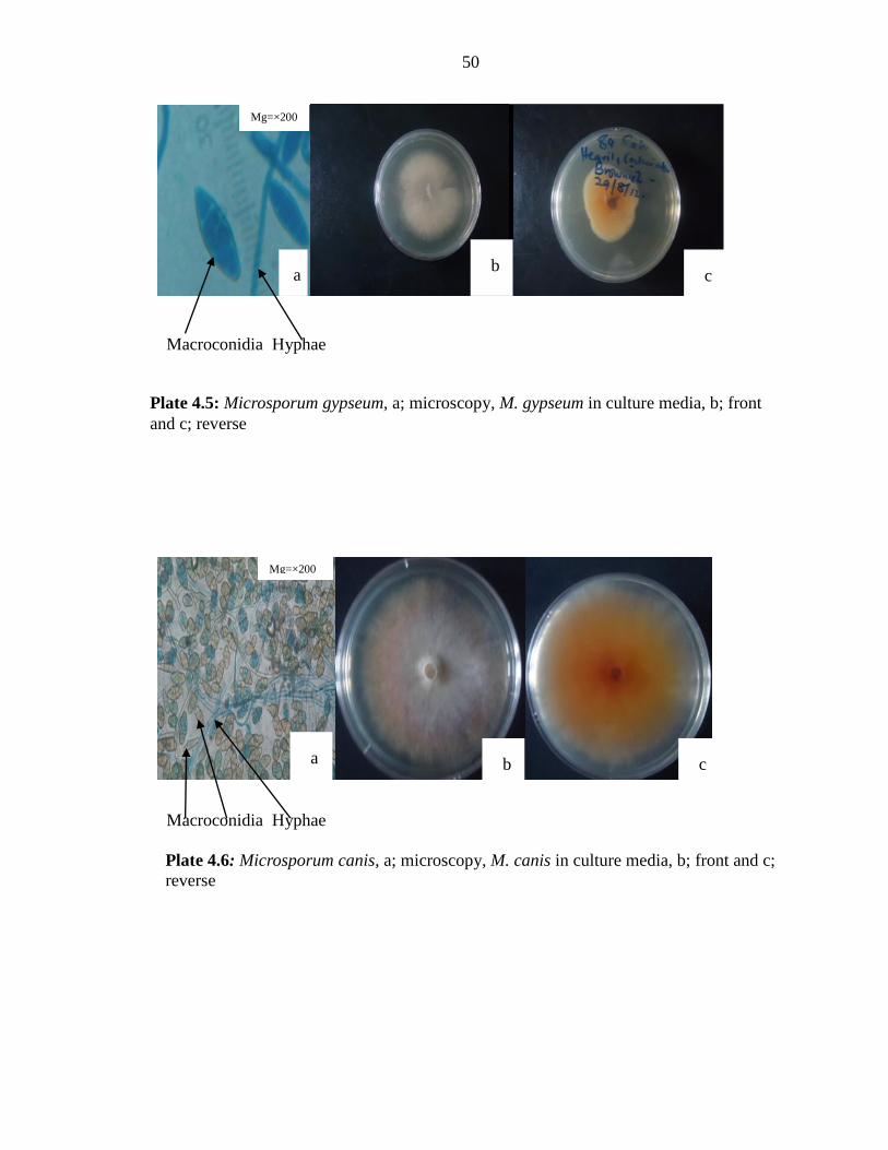
50
a
Plate 4.5: Microsporum gypseum, a; microscopy, M. gypseum in culture media, b; front
and c; reverse
Plate 4.6: Microsporum canis, a; microscopy, M. canis in culture media, b; front and c;
reverse
b c
a b c
Mg=×200
Mg=×200
Macroconidia Hyphae
Macroconidia Hyphae
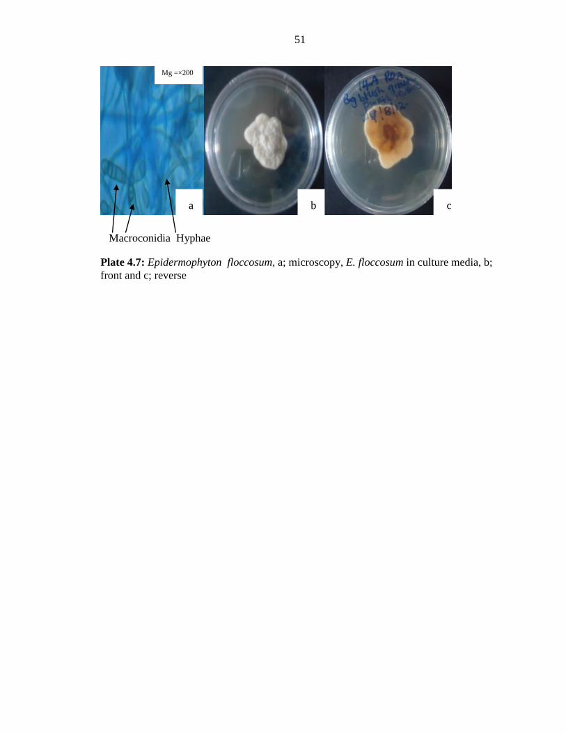
51
Plate 4.7: Epidermophyton floccosum, a; microscopy, E. floccosum in culture media, b;
front and c; reverse
a b c
Mg =×200
Macroconidia Hyphae
52
CHAPTER FIVE
DISCUSSION, CONCLUSIONS AND RECOMMENDATIONS
5.1 Discussion
5.1.1 Etiological agents of Tinea capitis
This study identified three genera of dermatological agents causing Tinea capitis. These
species included; Trichophyton tonsurans, Trichophyton mentagrophytes, Trichophyton
verrucosum, Trichophyton rubrum, Microsporum gypseum, Microsporum canis and
Epidermophyton flocossum. These dermatological agents detected in this study have also
been detected in previous studies carried out in Western Kenya, Libya and Nigeria on
Tinea capitis infection (Ayaya et al., 2001; Ellabib et al., 2002; Enemour and Amendu,
2009).
Isolation of different species of dermatophytes varies from one ecological niche to
another (Sajjan and Mangalgi, 2012). Trichophyton tonsurans was the most predominant
species isolated in this study. This observation was also made in the previous studies
carried out in Madagascar, Jamaica, Iraq and Kenya (Ayaya et al., 2001; Audonneau et
al., 2006; East-Innis et al., 2006; Al Samarai, 2007; Garg et al., 2009). The reason why
this dermatophyte is commonly isolated could be due to its resistance to adverse effects
of environmental conditions and therefore it can stay for a long time on surfaces making
it easily transferred by asymptomatic carriers. These results do not match with previous
studies carried out in Pakistan, Nepal, Ethiopia, Egypt, India and Kenya (Ahmed et al.,
2006; Jha et al., 2006; Woldeamanuel et al., 2005; Chepchirchir et al., 2009; Azab et al.,
2012; Grover et al., 2012) in which T. violaceum was the most frequent species isolated.
53
T. soudanense was predominant in studies carried out in Nigeria and Gabon (Ayanbimpe
et al., 2008 and Hogewoning et al., 2011). This difference in dermatophyte species
isolated could be due to difference in geographical location and changes in climatic
conditions. M. audonii was the most prevalent species noted in India and Nigeria (Avasn
et al., 2008 and Emele and Oyeka, 2008). M. canis, as the frequent isolate has been
reported in Egypt (Amer et al., 2007) while T. mentagrophytes and T. verroccusum were
common in studies conducted in India and Iran (Yazdanfar, 2010; Bose et al., 2011).
The etiological agents of Tinea capitis vary from one geographical location to another. In
Sub-Saharan West Africa, Microsporum audouinii and Trichophyton soudanense have
been isolated (Ayanbimpe et al., 2003; Anosikke et al., 2005) In the Western regions of
the world, Trichophyton tonsurans has emerged the predominant cause of tinea (Hay et
al.,1996; Warnock and Campbell, 1996; Gupta et al.,1998; Timen et al., 1999;
Ghannoum et al., 2003,). In a previous study conducted in Kisumu in Western part of
Kenya, the etiological agents isolated were T. violeceum, M. audonii and M. canis which
are zoophilic organisms (Schmeller, 1998).
T. tonsurans was the most commonly isolated organism in this study and this is similar to
the findings in the Western regions of the world and previous study conducted in Kenyan
school children in Eldoret ( Elewski, 2000; Ayaya et al., 2001). This distribution pattern
of dermatophytes infection in different parts of the world has been attributed to factors
such as climate, life-style, and prevalence of immunodeficiency diseases in the
community and also the reluctance of patients to seek treatment because of
54
embarrassment or minor nature of disease unless the condition becomes sufficiently
serious to affect the quality of life (Hashem al sheikh, 2009).
5.1.2 Prevalence of dermatological agents causing Tinea capitis in children aged 3-14
years
The prevalence of Tinea capitis in a particular area depends upon the environmental
conditions, personal hygiene and individual susceptibility. The isolation of different
species of dermatophytes also varies from one ecological niche to another, depending on
their primary natural habitat (Alsamaria, 2007). Findings from this study showed that
Trichophyton species, Microsporum species and Epidermophyton species were endemic
in the study area with varied prevalence. A trend that is similar to those previously
observed from other communities in tropical Africa (Enweani et al., 1996; Ayaya et al.,
2001; Hogewoning et al., 2013).
The prevalence of Tinea capitis (82 %) was higher than those obtained among Ethiopian
primary school children (59 %) (Woldeamanuel et al., 2005); Tanzania (4 %) (WHO,
2005) and (33.3 %) Kenya (Ayaya et al., 2001). The observed high prevalence could be
due to the fact that, the current study considered both symptomatic and asymptomatic
cases and also could be associated with high population growth hence frequent contact
with infected individuals coupled with low income levels of the people in the informal
settlement. The prevalence varied with sex and age. Males were highly infected
compared to girls. The high rates of hair cuts by boys from non specific barber shops and
frequent use of combs that are often shared and the heavy mingling with friends without
being conscious of personal hygiene are some of the factors that could be associated with
55
high infection rates contrary to girls (Menan et al., 2002; Adou-Bryn et al., 2004;
Ezeronye, 2005; Enendu and Ibe, 2005). The low prevalence rates in girls could be
associated with the fact that most of the girls especially older than 13 years prefer to plait
their hair rather than visiting barber shops. This could also be associated with the fact that
girls practice better hair, general personal hygiene and many do carry their own weaving
equipment hence reducing chances of possible contact with infective agents on hair
dresser’s hand (Ajao and Akintunde, 2005). These findings were contrary to those
obtained in Egypt and Nigeria that showed high prevalence rates among girls than boys, a
difference that was not statistically significant (Omar, 2000; Anosike et al., 2005). This
could be associated with the fact that contrary to Kenyan girls who practiced high levels
of personal hygiene, there was high prevalence of pediculosis among the females in
Egypt that was an indication of poor health habits and poor hygiene among the girls than
boys (Omar, 2000).
In relation to age, 3-5 and 6-8 had highest prevalence. Among the age group 6-8 years,
the prevalence (77.8 %) was similar to those obtained from previous studies in Kenya and
other sub-Saharan countries of 69 % to 78 % (Robertson and Wright, 1990; Ayaya et al.,
2001) (Table 4.1). This could be associated with poor hygiene at this age as well as the
absence of saturated fatty acids that provide a natural protective mechanism against
dermatophytoses (Chepchirchir et al., 2009). The low number of children in the age
category 12-14 who participated in the study could be associated with the fact that most
of them refused to go for skin scraping despite their parents’ consent.
56
The study also showed different prevalence of the species causing Tinea capitis among
the study subjects in the study area. Trichophyton species had the highest prevalence of
61.3 %, which could be due to the fact that most of the Trichophyton species are
anthropophilic and therefore their occurrence is high in human carriers. The Trichophyton
species which was observed to have the highest prevalence was Trichophyton tonsurans
(33.3 %). This could be explained by the fact that this dermatophyte is resistant to the
adverse impact of external conditions and asymptomatic carriers (Kalinowska, 2012)
transfer it. This rate was similar to that obtained in Nigeria (30.6 %) (Ayambimpe et al.,
2008) but contrarily lower than that previously obtained in Kenya (77.8 %) (Ayaya et al.,
2001).
For Trichophyton mentagrophytes (10.7 %), the findings were similar to those previously
obtained in studies in other sub-Saharan countries of 7.3 % - 15.7 % (Ayambimpe et al.,
2008; Emele and Oyeka, 2008). However, among this genus, Trichophyton verrucosum
and Trichophyton rubrum (8 %), were least isolated. These findings were similar to
previous studies that have shown a lowly maintained prevalence trend of this species in
Kenya and Nigeria ranging between 4 % and 9 % (Oyeka, 1990, and Ayaya et al., 2001).
The low prevalence of Trichophyton verrucosum among the children could be associated
with low rates of interaction with cattle which are the reservoirs for the dermatophyte
(Kalinowska, 2012) since these children mainly live in urban areas where cattle rearing is
rare. The low prevalence of Trichophyton rubrum can be explained by the fact that Tinea
capitis caused by Trichophyton rubrum is a rare event worldwide (Ziemer et al., 2005).
57
The Microsporum species; Microsporum gypseum and Microsporum canis were isolated
in this study. These species had a prevalence of 7.3 % and 6.0 % respectively that was
similar to those previously obtained in Nigeria of 6.9 % and 7.3 % respectively
(Ayambimpe et al., 2008). A low prevalence of Epidermophyton floccosum (7.3 %) was
also detected in this study. The observed rate was close to those previously obtained in
Nigeria and Libya of 4.3 % and 7.0 % respectively (Enweani et al., 1996; Ellabib et al.,
2002). Epidermophyton floccosum has been shown to cause sporadic cases of Tinea
capitis (Sberna et al., 1993) and asymptomatic carriage (Cuetara et al., 1998). It has been
further shown that changes in geographical distribution over time may be related to
increasing mobility of the population hence leading to the difference in prevalence of the
infection (Singal et al., 2001). The existence of multiple infections in this study
population could be associated with poor personal hygiene, suboptimal treatment, poor
drug adherence and abject poverty that predispose one to tinea infections (Alsamaria,
2007; Magill et al., 2007; Panackal et al., 2009).
5.1.3 Relationship between prevalence of Tinea capitis and various risk factors
One of the greatest problem hindering eradication and prevention of Tinea capitis is the
presence of healthy asymptomatic dermatophyte carriers. In this study, 43.7 % (21/48) of
the children without clinical symptoms of Tinea capitis were healthy carriers. However,
the difference among asymptomatic carriers varies highly between studies (Navarrete et
al., 2001). To evaluate the impact of employment status, socioeconomic status, income
and number of children in the family on Tinea capitis infection, a comparative study was
done. Employment status of the parents/ guardians was observed to have significant
influence on infection rates with children from unemployed parents being highly infected.

58
This showed that a low socioeconomic standard could be a risk factor to Tinea capitis
infections. The low prevalence rates among the children whose parents were employed
could probably be because these parents can afford to buy their own barber machines and
therefore the children do their hair shaving at home to avoid infection from nonspecific
barber shops. This finding concurred with previous studies (Figueroa et al., 1997;
Havlickova et al., 2004; Anosike et al., 2005; Ogunbiyi et al., 2005; Enemour and
Amendu, 2009; David et al., 2010; Nweke, 2010).
In this study, the number of children in a family was found to have no significant
influence on prevalence rates of Tinea capitis (p = 0.210). The association with
overcrowding probably stems from infectious nature of dermatophytes and sharing of
towel, combs, pillows and hats leading to an increased risk of interfamilial transmission
(Fathi and Al-samaria, 2000). In the current study, the prevalence of Tinea capitis among
the study subjects was significantly related (p < 0.001) to the sharing of personal effects
which might have largely contributed to high prevalence of the infection in children aged
3-14 years in Mathare informal settlement.
The study found knowledge on personal hygiene to be a significant risk factor (p < 0.001)
to prevalence of Tinea capitis among the study subjects. This was similar to a previous
study that found an association between the presence of dermatophytosis infection and
the habit of hygiene (Metintas et al., 2004). Majority (73.6 %) of the children did not
have access to information on personal hygiene that could have highly contributed to the
59
infection. Children brought up in clean environments with less crowding and reliable
water supply tend to suffer less from dermatophytes (Mark et al., 1999).
5.2 Conclusions
i) Based on the findings from this study, it shows that there is a very high prevalence (82%)
of Tinea capitis infections due to Trichophyton, Microsporum and Epidermophyton
species among school going children in informal settlement of Mathare with
Trichophyton species being the most prevalent among the dermatological agents.
ii) Low socio-economic background and poor personal hygiene were significant pre-
disposing factors of Tinea capitis among the study subjects with communal hair
shaving being the most significant risk factor to infection among boys.
iii) The prevalence of the infection was higher in lower age groups than the upper age
group of 12-14 years.
iv) The prevalence of the infection was higher in males than in females.
v) All the three dermatological agents causing Tinea capitis infection were isolated from
the samples collected from both symptomatic and asymptomatic subjects.
5.3 Recommendations
i) There is need for public awareness on the modes of spread and simple preventive
measures to reduce the prevalence of Tinea capitis in the informal settlement.
ii) There is a need for enforcing proper hygienic practices and education among school
going children and their parents/guardian to reduce the current trend of infection in
schools and by extension, the larger population.
60
iii) There is need to improve the living standards of people living in informal settlement
as one way to reduce the incidence of the infections in these areas.
iv) There is need for public health personnel to check antiseptic procedures used by the
barbers and advise them on the correct procedures to use in order to reduce
transmission of the infection.
v) There is need for adoption of mycological culture as the diagnostic procedure for
Tinea capitis in hospitals, health centres and schools for both symptomatic and
asymptomatic cases in order to determine the specific causative agent of Tinea capitis
and prevent transmission due to asymptomatic carriage.
61
REFERENCES
Abdel-Rahman, S.M., Herron, J., Fallon-Friedlander, S., Hauffe, S., Horowitz, A. and
Riviere, G.J. (2005). Pharmacokinetics of terbinafine in young children treated for Tinea
capitis. Pediatric Infectious Disease Journal; 24(10): 886-891.
Adamski, Z. and Batura Gabryel, H. (2007). Medical mycology for physicians and
students, 2nd
edition.Scientific publishing of Poznan medical University, Poznan, Poland,
p. 978-83.
Adou-Bryn, K.D., Assoumou, A., Haddad, R.N., Aka, B.R and Ouhon, J. (2004).
Epidemiology of Tinea capitis in Abidjan. Tropical Medicine; CotedÕIvoire. 64: 171–5.
Ahmed, I., Ahmed, Z. and Sarwat, N. (2006). Prevalence of Tinea capitis and
asymptomatic carriage amongst school going children. Journal of Pakistan Association of
Dermatologists; 16:215-219.
Ajao, A.O and Akintunde, C.A. (2005). Studies on the prevalence of Tinea capitis in Ile-
Ife Mycopathologia; 89: 43 – 48.
Aktas, E., Karakuzu, A. and Yiğit, N. (2009). Etiological agents of Tinea capitis in
Erzurum,Turkey. Journal of Medical Mycology; 19: 248-252.
Al Samarai A.G.M. (2007). Tinea Capitis among children: Public Health Implication.
Alexandria: infections and carriage. East Africa Mediteraen Health Journal. 6(5): 961-
967.
Ali, S., Graham, T. and Forgie, S. (2007). The assessment and management of Tinea
capitis in children. Pediatric Emergency Care; 23: 662-665.
Ameen, M. (2010). Epidemiology of superficial fungal infections. Clinical Dermatology;
28:197-201.
Ameh, I.G. and Okolo, R.U. (2004). Dermatophytoses among schoolchildren: domestic
animals as predisposing factor in Sokoto, Nigeria. Pakistan Journal of BioIogical
Science; 7: 1109-12.
Amer, A.E., Mohamed, K. and Alaa, A.E. (2007). Tinea capitis in Assuit Governorate: (A
clinical and mycological study). The Egyptian Journal of Hospital Medicine; 29:738-744.
Anosike, J.C., Keke, I.R. and Uwaezuoke, J.C. (2005). Prevalence and distribution of
ringworm infection in primary schools in parts of Eastern Nigeria. Journal of Applied
Science and Environmental Management; 9: 21-25
62
APHRC (2002).Population and health dynamics in Nairobi’s informal settlement; 64: 13-
14.
Arabatzis, M., Bruijnesteijn van Coppenraet, L.E., Kuijper, E.J., de Hoog, G.S.,
Lavrijsen, A.P. and Templeton, K. (2007). Diagnosis of common dermatophyte
infections by a novel multiplex real-time polymerase chain reaction
detection/identification scheme. British Journal of Dermatology; 157: 681-689.
Arenas R. (2002). Dermatofitosis en México. Revista Iberoamericana de Micologia;
19:63-7.
Arenas R. (2008). Micología médica ilustrada. 3rd ed. Mexico: Interamericana McGraw-
Hill; p. 61-94.
Asticcioli, S., Di Silverio, A., Sacco, L., Fusi, I., Vincenti, L. and Romero, E. (2008).
Dermatophyte infections in patients attending a tertiary care hospital in northern Italy.
New Microbiology; 31: 543-548.
Audonneau, N., Grosjean, P., Razanakolona, L.R., Andriantsinjovina, T. and Rapelanoro,
R. (2006). Tinea capitis in Madagascar: a survey in a primary school in Antsirabe.
Annales de Dermatologie et de Venereologie; 133:22-25.
Avasn, M. Y., Aruna, L. K., Ramakrishna, R.S., Hossain, K., Apta, C. D. and Karuna K.
(2008). Dermatophytes and other fungi associated with hair-scalp of primary school
children in Visakhapatnam, India: a case study and literature review. The Internet Journal
of Microbiology; 5: 2.
Ayanbimpe, G.M., Taghir, H., Diya, A. and Wapwera, S. (2008). Tinea capitis among
primary school children in some parts of central Nigeria. Mycoses; 51:336-40.
Ayanbimpe, G.M., Enweani, I. and, Solomon, E.G. (2003). Fungal infectionsin Jos: a-9
year study. African Journal of Clinical Microbiology; 4: 2-10.
Ayaya, S.O., Kamar, K.K. and Kakai, R. (2001). Aetiology of Tinea capitis. East Africa
Medical journal; 78:531-5.
Ayorinde, A.F., Adesanya, O.O. and Alaran, O.A. (2013). A Microbiological Study of
Dermatophyte Infection among Primary School Children in Mowe, Ogun State, Nigeria;
Current Research Journal of Biological Sciences; 5(5): 205-209.
Azab, M.M., Mahmoud, N.F., Abd, A. S., Alaa, E. D., Hosny, M.S, Shehata, A.S. and
Mohamed, R.W. (2012). Dermatophytes isolated from clinical samples of children
suffering from Tinea capitis in Ismailia, Egypt. Australian Journal of Basic and Applied
Sciences; 6:38-42.
63
Barry, L. and Hainer, M.D. (2003). Dermatophyte infections Medical University of
South Carolina Charleston, South Carolina. American Family Physician; 67(1): 101-109.
Bassiri, J. and Khaksar, A.A (2006). Aetiological agents of Tinea capitis in Tehran
(Iran). Mycoses; 49: 65-67.
Bennett, M.L., Fleischer, A.B., Loveless, J.W. and Feldman, S.R. (2000). Oral
griseofulvin remains the treatment of choice for Tinea capitis in children. Pediatric
Dermatology Journal; 17: 304– 309.
Betty, A.F., Daniel, F.S. and Alice, S.W. (2007). Laboratory methods in basic
microbiology, Bailey and Scotts Diagnostic Microbiology 12th edition Mosby Elsevier;
Chapter: 50; p. 629-717.
Bose, S., Kulkarni, S.G. and Akhter, I. (2011). The incidence of Tinea capitis in a tertiary
care rural hospital - a study. Journal of Clinical and Diagnostic Research; 5: 307-311.
Brajesh, K. J. and Mahadeva, S. M. (2013). Studies on invasive keratinophilic
dermatophytes of human hair. Journal of Drug Delivery and Therapeutics; 3(2): 70-74.
Brendan P. K. (2012) Superficial Fungal Infections. Pediatrics in Review; 33(4):22-40.
Caceres-Rios, H., Rueda, M., Ballona, R. and Bustamante, B. (2000). Comparison of
terbinafine and griseofulvin in the treatment of Tinea capitis. Journal of the American
Academy of Dermatology; 42:80-4.
Caputo, R., De Boulle, K., Del Rosso, J. and Nowicki, R. (2001). Prevalence of
superficial fungal infections among sports-active individuals: results from the Achilles
survey, a review of the literature. Journal of European Academy of Dermatology; 15:
312-6.
Centre for Food Security and Public Health (2005). College of Veterinary Medicine.
Retrieved from: http:// www.cfsph.Iastate.edu, (Accessed on: June 5, 2012).
Cerqueira, M.D.P., Neves, R.P., Magalhães, O.M.C.,Souza-Motta, C.M. and Queiroz,
L.A. (2005). Pathogenic aspects of Epidermophyton floccosum langeron et milochevitch
as possible etiological agent of Tinea capitis. Brazilian Journal of Microbiology. 36:36-
37.
Chan, Y.C. and Friedlander, S.F. (2004). New treatments for Tinea capitis. Current
Opinion in Infectious Disease; 17(2): 97-103.
Chen, B. and Friedlander, S. (2001). Tinea capitis update: A continuing conflict with an
old adversary. Current Opinion in Pediatrics; 13: 331-335.
64
Chepchirchir, A., Bii, C. and Ndinya-achola, J.O. (2009). Dermatophyte infections in
primary school children in kibera slums of Nairobi Correlation. International Journal of
Dermatology; 24(2): 116-119.
Côbo, E.C., Silva, J.C., Cota, U.A., Machado, J.R. and Castellano, L.R. (2010).
Evaluation of a modified microscopic direct diagnosis of dermatophytosis. Journal of
Microbiological Methods; 81: 205-207.
Corting, C.H. (2009). Fungal infections. In: Braun-Falco, O., Burgdorf, W.H.C., Plewig,
G., Wolf H.H. and Landthaler, M. 3rd ed. Springer Verlag, Italy: pp. 205-239.
Crow, B. and Odaba, E. (2010).Access to water in a Nairobi slum: Women’s work and
institutional learning. Water international; 35(6):733-747.
Cuetara, M. S., Del Palacio, A., Pereiro, M. and Noriega, A. R. (1998). Prevalence of
undetected Tinea capitis in prospective school survey in Madrid: emergence of new
causative fungi. British Journal of Dermatology; 138: 658–660.
Daniel, W.W. (1999). Biostatistics: A Foundation for Analysis in the Health Sciences. 7th
edition. New York: John Wiley and Sons. P. 9-14.
David, D.L., Edward, A., Zaruwa, M.Z. and Addass, P.A. (2010). Barbing Saloon
Associated Fungal Disease Infection in Mubi, Adamawa State-Nigeria. World Journal of
Medical Science; 5(1): 17- 21.
Devliotou-Panagiotidou, D. and Koussidou-Eremondi, T.H. (2004). Efficacy and
tolerability of 8 weeks' treatment with terbinafine in children with Tinea capitis caused by
Microsporum canis: a comparison of three doses. Journal of European Academy of
Dermatology; 18:155-159.
Dworacka-Kaszak, B. (2004). Dermatophytes. Keratophilic fungi and their role in
environment. Mikologia Lekarska; 11(4): 317-322.
East-Innis, A., Rainford, L., Dunwell, P., Barrett-Robinson, D. and Nicholson, A.M.
(2006). The Changing Pattern of Tinea Capitis in Jamaica. West Indian Medical journal;
55:85.
Elewski, B.E. (2000). Tinea capitis: A current perspective. Journal of American Academy
of Dermatology; 42: 1-20.
Ellabib, M.S., Khalifa, Z. and Kavanagh, K. (2002). Dermatophytes and other fungi
associated with skin mycoses in Tripoli, Libya. Mycoses; 45: 101–4.
Ellis D, Davis s, Alexiou H, Handke R. and Bartley R. (2007). Descriptions of medical
fungi, second edition, p. 61-167.
65
Elsayed, Z., Salem, M.I., Ibrahim, S. F., Yaser, M.M., Hamed, A. F., Abdel Hamid, E. H.,
Emam, F. M. and Abdel S. (2010). Applicability of Fourier Transform Infrared (FT-IR)
spectroscopy for rapid identification of some yeasts and dermatophytes isolated from
superficial fungal infection. Journal of Egypt Women Dermatology; 7: 105-110.
Emele, F.E. and Oyeka, C.A. (2008). Tinea capitis among primary school children in
Anambra state of Nigeria. Mycoses; 51:536–41.
Enany, M. E., khafagy, A.R., Madiha S. I., Marwa M. A. and Dalia T. H. (2013)
Identification of dermatophytes isolated from ringworm lesions of camels. Suez Canal
Veterinary Medicine Journal; 18(1):1-2.
Enemour, S.C and Amendu, A.S. (2009). Prevalence of superficial mycosis in primary
school children in Anyigba, Kogi state, Nigeria. African Journal of Microbiology
Research; 3: 63-65.
Enendu, N.E. and Ibe, S.N. (2005). Prevalence of Tinea capitis among primary school
pupils in Uli, Anambra state, Nigeria. African journal of applied zooogy and
enviromental bilology; 7:1-4.
Enweani, I.B., Ozan, C.C., Agbonlahor, D.E. and Ndip, R.N. (1996). Dermatophytosis in
school children in Ekpoma, Nigeria. Mycoses; 39: 303–5.
Ezeronye, O.U. (2005). Distribution of dermatomycoses in Cross River upstream bank of
Eastern Nigeria. In: Medical Mycology: The African Perspectives. Proceedings of an
international conference held at Hartenbosch, South Africa. p. 35.
Faithi, H.I. and Al Samarai, A.G.M. (2000). Prevalence of Tinea capitis among school
children in Iraq. Eastern Mediterranean Health Journal; 6: 128-137.
Figueroa, J. I., Hawraneck, T., Abraha, A. and Hay, R. J. (1997). Tinea capitis in south
western Ethiopia: a study of risk factors for infection and carriage. Internatinal Journal of
Dermatology; 3:16-20.
Fleece, D., Gaughan, J.P. and Aronoff, S.C. (2004). Griseofulvin versus terbinafine in the
treatment of Tinea capitis: A meta-analysis of randomized, clinical trials. Pediatric
Dermatology Journal; 114(5): 1312-1315.
Foster, K.W., Ghannoum, M.A. and Elewski, B.E. (2004). Epidemiologic surveillance of
cutaneous fungal infection in the UnitedStates from 1999 to 2002. Journal of America
Academy of Dermatology; 50:748–52.
Fuller, L., Child, F., Midgley, G. and Higgins, E. (2003). Diagnosis and management of
scalp ringworm. British Medical Journal; 326: 539-541.
Fuller, L.C., Smith, C.H., Cerio, R., Marsden, R.A., Midgley, G. and Beard, A.L. (2001).
66
A randomized comparison of 4 weeks of terbinafine vs. 8 weeks of griseofulvin for the
treatment of Tinea capitis. British Journal of Dermatology; 144: 321.
Fungal Research Trust (2011). How common are fungal diseases; London; 61(9): 2703-
2710, 2713-2714.
Garg, J., Tilak, R., Garg, A., Prakash, P., Gulati, A. and Nath, G. (2009). Rapid detection
of dermatophytes from skin and hair. Biomedical Research Notes; 2: 60.
Ghannoum, M., Isham, N., Hajjeh, R., Cano, M., Al-Hasawi, F., Yearick, D., Warner, J.,
Long, L., Jessup, C. and Elewski, B. (2003). Tinea capitis in Cleveland: survey of
elementary school students. Journal of American Academy of Dermatology; 48(2): 189-
193.
Ginter-Hanselmayer, G., Weger, W., Ilkit, M. and Smolle, J. (2007). Epidemiology of
Tinea capitis in Europe: current state and changing patterns. Mycoses; 50 (2):6-13.
Grover, C., Arora, P. and Manchanda, V. (2012). Comparative evaluation of
griseofulvin, terbinafine and fluconazole in the treatment of Tinea capitis. International
Journal of Dermatology; 51:455- 458.
Gupta, A.K .and Summerbell, R.C (1998). Increased incidence of Trichophyton tonsurans
Tinea capitis in Ontario Canada between1985 and 1996. Medical Mycology; 36; 55-60.
Gupta, A.K. and Cooper, E.A. (2008). Update in antifungal therapy of dermatophytosis.
Mycopathologia; 166(5):353–367.
Gupta, L.K. and Singhi, M.K. (2004). Wood's lamp. Indian Journal of Dermatology;
70:131.
H P A (2007). Tinea capitis in the United Kingdom: A report on its diagnosis,
management and prevention. London: East Africa Medical Journal; 78: 531–5.
Habif, T. (2004). Clinical dermatology (4th edition). Philadelphia: Mosby p. 428-433.
Habif, T., Campbell, J. Jr., Chapman, M., Dinulos, J. and Zug, K. (2005). Skin diseases:
diagnosis and treatment. 2nd edn. Philadelphia: Mosby; 75:121-132.
Hallgren, J., Petrini, B. and Wahlgren, C.F. (2004). Increasing Tinea capitis prevalence in
Stockholm reflects immigration. Medical Mycology; 42: 505-509.
Hashem al sheikh, M. (2009). Epidemiology of Dermatophytes in the Eastern Province of
Saudi Arabia. Research Journal of Microbiology; 4(6): 229-234.
Havlickova, B., Czaika, V.A. and Friedrich M. (2004). Epidemiological trends in skin
mycoses Worldwide. Mycoses; 51 (4): 2–15.
67
Havlickova, B., Czaika, V.A. and Friedrich, M. (2008). Epidemiological trends in skin
mycoses worldwide. Mycoses; 51(4):2-15.
Hay, R.J., Claydon, Y.M., de Silva, N., Midgley, G and Rossor, E. (1996).Tinea capitis in
South-east London: a new pattern ofinfection with public health implications. British
Journal of Dermatology; 135: 955-8.
Higgins, L., Fuller, C. and Smith, C.H. (2000). Guidelines for the management of Tinea
capitis. British Journal of Dermatology; 143:53-8.
Hogewoning, A.A., Adegnika, A.A., Bouwes, B.J.N., Yazdanbakhsh, M., Kremsner, P.G.,
van der Raaij-Helmer, E.M.H., Staats, C.C.G., Willemze, R. and Lavrijsen, A.P.M. (2011).
Prevalence and causative fungal species of Tinea capitis among schoolchildren in Gabon.
Mycoses; 54:354-359.
Hogewoning, A., Amoah A., Bavinck, J.N.B., Boakye, D., Yazdanbakhsh, M., Adegnika,
A., Smedt, S., Fonteyne, Y., Willemze, R. and Lavrijsen, A. (2013). Skin diseases among
schoolchildren in Ghana, Gabon, and Rwanda, International Journal of Dermatology; 52:
5.
Hryncewicz-Gwozdz, A., Plomer-Niezgoda, E. and Maj, J. (2005). Mycoses of feet,
hands and nails – epidemiology, symptoms, treatment. Mikologia Lekarska: 12: 57-62.
in Stockholm reflects immigration. Medical Mycology; 42:505-9.
Jankowska-Konsur, A., Dylg, M., Haryncewicz-Gwo, D.A., Plomer-Niezgoda E. and
Szepietowski J.C. (2011). A 5 year survey of dermatomycosis in south west Poland.
Mycoses; 54: 162-167.
Jha, B.N., Garg, V.K., Agrawal, S., Khanal, B. and Agarwalla, A. (2006). Tinea capitis
in eastern Nepal. International Journal of Dermatology; 45:100-102.
Johnson, B.A. and Nunley, J.R. (2000). Treatment of seborrheic dermatitis. American
Family Physician; 56: 345-349.
Joshi, R., Adamson's, F., Horatio, G. A. and Kligman's, J. (2011). Experiments and
Observations on Tinea Capitis. International Journal of Trichology; 3: 14-19.
Kalinowska, K. (2012). Epidemiology of Dermatomycoses in Poland over the Past
Decades. Mikologia Lekarska; 17(3): 165-168.
Kao, F.G. (2006). Tinea capitis. Retrieved January 29, 2006, from
http://www.eMedicine.com
Karimzadegan-Nia, M., Mir-Amin-Mohammadi, A., Bouzari, N. and Firooz, A. (2007).
Comparison of direct smear, culture and histology for the diagnosis of onychomycosis.
Australasian Journal of Dermatology; 48: 18-21.
68
Kawachi, Y., Ikegami, M., Takase, T. and Otsuka, F. (2010). Chronically recurrent and
disseminated Tinea faciei/corporis auto inoculation from asymptomatic Tinea capitis
carriage. Pediatric Dermatology; 27: 527–553.
Kemal, Ö., Halit, B., Perihan, Ö., Tuba, K. and Ümit, U. (2013). Tinea Capitis in Two
Sisters of a Wooly Hair Family; Journal of Turkish Academy of Dermatology; 7(1):137.
Kolivras, A., Lateur, N. and De Maubeuge, J. (2003). Tinea capitis in Brussels:
Epidemiology and new management strategy. Dermatology; 206:384-7.
Koné, M., Atindehou, K. K. and Traoré, D. (2002). Plantes et médecine traditionnelle
dans la région de Ferkessédougou (Côte d'Ivoire). Annals of Botany African Journal; 2:
13-21.
Krajewska-Kulak, E., Niczyporuk, W., Lukaszuk, C., Bartoszewicz, M., Roszkowska, I.
and Edyta, M. (2003). Difficulties in diagnosing and treating tinea in adults at the
department of dermatology in Bialystok, Poland. Dermatology Nursing; 15(6): 527-530,
534.
Larone, D.H. (2002). Medically important fungi. ASM Press, Washington, DC. 4th
Edition: p. 114-121.
Liu, D., Coloe, S., Baird, R. and Pedersen, J. (2000). Application of PCR to the
identification of dermatophyte fungi. Journal of Medical Microbiology; 49:493-7.
Magill, S.S., Manfredi, L., Swidersli, A., Cohen, B. and Merz, W.G. (2007). Isolation of
Trichophyton violaceum and Trichophyton soudanense in Baltimore, Maryland. Journal
of Clinical Microbiology; 45(2):461-65.
Mark, H. B., Andrew, J. F. and Thomas, V. J. (1999). The Merck Manual of Medical
information. Merck & Co. 4th
Edition: p. 979-980.
Marques, S.A., de Camargo, R.M.P., Fares, A.H.G., Takashi, R.M. and Stolf, H.O. (2005).
Tinea capitis: Epidemiological and ecological aspects of cases observed from 1983 to
2003 in the Botucatu Medical School, state of São Paulo-Brazil. Anais. Brasileiros.
Dermatologia; 80:42-56
Mcdonald, W. (2000), Guide for medical fungal morphology and identification. p. 70-74.
McPherson, M.E., Woodgyer, A.J., Simpson, K. and Chong, A.H. (2008). High
prevalence of Tinea capitis in newly arrived migrants at an English-language school,
Melbourne. Medical Journal of Australia; 189:13-6.
Menan, E.I., Zongo-Bonou, O. and Rouet, F. (2002). Tinea capitis in school children
from lvory Coast (western Africa). A 1998–1999 cross-sectional study. International
69
Journal of Dermatology; 41: 204–7.
Metintas, S., Kiraz, N., Arslantas, D., Akqun, Y. and Unsa, A. (2004). Frequency and risk
factors of dermatophytosis in students living in rural areas in Eskisehir, Turkey. Journal
of Clinical and Diagnostic Research; I: 476-482.
Möhrenschlager, M., Seidl, P.H., Ring, J. and Abeck, D. (2005). Pediatric Tinea capitis:
Recognition and management. American Journal of Clinical Dermatology; 6(4): 203-
213.
Molina D.A. (2011). Clinical, diagnostic and therapeutic aspects of dermatophytosis.
Enfermedades infecciosas y microbiologia clinica; 29 (3):33-39.
Mounsey, A. L. and Reed, S. W. (2009). Diagnosing and treating hair loss. American
Family Physician; 80: 356-362.
Mukherjee, P.K., Isham, N. and Ghannoum, M.A. (2011). Molecular Diagnostics in
Dermatology and Dermatopathology .Current Clinical Pathology, Infectious Diseases of
the Skin I: Dermatophytosis/ Onychomycosis. P. 311-337.
Navarrete, C.M., Zaror, C.L. and Moreno, V.M. (2001). Dermatofitosis en hogares de
menores de Valdivia, Chile. Aspectos clı´nicos-micolo´ gicos y epidemiolo´ gicos. Rev
Chil Cienc. Medical Biology; 11: 15–22.
Neji, S., Makni, F., Cheikhrouhou, F., Sellami, A., Sellami, H., Marreckchi, S., Turki, H.
and Ayadi A. (2008). Epidemiology of dermatophytoses in Sfax, Tunisia. Mycoses; 52:
534–538.
Nelson, M.M., Martin, A.G. and Hefferman, M.P. (2003). Superficial fungal infections:
dermatophytosis, onychomycosis, Tinea nigra, piedra. Dermatology in General Medicine;
2: 90-94.
Ngwogu, A.C. and Otokunefor, T.V. (2007). Epidemiology of dermatophytoses in a rural
community in Eastern Nigeria and review of literature from Africa. Mycopathologia;
164:149-58.
Nweke E.I. (2010). Dermatophytosis among children of Fulani/Hausa herdsmen living in
southeastern Nigeria. RevIberoam Micology; 27(4):191– 194.
Ogunbiyi, A.O., Owoaje, E. and Ndahi, A. (2005). Prevalence of skin disorders in school
children in Ibadan, Nigeria.Pediatric Dermatology; 22: 6-10.
Omar, A.A. (2000). Ringworm of the scalp in primary school children in Alexandria:
infections and carriage. East Africa Mediterranean Health Journal; 6(5):961-967

70
Oyeka, C.A. (1990). Tinea capitis in Awka local government area of Anambra State. West
African Journal of Medicine; 9: 120–3.
Panackal, A.A., Halpern, E. and Watson, A.J. (2009). Cutaneous fungal infections in the
United States. Internatinal Journal of Dermatology; 48(7):704-12.
Panasiti, V., Borroni, R.G., Devirgiliis, V., Rossi, M., Fabbrizio, L. and Masciangelo, R.
(2006). Comparison of diagnostic methods in the diagnosis of dermatomycoses and
onychomycoses. Mycoses; 49:26–9.
Panasiti, V., Devirgiliis, V., Borroni, R.G., Mancini, M., Curzio, M. and Rossi, M. (2007).
Epidemiology of dermatophytic infections in Rome, Italy: a retrospective study from
2002 to 2004. Medical Mycology; 45:57-60.
Rebollo, N., Lopez-Barcenas, A.P. and Arenas, R. (2008). Tinea capitis.
Departmento.de.dermatologia; 99:91-100.
Robert, R. and Pihet, M. (2008). Conventional methods for the diagnosis of
dermatophytes. Mycopathologia; 166: 295-306.
Roberts, B. and Friedlander, S. (2005). Tinea capitis: A treatment update. Pediatric
Annals; 34: 191-200.
Robertson, V.J. and Wright, S. (1990). A survey of Tinea capitis in primary school
children in Harare, Zimbabwe. Journal of Tropical Medicine and Hygiene; 93: 419–22.
Sarabi, K. (2008). Tinea capitis: A Review. Dermatology Nursing; 19(6):525-529.
Sajjan, A.G. and Mangalgi, S.S. (2012). Clinicomycological profile of Tinea capitis in
children residing in orphanages. International Journal of Biological and Medical
Research; 3(4): 2405-2407.
Sberna, F., Farella, V., Geti, V. (1993). Epidemiology of the dermatophytoses in the
Florence area of Italy: Mycopathologia; 122:153–162.
Schmelle, W. (1998). Community health workers reduce skin diseases in East African
children. International Journal of Dermatology; 37:370 – 377.
Schmeller, W., Baumgartner, S. and Dzikus, A. (1997). Dermatophytomycoses in
children in rural Kenya: Impact of Primary Health Care. Mycoses; 40: 55-63.
Seebacher, C., Bouchara, J.P. and Mignon, B. (2008). Updates on the epidemiology of
dermatophyte infections. Mycopathologia; 166:335-52.
Seema, B., kulkarni, S. and Irfaanakhter (2011). The incidence of Tinea capitis in a
tertiary care rural hospital. Journal of Clinical and Diagnostic Research; 5:307-311.
71
Sidat, M. M., Correia, D. and Buene, T. P (2007). Tinea capitis among children at one
suburban primary school in the city of Maputo, Mozambique. Revista Sociedade
Brasleira Medical Tropical Journal; 40: 473-475.
Silverberg, N.B., Weinberg, J.M. and DeLeo, V.A. (2002). Tinea capitis: Focus on
African American women. Journal of the American Academy of Dermatology; 46: 120-
124.
Simpanya, M.F. (2000). Dermatophytes: Their taxonomy, ecology and pathogenicity.
Mycology; 12: 1-12.
Singal, A., Rawat, S., Bhattacharya, S.N., Mohanty, S. and Baruah, M.C. (2001). Clinico-
myocological profile of Tinea capitis in North India and response to griseofulvin. Journal
of Dermatology; 28:22-6.
Summerbell, R.C., Weitzman, I. and Padhye, A.A. (2007). Trichophyton, Microsporum,
Epidermophyton, and Agents of Superficial Mycoses. In PR Murray (Ed.) Manual of
Clinical Microbiology. ASM Press, Washington, DC. 9th Edition: P. 1874-1897.
Szepietowski, J. and Baran, E. (2005). Epidemiology of mycoses. In: Outline of
mycology of nurses. E. Krajewska-Kulak (Ed.), 31-35, Czelej Publishing, Lublin, Poland,
p. 83- 89.
Timen, A., Bovee, L., Leentvaar-Kuijpers, A., Peerbooms, P.G. andCountinho, R.A
(1999). Tinea capitis in primary school age children in Southeastern Amsterdam:
primarily due toTrichophyton tonsurans; Mycopathologia; 157(4):379-81.
Tra Bi, F.H., Kouamé, N.F., Traoré, D. (2005). Utilisation of climbers in two forest
reserves in West Côted'Ivoire. In: Bongers F. P.M., Traoré D., Ed.Forest Climbing Plants
of West Africa:Diversity, Ecology and Management. CABI Publishing, Oxfords hire
(UK): p. 64-66.
Trivino-Duran, L., Torres-Rodriguez, J.M., Martinez-Roig, A., Cortina, C., Belver, V. and
Perez-Gonzalez, M. (2005). Prevalence of Tinea capitis and Tinea pedis in Barcelona
school children. Pediatric Infectious Disease Journal; 24(2): 137-141.
Uchida, T., Makimura, K., Ishihara, K., Goto, H., Tajiri, Y. and Okuma, M. (2009).
Comparative study of direct polymerase chain reaction, microscopic examination and
culture-based morphological methods for detection and identification of dermatophytes in
nail and skin samples. Journal of Dermatology; 36: 202-208.
Warnock, D.W. and Campbell, C.K (1996). Centenary review: medical mycology.
Mycology Research; 100: 1153-62.
72
WHO (2005). Epidemiology and management of common skin diseases in children in
developing countries. p. 6.
Woldeamanuel, Y., Leekassa, R. and Chryssanthou E. (2005). Prevalence of Tinea capitis
in Ethiopian school children. Mycoses; 48: 137-141.
Yazdanfar, A. (2010). Tinea capitis in primary school children in Hamedan (Westof Iran).
International Journal of Medicine and Medical Sciences; 2:29-33.
Yehia, M., El-Ammawi, T.S., Al-Mazidi, K.M., Abu El-Ela, M.A. and Al-Ajmi, H.S.
(2010). The Spectrum of Fungal Infections with a Special Reference to Dermatophytoses
in the Capital Area of Kuwait during 2000–2005: A Retrospective Analysis.
Mycopathologia; 169: 241–246.
Ziemer, A., Kohl, K. and Schröder G. (2005) Trichophyton rubrum-induced inflammatory
Tinea capitis in a 63-year-old man. Mycoses; 48(1):76-9.
73
APPENDICES
Appendix I
Questionnaire
This research is made for academic purpose. It is geared towards assessing the prevalence
of ring worms of the scalp in children aged 3-14 years in Mathare informal settlements of
Nairobi. The outcome will help in advising the policy makers and relevant disease control
programmes on the appropriate preventive measures to undertake to prevent the infection
in the area. It will also help in knowledge of the specific causal organism so as to help
therapists to administer the right type of therapy. You are kindly requested to provide
answers to these questions as honestly and precisely as possible. Response to these
questions will be treated as confidential and your identity will not be revealed to anyone.
Thank you.
PART A: PATIENT DEMOGRAPHIC CHARACTERISTICS
(Please don’t write your name)
SECTION 1(tick where appropriate)
1. AGE:
Between 3-5 years
Between6-8 years
Between 9-11 years
Between 12-14 years
2. SEX: MALE FEMALE
3. Class:

74
4. School:
5. Division:
6. EMPLOYMENT STATUS OF FATHER
Employed
Self employed
Unemployed
Retired
INCOME OF THE FATHER IN KSHS PER MONTH
KSH
7. EMPLOYMENT STATUS OF THE MOTHER
Employed
Self employed
Unemployed
Retired
INCOME OF THE MOTHER IN KSHS PER MONTH
KSH
8. Number of children in your family

75
PART B: KNOWLEDGE ON HYGIENIC PRACTICES IN RELATION TO
TINEA CAPITIS (RINGWORM) INFECTION.
QUESTIONS
1. Do you ever obtain information on personal hygiene? (Yes/No)
2. If yes, then where do you mostly obtain the information? (Parents, Teachers,
Friends, health care providers.)(Other specify)
3. Do you share combs with your friends/siblings?(Yes/No)
4. Do you share bathing towel with your friends/ siblings?( Yes/No)
5. Where do you normally have your hair shaving?
Barber shop
Home
Both
6. How often do you shave your hair?
Weekly
After two weeks
Monthly
7. Are you aware of ways through which ringworms can infect you?( Yes/No)
8. If the answer is yes, then which methods?
76
Appendix II
Ethical approval
77