Embed Size (px)
Citation preview

International Journal of
Molecular Sciences
Review
Dysfunction of the Neurovascular Unit in Ischemic Stroke:Highlights on microRNAs and Exosomes as Potential Biomarkersand Therapy
Timea Forró 1,2,* , Zoltán Bajkó 3,4, Adrian Bălas, a 5,6 and Rodica Bălas, a 3,4
�����������������
Citation: Forró, T.; Bajkó, Z.; Balas, a,
A.; Balas, a, R. Dysfunction of the
Neurovascular Unit in Ischemic
Stroke: Highlights on microRNAs and
Exosomes as Potential Biomarkers
and Therapy. Int. J. Mol. Sci. 2021, 22,
5621. https://doi.org/10.3390/
ijms22115621
Academic Editor: Ryszard Pluta
Received: 28 April 2021
Accepted: 23 May 2021
Published: 25 May 2021
Publisher’s Note: MDPI stays neutral
with regard to jurisdictional claims in
published maps and institutional affil-
iations.
Copyright: © 2021 by the authors.
Licensee MDPI, Basel, Switzerland.
This article is an open access article
distributed under the terms and
conditions of the Creative Commons
Attribution (CC BY) license (https://
creativecommons.org/licenses/by/
4.0/).
1 2nd Clinic of Neurology, Târgu Mures, County Emergency Clinical Hospital, 540136 Târgu Mures, , Romania2 Doctoral School, ‘George Emil Palade’ University of Medicine, Pharmacy, Science, and Technology of Târgu
Mures, , 540142 Târgu Mures, , Romania3 1st Clinic of Neurology, Târgu Mures, County Emergency Clinical Hospital, 540136 Târgu Mures, , Romania;
[email protected] (Z.B.); [email protected] (R.B.)4 Department of Neurology, ‘George Emil Palade’ University of Medicine, Pharmacy, Science, and Technology
of Târgu Mures, , 540142 Târgu Mures, , Romania5 Clinic of Neurosurgery, Târgu Mures, County Emergency Clinical Hospital, 540136 Târgu Mures, , Romania;
[email protected] Department of Neurosurgery, ‘George Emil Palade’ University of Medicine, Pharmacy, Science,
and Technology of Târgu Mures, , 540142 Târgu Mures, , Romania* Correspondence: [email protected]; Tel.: +40-758-454-845
Abstract: Ischemic stroke is a damaging cerebral vascular disease associated with high disability andmortality rates worldwide. In spite of the continuous development of new diagnostic and prognosticmethods, early detection and outcome prediction are often very difficult. The neurovascular unit(NVU) is a complex multicellular entity linking the interactions between neurons, glial cells, andbrain vessels. Novel research has revealed that exosome-mediated transfer of microRNAs plays animportant role in cell-to-cell communication and, thus, is integral in the multicellular crosstalk withinthe NVU. After a stroke, NVU homeostasis is altered, which induces the release of several potentialbiomarkers into the blood vessels. The addition of biological data representing all constituents ofthe NVU to clinical and neuroradiological findings can significantly advance stroke evaluation andprognosis. In this review, we present the current literature regarding the possible beneficial rolesof exosomes derived from the components of the NVU and multipotent mesenchymal stem cellsin preclinical studies of ischemic stroke. We also discuss the most relevant clinical trials on thediagnostic and prognostic roles of exosomes in stroke patients.
Keywords: neurovascular unit; exosomes; extracellular vesicles; microRNAs; stroke; brain ischemia;biomarkers; mesenchymal stem cells
1. Introduction
Stroke is a leading cause of death and disability worldwide [1]. It is classically definedas a neurological deficit due to an acute focal injury of the central nervous system (CNS)by a vascular cause [2]. Ischemic stroke, the most common type of stroke, is caused by anarrowing or blockage in an artery that supplies the brain, which leads to interruptionof blood flow in the corresponding territory [3], thereby causes irreversible cell damagein the ischemic core through oxygen deprivation and increased inflammation [4]. Theperi-infarct zone, also called the penumbra, contains cells at risk of further damage, butcan potentially be rescued if appropriate interventions are available [5]. Although plentifulneuroprotective agents have been studied in the past decades [6], timely reperfusion,including intravenous thrombolysis with tissue plasminogen activator (alteplase) [7] andthrombus removal by mechanical thrombectomy [8] remains the only immediate treatmentoption in acute ischemic stroke management.
Int. J. Mol. Sci. 2021, 22, 5621. https://doi.org/10.3390/ijms22115621 https://www.mdpi.com/journal/ijms
Int. J. Mol. Sci. 2021, 22, 5621 2 of 18
The prognosis of ischemic stroke is highly dependent on the interaction betweenclinical and demographic factors, such as age or stroke severity [9]. The addition ofbiological information, such as blood biomarkers or genetic polymorphisms, could furtherenhance the prognostic value of clinical data. A reliable outcome prediction method mightimprove decision-making processes and may be useful to improve and personalize strokecare [10].
Emerging data suggest that exosomes, which are nanosized (30–150 nm) endosome-derived vesicles, can be useful diagnostic, therapeutic, and prognostic markers in stroke [11].Along with shedding microvesicles or ectosomes (10–1000 nm) and apoptotic bodies (50–5000 nm), exosomes represent one of the three main subtypes of the extracellular vesi-cles (EVs) [12]. The biogenesis of exosomes is a finely calibrated process that includesfour stages: initiation, endocytosis, multivesicular body formation, and exosome secre-tion, which is the process in which these multivesicular bodies fuse with the plasmamembrane [13]. After being released from their cells of origin, exosomes can interact byendocytosis, fusion, or ligand–receptor interactions with recipient cells [14].
Secreted by most human cells, exosomes contribute to intercellular signaling viacell-to-cell communication in both physiological and pathological processes, and functionin different CNS diseases, such as stroke [15,16]. They also have the ability to cross theblood–brain barrier (BBB), thereby helping in the exchange between the CNS and theperipheral circulation [17]. Their lipid bilayer and aqueous core allow for the transport ofvarious molecules, including proteins, lipids, and nucleic acids from their parent cells toadjacent or distant recipient cells through paracrine or autocrine mechanisms [18]. The ex-osomal contents are published and continuously updated by multiple databases, includingVesiclepedia [19], EVpedia [20,21], and Exocarta [22,23]. Proteins that are uniquely foundin exosomes, such as tetraspanins (CD9, CD63, CD81, CD82), heat-shock proteins (Hsc70,Hsp90), proteins involved in membrane transport and fusion (GTPases, annexins, flotillin),in MVB biogenesis (Alix, TSG101), or related to lipids and phospholipases are often con-sidered to be markers for exosomes [24]. Exosomes are abundantly present in most bodyfluids, including blood (serum or plasma), cerebrospinal fluid, urine, breast milk, saliva,amnion fluid, semen, nasal secretion, bronchoalveolar lavage, synovial fluid, bile, aqueoushumor, and also in biological fluids produced under various disease conditions [25,26].
Valadi et al. first suggested that exosome-mediated transfer of microRNAs (miRs)could be a new mechanism of cell-to-cell communication [27]. Compared with cellular andfree miRs, these exosomal forms have several particular characteristics that define them inCNS homeostasis and diseases. They are more accessible, have distinct expression patterns,are more stable against degradation, and act as nanocarriers to deliver miRs and siRNAsto the CNS. Moreover, the brain’s cellular and tissue status can be directly monitoredby CNS-derived exosomal miRs through information carried from injured neural parentcells [14].
An increasing number of studies have emphasized the key role of the neurovascularunit (NVU) in ischemic stroke [28]. The NVU is a functional and morphological mul-ticellular entity involving neurons, glial cells (oligodendrocytes, astrocytes, microglia),extracellular matrix, and brain vessels (pericytes, endothelial cells, vascular smooth musclecells) [29], which acts to maintain homeostasis within the brain microenvironment [30].After an ischemic insult, the resulting hypoxia not only induces anaerobic metabolism, butalso contributes to the release of pro-inflammatory cytokines from glia and neurons leadingto neuroprotection or further neurotoxicity in the brain [31]. Glial cells are the primarycomponents of the peri-infarct area with important roles in post-stroke immune regulation.A large number of astrocytes, the most common glia type, survive after stroke and have reg-ulatory effects in response to ischemia [32]. Furthermore, the NVU induces a coordinatedresponse in order to preserve and re-establish blood flow and thereby diminish neuronaldamage [30]. Activation of the NVU elicits the release of plentiful potential biomark-ers into blood vessels, cerebrospinal fluid, and the extracellular space [33]. Moreover,through multiple interactions between the various components, the NVU has integrated
Int. J. Mol. Sci. 2021, 22, 5621 3 of 18
function in neurovascular repair, inflammatory immune response, BBB regulation, and cellpreservation during and after stroke [34]. Exosome secretion has been reported from allcomponents of the NVU [35]; under ischemic conditions, specific types of exosomes exertneuroprotective effects [36].
The NVU concept appears to be a promising pathway in stroke patient care as a sourceof biomarkers representing all elements of this unit, but also as a potential constituent ofnew diagnostic protocols. Our review highlights the possible beneficial roles of exosomesreleased by the individual components of the NVU and their therapeutic potential whenthey are derived from mesenchymal stem cells (MSCs) in preclinical studies of ischemicstroke. We also present the most relevant publications of recent years on the diagnosticand prognostic roles of exosomes in clinical trials involving stroke patients.
2. Preclinical Studies of Exosomes Derived from NVU Components
The NVU concept emphasizes the importance of not only the neurons but also the glialand vascular components. Exosomes are essential in the communication between theseelements. Brenna et al. characterized the different brain-derived EVs under physiologicaland ischemic conditions in mice. They found that microglia are the main contributors to thephysiological pool of brain EVs, while after experimental stroke, an increase in astrocyticEVs was observed [37]. In the following sections, we detail the roles of exosomes inischemic stroke from the NVU point of view, emphasizing every source of cells constitutingthis unit (see also Table 1).
2.1. Neural-Derived Exosomes (Ne-Exos)
Cortical neurons are able to release exosomes from their somato-dendritic compart-ments [38,39] in order to modulate local synaptic plasticity, trans-synaptic communication,post-stroke remodeling, and regeneration [35].
Song et al. reported the contribution of miR-181c-3p derived from cortical Ne-Exosto regulate neuroinflammation in rats after middle cerebral artery occlusion (MCAO).This effect was attributed to chemokine CXC motif ligand 1 (CXCL1), a target gene ofmiR-181-3p, which was downregulated by this miR in astrocytes [40]. Furthermore, miR-98derived from Ne-Exos acts as a post-ischemic endogenous protective factor by inhibitingmicroglial phagocytosis via the targeting of platelet-activating factor receptor (PAFR),thereby attenuating ischemia-induced neuronal death [41]. In addition, Xu et al. revealedthat neurons transfer miR-132 to endothelial cells via secreting exosomes. This miR main-tains brain vascular integrity and regulates the expression of vascular endothelial cadherin(VE-cadherin) by targeting eukaryotic elongation factor 2 kinase (eef2k) [42].
Neural stem cells (NSCs) provide neurotrophic support in various CNS diseases [43].Following ischemia, intravenous administration of NSC-derived exosomes (NSC-Exos)provides neuroprotection possibly by preserving astrocytic function [44], and improvesfunctional recovery by reducing neuronal apoptosis and microglial density [45]. Moreover,NSC-Exos promote tissue, cellular, and functional outcomes including lesion volumedecrease, hemispheric swelling, and brain atrophy in a thromboembolic mouse model ofstroke [46]. Similar results were reported in a porcine ischemic stroke model by the sameresearch group [47].
2.2. Exosomes Derived from Glial Components2.2.1. Oligodendrocyte-Derived Exosomes (Od-Exos)
Oligodendrocytes produce the myelin sheath and thus facilitate impulse conduction.Frühbeis et al. revealed a glutamate-dependent exosome release from oligodendrocytesmediated by Ca2+ through oligodendroglial N-methyl-D-aspartate (NMDA) and α-amino-3-hydroxy-5-methyl-4-isoxazolepropionic acid (AMPA) receptors. These vesicles transportdifferent cargos to neurons, thereby participating in bidirectional glial–neuron communica-tion and contributing to axonal integrity. Od-Exos also provide protection and metabolicsupport for neurons [48,49].
Int. J. Mol. Sci. 2021, 22, 5621 4 of 18
In addition, Fröhlich et al. reported that Od-Exos promote neuronal survival underconditions of oxygen–glucose deprivation (OGD), an in vitro model of cerebral ischemia.These exosomes appear to directly transfer antioxidant enzymes, such as catalase andsuperoxide dismutase 1 (SOD1), to neurons in order to enhance their stress tolerance.Moreover, they activate pro-survival signaling pathways, such as Akt and extracellularsignal-regulated protein kinases 1 and 2 (ERK1 and ERK2) [50].
2.2.2. Astrocyte-Derived Exosomes (As-Exos)
Astrocytes are the most numerous glial cell type of the CNS with crucial roles inthe regulation of innate and adaptive immune responses [51]. Beyond their involvementin proinflammatory responses, they participate in BBB maintenance and permeability,synaptic regulation, neural survival, and growth support, with many of their functionsbeing mediated by exosomes [35,52]. The aquaporin-4 (AQP4) water channels, which arehighly expressed on the end-feet of astrocytes, play a key role in brain water homeostasis.Following ischemia, the increased expression of proinflammatory mediators and the result-ing neurovascular inflammation lead to alterations in AQP4 expression causing cerebraledema [53]. Kitchen et al. revealed that targeting AQP4 not only reduces CNS edema butalso leads to accelerated functional recovery following hypoxia [54]. Recently, Sylvian et al.confirmed that targeting these water channels is efficacious in reducing brain swellingduring the early acute phase of cerebral ischemia using a photothrombotic stroke modelin mice. A part of this effect was attributed to an extra-osmotic change in brain energymetabolism, as indicated by the increased levels of glycogen [55].
As-Exos contain numerous miR species that are substantially different from the miRsdetectable in astrocytes, likely because of the existence of a mechanism that distributes cer-tain miRs to exosomes [56]. Moreover, different extracellular stimuli can modify the cargoof As-Exos. In response to trophic (adenosine triphosphate, ATP) or anti-inflammatory(interleukin-10, IL-10) stimuli, As-Exos contain proteins involved in promoting neuralsurvival, regulation of dendritic branching, neurite outgrowth, and synaptic transmis-sion, while in response to an inflammatory stimulus (IL-1β), they involve proteins thatparticipate in the regulation of peripheral immune response and immune cell tradingto the CNS [57]. As-Exos subjected to oxidative and heat stress are also altered in theircomposition; they are specifically enriched in protective HSP70 [58].
Pei et al. reported that As-Exos ameliorate ischemia-induced damage and inhibitapoptosis through the suppression of autophagy in neurons subjected to OGD [59]. Afollow-up study revealed the possible involvement of exosome-mediated miR-190b in thisprotection via the targeting of autophagy-related gene 7 (Atg7) [60]. Although excessive orinsufficient levels of autophagy promote cell death, moderate levels are pro-survival inresponse to stressful circumstances [61]. Chen et al. demonstrated that exosome-shuttledcircSHOC2, a circular RNA from ischemic pre-conditioned astrocytes, reduces neuronalapoptosis by targeting the miR-7670-3p/sirtuin 1 (SIRT1) axis to promote autophagy [62].Other As-Exo-derived miRs with similar effects are miR-361 and miR-34c. Both of thesemiRs suppress cell apoptosis and alleviate nerve damage in rats with ischemia/reperfusion(I/R) injury: miR-361 targets cathepsin B (CTSB) and downregulates the AMP-activatedprotein kinase/mammalian target of rapamycin (AMPK/mTOR) signaling pathway [63],while miR-34c targets Toll-like receptor 7 (TLR7) to downregulate the nuclear factor-kappaB (NF-κB)/mitogen-activated protein kinase (MAPK) axis [64].
Hira et al. affirmed that inhibition of semaphorin 3A (Sema3A) in the subacutephase of stroke suppressed astrocyte activation and negatively regulated miR-30c-2-3p andmiR-326-5p in As-Exos to promote axonal outgrowth and functional recovery in MCAOrats by increasing prostaglandin D2 synthase [65]. In addition, Wang et al. identified anoligomannose-binding protein, synapsin 1, which is released from astrocytes via exosomesunder stress conditions such as ischemia, and which can modulate neuronal outgrowthand survival as well as neuron–glia interactions [66]. It was also suggested that intra-arterial administration of miR-133b-enriched MSC-derived exosomes enhances post-stroke
Int. J. Mol. Sci. 2021, 22, 5621 5 of 18
neurological recovery, neurite outgrowth, and plasticity through a secondary release ofAs-Exos, possibly by downregulating the expression of Rab9 effector protein with Kelchmotifs (RABEPK) in rats subjected to MCAO [67]. Under hypoxic and ischemic conditions,astrocytes are capable of releasing exosomes containing non-pathogenic prion proteins,thereby improving neuronal survival [68].
Brain ischemic preconditioning (IPC), the adaptation of the brain to lethal ischemiawhen first exposed to mild doses of a sub-toxic stressor, is protective against ischemiccerebral injury [69]. One possible mechanism of IPC for neuroprotection is associated withan exosome-mediated miR-92b-3p transport from ischemic preconditioned astrocytes toneurons, which attenuates OGD-induced neuronal apoptosis and injury [70].
2.2.3. Microglia-Derived Exosomes (Mi-Exos)
Microglia are considered the principal immune cells of the brain; following ischemicinjury, they are the first to respond to the damage [71]. Microglial activation is essential forsynaptic remodeling, neurogenesis, and angiogenesis, thereby acting to improve functionalrecovery after stroke. It can be categorized into two different types: M1 or inflammatoryphenotype, and M2 or anti-inflammatory phenotype. Depending on the activation pheno-type, microglia can exert either cytotoxic or cytoprotective effects [72]. Mi-Exos modulateand spread inflammation, carry growth factors, and regulate synaptic activity [35].
Song et al. demonstrated that miR-124 derived from M2 Mi-Exos exerts neuropro-tective effects by promoting neural survival and attenuating ischemic brain injury, neuraldeficits, and apoptosis via the targeting of ubiquitin-specific protease 14 (USP14). At 72 hafter transient MCAO, this exosomal miR has been shown to be upregulated in the ischemicpenumbra region [73]. The same research group reported that M2 Mi-Exos also reduce glialscar formation and improve post-stroke recovery via miR-124, which inhibits the migrationand proliferation of astrocytes by reducing the expression of signal transducer and activa-tor of transcription 3 (STAT3), a direct target of miR-124, and phosphorylated-STAT3, theactivated form of STAT3. Moreover, this exosomal miR has been suggested to be involvedin the induction of astrocyte-to-neural progenitor cell transition by increasing Sox2 anddecreasing Notch1 expression [74].
Tian et al. investigated the protective role and pro-angiogenic effects of microglia inischemic stroke. They found that IL-4-polarized microglia might increase endothelial celltube formation by the secretion of exosomes that contain miR-26a [75]. In contrast, OGD-activated Mi-Exos induce significant brain microvascular endothelial cell (BMEC) damageand permeability through the upregulation of exosomal miR-424-5p by modulating thebasic fibroblast growth factor (FGF2)/STAT3 pathway [76].
In a recent study, Raffaele et al. stated that the infusion of pro-regenerative Mi-Exos inthe peri-infarct area restores protective microglia/macrophage functions, preventing theirsenescence at later stages of stroke, and enhances the maturation of G protein-coupledreceptor 17 (GPR17)-expressing oligodendrocyte precursor cells (OPCs), thereby increasingfunctional recovery. Moreover, these exosomes exert their beneficial effects on the matu-ration of OPCs possibly via the transmembrane tumor necrosis factor-α/tumor necrosisfactor receptor 2 (tmTNF/TNFR2) axis [77].
2.3. Exosomes Derived from Vascular Components2.3.1. Endothelial Cell-Derived Exosomes (Ec-Exos)
BMECs are a main cellular component of the BBB that form the interface betweencirculating blood and nervous tissue and regulate CNS homeostasis [78]. Exosomes derivedfrom BMECs present receptors for macromolecule transport across the BBB and modulateimmune and inflammatory responses [35]. They are particularly enriched with ribosomalproteins, histones, and proteins involved in exosome biogenesis and cell adhesion [79].
During acute ischemic brain injuries, vascular Ec-Exos are essential to neural repairand brain protection. Zhou et al. affirmed that intracerebral injection of Ec-Exos reducesinfarct volume, improves neurological outcome, and promotes neurogenesis by activating
Int. J. Mol. Sci. 2021, 22, 5621 6 of 18
neural progenitor cell proliferation and migration in the peri-infarct area, ipsilateral ventralsub-regions of subventricular zone, and dentate gyrus of the hippocampus in MCAOrats [80]. Another recent study demonstrated that brain Ec-Exos improve functionalmotor recovery and are pivotal for the regulation of synaptic plasticity and function.Moreover, they play a role in altering brain plasticity via miR-126-3p, which increasesneurite outgrowth and protects PC12 cells from nerve damage and apoptosis [81]. Zhanget al. reported that ischemic and non-ischemic cerebral Ec-Exos promote axonal growth ofcortical neurons by modulating miR-19a, miR-27a, miR-195, and miR-298 and targetingaxon-inhibitory proteins such as Sema6A, phosphatase and tensin homolog (PTEN), andras homolog family member A (RhoA) in recipient neurons. Moreover, ischemic Ec-Exoshave a more potent effect in mediating axonal homeostasis and plasticity [82].
Endothelial progenitor cells (EPCs) are known to modulate the functions of endothelialcells [83]. Exosomes derived from these cells (EPC-Exos) protect endothelial cells againstischemia by improving angiogenic function and decreasing apoptosis and reactive oxygenspecies production. These protective effects are enhanced by miR-210 mainly through thepromotion of mitochondrial function [84].
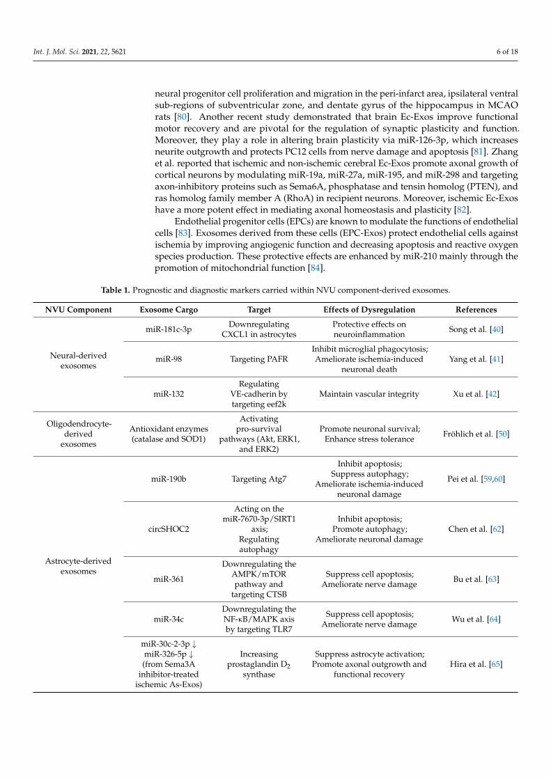
Table 1. Prognostic and diagnostic markers carried within NVU component-derived exosomes.
NVU Component Exosome Cargo Target Effects of Dysregulation References
Neural-derivedexosomes
miR-181c-3p DownregulatingCXCL1 in astrocytes
Protective effects onneuroinflammation Song et al. [40]
miR-98 Targeting PAFRInhibit microglial phagocytosis;Ameliorate ischemia-induced
neuronal deathYang et al. [41]
miR-132Regulating
VE-cadherin bytargeting eef2k
Maintain vascular integrity Xu et al. [42]
Oligodendrocyte-derived
exosomes
Antioxidant enzymes(catalase and SOD1)
Activatingpro-survival
pathways (Akt, ERK1,and ERK2)
Promote neuronal survival;Enhance stress tolerance Fröhlich et al. [50]
Astrocyte-derivedexosomes
miR-190b Targeting Atg7
Inhibit apoptosis;Suppress autophagy;
Ameliorate ischemia-inducedneuronal damage
Pei et al. [59,60]
circSHOC2
Acting on themiR-7670-3p/SIRT1
axis;Regulatingautophagy
Inhibit apoptosis;Promote autophagy;
Ameliorate neuronal damageChen et al. [62]
miR-361
Downregulating theAMPK/mTORpathway and
targeting CTSB
Suppress cell apoptosis;Ameliorate nerve damage Bu et al. [63]
miR-34cDownregulating theNF-κB/MAPK axisby targeting TLR7
Suppress cell apoptosis;Ameliorate nerve damage Wu et al. [64]
miR-30c-2-3p ↓miR-326-5p ↓(from Sema3A
inhibitor-treatedischemic As-Exos)
Increasingprostaglandin D2
synthase
Suppress astrocyte activation;Promote axonal outgrowth and
functional recoveryHira et al. [65]
Int. J. Mol. Sci. 2021, 22, 5621 7 of 18
Table 1. Cont.
NVU Component Exosome Cargo Target Effects of Dysregulation References
synapsin 1 -
Increase neurite outgrowthand survival;
Modulate neuron–gliainteraction
Wang et al. [66]
miR-92b-3p - Ameliorate ischemia-inducedneuronal apoptosis and injury Xu et al. [70]
Microglia-derivedexosomes
miR-124 fromMi2-Exos)
Targeting USP14Attenuate ischemic brain injury,
neural deficits, apoptosis;Promote neuronal survival
Song et al. [73]
DownregulatingSTAT3
Reduce glial scar formation;Improve post-stroke recovery;
Inhibit the migration andproliferation of astrocytes
Li et al. [74]
Increasing Sox2,decreasing Notch1
expression
Promote astrocyte-to-neuralprogenitor cell transition
miRNA-26a (fromIL-4 polarized
Mi-Exos)- Promote angiogenesis Tian et al. [75]
miR-424-5pRegulating theFGF2/STAT3
pathway
Induce cell damage andpermeability of BMECs Xie et al. [76]
Endothelialcell-derivedexosomes
miR-126-3p -Increase neurite outgrowth;
Protect PC12 cells from nervedamage and apoptosis
Gao et al. [81]
miR-27amiR-19amiR-298miR-195
Targeting Sema6A,PTEN, and RhoA
Promote the axonal growth ofcortical neurons Zhang et al. [82]
miR-126 -
Promote axon, myelin, andvascular density and M2
macrophage polarization indiabetic stroke;
Improve functional andcognitive functional outcomes
Venkat et al. [85]
Endothelialprogenitor
cell-derivedexosomes
miR-210Improving
mitochondrialfunction
Improve angiogenic function;Decrease apoptosis and reactive
oxygen species productionMa et al. [84]
miR-126
Downregulatingcleaved caspase-3;
UpregulatingVEGFR2
Promote neurogenesis andangiogenesis;
Improve neurological functionrecovery
Wang et al. [86]
Abbreviations: CXCL1, chemokine CXC motif ligand 1; PAFR, platelet activating factor receptor; VE-cadherin, vascular endothelialcadherin; eef2k, eukaryotic elongation factor 2 kinase; SOD1, superoxide dismutase 1; ERK1 and ERK2, extracellular signal-regulatedprotein kinases 1 and 2; Atg7, autophagy-related gene 7; SIRT1, sirtuin 1; AMPK, AMP-activated protein kinase; mTOR, mammalian targetof rapamycin; CTSB, cathepsin B; NF-κB, nuclear factor-kappa B; MAPK, mitogen-activated protein kinase; TLR7, toll-like receptor 7;Sema3A, semaphorin 3A; As-Exos, astrocytes-derived exosomes; Mi2-Exos, M2 microglia-derived exosomes; USP14, ubiquitin-specificprotease 14; STAT3, signal transducer and activator of transcription 3; IL-4, interleukin-4; FGF2, basic fibroblast growth factor; BMEC, brainmicrovascular endothelial cell; Sema 6A, semaphorin 6A; PTEN, phosphatase and tensin homolog; RhoA, ras homolog family member A;VEGFR2, vascular endothelial growth factor receptor 2.
The beneficial roles of exosomes have been investigated in diabetic mice. Ec-Exotreatment promoted vascular, myelin, and axonal density in the ischemic boundary zone,as well as M2 macrophage polarization, thereby improving neurological and cognitive
Int. J. Mol. Sci. 2021, 22, 5621 8 of 18
functional outcomes possibly mediated by miR-126 [85]. Enrichment with the same miRin EPC-Exos reduced acute injury by decreasing infarct volume and preserving cerebralmicrovascular density and blood flow, and improved neurological functional recoveryby accelerating neurogenesis and angiogenesis through the downregulation of cleavedcaspase-3 and upregulation of vascular endothelial growth factor receptor 2 (VEGFR2) [86].Moreover, these miR-126-enriched exosomes modulate the protective effects of moder-ate exercise on the brain against ischemia in both acute and chronic stages of ischemicinjury [87].
It was also reported that Ec-Exos from femoral arteries could directly protect neuronsagainst I/R injury by suppressing ischemia-induced cell cycle arrest and apoptosis inSH-SY5Y nerve cells [88].
2.3.2. Pericyte-Derived Exosomes (Pc-Exos)
Pericytes are isolated cells in close functional and anatomical contact with endothelialcells [89]. They exert trophic and neuroprotective activity and promote brain recovery,angiogenesis, and neurogenesis through exosome secretion [35]. These cells are also capableof generating MSCs in the perivascular area of lesioned or inflamed vessels [90]. Althoughhypoxic Pc-Exos have been shown to promote angiogenesis [91], to our best knowledgethere is no relevant information in the literature regarding the possible roles of theseexosomes during ischemic conditions.
3. Preclinical Studies of Mesenchymal Stem Cell-Derived Exosomes (MSC-Exos)
Restorative cell-based therapies, including intravenous administration of MSCs, im-prove functional outcomes after stroke [92]. The use of MSC-Exos as an alternative toMSCs offers several advantages, such as higher safety profile, less tumorigenicity, mini-mized occlusion of the microvascular system, lower immunogenicity, and the ability tocross biological barriers, i.e., the BBB [93]. They are mainly extracted from MSC subtypes,including bone marrow mesenchymal stem cells (BMSCs) and adipose-derived stem cells(ADSCs) [94].
Multiple studies suggested that MSC-Exos could be beneficial to promote post-strokerecovery due to their ability to mediate restorative effects involved in stroke, such asangiogenesis, neurogenesis, white matter restoration, oligodendrogenesis, and axonalsprouting (see also Table 2) [95].
3.1. Bone Marrow Mesenchymal Stem Cell-Derived Exosomes (BMSC-Exos)
Xin et al. demonstrated that intravenous administration of multipotent MSC-Exosimproves functional recovery and neurovascular plasticity in rats subjected to MCAO.This effect was attributed to an exosomal delivery of miR-133b from BMSCs to neuronsand astrocytes, which downregulates the expression of connective tissue growth factor(CTGF) and RhoA, thins the glial scar, and promotes neurite outgrowth [96–98]. Similarly,miR-17-92 cluster-enriched MSC-Exos increase functional recovery and neural plasticityafter stroke possibly by targeting PTEN to activate the phosphatidylinositol-3-kinase(PI3K)/Akt/mTOR/glycogen synthase kinase 3 beta (GSK-3β) signaling pathway [99].Additionally, these MSC-Exos enhance the axonal growth of cortical neurons by activatingthe same PTEN/mTOR signaling pathway [100].
Zhao et al. examined the anti-inflammatory effects of BMSC-Exos in acute brainischemia. These exosomes suppressed cysteinyl leukotriene receptor 2 (CysLT2R)-ERK1/2-mediated M1 microglial polarization, promoted microglial conversion into M2 phenotype,increased secretion of anti-inflammatory molecules, and decreased production of pro-inflammatory cytokines, thereby markedly attenuating ischemic brain injury in MCAOrats [101]. The same research group reported that miR-233-3p derived from these exosomespromotes neurological deficits by improving learning and memorizing abilities, and at-tenuates cerebral ischemic injury through the inhibition of pro-inflammatory responsesregulated by M1 microglial polarization via the targeting of CysLT2R [102]. Recently, Liu
Int. J. Mol. Sci. 2021, 22, 5621 9 of 18
et al. reported that BMSC-Exos ameliorate cerebral ischemic injury by suppressing NLRfamily pyrin domain containing 3 (NLRP3) inflammasome-mediated inflammation andpyroptosis via the modulation of microglial polarization [103]. Furthermore, exosomal miR-138-5p derived from BMSCs reduces post-ischemic neurological impairment by inhibitinginflammatory responses and promoting the proliferation of astrocytes through the negativeregulation of lipocalin 2 (LPCN2) [104]. In addition, miR-134-enriched BMSC-Exos sup-press oligodendrocyte apoptosis by downregulating caspase-8 after OGD treatment [105].Pan et al. revealed that the combination of miR-132-3p and MSC-Exos had beneficial effectson ameliorating ischemic brain injury. MiR-132-3p-enriched MSC-Exos protected endothe-lial cells from ischemia-induced apoptosis, oxidative stress, and tight junction disruptionthrough the repression of protein p120 Ras GTPase-activating protein (RASA1) expressionand activation of the Ras/PI3K/Akt/endothelial nitric oxide synthase (eNOS) signalingpathway [106]. Another recent study analyzed whether preconditioning of lithium-inducedMSCs modifies exosome secretion patterns. Due to lithium treatment, MSC-Exos displayedincreased levels of miR-1906, which inhibited the TLR4 and familiar proinflammatory sig-naling cascades, thereby enhancing neuroregeneration, neuroprotection, and neurologicalrecovery in ischemic mice [107].
Doeppner et al. studied the therapeutic effects of BMSC-Exos compared with BMSCsafter cerebral ischemia in mice, demonstrating that exosomes are not inferior to MSCs.They promote neuroregeneration and recovery, modulate peripheral immune responses,and induce long-term neuroprotection [108]. Moreover, Moon et al. revealed that treatmentwith BMSC-Exos was superior to that with BMSCs themselves. These exosomes containedvarious miRs essential for neurogenesis and angiogenesis, such as miR-184 and miR-210 [109]. It was also reported that administration of BMSC-Exos promoted recovery offine motor function of the hand in monkeys after cortical injury, with a return to pre-injury levels within the first 3–5 weeks of recovery [110]. Moreover, Go et al. suggestedthat EVs derived from MSCs reduce neuroinflammation and enhance functional recoveryafter cortical injury in aged Rhesus monkeys by shifting microglia towards restorativefunctions [111].
Safakheil et al. examined the effect of BMSC-Exos in combination with rosuvastatinin MCAO rats. This combination therapy reduced cell death and neuroinflammation andpromoted neuroprotection, thereby enhancing functional recovery after stroke [112].
3.2. Adipose-Derived Stem Cell-Derived Exosomes (ADSC-Exos)
It has been reported that miR-30d-5p and miR-126 levels are decreased in both pa-tients and animal models of ischemic stroke [113,114]. ADSC-Exos enriched with miR-30d-5p prevent cerebral ischemic injury by suppressing autophagy and promoting M2microglial/macrophage polarization [113]. Moreover, miR-126-overexpressing exosomesenhance neurogenesis and angiogenesis by increasing doublecortin and Von Willebrandfactor levels and suppress microglial activation and inflammatory response induced byischemia, thereby improving functional recovery [114].
Exosomal miR-181-5p derived from ADSCs promotes the angiogenesis of BMECs viathe targeting of transient receptor potential melastatin 7 (TRPM7) after OGD in rats. Further-more, these exosomes upregulate the expression of hypoxia-inducible factor 1α (HIF-1α)and VEGF and downregulate the expression of tissue inhibitor of metalloproteinase 3(TIMP3) [115]. In addition, ADSCs inhibit apoptosis and inflammation by miR-21-3psuppression, which contributes to methionine adenosyltransferase 2B (MAT2B) upregula-tion, possibly mediated by exosomes [116]. Moreover, pigment epithelium-derived factor(PEDF)-overexpressing ADSCs-Exos also suppress apoptosis and activate autophagy toameliorate cerebral ischemia [117]. Human ADSC-Exos increase the survival and prolifera-tion of immortalized HT-22 hippocampal neuronal cells after brain injury [118].

Int. J. Mol. Sci. 2021, 22, 5621 10 of 18
Table 2. Therapeutic potential of mesenchymal stem cell-derived exosomes.
MSC-Exos Exosome Cargo Target Effects of Dysregulation References
Bone marrowmesenchymal
stem cell-derivedexosomes
miR-133b
Downregulating CTGFand RhoA
Improve functional recovery andneurovascular plasticity;
Thin the glial scar;Promote neurite outgrowth
Xin et al. [96–98]
Downregulating RABEPK
Secondary release of As-Exos;Enhance post-stroke neurologicalrecovery, neurite outgrowth, and
plasticity
Xin et al. [67]
miR-17-92 cluster
Activating thePI3K/Akt/mTOR/GSK-3β pathway by targeting
PTEN
Improve functional recovery andneural plasticity Xin et al. [99]
Activating thePTEN/mTOR pathway Enhance axonal growth Zhang et al. [100]
miR-233-3p Targeting CysLT2R
Inhibit M1 microglialpolarization-mediated
pro-inflammatory response;Improve neurological deficits;
Ameliorate ischemic brain injury
Zhao et al. [102]
miR-138-5p Downregulating LPCN2Promote proliferation and inhibit
apoptosis of astrocytes;Reduce neurological impairment
Deng et al. [104]
miR-134Downregulating thecaspase-8-dependentapoptosis pathway
Suppress the apoptosis ofoligodendrocytes Xiao et al. [105]
miR-132-3p
Repressing RASA1;Activating the
Ras/PI3K/Akt/eNOSpathway
Protect endothelial cells fromischemia-induced apoptosis, oxidative
stress, and tight junction disruptionPan et al. [106]
miR-1906 (fromLi-induced MSCpreconditioning)
Inhibiting TLR4 andproinflammatory
signaling cascades
Enhance neuroregeneration,neuroprotection, and neurological
recoveryHaupt et al. [107]
Adipose-derivedstem cell-derived
exosomes
miR-30d-5p -Promote M2 microglia/macrophage
polarization;Suppress autophagy
Jiang et al. [113]
miR-126 Increase doublecortin andFvW levels
Improve neurogenesis, angiogenesis,and functional recovery after stroke;
Suppress microglial activation;Inhibit neuroinflammation
Geng et al. [114]
miR-181-5p
Targeting TRPM7;Upregulating HIF-1α and
VEGF;Downregulating TIMP3
Promote the angiogenesis of brainmicrovascular endothelial cells Yang et al. [115]
miR-21-3p(suppressing) Upregulating MAT2B Suppress apoptosis and inflammation
in neurons Li et al. [116]
miR-22-3pInhibiting
KDM6B-mediatedBMP2/BMF axis
Attenuate apoptosis and ischemic braininjury Zhang et al. [119]
Abbreviations: MSC-Exos, mesenchymal stem cells-derived exosomes; CTGF, connective tissue growth factor; RhoA, Ras homolog familymember A; RABEPK, Rab9 effector protein with Kelch motifs; As-Exos, astrocytes-derived exosomes; PI3K, phosphatidylinositol-3-kinase;mTOR, mammalian target of rapamycin; GSK-3β, glycogen synthase kinase 3 beta; PTEN, phosphatase and tensin homolog; CysLT2R,cysteinyl leukotriene receptor 2; LPCN2, lipocalin 2; RASA1, protein p120 Ras GTPase-activating protein; eNOS, endothelial nitricoxide synthase; Li, lithium; MSC, mesenchymal stem cell; TLR4, toll-like receptor 4; FvW, von Willebrand factor; TRPM7, transientreceptor potential melastatin 7; HIF-1 α, hypoxia-inducible factor 1α; VEGF, Vascular endothelial growth factor; TIMP3, tissue inhibitor ofmetalloproteinase 3; MAT2B, methionine adenosyltransferase 2B; KDM6B, lysine demethylase 6B; BMP2, bone morphogenic protein 2;BMF, Bcl-2 modifying factor.
Int. J. Mol. Sci. 2021, 22, 5621 11 of 18
Recently, Zhang et al. reported that miR-22-3p derived from ADSC-Exos attenuatesapoptosis and brain ischemia by inhibiting the lysine demethylase 6B (KDM6B)-mediatedbone morphogenic protein 2 (BMP2)/Bcl-2 modifying factor (BMF) axis in rats after I/Rinjury [119].
4. Exosomes in Stroke Patients
Multiple studies have investigated the diagnostic and prognostic role of exosomesand their cargos in acute ischemic stroke (AIS) patients [11]. One of the most commonexosomal contents are miRs, and each of them have a distinct role in the various molecularpathways involved in stroke.
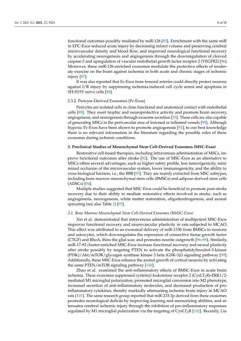
Ji et al. found that serum levels of exosomal miR-9 and miR-124 are elevated inAIS patients: both of these miRs were correlated to National Institutes of Health StrokeScale (NIHSS) scores, but also to infarct volume and serum concentrations of interleukin-6(IL-6) [120]. These miRs are highly expressed in the CNS and enhance neurogenesis [121,122];furthermore, they are promising biomarkers to evaluate the degree of damage causedby ischemic injury [120]. Another exosomal miR that is upregulated in AIS patientsand correlates with stroke severity is miR-223. Chen et al. revealed that miR-223 wasalso associated with stroke occurrence and poorer short-term prognosis [123]. MiR-134is a brain-specific, neuroprotective miR that plays an important role in dendritic spinegrowth control [124], neuritogenesis [125], and in memory and plasticity [126]. Zhoue et al.reported a significant increase in exosomal levels of miR-134 in AIS patients within 24 hof stroke onset: these levels were associated with infarct volume, NIHSS scores, andworse post-stroke prognosis, and additionally with the expression of serum IL-6 andplasma high-sensitivity C reactive protein (hs-CRP) [127]. In contrast, serum levels ofexosomal miR-152-3p were significantly lower in AIS patients, especially in those withNIHSS scores ≥7. Furthermore, the lowest levels of miR-152-3p were found in cases oflarge-artery atherosclerosis, and these levels were significantly lower in the acute phasethan in the chronic phase of stroke [128]. Taken together, miR-9, miR-124, miR-134, miR-152-3p, and miR-223 are associated with the severity of stroke; miR-134 and miR-223 withpoor prognosis; miR-9, miR-124, and miR-134 with the infarct volume and the level of IL-6(see Figure 1).
Int. J. Mol. Sci. 2021, 22, 5621 12 of 19
Figure 1. Exosomes as possible biomarkers in human studies. All the miRs listed in the central blue rectangle correlate to the NIHSS score. The three miRs listed in the orange circle correlate to infarct volume and serum concentrations of IL-6. The miRs in the green triangle are associated with poor prognosis. Nevertheless, all the elements representing clinical and paraclinical findings found around the blue rectangle correlate with each other.
Clinical assessments conducted on cell-free therapeutics remain extremely limited; however, there are plentiful in vitro and animal experiments exploring these therapies. MiR-124-overexpressing MSCs are among the rare exosome-based stroke curatives that have moved to phase I/II clinical trials (NCT03384433) [132].
Besides miRs, it has been demonstrated that exosomes released into the circulation after stroke can contain pro-inflammatory proteins, including CRP [133].
5. Future Directions and Conclusions The NVU is a conceptual model involving neurons, glial cells, and brain vessels, hav-
ing each of them a distinct contribution to the overall function of this structure. Exosomes are a newly defined way of interaction between these components due to their involve-ment in intercellular communication. Numerous studies discuss the role of these EVs se-creted by the constituents of the NVU, however, these are almost exclusively involving animal research and are not supported by clinical evaluation.
Ischemic stroke is a major cause of morbidity and mortality worldwide. Despite ad-vances in understanding the underlying pathophysiology, reperfusion is the only imme-diate treatment option for AIS patients. This justifies the unmet clinical need for further studies aimed to develop new therapeutics in this field. Recent advances in experimental approaches of drug discovery, including high-throughput screening [134] and computer-aided drug design [135], can provide novel insights into the identification and validation of potential therapeutic targets not only in various neurodegenerative diseases, but also in stroke. Moreover, these techniques can also serve as a basis for the target validation of exosomes in future studies.
Apart from the above-mentioned, in vitro models that can precisely simulate the NVU can help us to expand our understanding of the complex interactions between all components of this unit during ischemic stroke. Microfluidic organ-on-a-chip models, such as perfused BBB on-a-chip [136] and human brain microvessel-on-a-chip [137], have been demonstrated to be amenable for optical advanced imaging, which makes them use-ful in studying molecular transport mechanisms involved in the transcytosis of nanopar-ticles, viruses, or biologicals across the BBB. These platforms enable real-time monitoring
Figure 1. Exosomes as possible biomarkers in human studies. All the miRs listed in the central bluerectangle correlate to the NIHSS score. The three miRs listed in the orange circle correlate to infarctvolume and serum concentrations of IL-6. The miRs in the green triangle are associated with poorprognosis. Nevertheless, all the elements representing clinical and paraclinical findings found aroundthe blue rectangle correlate with each other.
Int. J. Mol. Sci. 2021, 22, 5621 12 of 18
Two additional miRs that can be found in the exosomes of stroke patients are miR-21-5p and miR-30-5p. These are apoptosis-related miRs that can distinguish the hyperacutephase of ischemic stroke from the subacute and recovery phases [129]. Moreover, plasmaexosomal miR-422a and miR-125b-2-3p were also demonstrated as monitoring and di-agnostic markers of stroke patients, with the assertion that the combined use of thesetwo may be powerful for determining stroke stage [130]. Progression of asymptomaticcarotid artery stenosis with >50% luminal narrowing is considered a potential risk factorfor stroke or transient ischemic attack during follow-up. Dolz et al. reported significantlyhigher expression of exosomal miR-199b-3p, miR-27b-3p, miR-130a-3p, miR-221-3p, andmiR-24-3p in these patients [131].
Clinical assessments conducted on cell-free therapeutics remain extremely limited;however, there are plentiful in vitro and animal experiments exploring these therapies.MiR-124-overexpressing MSCs are among the rare exosome-based stroke curatives thathave moved to phase I/II clinical trials (NCT03384433) [132].
Besides miRs, it has been demonstrated that exosomes released into the circulationafter stroke can contain pro-inflammatory proteins, including CRP [133].
5. Future Directions and Conclusions
The NVU is a conceptual model involving neurons, glial cells, and brain vessels, hav-ing each of them a distinct contribution to the overall function of this structure. Exosomesare a newly defined way of interaction between these components due to their involvementin intercellular communication. Numerous studies discuss the role of these EVs secretedby the constituents of the NVU, however, these are almost exclusively involving animalresearch and are not supported by clinical evaluation.
Ischemic stroke is a major cause of morbidity and mortality worldwide. Despiteadvances in understanding the underlying pathophysiology, reperfusion is the only imme-diate treatment option for AIS patients. This justifies the unmet clinical need for furtherstudies aimed to develop new therapeutics in this field. Recent advances in experimentalapproaches of drug discovery, including high-throughput screening [134] and computer-aided drug design [135], can provide novel insights into the identification and validationof potential therapeutic targets not only in various neurodegenerative diseases, but alsoin stroke. Moreover, these techniques can also serve as a basis for the target validation ofexosomes in future studies.
Apart from the above-mentioned, in vitro models that can precisely simulate theNVU can help us to expand our understanding of the complex interactions between allcomponents of this unit during ischemic stroke. Microfluidic organ-on-a-chip models, suchas perfused BBB on-a-chip [136] and human brain microvessel-on-a-chip [137], have beendemonstrated to be amenable for optical advanced imaging, which makes them useful instudying molecular transport mechanisms involved in the transcytosis of nanoparticles,viruses, or biologicals across the BBB. These platforms enable real-time monitoring ofpermeability changes, thereby offering an opportunity to examine the exosome’s releaseand penetration during stroke, as well as ischemia-induced neuroinflammation. Further-more, 3D cultures and organoids are also capable to mimic BBB dysfunction thus might bepotentially used in stroke modeling and therapy development [138]. There is recent pre-liminary preclinical evidence that cerebral organoid transplantation might be an effectiveintervention for stroke treatment [139].
Early stroke diagnosis and prognosis prediction are often challenging. Brain ischemiais a heterogeneous process that cannot be characterized by a single biomarker. Therefore,diagnostic panels should be composed of multiple biomarkers representing distinct patho-physiological processes, including inflammatory response, BBB disintegration and brainedema, necrotic and apoptotic cell death, oxidative stress, and thrombosis [33].
A better understanding of the pathological aspects of exosomes and their cargoswill contribute to stroke diagnosis, outcome prediction, and therapy, and thus to theimprovement of patient care. In the future, complex diagnostic stroke protocols should
Int. J. Mol. Sci. 2021, 22, 5621 13 of 18
include not only clinical and neuroradiological findings, but also biomarkers representativeof all elements of the NVU.
Author Contributions: Conceptualization, T.F., Z.B., and R.B.; methodology, T.F. and R.B.; formalanalysis, T.F. and R.B.; data curation, T.F., Z.B., and A.B.; visualization, T.F.; writing—original draftpreparation, T.F.; writing—review and editing, T.F., Z.B., and R.B.; supervision, A.B. and R.B.; projectadministration, A.B.; funding acquisition, A.B. All authors have read and agreed to the publishedversion of the manuscript.
Funding: This work was supported by the University of Medicine, Pharmacy, Science, and Technol-ogy ‘George Emil Palade’ of Târgu Mures, Research Grant number 10126/2/17.12.2020.
Institutional Review Board Statement: Not applicable.
Informed Consent Statement: Not applicable.
Data Availability Statement: Not applicable.
Acknowledgments: We thank Ádám Dénes, head of the Momentum Laboratory of Neuroimmunol-ogy at the Institute of Experimental Medicine of the Hungarian Academy of Sciences (MTA KOKI),for providing scientific guidance and comments on the article.
Conflicts of Interest: The authors declare no conflict of interest.
References1. Katan, M.; Luft, A. Global burden of stroke. Semin. Neurol. 2018, 38, 208–211. [CrossRef]2. Sacco, R.L.; Kasner, S.E.; Broderick, J.P.; Caplan, L.R.; Connors, J.J.; Culebras, A.; Elkind, M.S.; George, M.G.; Hamdan, A.D.;
Higashida, R.T.; et al. An updated definition of stroke for the 21st century: A statement for healthcare professionals from theAmerican Heart Association/American Stroke Association. Stroke 2013, 44, 2064–2089. [CrossRef]
3. World Health Organisation. Neurological Disorders: Public Health Challenges; WHO Press: Geneva, Switzerland, 2006; pp. 151–163.4. Kamel, H.; Iadecola, C. Brain-immune interactions and ischemic stroke: Clinical implications. Arch. Neurol. 2012, 69, 576–581.
[CrossRef]5. Ramos-Cabrer, P.; Campos, F.; Sobrino, T.; Castillo, J. Targeting the ischemic penumbra. Stroke 2011, 42, S7–S11. [CrossRef]6. Chamorro, Á.; Dirnagl, U.; Urra, X.; Planas, A.M. Neuroprotection in acute stroke: Targeting excitotoxicity, oxidative and
nitrosative stress, and inflammation. Lancet Neurol. 2016, 15, 869–881. [CrossRef]7. Berge, E.; Whiteley, W.; Audebert, H.; De Marchis, G.M.; Fonseca, A.C.; Padiglioni, C.; de la Ossa, N.P.; Strbian, D.; Tsivgoulis, G.;
Turc, G. European Stroke Organisation (ESO) guidelines on intravenous thrombolysis for acute ischaemic stroke. Eur. Stroke J.2021, 6, I–LXII. [CrossRef]
8. Turc, G.; Bhogal, P.; Fischer, U.; Khatri, P.; Lobotesis, K.; Mazighi, M.; Schellinger, P.D.; Toni, D.; de Vries, J.; White, P.; et al.European stroke organisation (ESO)—European society for minimally invasive neurological therapy (ESMINT) guidelines onmechanical thrombectomy in acute ischaemic strokeendorsed by stroke alliance for Europe (SAFE). Eur. Stroke J. 2019, 4, 6–12.[CrossRef] [PubMed]
9. Appelros, P.; Nydevik, I.; Viitanen, M. Poor outcome after first-ever stroke: Predictors for death, dependency, and recurrent strokewithin the first year. Stroke 2003, 34, 122–126. [CrossRef] [PubMed]
10. Bustamante, A.; García-Berrocoso, T.; Rodriguez, N.; Llombart, V.; Ribó, M.; Molina, C.; Montaner, J. Ischemic stroke outcome:A review of the influence of post-stroke complications within the different scenarios of stroke care. Eur. J. Intern. Med. 2016,29, 9–21. [CrossRef] [PubMed]
11. Jafarzadeh-Esfehani, R.; Soudyab, M.; Parizadeh, S.M.; Jaripoor, M.E.; Nejad, P.S.; Shariati, M.; Nabavi, A.S. Circulating exosomesand their role in stroke. Curr. Drug Targets 2020, 21, 89–95. [CrossRef] [PubMed]
12. Kalra, H.; Drummen, G.P.; Mathivanan, S. Focus on extracellular vesicles: Introducing the next small big thing. Int. J. Mol. Sci.2016, 17, 170. [CrossRef]
13. Théry, C.; Zitvogel, L.; Amigorena, S. Exosomes: Composition, biogenesis and function. Nat. Rev. Immunol. 2002, 2, 569–579.[CrossRef]
14. Xia, X.; Wang, Y.; Huang, Y.; Zhang, H.; Lu, H.; Zheng, J.C. Exosomal miRNAs in central nervous system diseases: Biomarkers,pathological mediators, protective factors and therapeutic agents. Prog. Neurobiol. 2019, 183, 101694. [CrossRef]
15. Yáñez-Mó, M.; Siljander, P.R.; Andreu, Z.; Zavec, A.B.; Borràs, F.E.; Buzas, E.I.; Buzas, K.; Casal, E.; Cappello, F.; Carvalho, J.; et al.Biological properties of extracellular vesicles and their physiological functions. J. Extracell. Vesicles 2015, 4, 27066. [CrossRef][PubMed]
16. Zhang, Z.G.; Chopp, M. Exosomes in stroke pathogenesis and therapy. J. Clin. Investig. 2016, 126, 1190–1197. [CrossRef]17. Johnsen, K.B.; Gudbergsson, J.M.; Skov, M.N.; Pilgaard, L.; Moos, T.; Duroux, M. A comprehensive overview of exosomes as drug
delivery vehicles—Endogenous nanocarriers for targeted cancer therapy. Biochim. Biophys. Acta 2014, 1846, 75–87. [CrossRef]
Int. J. Mol. Sci. 2021, 22, 5621 14 of 18
18. Bălas, a, A.; S, erban, G.; Chinezu, R.; Hurghis, , C.; Tămas, , F.; Manu, D. The involvement of exosomes in glioblastoma development,diagnosis, prognosis, and treatment. Brain Sci. 2020, 10, 553. [CrossRef]
19. Kalra, H.; Simpson, R.J.; Ji, H.; Aikawa, E.; Altevogt, P.; Askenase, P.; Bond, V.C.; Borràs, F.E.; Breakefield, X.; Budnik, V.; et al.Vesiclepedia: A compendium for extracellular vesicles with continuous community annotation. PLoS Biol. 2012, 10, e1001450.[CrossRef]
20. Kim, D.K.; Lee, J.; Simpson, R.J.; Lötvall, J.; Gho, Y.S. EVpedia: A community web resource for prokaryotic and eukaryoticextracellular vesicles research. Semin. Cell Dev. Biol. 2015, 40, 4–7. [CrossRef]
21. Kim, D.K.; Kang, B.; Kim, O.Y.; Choi, D.S.; Lee, J.; Kim, S.R.; Go, G.; Yoon, Y.J.; Kim, J.H.; Jang, S.C.; et al. EVpedia: An integrateddatabase of high-throughput data for systemic analyses of extracellular vesicles. J. Extracell. Vesicles 2013, 2, 20384. [CrossRef]
22. Mathivanan, S.; Simpson, R.J. ExoCarta: A compendium of exosomal proteins and RNA. Proteomics 2009, 9, 4997–5000. [CrossRef][PubMed]
23. Mathivanan, S.; Fahner, C.J.; Reid, G.E.; Simpson, R.J. ExoCarta 2012: Database of exosomal proteins, RNA and lipids. NucleicAcids Res. 2012, 40, D1241–D1244. [CrossRef] [PubMed]
24. Kalani, A.; Tyagi, A.; Tyagi, N. Exosomes: Mediators of neurodegeneration, neuroprotection and therapeutics. Mol. Neurobiol.2014, 49, 590–600. [CrossRef]
25. Mincheva-Nilsson, L.; Baranov, V.; Nagaeva, O.; Dehlin, E. Isolation and characterization of exosomes from cultures of tissueexplants and cell lines. Curr. Protoc. Immunol. 2016, 115, 14.42.1–14.42.21. [CrossRef]
26. Witwer, K.W.; Buzás, E.I.; Bemis, L.T.; Bora, A.; Lässer, C.; Lötvall, J.; Nolte-’t Hoen, E.N.; Piper, M.G.; Sivaraman, S.; Skog, J.; et al.Standardization of sample collection, isolation and analysis methods in extracellular vesicle research. J. Extracell. Vesicles 2013,2, 20360. [CrossRef]
27. Valadi, H.; Ekstrom, K.; Bossios, A.; Sjostrand, M.; Lee, J.J.; Lotvall, J.O. Exosome-mediated transfer of mRNAs and microRNAs isa novel mechanism of genetic exchange between cells. Nat. Cell Biol. 2007, 9, 654–659. [CrossRef]
28. Ozaki, T.; Nakamura, H.; Kishima, H. Therapeutic strategy against ischemic stroke with the concept of neurovascular unit.Neurochem. Int. 2019, 126, 246–251. [CrossRef]
29. Potjewyd, G.; Moxon, S.; Wang, T.; Domingos, M.; Hooper, N.M. Tissue engineering 3D neurovascular units: A biomaterials andbioprinting perspective. Trends Biotechnol. 2018, 36, 457–472. [CrossRef]
30. Posada-Duque, R.A.; Barreto, G.E.; Cardona-Gomez, G.P. Protection after stroke: Cellular effectors of neurovascular unit integrity.Front. Cell Neurosci. 2014, 8, 231. [CrossRef]
31. Mukandala, G.; Tynan, R.; Lanigan, S.; O’Connor, J.J. The effects of hypoxia and inflammation on synaptic signaling in the CNS.Brain Sci. 2016, 6, 6. [CrossRef]
32. Xu, S.; Lu, J.; Shao, A.; Zhang, J.H.; Zhang, J. Glial cells: Role of the immune response in ischemic stroke. Front. Immunol. 2020,11, 294. [CrossRef]
33. Steliga, A.; Kowianski, P.; Czuba, E.; Waskow, M.; Morys, J.; Lietzau, G. Neurovascular unit as a source of ischemic strokebiomarkers-limitations of experimental studies and perspectives for clinical application. Transl. Stroke Res. 2020, 11, 553–579.[CrossRef]
34. Wang, L.; Xiong, X.; Zhang, L.; Shen, J. Neurovascular Unit: A critical role in ischemic stroke. CNS Neurosci. Ther. 2021, 27, 7–16.[CrossRef]
35. Zagrean, A.M.; Hermann, D.M.; Opris, I.; Zagrean, L.; Popa-Wagner, A. Multicellular crosstalk between exosomes and theneurovascular unit after cerebral ischemia. Therapeutic implications. Front. Neurosci. 2018, 12, 811. [CrossRef]
36. Holm, M.M.; Kaiser, J.; Schwab, M.E. Extracellular vesicles: Multimodal envoys in neural maintenance and repair. Trends Neurosci.2018, 41, 360–372. [CrossRef] [PubMed]
37. Brenna, S.; Altmeppen, H.C.; Mohammadi, B.; Rissiek, B.; Schlink, F.; Ludewig, P.; Krisp, C.; Schlüter, H.; Failla, A.V.; Schneider,C.; et al. Characterization of brain-derived extracellular vesicles reveals changes in cellular origin after stroke and enrichment ofthe prion protein with a potential role in cellular uptake. J. Extracell. Vesicles 2020, 9, 1809065. [CrossRef] [PubMed]
38. Fauré, J.; Lachenal, G.; Court, M.; Hirrlinger, J.; Chatellard-Causse, C.; Blot, B.; Grange, J.; Schoehn, G.; Goldberg, Y.; Boyer, V.;et al. Exosomes are released by cultured cortical neurones. Mol. Cell Neurosci. 2006, 31, 642–648. [CrossRef]
39. Lachenal, G.; Pernet-Gallay, K.; Chivet, M.; Hemming, F.J.; Belly, A.; Bodon, G.; Blot, B.; Haase, G.; Goldberg, Y.; Sadoul, R.Release of exosomes from differentiated neurons and its regulation by synaptic glutamatergic activity. Mol. Cell Neurosci. 2011,46, 409–418. [CrossRef] [PubMed]
40. Song, H.; Zhang, X.; Chen, R.; Miao, J.; Wang, L.; Cui, L.; Ji, H.; Liu, Y. Cortical neuron-derived exosomal MicroRNA-181c-3p inhibits neuroinflammation by downregulating CXCL1 in astrocytes of a rat model with ischemic brain injury.Neuroimmunomodulation 2019, 26, 217–233. [CrossRef]
41. Yang, J.; Cao, L.L.; Wang, X.P.; Guo, W.; Guo, R.B.; Sun, Y.Q.; Xue, T.F.; Cai, Z.Y.; Ji, J.; Cheng, H.; et al. Neuronal extracellularvesicle derived miR-98 prevents salvageable neurons from microglial phagocytosis in acute ischemic stroke. Cell Death Dis. 2021,12, 23. [CrossRef] [PubMed]
42. Xu, B.; Zhang, Y.; Du, X.F.; Li, J.; Zi, H.X.; Bu, J.W.; Yan, Y.; Han, H.; Du, J.L. Neurons secrete miR-132-containing exosomes toregulate brain vascular integrity. Cell Res. 2017, 27, 882–897. [CrossRef]
43. Marsh, S.E.; Blurton-Jones, M. Neural stem cell therapy for neurodegenerative disorders: The role of neurotrophic support.Neurochem. Int. 2017, 106, 94–100. [CrossRef] [PubMed]
Int. J. Mol. Sci. 2021, 22, 5621 15 of 18
44. Sun, X.; Jung, J.H.; Arvola, O.; Santoso, M.R.; Giffard, R.G.; Yang, P.C.; Stary, C.M. Stem cell-derived exosomes protect astrocytecultures from in vitro ischemia and decrease injury as post-stroke intravenous therapy. Front. Cell Neurosci. 2019, 13, 394.[CrossRef] [PubMed]
45. Mahdavipour, M.; Hassanzadeh, G.; Seifali, E.; Mortezaee, K.; Aligholi, H.; Shekari, F.; Sarkoohi, P.; Zeraatpisheh, Z.; Nazari, A.;Movassaghi, S.; et al. Effects of neural stem cell-derived extracellular vesicles on neuronal protection and functional recovery inthe rat model of middle cerebral artery occlusion. Cell Biochem. Funct. 2020, 38, 373–383. [CrossRef]
46. Webb, R.L.; Kaiser, E.E.; Scoville, S.L.; Thompson, T.A.; Fatima, S.; Pandya, C.; Sriram, K.; Swetenburg, R.L.; Vaibhav, K.; Arbab,A.S.; et al. Human neural stem cell extracellular vesicles improve tissue and functional recovery in the murine thromboembolicstroke model. Transl. Stroke Res. 2018, 9, 530–539. [CrossRef] [PubMed]
47. Webb, R.L.; Kaiser, E.E.; Jurgielewicz, B.J.; Spellicy, S.; Scoville, S.L.; Thompson, T.A.; Swetenburg, R.L.; Hess, D.C.; West, F.D.;Stice, S.L. Human neural stem cell extracellular vesicles improve recovery in a porcine model of ischemic stroke. Stroke 2018,49, 1248–1256. [CrossRef] [PubMed]
48. Frühbeis, C.; Fröhlich, D.; Kuo, W.P.; Krämer-Albers, E.M. Extracellular vesicles as mediators of neuron-glia communication.Front. Cell Neurosci. 2013, 7, 182. [CrossRef]
49. Frühbeis, C.; Fröhlich, D.; Kuo, W.P.; Amphornrat, J.; Thilemann, S.; Saab, A.S.; Kirchhoff, F.; Möbius, W.; Goebbels, S.; Nave,K.A.; et al. Neurotransmitter-triggered transfer of exosomes mediates oligodendrocyte-neuron communication. PLoS Biol. 2013,11, e1001604. [CrossRef]
50. Fröhlich, D.; Kuo, W.P.; Frühbeis, C.; Sun, J.J.; Zehendner, C.M.; Luhmann, H.J.; Pinto, S.; Toedling, J.; Trotter, J.; Krämer-Albers,E.M. Multifaceted effects of oligodendroglial exosomes on neurons: Impact on neuronal firing rate, signal transduction and generegulation. Philos. Trans. R. Soc. Lond. B Biol. Sci. 2014, 369, 20130510. [CrossRef]
51. Colombo, E.; Farina, C. Astrocytes: Key regulators of neuroinflammation. Trends Immunol. 2016, 37, 608–620. [CrossRef]52. Zhang, Z.G.; Buller, B.; Chopp, M. Exosomes—Beyond stem cells for restorative therapy in stroke and neurological injury. Nat.
Rev. Neurol. 2019, 15, 193–203. [CrossRef]53. Patabendige, A.; Singh, A.; Jenkins, S.; Sen, J.; Chen, R. Astrocyte activation in neurovascular damage and repair following
ischaemic stroke. Int. J. Mol. Sci. 2021, 22, 4280. [CrossRef] [PubMed]54. Kitchen, P.; Salman, M.M.; Halsey, A.M.; Clarke-Bland, C.; MacDonald, J.A.; Ishida, H.; Vogel, H.J.; Almutiri, S.; Logan, A.;
Kreida, S.; et al. Targeting aquaporin-4 subcellular localization to treat central nervous system edema. Cell 2020, 181, 784–799.e19.[CrossRef]
55. Sylvain, N.J.; Salman, M.M.; Pushie, M.J.; Hou, H.; Meher, V.; Herlo, R.; Peeling, L.; Kelly, M.E. The effects of trifluoperazine onbrain edema, aquaporin-4 expression and metabolic markers during the acute phase of stroke using photothrombotic mousemodel. Biochim. Biophys. Acta Biomembr. 2021, 1863, 183573. [CrossRef]
56. Jovicic, A.; Gitler, A.D. Distinct repertoires of microRNAs present in mouse astrocytes compared to astrocyte-secreted exosomes.PLoS ONE 2017, 12, e0171418. [CrossRef] [PubMed]
57. Datta Chaudhuri, A.; Dasgheyb, R.M.; DeVine, L.R.; Bi, H.; Cole, R.N.; Haughey, N.J. Stimulus-dependent modifications inastrocyte-derived extracellular vesicle cargo regulate neuronal excitability. Glia 2020, 68, 128–144. [CrossRef]
58. Taylor, A.R.; Robinson, M.B.; Gifondorwa, D.J.; Tytell, M.; Milligan, C.E. Regulation of heat shock protein 70 release in astrocytes:Role of signaling kinases. Dev. Neurobiol. 2007, 67, 1815–1829. [CrossRef]
59. Pei, X.; Li, Y.; Zhu, L.; Zhou, Z. Astrocyte-derived exosomes suppress autophagy and ameliorate neuronal damage in experimentalischemic stroke. Exp. Cell Res. 2019, 382, 111474. [CrossRef]
60. Pei, X.; Li, Y.; Zhu, L.; Zhou, Z. Astrocyte-derived exosomes transfer miR-190b to inhibit oxygen and glucose deprivation-inducedautophagy and neuronal apoptosis. Cell Cycle 2020, 19, 906–917. [CrossRef] [PubMed]
61. Kang, C.; Avery, L. To be or not to be, the level of autophagy is the question: Dual roles of autophagy in the survival response tostarvation. Autophagy 2008, 4, 82–84. [CrossRef]
62. Chen, W.; Wang, H.; Zhu, Z.; Feng, J.; Chen, L. Exosome-shuttled circSHOC2 from IPASs regulates neuronal autophagy andameliorates ischemic brain injury via the miR-7670-3p/SIRT1 axis. Mol. Ther. Nucleic Acids 2020, 22, 657–672. [CrossRef] [PubMed]
63. Bu, X.; Li, D.; Wang, F.; Sun, Q.; Zhang, Z. Protective role of astrocyte-derived exosomal microRNA-361 in cerebral ischemic-reperfusion injury by regulating the AMPK/mTOR signaling pathway and targeting CTSB. Neuropsychiatr. Dis. Treat. 2020,16, 1863–1877. [CrossRef]
64. Wu, W.; Liu, J.; Yang, C.; Xu, Z.; Huang, J.; Lin, J. Astrocyte-derived exosome-transported microRNA-34c is neuroprotectiveagainst cerebral ischemia/reperfusion injury via TLR7 and the NF-κB/MAPK pathways. Brain Res. Bull. 2020, 163, 84–94.[CrossRef] [PubMed]
65. Hira, K.; Ueno, Y.; Tanaka, R.; Miyamoto, N.; Yamashiro, K.; Inaba, T.; Urabe, T.; Okano, H.; Hattori, N. Astrocyte-derived exo-somes treated with a semaphorin 3a inhibitor enhance stroke recovery via prostaglandin D2 synthase. Stroke 2018, 49, 2483–2494.[CrossRef]
66. Wang, S.; Cesca, F.; Loers, G.; Schweizer, M.; Buck, F.; Benfenati, F.; Schachner, M.; Kleene, R. Synapsin I is an oligomannose-carrying glycoprotein, acts as an oligomannose-binding lectin, and promotes neurite outgrowth and neuronal survival whenreleased via glia-derived exosomes. J. Neurosci. 2011, 31, 7275–7290. [CrossRef]
Int. J. Mol. Sci. 2021, 22, 5621 16 of 18
67. Xin, H.; Wang, F.; Li, Y.; Lu, Q.E.; Cheung, W.L.; Zhang, Y.; Zhang, Z.G.; Chopp, M. Secondary release of exosomes from astrocytescontributes to the increase in neural plasticity and improvement of functional recovery after stroke in rats treated with exosomesharvested from MicroRNA 133b-overexpressing multipotent mesenchymal stromal cells. Cell Transplant. 2017, 26, 243–257.[CrossRef]
68. Guitart, K.; Loers, G.; Buck, F.; Bork, U.; Schachner, M.; Kleene, R. Improvement of neuronal cell survival by astrocyte-derivedexosomes under hypoxic and ischemic conditions depends on prion protein. Glia 2016, 64, 896–910. [CrossRef]
69. Thushara Vijayakumar, N.; Sangwan, A.; Sharma, B.; Majid, A.; Rajanikant, G.K. Cerebral ischemic preconditioning: The road sofar. Mol. Neurobiol. 2016, 53, 2579–2593. [CrossRef] [PubMed]
70. Xu, L.; Cao, H.; Xie, Y.; Zhang, Y.; Du, M.; Xu, X.; Ye, R.; Liu, X. Exosome-shuttled miR-92b-3p from ischemic preconditionedastrocytes protects neurons against oxygen and glucose deprivation. Brain Res. 2019, 1717, 66–73. [CrossRef]
71. Ma, Y.; Wang, J.; Wang, Y.; Yang, G.Y. The biphasic function of microglia in ischemic stroke. Prog. Neurobiol. 2017, 157, 247–272.[CrossRef] [PubMed]
72. Tang, Y.; Le, W. Differential roles of M1 and M2 microglia in neurodegenerative diseases. Mol. Neurobiol. 2016, 53, 1181–1194.[CrossRef] [PubMed]
73. Song, Y.; Li, Z.; He, T.; Qu, M.; Jiang, L.; Li, W.; Shi, X.; Pan, J.; Zhang, L.; Wang, Y.; et al. M2 microglia-derived exosomes protectthe mouse brain from ischemia-reperfusion injury via exosomal miR-124. Theranostics 2019, 9, 2910–2923. [CrossRef] [PubMed]
74. Li, Z.; Song, Y.; He, T.; Wen, R.; Li, Y.; Chen, T.; Huang, S.; Wang, Y.; Tang, Y.; Shen, F.; et al. M2 microglial small extracellularvesicles reduce glial scar formation via the miR-124/STAT3 pathway after ischemic stroke in mice. Theranostics 2021, 11, 1232–1248.[CrossRef]
75. Tian, Y.; Zhu, P.; Liu, S.; Jin, Z.; Li, D.; Zhao, H.; Zhu, X.; Shu, C.; Yan, D.; Dong, Z. IL-4-polarized BV2 microglia cells promoteangiogenesis by secreting exosomes. Adv. Clin. Exp. Med. 2019, 28, 421–430. [CrossRef]
76. Xie, L.; Zhao, H.; Wang, Y.; Chen, Z. Exosomal shuttled miR-424-5p from ischemic preconditioned microglia mediates cerebralendothelial cell injury through negatively regulation of FGF2/STAT3 pathway. Exp. Neurol. 2020, 333, 113411. [CrossRef][PubMed]
77. Raffaele, S.; Gelosa, P.; Bonfanti, E.; Lombardi, M.; Castiglioni, L.; Cimino, M.; Sironi, L.; Abbracchio, M.P.; Verderio, C.; Fumagalli,M. Microglial vesicles improve post-stroke recovery by preventing immune cell senescence and favouring oligodendrogenesis.Mol. Ther. 2021, 29, 1439–1458. [CrossRef] [PubMed]
78. Yu, Q.J.; Tao, H.; Wang, X.; Li, M.C. Targeting brain microvascular endothelial cells: A therapeutic approach to neuroprotectionagainst stroke. Neural. Regen. Res. 2015, 10, 1882–1891. [CrossRef]
79. Dozio, V.; Sanchez, J.C. Characterisation of extracellular vesicle-subsets derived from brain endothelial cells and analysis of theirprotein cargo modulation after TNF exposure. J. Extracell. Vesicles 2017, 6, 1302705. [CrossRef]
80. Zhou, S.; Gao, B.; Sun, C.; Bai, Y.; Cheng, D.; Zhang, Y.; Li, X.; Zhao, J.; Xu, D. Vascular endothelial cell-derived exosomes protectneural stem cells against ischemia/reperfusion Injury. Neuroscience 2020, 441, 184–196. [CrossRef]
81. Gao, B.; Zhou, S.; Sun, C.; Cheng, D.; Zhang, Y.; Li, X.; Zhang, L.; Zhao, J.; Xu, D.; Bai, Y. Brain endothelial cell-derived exosomesinduce neuroplasticity in rats with ischemia/reperfusion injury. ACS Chem. Neurosci. 2020, 11, 2201–2213. [CrossRef]
82. Zhang, Y.; Qin, Y.; Chopp, M.; Li, C.; Kemper, A.; Liu, X.; Wang, X.; Zhang, L.; Zhang, Z.G. Ischemic cerebral endothelialcell-derived exosomes promote axonal growth. Stroke 2020, 51, 3701–3712. [CrossRef]
83. Werner, N.; Nickenig, G. Endothelial progenitor cells in health and atherosclerotic disease. Ann. Med. 2007, 39, 82–90. [CrossRef]84. Ma, X.; Wang, J.; Li, J.; Ma, C.; Chen, S.; Lei, W.; Yang, Y.; Liu, S.; Bihl, J.; Chen, C. Loading MiR-210 in endothelial progenitor
cells derived exosomes boosts their beneficial effects on hypoxia/reoxygeneation-injured human endothelial cells via protectingmitochondrial function. Cell Physiol. Biochem. 2018, 46, 664–675. [CrossRef]
85. Venkat, P.; Cui, C.; Chopp, M.; Zacharek, A.; Wang, F.; Landschoot-Ward, J.; Shen, Y.; Chen, J. MiR-126 mediates brain endothelialcell exosome treatment-induced neurorestorative effects after stroke in type 2 diabetes mellitus mice. Stroke 2019, 50, 2865–2874.[CrossRef]
86. Wang, J.; Chen, S.; Zhang, W.; Chen, Y.; Bihl, J.C. Exosomes from miRNA-126-modified endothelial progenitor cells alleviate braininjury and promote functional recovery after stroke. CNS Neurosci. Ther. 2020, 26, 1255–1265. [CrossRef]
87. Wang, J.; Liu, H.; Chen, S.; Zhang, W.; Chen, Y.; Yang, Y. Moderate exercise has beneficial effects on mouse ischemic stroke byenhancing the functions of circulating endothelial progenitor cell-derived exosomes. Exp. Neurol. 2020, 330, 113325. [CrossRef]
88. Xiao, B.; Chai, Y.; Lv, S.; Ye, M.; Wu, M.; Xie, L.; Fan, Y.; Zhu, X.; Gao, Z. Endothelial cell-derived exosomes protect SH-SY5Y nervecells against ischemia/reperfusion injury. Int. J. Mol. Med. 2017, 40, 1201–1209. [CrossRef]
89. Muoio, V.; Persson, P.B.; Sendeski, M.M. The neurovascular unit—Concept review. Acta Physiol. 2014, 210, 790–798. [CrossRef]90. Caplan, A.I. MSCs: The sentinel and safe-guards of injury. J. Cell. Physiol. 2016, 231, 1413–1416. [CrossRef]91. Mayo, J.N.; Bearden, S.E. Driving the hypoxia-inducible pathway in human pericytes promotes vascular density in an exosome-
dependent manner. Microcirculation 2015, 22, 711–723. [CrossRef]92. Zhang, Z.G.; Chopp, M. Neurorestorative therapies for stroke: Underlying mechanisms and translation to the clinic. Lancet Neurol.
2009, 8, 491–500. [CrossRef]93. Gowen, A.; Shahjin, F.; Chand, S.; Odegaard, K.E.; Yelamanchili, S.V. Mesenchymal stem cell-derived extracellular vesicles:
Challenges in clinical applications. Front. Cell Dev. Biol. 2020, 8, 149. [CrossRef]
Int. J. Mol. Sci. 2021, 22, 5621 17 of 18
94. Gutiérrez-Fernández, M.; Rodríguez-Frutos, B.; Ramos-Cejudo, J.; Teresa Vallejo-Cremades, M.; Fuentes, B.; Cerdán, S.; Díez-Tejedor, E. Effects of intravenous administration of allogenic bone marrow- and adipose tissue-derived mesenchymal stem cellson functional recovery and brain repair markers in experimental ischemic stroke. Stem Cell Res. Ther. 2013, 4, 11. [CrossRef]
95. Otero-Ortega, L.; Laso-García, F.; Gómez-de Frutos, M.; Fuentes, B.; Diekhorst, L.; Díez-Tejedor, E.; Gutiérrez-Fernández, M. Roleof exosomes as a treatment and potential biomarker for stroke. Transl. Stroke Res. 2019, 10, 241–249. [CrossRef]
96. Xin, H.; Li, Y.; Buller, B.; Katakowski, M.; Zhang, Y.; Wang, X.; Shang, X.; Zhang, Z.G.; Chopp, M. Exosome-mediated transferof miR-133b from multipotent mesenchymal stromal cells to neural cells contributes to neurite outgrowth. Stem Cells 2012,30, 1556–1564. [CrossRef]
97. Xin, H.; Li, Y.; Cui, Y.; Yang, J.J.; Zhang, Z.G.; Chopp, M. Systemic administration of exosomes released from mesenchymalstromal cells promote functional recovery and neurovascular plasticity after stroke in rats. J. Cereb. Blood Flow Metab. 2013,33, 1711–1715. [CrossRef]
98. Xin, H.; Li, Y.; Liu, Z.; Wang, X.; Shang, X.; Cui, Y.; Zhang, Z.G.; Chopp, M. MiR-133b promotes neural plasticity and functional re-covery after treatment of stroke with multipotent mesenchymal stromal cells in rats via transfer of exosome-enriched extracellularparticles. Stem Cells 2013, 31, 2737–2746. [CrossRef]
99. Xin, H.; Katakowski, M.; Wang, F.; Qian, J.Y.; Liu, X.S.; Ali, M.M.; Buller, B.; Zhang, Z.G.; Chopp, M. MicroRNA cluster miR-17-92cluster in exosomes enhance neuroplasticity and functional recovery after stroke in rats. Stroke 2017, 48, 747–753. [CrossRef]
100. Zhang, Y.; Chopp, M.; Liu, X.S.; Katakowski, M.; Wang, X.; Tian, X.; Wu, D.; Zhang, Z.G. Exosomes derived from mesenchymalstromal cells promote axonal growth of cortical neurons. Mol. Neurobiol. 2017, 54, 2659–2673. [CrossRef]
101. Zhao, Y.; Gan, Y.; Xu, G.; Yin, G.; Liu, D. MSCs-Derived exosomes attenuate acute brain injury and inhibit microglial inflammationby reversing CysLT2R-ERK1/2 mediated microglia M1 polarization. Neurochem. Res. 2020, 45, 1180–1190. [CrossRef]
102. Zhao, Y.; Gan, Y.; Xu, G.; Hua, K.; Liu, D. Exosomes from MSCs overexpressing microRNA-223-3p attenuate cerebral ischemiathrough inhibiting microglial M1 polarization mediated inflammation. Life Sci. 2020, 260, 118403. [CrossRef] [PubMed]
103. Liu, X.; Zhang, M.; Liu, H.; Zhu, R.; He, H.; Zhou, Y.; Zhang, Y.; Li, C.; Liang, D.; Zeng, Q.; et al. Bone marrow mesenchymal stemcell-derived exosomes attenuate cerebral ischemia-reperfusion injury-induced neuroinflammation and pyroptosis by modulatingmicroglia M1/M2 phenotypes. Exp. Neurol. 2021, 341, 113700. [CrossRef]
104. Deng, Y.; Chen, D.; Gao, F.; Lv, H.; Zhang, G.; Sun, X.; Liu, L.; Mo, D.; Ma, N.; Song, L.; et al. Exosomes derived from microRNA-138-5p-overexpressing bone marrow-derived mesenchymal stem cells confer neuroprotection to astrocytes following ischemicstroke via inhibition of LCN2. J. Biol. Eng. 2019, 13, 71. [CrossRef]
105. Xiao, Y.; Geng, F.; Wang, G.; Li, X.; Zhu, J.; Zhu, W. Bone marrow-derived mesenchymal stem cells-derived exosomes preventoligodendrocyte apoptosis through exosomal miR-134 by targeting caspase-8. J. Cell. Biochem. 2019, 120, 2109–2118. [CrossRef]
106. Pan, Q.; Kuang, X.; Cai, S.; Wang, X.; Du, D.; Wang, J.; Wang, Y.; Chen, Y.; Bihl, J.; Chen, Y.; et al. miR-132-3p priming enhancesthe effects of mesenchymal stromal cell-derived exosomes on ameliorating brain ischemic injury. Stem Cell Res. Ther. 2020, 11, 260.[CrossRef] [PubMed]
107. Haupt, M.; Zheng, X.; Kuang, Y.; Lieschke, S.; Janssen, L.; Bosche, B.; Jin, F.; Hein, K.; Kilic, E.; Venkataramani, V.; et al. Lithiummodulates miR-1906 levels of mesenchymal stem cell-derived extracellular vesicles contributing to poststroke neuroprotection bytoll-like receptor 4 regulation. Stem Cells Transl. Med. 2021, 10, 357–373. [CrossRef]
108. Doeppner, T.; Herz, J.; Görgens, A.; Schlechter, J.; Ludwig, A.K.; Radtke, S.; de Miroschedji, K.; Horn, P.A.; Giebel, B.; Her-mann, D.M. Extracellular Vesicles Improve Post-Stroke Neuroregeneration and Prevent Postischemic Immunosuppression.Stem Cells Transl. Med. 2015, 4, 1131–1143. [CrossRef]
109. Moon, G.J.; Sung, J.H.; Kim, D.H.; Kim, E.H.; Cho, Y.H.; Son, J.P.; Cha, J.M.; Bang, O.Y. Application of mesenchymal stemcell-derived extracellular vesicles for stroke: Biodistribution and MicroRNA study. Transl. Stroke Res. 2019, 10, 509–521. [CrossRef][PubMed]
110. Moore, T.L.; Bowley, B.G.E.; Pessina, M.A.; Calderazzo, S.M.; Medalla, M.; Go, V.; Zhang, Z.G.; Chopp, M.; Finklestein, S.;Harbaugh, A.G.; et al. Mesenchymal derived exosomes enhance recovery of motor function in a monkey model of cortical injury.Restor. Neurol. Neurosci. 2019, 37, 347–362. [CrossRef]
111. Go, V.; Bowley, B.G.E.; Pessina, M.A.; Zhang, Z.G.; Chopp, M.; Finklestein, S.P.; Rosene, D.L.; Medalla, M.; Buller, B.; Moore, T.L.Extracellular vesicles from mesenchymal stem cells reduce microglial-mediated neuroinflammation after cortical injury in agedRhesus monkeys. GeroScience 2020, 42, 1–17. [CrossRef]
112. Safakheil, M.; Safakheil, H. The effect of exosomes derived from bone marrow stem cells in combination with rosuvastatin onfunctional recovery and neuroprotection in rats after ischemic stroke. J. Mol. Neurosci. 2020, 70, 724–737. [CrossRef]
113. Jiang, M.; Wang, H.; Jin, M.; Yang, X.; Ji, H.; Jiang, Y.; Zhang, H.; Wu, F.; Wu, G.; Lai, X.; et al. Exosomes from MiR-30d-5p-ADSCsreverse acute ischemic stroke-induced, autophagy-mediated brain injury by promoting M2 microglial/macrophage polarization.Cell. Physiol. Biochem. 2018, 47, 864–878. [CrossRef]
114. Geng, W.; Tang, H.; Luo, S.; Lv, Y.; Liang, D.; Kang, X.; Hong, W. Exosomes from miRNA-126-modified ADSCs promotesfunctional recovery after stroke in rats by improving neurogenesis and suppressing microglia activation. Am. J. Transl. Res. 2019,11, 780–792.
115. Yang, Y.; Cai, Y.; Zhang, Y.; Liu, J.; Xu, Z. Exosomes secreted by adipose-derived stem cells contribute to angiogenesis ofbrain microvascular endothelial cells following oxygen-glucose deprivation in vitro through MicroRNA-181b/TRPM7 axis.J. Mol. Neurosci. 2018, 65, 74–83. [CrossRef]
Int. J. Mol. Sci. 2021, 22, 5621 18 of 18
116. Li, C.; Fei, K.; Tian, F.; Gao, C.; Yang, S. Adipose-derived mesenchymal stem cells attenuate ischemic brain injuries in rats bymodulating miR-21-3p/MAT2B signaling transduction. Croat. Med. J. 2019, 60, 439–448. [CrossRef]
117. Huang, X.; Ding, J.; Li, Y.; Liu, W.; Ji, J.; Wang, H.; Wang, X. Exosomes derived from PEDF modified adipose-derived mesenchymalstem cells ameliorate cerebral ischemia-reperfusion injury by regulation of autophagy and apoptosis. Exp. Cell Res. 2018,371, 269–277. [CrossRef] [PubMed]
118. El Bassit, G.; Patel, R.S.; Carter, G.; Shibu, V.; Patel, A.A.; Song, S.; Murr, M.; Cooper, D.R.; Bickford, P.C.; Patel, N.A. MALAT1 inhuman adipose stem cells modulates survival and alternative splicing of PKCδII in HT22 cells. Endocrinology 2017, 158, 183–195.[CrossRef] [PubMed]
119. Zhang, Y.; Liu, J.; Su, M.; Wang, X.; Xie, C. Exosomal microRNA-22-3p alleviates cerebral ischemic injury by modulatingKDM6B/BMP2/BMF axis. Stem Cell Res. Ther. 2021, 12, 111. [CrossRef] [PubMed]
120. Ji, Q.; Ji, Y.; Peng, J.; Zhou, X.; Chen, X.; Zhao, H.; Xu, T.; Chen, L.; Xu, Y. Increased brain-specific MiR-9 and MiR-124 in the serumexosomes of acute ischemic stroke patients. PLoS ONE 2016, 11, e0163645. [CrossRef]
121. Madelaine, R.; Sloan, S.A.; Huber, N.; Notwell, J.H.; Leung, L.C.; Skariah, G.; Halluin, C.; Pasca, S.P.; Bejerano, G.; Krasnow, M.A.;et al. MicroRNA-9 couples brain neurogenesis and angiogenesis. Cell Rep. 2017, 20, 1533–1542. [CrossRef]
122. Sun, Y.; Luo, Z.M.; Guo, X.M.; Su, D.F.; Liu, X. An updated role of microRNA-124 in central nervous system disorders: A review.Front. Cell. Neurosci. 2015, 9, 193. [CrossRef]
123. Chen, Y.; Song, Y.; Huang, J.; Qu, M.; Zhang, Y.; Geng, J.; Zhang, Z.; Liu, J.; Yang, G.Y. Increased circulating exosomal miRNA-223is associated with acute ischemic stroke. Front. Neurol. 2017, 8, 57. [CrossRef]
124. Schratt, G.M.; Tuebing, F.; Nigh, E.A.; Kane, C.G.; Sabatini, M.E.; Kiebler, M.; Greenberg, M.E. A brain-specific microRNAregulates dendritic spine development. Nature 2006, 439, 283–289. [CrossRef] [PubMed]
125. Coolen, M.; Bally-Cuif, L. MicroRNAs in brain development and physiology. Curr. Opin. Neurobiol. 2009, 19, 461–470. [CrossRef]126. Gao, J.; Wang, W.Y.; Mao, Y.W.; Gräff, J.; Guan, J.S.; Pan, L.; Mak, G.; Kim, D.; Su, S.C.; Tsai, L.H. A novel pathway regulates
memory and plasticity via SIRT1 and miR-134. Nature 2010, 466, 1105–1109. [CrossRef]127. Zhou, J.; Chen, L.; Chen, B.; Huang, S.; Zeng, C.; Wu, H.; Chen, C.; Long, F. Increased serum exosomal miR-134 expression in the
acute ischemic stroke patients. BMC Neurol. 2018, 18, 198. [CrossRef]128. Song, P.; Sun, H.; Chen, H.; Wang, Y.; Zhang, Q. Decreased serum exosomal miR-152-3p contributes to the progression of acute
ischemic stroke. Clin. Lab. 2020, 66. [CrossRef]129. Wang, W.; Li, D.B.; Li, R.Y.; Zhou, X.; Yu, D.J.; Lan, X.Y.; Li, J.P.; Liu, J.L. Diagnosis of hyperacute and acute ischemic stroke: The
potential utility of exosomal MicroRNA-21-5p and MicroRNA-30a-5p. Cerebrovasc. Dis. 2018, 45, 204–212. [CrossRef]130. Li, D.B.; Liu, J.L.; Wang, W.; Li, R.Y.; Yu, D.J.; Lan, X.Y.; Li, J.P. Plasma exosomal miR-422a and miR-125b-2-3p serve as Biomarkers
for ischemic stroke. Curr. Neurovasc. Res. 2017, 14, 330–337. [CrossRef]131. Dolz, S.; Górriz, D.; Tembl, J.I.; Sánchez, D.; Fortea, G.; Parkhutik, V.; Lago, A. Circulating MicroRNAs as novel biomarkers of
stenosis progression in asymptomatic carotid stenosis. Stroke 2017, 48, 10–16. [CrossRef]132. Shahid Beheshti University of Medical Sciences. Safety and Efficacy of Allogenic Mesenchymal Stem Cells Derived Exosome on
Disability of Patients with Acute Ischemic Stroke: A Randomized, Single-blind, Placebo-Controlled, Phase 1, 2 Trial. IdentifierNCT03384433. Available online: https://clinicaltrials.gov/ct2/show/NCT03384433 (accessed on 28 April 2021).
133. Couch, Y.; Akbar, N.; Davis, S.; Fischer, R.; Dickens, A.M.; Neuhaus, A.A.; Burgess, A.I.; Rothwell, P.M.; Buchan, A.M.inflammatory stroke extracellular vesicles induce macrophage activation. Stroke 2017, 48, 2292–2296. [CrossRef]
134. Aldewachi, H.; Al-Zidan, R.N.; Conner, M.T.; Salman, M.M. High-throughput screening platforms in the discovery of noveldrugs for neurodegenerative diseases. Bioengineering 2021, 8, 30. [CrossRef]
135. Salman, M.M.; Al-Obaidi, Z.; Kitchen, P.; Loreto, A.; Bill, R.M.; Wade-Martins, R. Advances in applying computer-aided drugdesign for neurodegenerative diseases. Int. J. Mol. Sci. 2021, 22, 4688. [CrossRef]
136. Wevers, N.R.; Kasi, D.G.; Gray, T.; Wilschut, K.J.; Smith, B.; van Vught, R.; Shimizu, F.; Sano, Y.; Kanda, T.; Marsh, G.; et al.A perfused human blood-brain barrier on-a-chip for high-throughput assessment of barrier function and antibody transport.Fluids Barriers CNS 2018, 15, 23. [CrossRef]
137. Salman, M.M.; Marsh, G.; Kusters, I.; Delincé, M.; Di Caprio, G.; Upadhyayula, S.; de Nola, G.; Hunt, R.; Ohashi, K.G.; Gray, T.;et al. Design and validation of a human brain endothelial microvessel-on-a-chip open microfluidic model enabling advancedoptical imaging. Front. Bioeng. Biotechnol. 2020, 8, 573775. [CrossRef] [PubMed]
138. Nzou, G.; Wicks, R.T.; VanOstrand, N.R.; Mekky, G.A.; Seale, S.A.; El-Taibany, A.; Wicks, E.E.; Nechtman, C.M.; Marrotte, E.J.;Makani, V.S.; et al. Multicellular 3D neurovascular unit model for assessing hypoxia and neuroinflammation induced blood-brainbarrier dysfunction. Sci. Rep. 2020, 10, 9766. [CrossRef]
139. Wang, S.N.; Wang, Z.; Xu, T.Y.; Cheng, M.H.; Li, W.L.; Miao, C.Y. Cerebral organoids repair ischemic stroke brain injury. Transl.Stroke Res. 2020, 11, 983–1000. [CrossRef]


![Imaging in Neurovascular conflicts [Neurovascular compression syndrome ]](https://img.dokumen.tips/doc/110x75/559b6a361a28ab2c188b4611/imaging-in-neurovascular-conflicts-neurovascular-compression-syndrome-.jpg)