Embed Size (px)
Citation preview

The
Jour
nal o
f Cel
l Bio
logy
JCB
©
The Rockefeller University Press, 0021-9525/2004/08/455/9 $8.00The Journal of Cell Biology, Volume 166, Number 4, August 16, 2004 455–463http://www.jcb.org/cgi/doi/10.1083/jcb.200312048 455
Report
Dynamic targeting of the replication machinery to sites of DNA damage
David A. Solomon,
1
M. Cristina Cardoso,
2
and Erik S. Knudsen
1
1
Department of Cell Biology, Vontz Center for Molecular Studies, University of Cincinnati College of Medicine, Cincinnati, OH 45267
2
Max Delbrück Center for Molecular Medicine, D-13125 Berlin, Germany
omponents of the DNA replication machinery local-ize into discrete subnuclear foci after DNA damage,where they play requisite functions in repair pro-
cesses. Here, we find that the replication factors proliferat-ing cell nuclear antigen (PCNA) and RPAp34 dynamicallyexchange at these repair foci with discrete kinetics, andthis behavior is distinct from kinetics during DNA replica-tion. Posttranslational modification is hypothesized to tar-get specific proteins for repair, and we find that accumula-
C
tion and stability of PCNA at sites of damage requiresmonoubiquitination. Contrary to the popular notion thatphosphorylation on the NH
2
terminus of RPAp34 directsthe protein for repair, we demonstrate that phosphorylationby DNA-dependent protein kinase enhances RPAp34 turn-over at repair foci. Together, these findings support a dy-namic exchange model in which multiple repair factorsregulated by specific modifications have access to and rap-idly turn over at sites of DNA damage.
Introduction
The cellular response to DNA damage requires a wide rangeof protein factors to ensure the fidelity of genetic materialpassed on to daughter cells. These factors include detectionand signaling proteins to warn that damage has occurred,checkpoint response proteins to inhibit cell division allowingtime for repair, and the plethora of proteins that function inthe repair of genetic lesions (Rouse and Jackson, 2002).Apart from its function in the replication of the genome,the DNA replication machinery plays an essential role inthe repair of damaged DNA. The heterotrimeric replicationprotein A (RPA) complex has been implicated in the damagedetection process, whereas factors including proliferating cellnuclear antigen (PCNA), DNA polymerases, and DNAligases are known to be important in the resynthesis of ex-cised nucleotide sequences (Shivji et al., 1992; He et al.,1995; Zou and Elledge, 2003). Though DNA replicationand repair both involve DNA synthesis, we speculated thatclassical replication proteins might behave distinctly during
these two processes and that discrete signaling pathwaysmight underlie these differences.
Results and discussion
To investigate the action of specific replication factors duringDNA replication and repair in living cells, we generated Rat-1and U2OS cell lines stably expressing GFP fused to thereplication proteins PCNA and RPAp34 (the 34-kD subunitof the replication protein A complex, also termed RPA2).The GFP-PCNA fusion protein colocalized with sites ofBrdU incorporation in S-phase cells and was homoge-neously distributed throughout the nucleoplasm duringother phases of the cell cycle (Fig. S1, available at http://www.jcb.org/cgi/content/full/jcb.200312048/DC1). GFP-RPAp34 displayed similar localization but was more difficultto detect at sites of replication until the formation of largereplication clusters during late S-phase (Fig. S1). This findingis likely due to the transient role of RPA in DNA replicationand rapid turnover at replication sites (Sporbert et al., 2002).Similar localization and behavior of these GFP-fusion proteinsduring replication in other cell systems has been documented(Leonhardt et al., 2000; Sporbert et al., 2002). As such, thesecell lines provide an effective model for studying the action ofthe replication machinery in the DNA damage response.
The online version of this article includes supplemental material.Address correspondence to E.S. Knudsen, Dept. of Cell Biology, VontzCenter for Molecular Studies, University of Cincinnati College ofMedicine, 3125 Eden Ave., Cincinnati, OH 45267. Tel.: (513) 558-8885.Fax: (513) 558-4454. email: [email protected]; or D.A. Solomon,email: [email protected]. Solomon’s present address is Dept. of Cell Biology, LombardiCancer Center, Georgetown University School of Medicine, Washington,DC 20057.Key words: proliferating cell nuclear antigen; replication protein A;cisplatin; FRAP; foci
Abbreviations used in this paper: APH, aphidicolin; CDDP, cisplatin orcis-diamminedichloroplatinum II; DNA-PK, DNA-dependent proteinkinase; HU, hydroxyurea; PCNA, proliferating cell nuclear antigen.
on February 16, 2018
jcb.rupress.orgD
ownloaded from
http://doi.org/10.1083/jcb.200312048Supplemental material can be found at:

456 The Journal of Cell Biology
|
Volume 166, Number 4, 2004
Numerous proteins have been identified to relocalizewithin the nucleus in response to genotoxic insult. For ex-ample, ionizing radiation induces the formation of nuclearfoci enriched in proteins including BRCA1, 53BP1, and theRad50/Mre11/Nbs1 complex (Maser et al., 1997; Wanget al., 2002). Therefore, the localization of PCNA andRPAp34 in response to DNA damage was examined (Fig.1). Upon treatment of the stable lines with cis-diammine-dichloroplatinum II (CDDP), a chemotherapeutic agentthat induces platinum-DNA (Pt-DNA) adducts (Siddik,2003), the GFP-fusion proteins accumulated into discretesubnuclear foci (Fig. 1 A). GFP-RPAp34 and GFP-PCNAalso accumulated into numerous subnuclear foci in responseto acute UV irradiation (Fig. 1 B and not depicted), consis-tent with reports on the function of these replication factorsin the nucleotide excision repair of pyrimidine dimers (He etal., 1995; Riedl et al., 2003). Focus formation of GFP-RPAp34 and GFP-PCNA was restricted to microdomains ofthe nucleus that were irradiated through pores in a polycar-
bonate filter, indicating that the nucleation of these factorsin response to UV irradiation is not a pan-nuclear event(Fig. 1 C and Fig. S1). Although GFP-RPAp34 was largelydiffuse in replicating cells, it assembled into discrete fociwhen DNA replication was stalled with hydroxyurea (HU)or aphidicolin (APH; Fig. 1 D). These foci presumably rep-resent the accumulation of RPA at regions of single-strandedDNA occurring at stalled or collapsed replication forks, asappearance of these foci preceded the development of strandbreaks (Fig. 1 E) indicated by the accumulation of H2AXphosphorylation (
�
-H2AX; Ward and Chen, 2001). The to-poisomerase inhibitor camptothecin also induced the forma-tion of GFP-RPAp34 foci that colocalized with
�
-H2AXfoci (Fig. 1 F). Insult with CDDP or UV irradiation in-duced the formation of GFP-RPAp34 foci irrespective ofongoing cell cycle progression (Fig. 1 G). In contrast, chal-lenge with HU only induced GFP-RPAp34 and GFP-PCNA foci formation in cells progressing into S-phase (Fig.1 G and not depicted). Together, these results demonstrate
Figure 1. Visualization of replication factors in the DNA damage response. (A) CDDP induces the relocalization of GFP-PCNA and GFP-RPAp34, but not GFP-histone H2B, into discrete foci within the nucleus of stably expressing Rat-1 cells. Bar, 5 �m. (B) RPAp34 rapidly accumulates into foci in response to UV irradiation. (C) Cells expressing GFP-RPAp34 were exposed to UV irra-diation through a porous polycarbonate filter. GFP-RPAp34 only accumulates into foci within the irradiated micro-domains 3 �m in diameter. (D) RPAp34 accumulates into foci in response to the stalling of DNA replication with HU or APH. (E) RPAp34 foci appear before the accumulation of H2AX phosphorylation after HU treatment. (F) Camptothecin (CPT) induces the formation of RPAp34 foci that colocalize with �-H2AX foci. (G) Quantitation of the number of cells with punctate versus diffuse localization of GFP-RPAp34 in asynchronous (left) or serum-starved (right) cultures after various stresses over time. 200 cells were counted at each time point from each of three independent experiments. After 48 h serum-starvation, 4% of the unstressed cells labeled BrdU positive during the 12-h time course of the experiment.
on February 16, 2018
jcb.rupress.orgD
ownloaded from

In vivo dynamics of DNA damage repair |
Solomon et al. 457
that replication factors are recruited to sites of genetic insult,independent of ongoing DNA replication.
Given its clinical relevance and complexity of action, weextended the analyses of PCNA and RPA recruitment inthe response to CDDP. Initially, we determined the kinet-
ics of formation of Pt-DNA adducts. Using an antibodyspecific for Pt-DNA (Tilby et al., 1991), we found that Pt-DNA adducts were detectable within 2 h of exposure toCDDP (Fig. 2 A). Corresponding analyses of PCNA andRPA indicated that peak focal recruitment coincided with
Figure 2. Kinetic induction of repair foci by cisplatin. (A) Cells were treated with 16 �M CDDP, and total DNA was analyzed for Pt-DNA levels at the indicated times by dot blotting (right). Cells were concurrently scored for PCNA/RPAp34 foci and �-H2AX reactivity (left). (B) Representative images of GFP-RPAp34 and Pt-DNA adducts after 4 h of CDDP damage. (C–E) Representative images of GFP-PCNA and �-H2AX (C), MSH2 (D), or ERCC1 (E) localization in replicating, undamaged, and CDDP-treated cells. (F) Cells were treated with 16 �M CDDP for 4 h and then cultured in the absence of CDDP for the indicated times. DNA was analyzed for Pt-DNA levels by dot blotting (right), and cells were concurrently scored for PCNA/RPAp34 foci and �-H2AX reactivity (left). The error bars are the SD in the cell, counting from three independent experiments.
on February 16, 2018
jcb.rupress.orgD
ownloaded from

458 The Journal of Cell Biology
|
Volume 166, Number 4, 2004
maximal levels of Pt-DNA (Fig. 2 A). By microscopicanalyses we found that Pt-DNA was relatively dispersedthroughout the nucleus, whereas both GFP-PCNA andGFP-RPAp34 accumulated at discrete subnuclear foci aftertreatment with CDDP (Fig. 2 B). This result led us to spec-ulate that the RPA/PCNA foci represented discrete sites ofactive repair. It has been previously shown that the repairof Pt-DNA adducts involves DNA strand breaks (Siddik,2003), and we found that RPA/PCNA foci temporallycolocalized with
�
-H2AX (Fig. 2, A and C). To furtherdetermine the nature of these foci, the coordinate localiza-tion of GFP-PCNA and GFP-RPAp34 with other factorsknown to contribute to the repair of Pt-DNA adducts wasanalyzed. The repair of Pt-DNA adducts is a complex pro-cess that likely involves both nucleotide excision repair andmismatch repair (Siddik, 2003). We found that the mis-match repair factor MSH2 colocalized with GFP-PCNA atreplication foci in S-phase cells and similarly colocalizedwith GFP-PCNA and GFP-RPAp34 at foci induced byCDDP damage (Fig. 2 D and not depicted). The nucle-otide excision repair factor ERCC1 was largely diffuse inunstressed cells, including those in S-phase, but accumu-lated into numerous foci after CDDP damage that colocal-ized with GFP-PCNA and GFP-RPAp34 foci (Fig. 2 E andnot depicted). These data indicate that the foci represent re-pair factories and led us to question their persistence as afunction of repair. CDDP damage is difficult to efficientlyrepair, and the presence of Pt-DNA was greatly diminishedyet clearly persisted above background levels by 48 h (Fig. 2F). Concurrent with the decreased levels of Pt-DNA, weobserved a decline in the number of cells with focal RPA/PCNA and
�
-H2AX reactivity (Fig. 2 F), suggesting thecompletion of repair was being achieved.
Numerous studies have demonstrated the localization ofrepair factors to foci in fixed cells after damage. However,very little is known about the kinetic behavior of replicationfactors during DNA repair processes in living cells. To studythis dynamic behavior, we performed FRAP analysis. Afterirreversible bleaching of the GFP signal within a defined re-gion of the nucleus, the recovery of fluorescence intensityinto the bleached region was assessed (Fig. S2, available athttp://www.jcb.org/cgi/content/full/jcb.200312048/DC1).These analyses revealed discrete dynamic properties of repli-cation factors dependent on their biological action (Fig. 3).In cycling cells not in S-phase, GFP-PCNA was diffuse andexhibited highly mobile behavior with recovery reaching50% in 260 ms. In contrast, PCNA assembled into replica-tion foci readily bleached and exhibited recovery over longerintervals (
t
1/2
of
�
25 s). Surprisingly, GFP-PCNA at repairfoci after insult with 16
�
M CDDP was highly immobileand turned over only on the order of minutes (Fig. 3, A andB). Thus, the kinetic behavior of subnuclear PCNA foci isdistinct dependent on the biological process, demonstratinga separation of function between replication and repair. Ininterphase nuclei, GFP-RPAp34 was diffuse and highly mo-bile with recovery reaching 50% in 170 ms. To enrich forS-phase cells and render GFP-RPAp34 focal, cells were syn-chronized with APH for 8 h and then released into a replica-tive state before imaging. Under these conditions, GFP-
RPAp34 was focal but still highly mobile, as indicated byrapid recovery after bleaching (
t
1/2
of 220 ms). Treatmentwith CDDP increased GFP-RPAp34 retention, but turn-over was still seen with 50% recovery in 660 ms (Fig. 3 C).Thus, like PCNA, RPA is more stably associated at repairthan replicative foci. Interestingly, these studies also pro-vided the opportunity to directly compare the kinetic behav-ior of these replication factors in the repair process. Fromthe data it is clear that RPAp34 is a highly dynamic partici-pant in the DNA damage detection and repair process,whereas PCNA is a more static component of the repairmachinery.
To understand the molecular basis associated with the re-cruitment of replication factors to repair foci, we investi-gated the impact of specific posttranslational modificationson their dynamic behavior. Subunits of the RPA complexare known to be phosphorylated by kinases activated in theDNA damage response (e.g., ATM, ATR, and DNA-depen-dent protein kinase [DNA-PK]) or during progression intoS-phase (e.g., CDK/cyclins) (Dutta and Stillman, 1992; Niuet al., 1997; Shao et al., 1999; Wang et al., 2001; Barr etal., 2003). It has been hypothesized that such modificationsmay direct RPA association with repair versus replicationcomplexes (Shao et al., 1999; Wang et al., 2001; Barr et al.,2003; Vassin et al., 2004). As an initial screen for phospho-modifications of RPA behavior, we used an array of phar-macological kinase inhibitors. Specifically, we used caffeine(an inhibitor of PI3 kinases with highest specificity forATM and ATR), PD98059 (a MAPK kinase inhibitor),roscovitine (a CDK inhibitor), and LY294002 (anotherPI3 kinase inhibitor with highest specificity for DNA-PK;Meijer et al., 1997; Izzard et al., 1999; Sarkaria et al., 1999;Walker et al., 2000). Roscovitine and PD98059 did notqualitatively or quantitatively influence the behavior ofRPAp34 after CDDP damage (Fig. 4 A). However, caffeineblocked foci assembly after CDDP damage, as has recentlybeen reported after IR irradiation (Barr et al., 2003), andresulted in a corresponding increase in protein mobility asassessed by FRAP (Fig. 4 A). In contrast, we found thatLY294002 had little effect on foci formation but markedlyimpaired exchange after CDDP damage (Fig. 4 A). Al-though the use of such pharmacological agents does notpreclude indirect effects, these results indicate that specificmodifications may have discrete impacts on RPA dynamics.Thus, point mutations in GFP-RPAp34 were constructedin which either the two CDK phosphorylation sites (Ser-23and Ser-29) or the defined DNA-PK phosphorylation sites(Thr-21 and Ser-33) were mutated to alanine (Dutta andStillman, 1992; Niu et al., 1997). Interestingly, the assem-bly of these mutants into repair foci after DNA damage wasnot qualitatively impacted, nor was there a significant im-pact on their behavior at replication foci (Fig. S3, availableat http://www.jcb.org/cgi/content/full/jcb.200312048/DC1).However, when examined by FRAP analysis the DNA-PKphosphorylation site mutant failed to exhibit rapid turn-over at repair foci induced by CDDP insult (
t
1/2
of 1.8 s),unlike the wild-type or CDK phosphorylation site mutantwith
t
1/2
values of 540 and 590 ms, respectively (Fig. 4, Band C). To further test the action of DNA-PK in regulating
on February 16, 2018
jcb.rupress.orgD
ownloaded from

In vivo dynamics of DNA damage repair |
Solomon et al. 459
RPA behavior, wild-type GFP-RPAp34 was transfectedinto the glioblastoma cell line M059J deficient in DNA-PKactivity and also into the cell line M059K restored with theDNA-PK catalytic subunit. We observed no qualitative dif-ference in focus formation induced by various stresses (Fig.4 D and not depicted), which is consistent with recent re-ports (Barr et al., 2003). However, when analyzed by pho-tobleaching analysis a significant difference in the exchangeat repair foci was detected (Fig. 4 D). GFP-RPAp34 exhib-ited rapid turnover in the DNA-PK–proficient cell lineM059K (
t
1/2
of 680 ms), whereas turnover was drastically
impaired in M059J cells (
t
1/2
of 3.1 s). Together, these dataclearly demonstrate that DNA-PK plays a direct role inpromoting RPAp34 turnover at repair foci, presumably byaltering its binding affinity for substrates such as single-stranded DNA or other repair factors. These results providea dynamic framework for RPA modification in the regula-tion of repair and led us to speculate that similar modifica-tions may dictate the behavior of PCNA.
In contrast to RPA, which is highly phosphorylated afterDNA damage, there is little evidence that PCNA is regulatedvia direct phosphorylation. However, we used kinase inhibi-
Figure 3. Components of the replication machinery turn over at repair foci with distinct kinetics. (A) FRAP on undamaged, replicating, and CDDP-treated Rat-1 cells expressing GFP-PCNA and GFP-RPAp34. Pseudocolored insets display the intensity profile of the photobleached region boxed in white, 2.9 �m � 2.9 �m. (B and C) Quantitative FRAP analysis of GFP-PCNA (B) and GFP-RPAp34 (C) in undamaged, replicating, and CDDP-treated (4 h) Rat-1 cells. These replication proteins exhibit distinct dynamic properties and recover more slowly during repair than replication. The error bars are the SD in the FRAP measurements.
on February 16, 2018
jcb.rupress.orgD
ownloaded from

460 The Journal of Cell Biology
|
Volume 166, Number 4, 2004
tors to examine the consequence of attenuating signalingpathways that may have indirect influence on PCNA dynam-ics (e.g., via RPA modulation). As with RPAp34, caffeinetreatment inhibited the recruitment of PCNA to subnuclearfoci and resulted in highly mobile protein behavior (Fig. 5 A).Similarly, roscovitine and PD98059 had little influence onPCNA retention (Fig. 5 A). Surprisingly, LY294002 signifi-
cantly enhanced PCNA turnover at repair foci (Fig. 5 A), sug-gesting that the action of DNA-PK has opposing effects onPCNA versus RPA dynamics. To determine if direct influ-ences on PCNA could mimic the effect of either caffeine orLY294002, we sought to examine if any posttranslationalmodification is responsible for dictating PCNA function inrepair. It has been recently shown in
Saccharomyces cerevisiae
Figure 4. Phosphorylation by DNA-PK enhances turnover of RPAp34 at repair foci. (A) Localization and quantitative FRAP analysis of U2OS cells stably expressing GFP-RPAp34 after damage with 16 �M CDDP in combination with the indicated drug for 4 h. (B) FRAP on U2OS cells stably expressing GFP-RPAp34 wild type, a CDK phosphorylation site mutant (S23A and S29A), or a DNA-PK phosphorylation site mutant (T21A and S33A) after damage with 16 �M CDDP for 4 h. Pseudocolored insets display the intensity profile of the photobleached region boxed in white, 2.9 �m � 2.9 �m. (C) Quantitative FRAP analysis of cells in B. (D) Representative images of endogenous RPA (left) and quantitative FRAP analysis of GFP-RPAp34 (right) expressed in M059J and M059K cells after damage with 16 �M CDDP for 4 h. The error bars are the SD in the FRAP measurements.
on February 16, 2018
jcb.rupress.orgD
ownloaded from
In vivo dynamics of DNA damage repair |
Solomon et al. 461
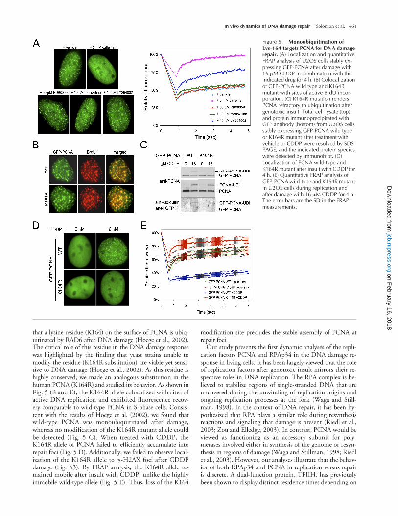
that a lysine residue (K164) on the surface of PCNA is ubiq-uitinated by RAD6 after DNA damage (Hoege et al., 2002).The critical role of this residue in the DNA damage responsewas highlighted by the finding that yeast strains unable tomodify the residue (K164R substitution) are viable yet sensi-tive to DNA damage (Hoege et al., 2002). As this residue ishighly conserved, we made an analogous substitution in thehuman PCNA (K164R) and studied its behavior. As shown inFig. 5 (B and E), the K164R allele colocalized with sites ofactive DNA replication and exhibited fluorescence recov-ery comparable to wild-type PCNA in S-phase cells. Consis-tent with the results of Hoege et al. (2002), we found thatwild-type PCNA was monoubiquitinated after damage,whereas no modification of the K164R mutant allele couldbe detected (Fig. 5 C). When treated with CDDP, theK164R allele of PCNA failed to efficiently accumulate intorepair foci (Fig. 5 D). Additionally, we failed to observe local-ization of the K164R allele to
�
-H2AX foci after CDDPdamage (Fig. S3). By FRAP analysis, the K164R allele re-mained mobile after insult with CDDP, unlike the highlyimmobile wild-type allele (Fig. 5 E). Thus, loss of the K164
modification site precludes the stable assembly of PCNA atrepair foci.
Our study presents the first dynamic analyses of the repli-cation factors PCNA and RPAp34 in the DNA damage re-sponse in living cells. It has been largely viewed that the roleof replication factors after genotoxic insult mirrors their re-spective roles in DNA replication. The RPA complex is be-lieved to stabilize regions of single-stranded DNA that areuncovered during the unwinding of replication origins andongoing replication processes at the fork (Waga and Still-man, 1998). In the context of DNA repair, it has been hy-pothesized that RPA plays a similar role during resynthesisreactions and signaling that damage is present (Riedl et al.,2003; Zou and Elledge, 2003). In contrast, PCNA would beviewed as functioning as an accessory subunit for poly-merases involved either in synthesis of the genome or resyn-thesis in regions of damage (Waga and Stillman, 1998; Riedlet al., 2003). However, our analyses illustrate that the behav-ior of both RPAp34 and PCNA in replication versus repairis discrete. A dual-function protein, TFIIH, has previouslybeen shown to display distinct residence times depending on
Figure 5. Monoubiquitination of Lys-164 targets PCNA for DNA damage repair. (A) Localization and quantitative FRAP analysis of U2OS cells stably ex-pressing GFP-PCNA after damage with 16 �M CDDP in combination with the indicated drug for 4 h. (B) Colocalization of GFP-PCNA wild type and K164R mutant with sites of active BrdU incor-poration. (C) K164R mutation renders PCNA refractory to ubiquitination after genotoxic insult. Total cell lysate (top) and protein immunoprecipitated with GFP antibody (bottom) from U2OS cells stably expressing GFP-PCNA wild type or K164R mutant after treatment with vehicle or CDDP were resolved by SDS-PAGE, and the indicated protein species were detected by immunoblot. (D) Localization of PCNA wild type and K164R mutant after insult with CDDP for 4 h. (E) Quantitative FRAP analysis of GFP-PCNA wild-type and K164R mutant in U2OS cells during replication and after damage with 16 �M CDDP for 4 h. The error bars are the SD in the FRAP measurements.
on February 16, 2018
jcb.rupress.orgD
ownloaded from
462 The Journal of Cell Biology
|
Volume 166, Number 4, 2004
its action in transcription versus repair (Hoogstraten etal., 2002). Interestingly, TFIIH displayed longer residencetimes during repair than transcription, similar to our obser-vations with PCNA and RPAp34 during repair versus rep-lication. However, in the case of TFIIH there is a welldocumented biochemical distinction between the repair andtranscriptional complex (Svejstrup et al., 1995). It is lessclear what might underlie the differences in PCNA andRPAp34 behavior between replication and repair. In the caseof PCNA, we speculate that the types of DNA polymerasesassociated with the sliding clamp, or alternatively associationwith other repair proteins (e.g., MSH2 or Rad9), may be aprimary determinant.
Several studies have described specific signaling pathwaysthat regulate the biological activity of replication factors.Specifically, phosphorylation of RPA subunits after geno-toxic insult has been observed (Shao et al., 1999; Wang etal., 2001; Barr et al., 2003). These reports have speculatedthat phosphorylation enhances RPA affinity for damagedor single-stranded DNA, effectively stabilizing a denaturedDNA structure that RPA has been predicted to play a staticrole in maintaining. Our results strongly question thismodel. RPAp34 was observed to rapidly exchange at repairfoci with significantly faster kinetics than other repair factors(i.e., PCNA). Contrary to expectation, we found that phos-phorylation of RPAp34 by DNA-PK was not required forfocus formation. Rather, this phosphorylation event destabi-lized RPAp34 at sites of damage. The use of increasing theturnover of RPAp34 at repair foci is at present unknown.However, several tantalizing possibilities exist, such as a rolefor phosphorylated RPA in signaling for the recruitment ofadditional factors from the nucleoplasm or to facilitate ac-cessibility of repair factors to the region of damage. Alterna-tively, phosphorylation by DNA-PK may be required for ef-ficient functioning of RPAp34 in repair processes. Cellsdeficient in DNA-PK activity have defects in DNA repairand V(D)J recombination, and an inability to rapidly modu-late RPA–DNA interactions may explain these defects. Ad-ditionally, we have delineated a posttranslational mechanismby which PCNA is targeted to or stabilized at repair foci. Al-though there appears to be no significant modification ofhuman PCNA during replication or other phases of the cellcycle, modification by ubiquitin after DNA damage is essen-tial for the stable assembly of PCNA at repair foci. This re-sult provides an explanation for the lack of viability in yeastharboring the K164R allele after DNA damage (Hoege etal., 2002). Notably, ubiquitination also plays a role in tar-geting the Fanconi anemia protein FANCD2 to foci afterDNA damage (Garcia-Higuera et al., 2001). It will be inter-esting to further examine the critical role of ubiquitinationin the DNA damage response.
The dynamic behavior of repair factors in living cells wasfirst demonstrated by Houtsmuller et al. (1999) who ob-served that the endonuclease complex ERCC1-XPF rapidlyexchanges at sites of UV-induced damage. However, not allproteins behave identically at sites of damage, as Nbs1 islargely tethered whereas Chk2 remains highly mobile (com-parable to our observations with PCNA and RPAp34, re-spectively; Lukas et al., 2003). In this context, our data sup-port a dynamic exchange model in which multiple repair
factors have access to and rapidly turn over at sites of geneticinsult. Further analysis of the DNA damage response in liv-ing cells will continue to provide powerful insight into thecomplex process of repair essential for the maintenance ofgenomic stability.
Materials and methods
Plasmids and mutagenesis
Expression plasmids encoding GFP-PCNA, GFP-RPAp34, and GFP-histoneH2B have been described previously (Leonhardt et al., 2000; Phair andMisteli, 2000; Sporbert et al., 2002). Lys-164 of GFP-PCNA was mutated toArg, and multiple NH
2
-terminal phosphorylation sites of GFP-RPAp34(Thr-21, Ser-23, Ser-29, and Ser-33) were mutated to Ala and confirmed bysequencing.
Cell culture and generation of stable cell lines
Rat-1, U2OS, and M059 cells were grown in DME supplemented with10% FBS. Rat-1 and U2OS cells were transfected with GFP-PCNA, GFP-RPAp34, and GFP-histone H2B along with pBABE-Puro using FuGene 6(Roche), and multiple independent stable cell lines were established afterpuromycin selection. Transient transfection of M059 cells was performedusing FuGene 6. Clinical grade CDDP was obtained from Bristol Oncol-ogy. All other drugs were obtained from commercially available sources.
Immunoprecipitation and immunoblotting
Cells were lysed in NET-N supplemented with protease inhibitors, andclarified lysate was immunoprecipitated with a monoclonal GFP antibody(B-2; Santa Cruz Biotechnology, Inc.). Proteins were detected with the fol-lowing antibodies: PCNA (FL-261; Santa Cruz Biotechnology, Inc.), RPA(Ab-3; Oncogene Research Products), and ubiquitin (U5379; Sigma-Aldrich).
BrdU labeling and immunofluorescence
Asynchronously growing cells were pulsed with BrdU for 15 min, and sitesof BrdU incorporation were visualized as described previously (Angus etal., 2003). For detection of specific DNA lesions, cells were fixed in 3.7%formaldehyde and probed with antibodies to cyclobutane pyrimidinedimers (T. Matsunaga, Osaka University, Osaka, Japan) or Pt-DNA adducts(M. Tilby, University of Newcastle upon Tyne, Newcastle, UK). For allother immunofluorescence, cells were fixed in cold methanol for 20 minand probed with antibodies to RPA (Ab-3; Oncogene Research Products),MSH2 (N-20; Santa Cruz Biotechnology, Inc.), ERCC1 (FL-297; Santa CruzBiotechnology, Inc.), or phospho-histone H2A.X Ser-139 (JBW301; Up-state Biotechnology). Imaging was performed on a microscope (model Mi-crophot-FX; Nikon) using a plan-apochromat 60
�
1.4 NA objective.
UV microirradiation
UVC radiation was delivered using a low-pressure mercury lamp withmaximal output at 254 nm. Polycarbonate filters containing either 3- or5-
�
m diameter pores were gently laid over the cells before UV irradiation.
Dot-blot assay of Pt-DNA adduct levels
4
�
g of genomic DNA isolated from U2OS cells after CDDP damage wasextensively boiled and sheared by sonication, transferred onto a nylonmembrane, and probed with the Pt-DNA adduct antibody.
Live-cell imaging and FRAP
Live-cell imaging and photobleaching analysis was performed at 37
�
C on aconfocal microscope (model LSM510 Axiovert; Carl Zeiss MicroImaging,Inc.) using the 488-nm line of an argon laser (25 mW nominal output, pho-tomultiplier detection through a pinhole diameter of 96
�
m and a 505-nmlong-pass filter) as described previously (Phair and Misteli, 2000; Angus etal., 2003).
Online supplemental material
Three supplemental figures and accompanying figure legends are availableonline at http:www.jcb.org/cgi/conent/full/jcb.200312048/DC1.
We thank Nancy Kleene, Robert Hennigan, Michael Tilby, Tsukasa Matsu-naga, Yoli Sanchez, and Kathleen Dixon for kindly providing reagents andtechnical assistance. We thank Andre Nussenzweig, Yoli Sanchez, PeterStambrook, and Karen Knudsen for critical reading of the manuscript.
on February 16, 2018
jcb.rupress.orgD
ownloaded from
In vivo dynamics of DNA damage repair |
Solomon et al. 463
M.C. Cardoso is funded by the Deutsche Forschungsgemeinschaft, andE.S. Knudsen is funded by the American Cancer Society (grant RSG-01-254-ECG) and National Cancer Institute (grant CA-106471).
Submitted: 5 December 2003Accepted: 10 June 2004
References
Angus, S.P., D.A. Solomon, L. Kuschel, R.F. Hennigan, and E.S. Knudsen. 2003.Retinoblastoma tumor suppressor: analyses of dynamic behavior in livingcells reveal multiple modes of regulation.
Mol. Cell. Biol.
23:8172–8188.Barr, S.M., C.G. Leung, E.E. Chang, and K.A. Cimprich. 2003. ATR kinase activ-
ity regulates the intranuclear translocation of ATR and RPA following ioniz-ing radiation.
Curr. Biol.
13:1047–1051.Dutta, A., and B. Stillman. 1992. cdc2 family kinases phosphorylate a human cell
DNA replication factor, RPA, and activate DNA replication.
EMBO J.
11:2189–2199.
Garcia-Higuera, I., T. Taniguchi, S. Ganesan, M.S. Meyn, C. Timmers, J. Hejna,M. Grompe, and A.D. D’Andrea. 2001. Interaction of the Fanconi anemiaproteins and BRCA1 in a common pathway.
Mol. Cell.
7:249–262.He, Z., L.A. Henricksen, M.S. Wold, and C.J. Ingles. 1995. RPA involvement in
the damage-recognition and incision steps of nucleotide excision repair.
Na-ture.
374:566–569.Hoege, C., B. Pfander, G.L. Moldovan, G. Pyrowolakis, and S. Jentsch. 2002.
RAD6-dependent DNA repair is linked to modification of PCNA by ubiq-uitin and SUMO.
Nature.
419:135–141.Hoogstraten, D., A.L. Nigg, H. Heath, L.H. Mullenders, R. van Driel, J.H. Hoeij-
makers, W. Vermeulen, and A.B. Houtsmuller. 2002. Rapid switching ofTFIIH between RNA polymerase I and II transcription and DNA repair invivo.
Mol. Cell.
10:1163–1174.Houtsmuller, A.B., S. Rademakers, A.L. Nigg, D. Hoogstraten, J.H. Hoeijmakers,
and W. Vermeulen. 1999. Action of DNA repair endonuclease ERCC1/XPF in living cells.
Science.
284:958–961.Izzard, R.A., S.P. Jackson, and G.C. Smith. 1999. Competitive and noncompetitive
inhibition of the DNA-dependent protein kinase.
Cancer Res.
59:2581–2586.Leonhardt, H., H.P. Rahn, P. Weinzierl, A. Sporbert, T. Cremer, D. Zink, and
M.C. Cardoso. 2000. Dynamics of DNA replication factories in living cells.
J. Cell Biol.
149:271–280.Lukas, C., J. Falck, J. Bartkova, J. Bartek, and J. Lukas. 2003. Distinct spatiotem-
poral dynamics of mammalian checkpoint regulators induced by DNA dam-age.
Nat. Cell Biol.
5:255–260.Maser, R.S., K.J. Monsen, B.E. Nelms, and J.H. Petrini. 1997. hMre11 and
hRad50 nuclear foci are induced during the normal cellular response toDNA double-strand breaks.
Mol. Cell. Biol.
17:6087–6096.Meijer, L., A. Borgne, O. Mulner, J.P. Chong, J.J. Blow, N. Inagaki, M. Inagaki,
J.G. Delcros, and J.P. Moulinoux. 1997. Biochemical and cellular effects ofroscovitine, a potent and selective inhibitor of the cyclin-dependent kinasescdc2, cdk2 and cdk5.
Eur. J. Biochem.
243:527–536.Niu, H., H. Erdjument-Bromage, Z.Q. Pan, S.H. Lee, P. Tempst, and J. Hurwitz.
1997. Mapping of amino acid residues in the p34 subunit of human single-
stranded DNA-binding protein phosphorylated by DNA-dependent proteinkinase and Cdc2 kinase in vitro.
J. Biol. Chem.
272:12634–12641.Phair, R.D., and T. Misteli. 2000. High mobility of proteins in the mammalian
cell nucleus.
Nature.
404:604–609.Riedl, T., F. Hanaoka, and J.M. Egly. 2003. The comings and goings of nucleotide
excision repair factors on damaged DNA.
EMBO J.
22:5293–5303.Rouse, J., and S.P. Jackson. 2002. Interfaces between the detection, signaling, and
repair of DNA damage.
Science.
297:547–551.Sarkaria, J.N., E.C. Busby, R.S. Tibbetts, P. Roos, Y. Taya, L.M. Karnitz, and R.T.
Abraham. 1999. Inhibition of ATM and ATR kinase activities by the radio-sensitizing agent, caffeine.
Cancer Res.
59:4375–4382.Shao, R.G., C.X. Cao, H. Zhang, K.W. Kohn, M.S. Wold, and Y. Pommier.
1999. Replication-mediated DNA damage by camptothecin induces phos-phorylation of RPA by DNA-dependent protein kinase and dissociates RPA:DNA-PK complexes.
EMBO J.
18:1397–1406.Shivji, K.K., M.K. Kenny, and R.D. Wood. 1992. Proliferating cell nuclear anti-
gen is required for DNA excision repair.
Cell.
69:367–374.Siddik, Z.H. 2003. Cisplatin: mode of cytotoxic action and molecular basis of re-
sistance.
Oncogene.
22:7265–7279.Sporbert, A., A. Gahl, R. Ankerhold, H. Leonhardt, and M.C. Cardoso. 2002.
DNA polymerase clamp shows little turnover at established replication sitesbut sequential de novo assembly at adjacent origin clusters.
Mol. Cell.
10:1355–1365.
Svejstrup, J.Q., Z. Wang, W.J. Feaver, X. Wu, D.A. Bushnell, T.F. Donahue, E.C.Friedberg, and R.D. Kornberg. 1995. Different forms of TFIIH for tran-scription and DNA repair: holo-TFIIH and a nucleotide excision re-pairosome.
Cell.
80:21–28.Tilby, M.J., C. Johnson, R.J. Knox, J. Cordell, J.J. Roberts, and C.J. Dean. 1991.
Sensitive detection of DNA modifications induced by cisplatin and carbo-platin in vitro and in vivo using a monoclonal antibody.
Cancer Res.
51:123–129.
Vassin, V.M., M.S. Wold, and J.A. Borowiec. 2004. Replication protein A (RPA)phosphorylation prevents RPA association with replication centers.
Mol.Cell. Biol.
24:1930–1943.Waga, S., and B. Stillman. 1998. The DNA replication fork in eukaryotic cells.
Annu. Rev. Biochem.
67:721–751.Walker, E.H., M.E. Pacold, O. Perisic, L. Stephens, P.T. Hawkins, M.P. Wy-
mann, and R.L. Williams. 2000. Structural determinants of phosphoinosi-tide 3-kinase inhibition by wortmannin, LY294002, quercetin, myricetin,and staurosporine.
Mol. Cell.
6:909–919.Wang, B., S. Matsuoka, P.B. Carpenter, and S.J. Elledge. 2002. 53BP1, a mediator
of the DNA damage checkpoint.
Science.
298:1435–1438.Wang, H., J. Guan, A.R. Perrault, Y. Wang, and G. Iliakis. 2001. Replication pro-
tein A2 phosphorylation after DNA damage by the coordinated action ofataxia telangiectasia-mutated and DNA-dependent protein kinase.
CancerRes.
61:8554–8563.Ward, I.M., and J. Chen. 2001. Histone H2AX is phosphorylated in an ATR-
dependent manner in response to replicational stress.
J. Biol. Chem.
276:47759–47762.
Zou, L., and S.J. Elledge. 2003. Sensing DNA damage through ATRIP recognitionof RPA-ssDNA complexes.
Science.
300:1542–1548.
on February 16, 2018
jcb.rupress.orgD
ownloaded from


![Genome-Wide Analysis of the Core DNA Replication ......Genome Analysis Genome-Wide Analysis of the Core DNA Replication Machinery in the Higher Plants Arabidopsis and Rice1[W][OA]](https://img.dokumen.tips/doc/110x75/5f05685d7e708231d412d002/genome-wide-analysis-of-the-core-dna-replication-genome-analysis-genome-wide.jpg)