Embed Size (px)
Citation preview

ARTICLE OPEN
LYMPHOMA
Dual targeting of the DNA damage response pathway and BCL-2 in diffuse large B-cell lymphomaAlessandra Rossi1, Stefania Orecchioni2, Paolo Falvo2, Valentina Tabanelli3, Elena Baiardi 1, Claudio Agostinelli4,5, Federica Melle3,Giovanna Motta3, Angelica Calleri3, Stefano Fiori3, Chiara Corsini2, Beatrice Casadei6, Saveria Mazzara3, Umberto Vitolo 7,Francesco Bertolini2, Pier Luigi Zinzani 6, Myriam Alcalay8,9, Pier Giuseppe Pelicci8,9, Stefano Pileri3, Corrado Tarella 1,10✉ andEnrico Derenzini 1,10✉
© The Author(s) 2021
Standard chemotherapies for diffuse large B-cell lymphoma (DLBCL), based on the induction of exogenous DNA damage andoxidative stress, are often less effective in the presence of increased MYC and BCL-2 levels, especially in the case of double hit (DH)lymphomas harboring rearrangements of the MYC and BCL-2 oncogenes, which enrich for a patient’s population characterized byrefractoriness to anthracycline-based chemotherapy. Here we hypothesized that adaptive mechanisms to MYC-induced replicativeand oxidative stress, consisting in DNA damage response (DDR) activation and BCL-2 overexpression, could represent the biologicbasis of the poor prognosis and chemoresistance observed in MYC/BCL-2-positive lymphoma. We first integrated targeted geneexpression profiling (T-GEP), fluorescence in situ hybridization (FISH) analysis, and characterization of replicative and oxidative stressbiomarkers in two independent DLBCL cohorts. The presence of oxidative DNA damage biomarkers identified a poor prognosisdouble expresser (DE)-DLBCL subset, characterized by relatively higher BCL-2 gene expression levels and enrichment for DHlymphomas. Based on these findings, we tested therapeutic strategies based on combined DDR and BCL-2 inhibition, confirmingefficacy and synergistic interactions in in vitro and in vivo DH-DLBCL models. These data provide the rationale for precision-therapystrategies based on combined DDR and BCL-2 inhibition in DH or DE-DLBCL.
Leukemia; https://doi.org/10.1038/s41375-021-01347-6
INTRODUCTIONDiffuse Large B-cell lymphoma (DLBCL) is the most common non-Hodgkin lymphoma (NHL) subtype, and yet 40% of patients areresistant to current therapies [1–3], which are still based on theinduction of exogenous DNA damage with anthracycline-basedchemotherapy regimens representing the standard of care [1–3].The cell of origin (COO) determined by gene expression profiling(GEP) is a well-established prognostic predictor in DLBCL [4–7]. Ingeneral, DLBCLs with a GEP signature related to activated B-cellcells (ABC subgroup) have a worse outcome compared to theirgerminal center B-cell (GCB) counterparts [4–7], and display higherexpression levels of MYC and BCL2 [8] and oncogenic addiction tonuclear factor kappa-B (NF-kB) signaling [9]. However, despite itsprognostic relevance, COO-based precision therapy has not yettranslated into meaningful clinical benefits [10–12]. Besides the
COO, overexpression or genomic rearrangements of the MYC andBCL-2 oncogenes are powerful negative prognostic factors inDLBCL [13, 14]. Due to the fact that concurrent MYC and BCL-2rearrangements enrich for a patient’s population characterized byrefractoriness to standard anthracycline-based chemotherapy,these lymphomas are now classified as a separate disease entity(HG-BCL w/DH) [15] and currently treated with more intensivechemotherapy regimens, representing a major unmet need inlymphoma therapy [16, 17]. On the other hand, the observationthat a fraction of HG-BCL w/DH can be cured with standardtherapies underlines the concept that the mechanisms underlyingchemoresistance in MYC/BCL-2 positive DLBCL are still poorlydefined. Recent evidence suggests that MYC-positive tumors arecharacterized by replicative and oxidative stress leading toinherent DNA damage and genomic instability [18–20]. Constitu-
Received: 1 November 2020 Revised: 28 June 2021 Accepted: 8 July 2021
1Onco-Hematology Division, IEO European Institute of Oncology IRCCS, Milan, Italy. 2Laboratory of Hematology-Oncology, IEO European Institute of Oncology IRCCS, Milan, Italy.3Division of diagnostic Haematopathology, IEO European Institute of Oncology IRCCS, Milan, Italy. 4Hematopathology Unit, Department of Experimental, Diagnostic, and SpecialtyMedicine (DIMES), Bologna University School of Medicine, Bologna, Italy. 5Haematopathology Unit, IRCCS Azienda Ospedaliero-Universitaria di Bologna, Bologna, Italy. 6IRCCSAzienda Ospedaliero-Universitaria di Bologna, Institute of Hematology and Medical Oncology “L. e A. Seragnoli”, Department of Experimental, Diagnostic, and Specialty Medicine(DIMES), University of Bologna, Bologna, Italy. 7Multidisciplinary Oncology Outpatient Clinic, Candiolo Cancer Institute, FPO-IRCCS, Candiolo, Italy. 8Department of ExperimentalOncology, IEO European Institute of Oncology IRCCS, Milan, Italy. 9Department of Oncology and Hemato-Oncology, University of Milan, Milan, Italy. 10Department of HealthSciences, University of Milan, Milan, Italy. ✉email: [email protected]; [email protected]
www.nature.com/leuLeukemia

tive activation of the DNA damage response (DDR) pathway is oneof the main mechanisms by which cancer cells cope withreplicative stress, avoiding intolerable levels of endogenous DNAdamage [21–23]. On the other hand, it is well known that BCL-2overexpression synergizes with MYC in driving B-cell lymphoma-genesis, by counteracting MYC-related proapoptotic effects andoxidative stress [24–28]. Of note, constitutive DDR activationcorrelates with MYC levels predicting poor prognosis [29], andtherapeutic approaches targeting DDR through inhibition of theAtaxia teleangiectasia and Rad3 related (ATR)-checkpoint kinase 1/2 (CHK1/2) axis showed efficacy in preclinical models of MYC-positive DLBCL, including those with TP53 mutations/deletionsand CDKN2A loss which are mechanistically linked to anthracyclineresistance [30–33].Since chemotherapy exerts its cytotoxic effects through
exogenous DNA damage and induction of reactive oxygen species(ROS), adaptive mechanisms to replicative and oxidative stress(consisting in DDR activation and upregulation of antioxidantcapacity), could represent the biologic basis of the poor prognosisand chemoresistance observed in MYC/BCL-2-double expresserDLBCL and HG-BCL w/DH.In an effort to design specific therapies for MYC/BCL-2 positive
DLBCL, we first integrated targeted-GEP (T-GEP), fluorescencein situ hybridization (FISH) analysis, and functional characterizationof replicative and oxidative stress biomarkers in two independentDLBCL cohorts. Since the presence of oxidative DNA damagebiomarkers identified a poor prognosis DE-DLBCL subset, whichwas characterized by relatively higher BCL-2 gene expressionlevels and enrichment in HG-BCL w/DH, we then testedtherapeutic strategies based on combined DDR and BCL-2inhibition, confirming efficacy and synergistic interactions inin vitro and in vivo HG-BCL w/DH models. These data providethe rationale for novel precision therapy strategies based oncombined DDR and BCL-2 inhibition in double-hit DLBCL.
Table 1. Patients characteristics.
Factor DLCL04(N= 69)
Real life(n= 66)
p value Combined(n= 135)
COO
ABC 21 12 0.1 33
GCB/UNCL 48 54 102
Age 51 (18–63) 64(17–85)
<0.01 57 (17–85)
IPI
0–1 – 17 0.2 17
2–3 57 45 102
4–5 12 4 16
Treatment
R-CHOP-like 39 66 – 105
R-CHOP-like + ASCT
30 – 30
MYC/BCL-2 status
Nano
DE 18 17 0.9 35
Non-DE 51 49 100
DH 4 3 0.9 7
γH2AX/8-OHDGDE 38 31 0.39 69
NON DE 31 35 66
COO Cell of origin, IPI international prognostic index, DH double hit, ASCTautologous stem cell transplant, nano NanoString, DE double expresser,ABC activated B-cell, GCB/UNCL germinal center B-cell/unclassified.
Fig. 1 Study cohorts. A On the left, the discovery cohort is represented. 94 stage III-IV DLBCL patients enrolled in the DLCL04 trial withavailable FFPE tissue were initially considered in this analysis. T-GEP success rate was 92.6% (n= 87), with seven cases not yielding enoughhigh-quality mRNA to undergo successful GEP assessment. Only cases of non-otherwise specified (NOS) histology (including those originallydiagnosed as DLBCL-NOS and nowadays included in the HG-BCL provisional category) were considered; therefore 11 cases classified indifferent DLBCL categories were excluded. In seven cases that were not evaluable (NE), we could not retrieve enough tissue for additionalimmunohistochemistry studies and these cases were excluded. Sixty-nine NOS-DLBCL FFPE patient samples from the DLC04 trial were finallyincluded in this study. B On the right a “real-life” validation cohort including 66 consecutive DLBCL NOS cases with available FFPE tissue for T-GEP and IHC, treated with R-CHOP/CHOP-like regimens. Success rates of T-GEP, and cases excluded for histologic classification or IHC tissueavailability issues are detailed in the figure.
A. Rossi et al.
2
Leukemia
1234567890();,:

Fig. 2 Oxidative and replicative stress biomarkers in poor prognosis DLBCL subsets. A In the upper panel, bar graph indicating theproportion of 8-OhDG positive and negative cases according to the expression levels of γH2AX in the discovery (left) and real-life cohort(right). In the lower panel, the proportion of γH2AX positive and negative cases according to the expression levels of 8-OhDG in the discovery(left) and real-life cohort (right). P value was calculated with the chi-square test. A p value <0.05 was considered as statistically significant: *<0.05, ** <0.01. POS (positive); NEG (negative). See also Figure S1. B Representative examples of 8-OHdG and γH2AX positive (right panel) andnegative (left panel) DLBCLs. Original magnification, 600x. C OS of the discovery cohort (DLCL04; n= 69) according to the MYC/BCL-2 status asdetermined by T-GEP and to γH2AX/8-OhDG levels as assessed by IHC. DLBCL showing increased levels of MYC and BCL-2 mRNA by T-GEP(MYC/BCL-2 DE) and concurrent overexpression of γH2AX and 8-OhDG (DE-OX_high subgroup) are characterized by a worse outcomecompared to all other subgroups. P values were calculated with the log-rank test. See also Figures S2 and S3. D OS of the real-life validationcohort (n= 66) according to the MYC/BCL-2 status as determined by T-GEP and to γH2AX/8-OhDG levels as assessed by IHC. DLBCL showingincreased levels of MYC and BCL-2 mRNA by T-GEP (MYC/BCL-2 DE) and concurrent overexpression of γH2AX and 8-OhDG (DE-OX_highsubgroup) are characterized by a worse outcome compared to all other subgroups. P values were calculated with the log rank test. See alsoFigure S3. E Bar graphs representing the proportions of different DLBCL subsets in the DE-OX_high and DE-OX_low subgroups (analysis of thewhole cohort: discovery+ validation), showing relative increase in ABC cases and significant enrichment HG-BCL w/DH in the DE-OX_highsubgroup. See also Table 2 for detailed statistics. See also Figure S3. F Box plot graph showing BCL-2 mRNA levels as assessed by T-GEP in theDE-OX_high vs DE-OX_low subgroup. Differences between groups were calculated with the Student T test. *p < 0.05, **p < 0.01.
A. Rossi et al.
3
Leukemia

METHODSPatientsIn the present study, we analyzed two independent patients cohorts: 69patients from the DLCL04 study [34], a prospective randomized phase 3clinical trial investigating the role of first-line autologous stem celltransplant (ASCT) consolidation after chemoimmunotherapy in CD20+DLBCL, and 66 patients from a real-life cohort treated with R-CHOP/CHOP-like regimens at S. Orsola-Malpighi Hospital, Bologna (Italy), from 2007 to2012. Patients characteristics are summarized in Table 1.The study flowchart is depicted in Fig. 1 and additional details are
provided in supplement.This study was approved by the Institutional Review Boards of the
participating centers, in accordance with the Declaration of Helsinki.
Targeted gene expression profiling (T-GEP) panelGene expression was measured on the NanoString nCounter AnalysisSystem (NanoString Technologies, Seattle, WA, USA). The original T-GEPpanel contains 22 genes: 15 genes used to assign COO subtype [6], 5housekeeping genes (UBXN4, ISY1, R3HDM1, WDR55, TRIM56); and theadditional genes of interest: MYC and BCL-2. The complete list of genes,target sequences, and detailed methods are available in supplement.
Immunohistochemistry and fluorescence in situ hybridization(FISH)Immunohistochemistry (IHC) was centralized in Milan for the DLCL04 trialand in Bologna for the real-life control group. Primary antibody source anddilutions are shown in Table S1 and a detailed description of IHC and FISHmethods is provided in supplement.The cut-off values of 50% and 40% positive neoplastic cells were applied
for BCL2 and MYC respectively [14], and 50% for γH2AX and 8-OHdG.
Reagents, in vitro assays, and cell linesPrexasertib was provided by the Eli Lilly company for in vitro studies, andwas purchased from Selleckchem (Houston, Tx) for in vivo studies.AZD7762, MK-8776, Venetoclax and the MCL-1 inhibitor S63845 werepurchased from Selleckchem (Houston, TX).Detailed information on cell lines, 8-OHdG ELISA assay, Caspase 3/7
assay, cell cycle analyses, qPCR assays, proliferation assays (Cell Titer Glo,Promega), and western blot antibodies are provided in supplement. TheTet-OFF MYC P-4936 cell line [35, 36], which carries a conditional,tetracycline-regulated MYC promoter, was provided by Dr. A. Younes lab(Memorial Sloan Kettering Cancer Center, New York, NY). Inducibleoverexpression of BCL2 in the SUDHL5 (BCL2 negative) cell line wasperformed by using the Cellecta InDOXible Tet-Activated cDNA LentiviralExpression System (custom Cellecta). Detailed methods for in vitro studiesare provided in supplement.
High-throughput screening experimentsHigh-throughput drug screening experiments were performed as pre-viously described [37]. Plates were imaged after incubation with AlamarBlue on the LEADseekerTM Multimodality Imaging System (GE Healthcare,Piscataway, NJ). In order to evaluate synergy, we compared the observedactivity to the expected activity of the combination at that dose levelunder Bliss independence model [38, 39]. HTS experiments and analyseswere performed at the Memorial Sloan Kettering Cancer Center, New York.Detailed information is available in supplement.
In vivo studiesExperiments involving animals were approved by the Italian Ministry ofHealth and have been performed in accordance with the applicable Italianlaws (D.L.vo 26/14 and following amendments), the Institutional AnimalCare and Use Committee, the institutional guidelines of the EuropeanInstitute of Oncology and the ARRIVE guidelines [40].The luciferase-expressing patient-derived xenograft (PDX) line DFBL-
69487-V3-mCLP [41] was obtained from the Public Repository ofXenografts (www.proxe.org). Detailed information is available insupplement.
Statistical analysisSurvival data and correlations were analyzed retrospectively. For survivalanalysis, we used the Kaplan-Meier method [42] to estimate overall survival(OS). The two-tailed Student t test and Wilcoxon Rank test were used toestimate statistical significance. Correlations and differences in patients’characteristics were analyzed with the chi-square and Fisher’s exact test.The PRISM software was used for the statistical analyses (v7). Significancewas set at P < 0.05. Combination index analysis was performed using theChou-Talalay method [43].T-GEP statistical analyses were calculated with the R software (v3.5.0)
[44]. A detailed description is provided in supplement.
RESULTSReplicative and oxidative stress biomarkers identify poorprognosis subsets of double expresser DLBCLIn order to investigate the relationship between COO classifica-tion, MYC/BCL-2 status, and replicative/oxidative stress biomar-kers, we first profiled two independent case series ofchemoimmunotherapy-treated DLBCL with T-GEP, FISH, andimmunohistochemistry: a discovery cohort from theDLC04 study [34], and a validation cohort of patients treated inreal-life clinical practice. Patients characteristics are shown inTable 1 and were similar in the two cohorts (except for medianage, significantly higher in the real-life cohort), with no significantdifferences in the overall outcome (Figure S1). For T-GEP studies,we used a digital multiplex gene expression profiling platform(NanoString Technology) with a panel containing 22 genes (15genes for COO subtyping according to LST algorithm [6] plus MYCand BCL2 and 5 housekeeping genes). T-GEP, FISH and immuno-histochemistry (IHC) profiling for c-MYC, BCL-2, the phosphory-lated form of H2AX at S139 (γH2AX, a biomarker of DNA damageand DDR activation) [45, 46] and 8-hydroxy-2’-deoxyguanosine (8-OHdG, an oxidative DNA damage marker) [47] were available in 69patients from the DLC04 study [34], and 66 patients treated in thereal-life cohort (Fig. 1 A, B).The two cohorts displayed similar patterns of γH2AX and 8-
OHdG expression (cut-off value 50% of positive cells, with 55%and 47% of cases showing dual nuclear positivity for γH2AX and 8-OHdG in the DLCL04 and real-life cohort respectively). Theexpression levels of γH2AX and 8-OHdG were significantlycorrelated, with the 8-OHdG positive subgroup showing asignificantly higher fraction of γH2AX positive cases, as comparedto the 8-OHdG negative subset. In line with this observation, theproportion of 8-OhDG-positive samples was significantly increasedin the γH2AX-positive subset in both cohorts (Fig. 2 A,B).In order to define the prognostic implications of γH2AX and 8-
OHdG expression with respect to the COO and MYC/BCL-2 status,
Table 2. Characteristics of the DE-OX_low and DE-OX-high subgroups(analysis of the whole cohort: discovery+ validation).
DE-OX_low(n= 15)
DE-OX_high(n= 20)
p value
Treatment
R-CHOP_like 10 13 0.99
R-CHOP_like+ ASCT 5 7
COO
GCB 5 6 0.99
UNCL 5 4 0.45
ABC 5 10 0.49
DHa 0 6 0.027
DH+ ABC 5 16 0.013
COO cell of origin, DH double hit, ASCT autologous stem cell transplant, DEdouble expresser, ABC activated B-cell, GCB/UNCL germinal center B-cell/unclassified.P value was calculated with the Fisher’s exact test.aThe remaining 1 DH case clustered in the non-DE subset. All DH cases were ofGCB origin.
A. Rossi et al.
4
Leukemia

we analyzed the impact of these variables on OS rates in the twocohorts.Although there was a significant correlation between MYC and
BCL-2 mRNA (assessed with T-GEP) and protein levels (assessed byIHC) (Figure S2A), MYC/BCL-2 status evaluated by T-GEP out-performed IHC for prognostic stratification in both cohorts(Table S2 and Figure S2B) (patients were classified as high andlow MYC or BCL-2 expressers based on the median normalizedMYC and BCL-2 mRNA levels in the respective cohorts).In both patients cohorts, dual nuclear positivity for γH2AX and
8-OHdG identified a MYC/BCL-2 mRNA Double Expresser (DE)-
DLBCL subset characterized by dismal outcome (hereafter definedas DE-OX_high) (Fig. 2 C, D). Interestingly MYC/BCL-2 mRNA DEcases with low oxidative DNA damage (DE-OX_low) had a veryfavorable outcome, with OS rates comparable to non-DE cases(Fig. 2 C, D). Similar trends were observed in both cohorts (Fig. 2 C,D) and a cumulative analysis (n= 135 patients) confirmed asignificantly worse OS rate for the DE-OX_high subgroup,compared to all other subgroups (Figure S3A).To further define the biologic characteristics of the DE-OX_high
subgroup we investigated COO subtyping and the presence ofMYC and BCL-2 rearrangements (HG-BCL w/DH) across different
A. Rossi et al.
5
Leukemia
patients’ subsets (DE-OX_high vs DE-OX_low). Interestingly wefound that the DE-OX_high subgroups were enriched in HG-BCLsw/DH (Fig. 2E), showed a relative increase in ABC cases, and werecharacterized by relatively higher BCL-2 mRNA levels compared tothe DE-OX_low subsets (Fig. 2F). Notably, all but one HG-BCL w/DH clustered in the DE-OX_high subgroups (Table 2). Theoutcome of DE-OX_HIGH subset was dismal irrespective of theDH status, with similarly poor OS rates observed in DH and non-DH cases (Figure S3B). These data indicate that the expression ofreplicative and oxidative stress biomarkers such as γH2AX and 8-OHdG could define a subset of MYC/BCL-2 DE DLBCL with specificmolecular features and characterized by a worse outcome.
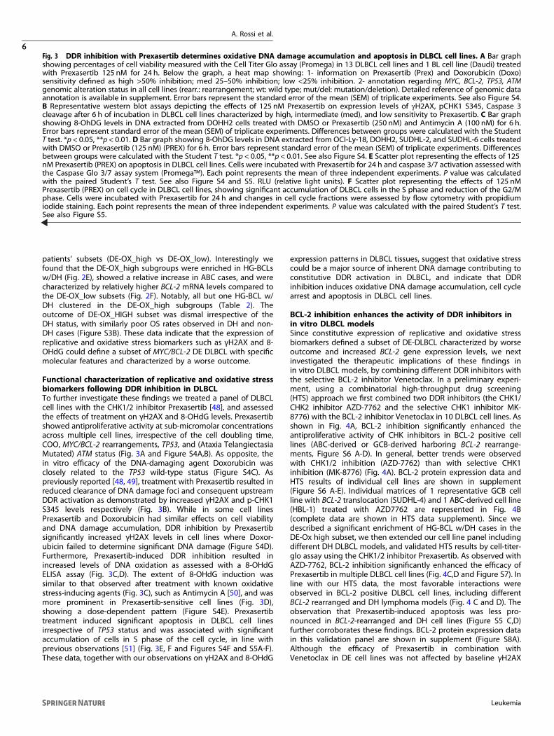
Functional characterization of replicative and oxidative stressbiomarkers following DDR inhibition in DLBCLTo further investigate these findings we treated a panel of DLBCLcell lines with the CHK1/2 inhibitor Prexasertib [48], and assessedthe effects of treatment on γH2AX and 8-OHdG levels. Prexasertibshowed antiproliferative activity at sub-micromolar concentrationsacross multiple cell lines, irrespective of the cell doubling time,COO, MYC/BCL-2 rearrangements, TP53, and (Ataxia TelangiectasiaMutated) ATM status (Fig. 3A and Figure S4A,B). As opposite, thein vitro efficacy of the DNA-damaging agent Doxorubicin wasclosely related to the TP53 wild-type status (Figure S4C). Aspreviously reported [48, 49], treatment with Prexasertib resulted inreduced clearance of DNA damage foci and consequent upstreamDDR activation as demonstrated by increased γH2AX and p-CHK1S345 levels respectively (Fig. 3B). While in some cell linesPrexasertib and Doxorubicin had similar effects on cell viabilityand DNA damage accumulation, DDR inhibition by Prexasertibsignificantly increased γH2AX levels in cell lines where Doxor-ubicin failed to determine significant DNA damage (Figure S4D).Furthermore, Prexasertib-induced DDR inhibition resulted inincreased levels of DNA oxidation as assessed with a 8-OHdGELISA assay (Fig. 3C,D). The extent of 8-OHdG induction wassimilar to that observed after treatment with known oxidativestress-inducing agents (Fig. 3C), such as Antimycin A [50], and wasmore prominent in Prexasertib-sensitive cell lines (Fig. 3D),showing a dose-dependent pattern (Figure S4E). Prexasertibtreatment induced significant apoptosis in DLBCL cell linesirrespective of TP53 status and was associated with significantaccumulation of cells in S phase of the cell cycle, in line withprevious observations [51] (Fig. 3E, F and Figures S4F and S5A-F).These data, together with our observations on γH2AX and 8-OHdG
expression patterns in DLBCL tissues, suggest that oxidative stresscould be a major source of inherent DNA damage contributing toconstitutive DDR activation in DLBCL, and indicate that DDRinhibition induces oxidative DNA damage accumulation, cell cyclearrest and apoptosis in DLBCL cell lines.
BCL-2 inhibition enhances the activity of DDR inhibitors inin vitro DLBCL modelsSince constitutive expression of replicative and oxidative stressbiomarkers defined a subset of DE-DLBCL characterized by worseoutcome and increased BCL-2 gene expression levels, we nextinvestigated the therapeutic implications of these findings inin vitro DLBCL models, by combining different DDR inhibitors withthe selective BCL-2 inhibitor Venetoclax. In a preliminary experi-ment, using a combinatorial high-throughput drug screening(HTS) approach we first combined two DDR inhibitors (the CHK1/CHK2 inhibitor AZD-7762 and the selective CHK1 inhibitor MK-8776) with the BCL-2 inhibitor Venetoclax in 10 DLBCL cell lines. Asshown in Fig. 4A, BCL-2 inhibition significantly enhanced theantiproliferative activity of CHK inhibitors in BCL-2 positive celllines (ABC-derived or GCB-derived harboring BCL-2 rearrange-ments, Figure S6 A-D). In general, better trends were observedwith CHK1/2 inhibition (AZD-7762) than with selective CHK1inhibition (MK-8776) (Fig. 4A). BCL-2 protein expression data andHTS results of individual cell lines are shown in supplement(Figure S6 A-E). Individual matrices of 1 representative GCB cellline with BCL-2 translocation (SUDHL-4) and 1 ABC-derived cell line(HBL-1) treated with AZD7762 are represented in Fig. 4B(complete data are shown in HTS data supplement). Since wedescribed a significant enrichment of HG-BCL w/DH cases in theDE-Ox high subset, we then extended our cell line panel includingdifferent DH DLBCL models, and validated HTS results by cell-titer-glo assay using the CHK1/2 inhibitor Prexasertib. As observed withAZD-7762, BCL-2 inhibition significantly enhanced the efficacy ofPrexasertib in multiple DLBCL cell lines (Fig. 4C,D and Figure S7). Inline with our HTS data, the most favorable interactions wereobserved in BCL-2 positive DLBCL cell lines, including differentBCL-2 rearranged and DH lymphoma models (Fig. 4 C and D). Theobservation that Prexasertib-induced apoptosis was less pro-nounced in BCL-2-rearranged and DH cell lines (Figure S5 C,D)further corroborates these findings. BCL-2 protein expression datain this validation panel are shown in supplement (Figure S8A).Although the efficacy of Prexasertib in combination withVenetoclax in DE cell lines was not affected by baseline γH2AX
Fig. 3 DDR inhibition with Prexasertib determines oxidative DNA damage accumulation and apoptosis in DLBCL cell lines. A Bar graphshowing percentages of cell viability measured with the Cell Titer Glo assay (Promega) in 13 DLBCL cell lines and 1 BL cell line (Daudi) treatedwith Prexasertib 125 nM for 24 h. Below the graph, a heat map showing: 1- information on Prexasertib (Prex) and Doxorubicin (Doxo)sensitivity defined as high >50% inhibition; med 25–50% inhibition; low <25% inhibition. 2- annotation regarding MYC, BCL-2, TP53, ATMgenomic alteration status in all cell lines (rearr.: rearrangement; wt: wild type; mut/del: mutation/deletion). Detailed reference of genomic dataannotation is available in supplement. Error bars represent the standard error of the mean (SEM) of triplicate experiments. See also Figure S4.B Representative western blot assays depicting the effects of 125 nM Prexasertib on expression levels of γH2AX, pCHK1 S345, Caspase 3cleavage after 6 h of incubation in DLBCL cell lines characterized by high, intermediate (med), and low sensitivity to Prexasertib. C Bar graphshowing 8-OhDG levels in DNA extracted from DOHH2 cells treated with DMSO or Prexasertib (250 nM) and Antimycin A (100 nM) for 6 h.Error bars represent standard error of the mean (SEM) of triplicate experiments. Differences between groups were calculated with the StudentT test. *p < 0.05, **p < 0.01. D Bar graph showing 8-OhDG levels in DNA extracted from OCI-Ly-18, DOHH2, SUDHL-2, and SUDHL-6 cells treatedwith DMSO or Prexasertib (125 nM) (PREX) for 6 h. Error bars represent standard error of the mean (SEM) of triplicate experiments. Differencesbetween groups were calculated with the Student T test. *p < 0.05, **p < 0.01. See also Figure S4. E Scatter plot representing the effects of 125nM Prexasertib (PREX) on apoptosis in DLBCL cell lines. Cells were incubated with Prexasertib for 24 h and caspase 3/7 activation assessed withthe Caspase Glo 3/7 assay system (Promega™). Each point represents the mean of three independent experiments. P value was calculatedwith the paired Student’s T test. See also Figure S4 and S5. RLU (relative light units). F Scatter plot representing the effects of 125 nMPrexasertib (PREX) on cell cycle in DLBCL cell lines, showing significant accumulation of DLBCL cells in the S phase and reduction of the G2/Mphase. Cells were incubated with Prexasertib for 24 h and changes in cell cycle fractions were assessed by flow cytometry with propidiumiodide staining. Each point represents the mean of three independent experiments. P value was calculated with the paired Student’s T test.See also Figure S5.
A. Rossi et al.
6
Leukemia

and 8-OHdG levels, cell lines with low levels of γH2AX and 8-OHdGshowed a trend toward a decreased sensitivity to single-agentVenetoclax (Figure S8B-G). Combinatory treatment with Prexaser-tib and Venetoclax synergistically induced apoptosis as demon-strated by time-dependent increase in caspase 3/7 cleavage inBCL-2 positive ABC and DH lymphoma cell lines (Fig. 4E). In linewith the data shown in Fig. 2 and with the role of BCL-2 inregulating the oxidative stress response, these changes wereassociated with early induction of oxidative DNA damage (Fig. 4F).Taken together these data indicate that selective BCL-2 blockade
enhances the in vitro activity of CHK inhibitors in multiple BCL-2positive DLBCL models including HG-BCL w/DH, by inducingoxidative DNA damage and apoptosis. Of note, single-agentPrexasertib or oxidative stress-inducing agents such as doxorubi-cin or H2O2 did not determine significant BCL-2 induction ineither BCL-2 negative or BCL-2 positive cell lines (Figure S9A).Finally, the MCL-1 inhibitor S68345 enhanced Prexasertib efficacyin BCL-2 negative cell lines (Figure S9B), suggesting that dualblockade of the DDR and alternative BCL-2 family members couldbe of value in DLBCLs with low BCL-2 expression.
A. Rossi et al.
7
Leukemia

Differential role of MYC and BCL-2 in modulating the efficacyof checkpoint kinase inhibitionTo determine the effects of BCL-2 overexpression on thetherapeutic efficacy of DDR inhibition, we generated a Tet-oninducible system to overexpress BCL-2 in the Prexasertib-sensitiveBCL-2 negative cell line SUDHL-5. Cells were preincubated withdoxycycline 1 μg/ml for 24 h to overexpress BCL-2, and then weretreated with DMSO or Prexasertib for 24 h (Figure S10A). BCL-2overexpression, while not exerting significant effects on cellproliferation in untreated cells, significantly decreased the efficacyof Prexasertib in this system, indicating that BCL-2 promotesresistance to DDR inhibition (Fig. 5A and S10A). These changeswere associated with an attenuated induction of γH2AX and witha decreased caspase 3 cleavage in cells overexpressing BCL-2(Fig. 5B,C and S10A,B). BCL-2 overexpression did not determinesignificant changes in cell cycle dynamics under Prexasertibtreatment (Fig. 5D).To assess the role of MYC in regulating the antiproliferative
activity of Prexasertib, we used the P-4936 cell line, which carries aconditional, tetracycline-regulated (Tet-OFF) MYC promoter[35, 36]. Cells were pre-treated with DMSO or with doxycyclinefor 6 h to abrogate MYC expression, and then incubated withPrexasertib for 24 h (Fig. 5E-H and S11A-C). MYC silencing withdoxycycline significantly reduced cell proliferation and baselineγH2AX expression (Figure S11A-C). Importantly MYC depletionreduced the antiproliferative effects of Prexasertib in these cells,which was associated with impaired γH2AX induction, decreasedcaspase 3 cleavage, and attenuated effects on cell cycle dynamics(Fig. 5E–H). These data suggest that MYC and BCL-2 may modulatethe sensitivity to CHK1 inhibition in opposite ways: in fact, whilehigh MYC expression could be associated with increased DDRactivation and enhanced susceptibility to DDRi-induced DNAdamage, overexpression of BCL-2 may significantly decrease thetherapeutic activity of DDR inhibitors, providing mechanisticrationale of dual blockade of DDR and BCL-2 in DE and DHlymphomas.
BCL-2 inhibition enhances the activity of DDR inhibitorsin vivo in DH PDX modelsTo assess whether our in vitro results could be confirmed in in vivolymphoma models, we used a PDX mouse model harboring adouble MYC and BCL-2 rearrangement and a 17p deletion (TP53
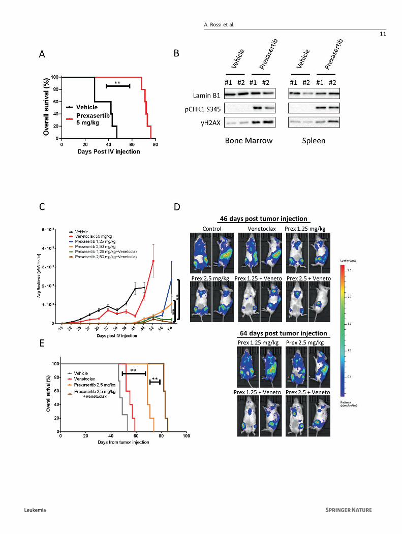
loss) [42]. Treatment with Prexasertib as single-agent significantlyextended the survival of mice bearing DH lymphomas (Fig. 6A). Ofnote, a short course 3-week therapy schedule was sufficient toextend survival of several weeks in this mouse model. In order toassess the in vivo effects of CHK inhibition, lysates from bonemarrow and spleens harvested after 6 h of vehicle or Prexasertibadministration were subjected to western blotting. In line with ourin vitro data, Prexasertib treatment resulted in increased γH2AXand p-CHK1 s345 levels, which are established biomarkers of DDRinhibition, indicative of DNA damage accumulation and ATR-dependent CHK1 phosphorylation (Fig. 6B). In an independentexperiment, we investigated the efficacy of Prexasertib incombination with the BCL-2 inhibitor Venetoclax. In vivocombination therapy with Prexasertib and Venetoclax exertedsynergistic effects in our DH PDX model. Prexasertib as single-agent confirmed high antitumor activity resulting in extendedsurvival. On the contrary, Venetoclax alone had no substantialantitumor activity. However, the combination of Prexasertib andVenetoclax resulted in enhanced tumor growth inhibition andprolonged survival, as compared to either drug administered assingle agent (Fig. 6C–E). Interestingly these synergistic effectswere observed after only one cycle (3 weeks) of combinedtreatment. We did not observe significant weight loss in micetreated with Prexasertib, Venetoclax, or the combination (Fig-ure S12 A and B). Collectively these data suggest that combinedDDR and BCL-2 inhibition could be an effective treatment strategyin DH lymphoma models, including those with defective p53 axis.
DISCUSSIONIn an effort to understand the functional basis of the intrinsicchemoresistance associated with increased MYC and BCL-2 levelsin a significant fraction of DLBCL, we hypothesized that: 1)Overexpression of DDR and oxidative DNA damage markers(γH2AX and 8-OHdG) could identify poor prognosis DLBCLsubsets. 2) Pharmacologic inhibition of the DDR and antioxidantresponse through combined CHK1/2 and BCL-2 blockade couldunleash endogenous replicative and oxidative stress resulting insynergistic therapeutic activity in MYC/BCL-2 positive DLBCL.To address these hypotheses, we first profiled two independent
DLBCL cohorts with T-GEP, FISH, and IHC, in order to define COOsubtyping, MYC, and BCL-2 status and expression levels of
Fig. 4 BCL-2 blockade enhances antilymphoma activity of DDR inhibitors in vitro. A High throughput screening of the checkpoint kinaseinhibitors AZD7762 or MK-8776 combined with the BCL-2 inhibitor Venetoclax (ABT-199) in 10 DLBCL cell lines. Box plot graphs summarizethe results of the combinatorial drug screening analyzed with the Bliss model in 10 DLBCL cell lines treated for 72 h with the indicated drugcombinations. The Y axis indicates the ratio between observed and expected inhibition in a log scale. Values above the “0” line indicateenhanced antiproliferative effects for the combinations. See HTS statistical analysis in the methods section and supplemental data for detailedinformation. See also Figure S6. B Representative examples of combination experiments of AZD7762 plus Venetoclax in HBL-1 and SUDHL-4cells. Combination responses are examined using a 8 × 8 viability matrix representing the ratio between observed/expected inhibition in logscale, which was measured after 72 h post treatment. Full data are shown in supplement. C High throughput screening results were confirmedby independent experiments with the CHK1/2 inhibitor Prexasertib and Venetoclax, using the Cell Titer Glo assay in 13 DLBCL cell lines, andresults were calculated using Combination Index analysis. Three doses of Prexasertib (12.5, 25, 50 nM) were combined with three doses ofVenetoclax (12.5, 25, 50 nM) for 24 h, allowing three combinatory values for each cell line. Values below 1 indicate synergistic interactions. ABCcell lines overexpressing BCL-2 are depicted in red, GCB in blue, DH in light gray, BCL-2 negative in dark gray. See also Figures S7 and S8. DRepresentative examples of Cell Titer Glo experiments in two BCL-2 positive (DOHH2, HBL-1), and 2 BCL-2 negative (OCILY-7, SUDHL-5) celllines. Cells were incubated with increasing concentrations of Prexasertib (PREX) and Venetoclax (12.5, 25, 50 nM), and cell viability wasassessed after 24 h. Error bars represent standard error of the mean (S.E.M) of triplicate experiments. Differences between groups werecalculated with the Student T test. *p < 0.05, **p < 0.01. See also Figures S7, S8 and S9. E Graph showing fold changes over time in caspase 3/7activity in BCL-2 positive (DOHH2, HBL-1) vs BCL-2 negative (OCILY-7, SUDHL-5) cell lines treated with Prexasertib, Venetoclax, or thecombination. Cells were treated with 50 nM of Prexasertib, Venetoclax and the combination for the indicated time. Error bars representstandard error of the mean (S.E.M) of triplicate experiments. Differences between groups (combo vs Venetoclax and Prexasertib as singleagents) were calculated with the Student T test. *p < 0.05, **p < 0.01. F Bar graph showing early changes in DNA oxidation levels in DOHH2and OCILY-7 cells treated with Prexasertib, Venetoclax or the combination for 1 h. After treatment, DNA was extracted and 8-OHdG levels wereassessed with a dedicated ELISA assay. The combination determined synergistic induction of 8-OHdG in the BCL-2 positive DOHH2 cells andwas ineffective in the BCL-2 negative cell line OCILY-7. Error bars represent standard error of the mean (S.E.M) of triplicate experiments.Differences between groups were calculated with the Student T test. *p < 0.05, **p < 0.01.
A. Rossi et al.
8
Leukemia

biomarkers of DDR activation and oxidative DNA damage (γH2AXand 8-OHdG). We demonstrated that: 1) expression levels of theDNA damage marker γH2AX and the oxidative DNA damagemarker 8-OHdG are tightly associated, suggesting that oxidative
stress could be a major source of inherent DNA damagecontributing to constitutive DDR activation in DLBCL (Fig. 2); 2)Dual positivity for γH2AX and 8-OHdG is significantly associatedwith adverse outcome in the MYC/BCL-2 positive DLBCL subset
A. Rossi et al.
9
Leukemia

(Fig. 2); 3) MYC/BCL-2 mRNA DE DLBCL overexpressing γH2AX and8-OHdG (DE-OX high) are enriched of ABC and DH cases, and arecharacterized by increased BCL-2 mRNA expression compared totheir γH2AX and 8-OHdG negative DE counterparts. Importantly allbut one HG-BCL w/DH cases clustered in the DE-OX highsubgroup (Fig. 2, Table 2). These data indicate that a subgroupof ABC DLBCL and HG-BCL w/DH are characterized by high levelsof inherent oxidative DNA damage, these features beingassociated with increased BCL-2 expression levels. Given the poorprognosis of these DLBCL subsets, the well-established oncogeniccooperation between MYC and BCL-2, and the known role of BCL-2 in oxidative stress response, these data are in line with a modelwhereby BCL-2 overexpression and constitutive DDR activationcould provide a tolerance mechanism to MYC-induced replicativeand oxidative stress. Since antracyclines exert their cytotoxicactivity at least in part by increasing ROS levels [52, 53] anddetermining oxidative DNA damage, lymphoma subsets display-ing inherent tolerance to oxidative DNA damage throughconstitutive DDR activation and BCL-2 overexpression could beintrinsically resistant to current antracycline-based chemothera-peutic regimens. The results of our in vitro experiments supportthis hypothesis since DDR inhibition by Prexasertib determinedoxidative DNA damage accumulation (Fig. 3), which was furtherenhanced by the addition of Venetoclax (Fig. 4). Notably,treatment with ROS-inducing agents (Antimycin A) and Prexaser-tib determined accumulation of oxidative DNA damage to similarextents, suggesting that constitutive DDR activation could have amajor role in preventing intolerable levels of DNA damage andgenomic instability induced by endogenous oxidative stress(Fig. 3). BCL-2 blockade with Venetoclax enhanced the antilym-phoma activity of checkpoint kinase inhibitors in multiple BCL-2positive cell lines (including ABC and DH DLBCL models) resultingin increased apoptosis (Fig. 4). While enforced BCL-2 expression
significantly decreased the efficacy of single-agent DDR inhibitionby attenuating DNA damage accumulation and apoptosis induc-tion (Fig. 5), on the contrary, MYC overexpression was associatedwith increased sensitivity to DDR inhibitors, and enhancedapoptotic response in line with previous reports [21–23].Interestingly, while BCL-2 overexpression did not exert significanteffects on cell proliferation, ectopic MYC expression wasassociated with increased cell proliferation and enhanced γH2AXexpression, indicative of increased replicative stress and DDRactivation (Figure S11). These observations underline the intrinsiccorrelation between MYC and the DDR, and the importance ofdual targeting of the DDR and BCL-2 in MYC/BCL-2 positivelymphoma in order to maximize the therapeutic efficacy. Thesedata were confirmed in vivo, in a double hit PDX model with TP53loss (Fig. 6). Interestingly single-agent Venetoclax had negligibleantilymphoma activity in vivo in line with data from early phaseclinical trials in DLBCL [54]. The recent demonstration of synergybetween Venetoclax and Tygecycline in DH lymphoma models isin line with our findings, indicating that therapeutic strategiesbased on synthetic lethal targeting of oxidative stress could be ofvalue in DH-DLBCL [55].In summary, these data indicate that increased tolerance to
replicative and oxidative stress through DDR activation and BCL-2overexpression could be a unifying feature of poor prognosis MYCpositive DLBCL subsets such as ABC and HG-BCL w/DH, whichcould be the basis for a tailored therapeutic approach. In this light,novel therapies based on dual targeting of DDR and antioxidantresponse could determine significant improvements in DLBCLtherapy. This strategy, based on unleashing endogenous MYC-related replicative and oxidative stress rather than inducingexogenous DNA damage, represents a significant innovation,which could provide less toxic alternatives to conventionalchemotherapy.
Fig. 5 Role of BCL-2 and MYC in modulating sensitivity to DDR inhibition. A Cell Titer Glo assay showing the effects of 3 doses ofPrexasertib (3, 6, and 12 nM) for 24 h in the absence (EMPTY VECTOR) or in the presence of BCL-2 (BCL-2 EXP). SUDHL-5 cells (transfected withEMPTY VECTOR or a BCL-2 TET-ON inducible system (BCL-2 EXP) were preincubated with doxycycline 1 μg/ml for 24 h and then treated withPrexasertib at the indicated doses (see also Figure S10A). Error bars represent standard error of the mean (S.E.M) of triplicate experiments.Differences between groups were calculated with the Student T test. *p < 0.05, **p < 0.01. B On the left, representative western blot assayshowing the effects of Prexasertib (6 nM) (PREX) on γH2AX induction and caspase 3 cleavage (CL. CASP 3) in the presence or absence of BCL-2,after 6 and 24 h of incubation. On the right, quantitative densitometry analyses (ImageJ software, western blots are shown in Figure S10B)showing normalized γH2AX levels vs vinculin after 6 h of incubation with Prexasertib in the presence or absence of BCL-2: γH2AX induction byPrexasertib was significantly attenuated in the presence of BCL-2. Error bars represent standard error of the mean (S.E.M) of triplicateexperiments. Differences between groups were calculated with the Student T test. *p < 0.05, **p < 0.01. C Graph showing fold changes incaspase 3/7 activity in SUDHL-5 cells treated with increasing doses of Prexasertib (3,6,12 nM) for 12 h in the presence or absence of BCL-2(BCL-2 EXP vs EMPTY VECTOR), showing significant inhibition of caspase 3/7 cleavage in BCL-2 overexpressing SUDHL-5 cells. Error barsrepresent standard error of the mean (S.E.M) of triplicate experiments. Differences between groups were calculated with the Student T test. *p< 0.05, **p < 0.01. D Bar graph showing the effects of DMSO or Prexasertib (PREX) on cell cycle phases in SUDHL-5 cells in the presence orabsence of BCL-2. After overexpression of BCL-2 (BCL-2) or the empty vector (EMPTY) for 24 h, cells were incubated with 12 nM Prexasertib foradditional 24 h and cell cycle phases assessed by flow cytometry (propidium iodide staining). Error bars represent standard error of the mean(S.E.M) of triplicate experiments. Differences between groups were calculated with the Student T test. *p < 0.05, **p < 0.01. E Cell Titer Gloassay showing the effects of three doses of Prexasertib (25, 50, and 100 nM) for 24 h in the absence of MYC (MYC OFF) or in the presence ofMYC (MYC-ON) in P-4936 cells. P-4936 cells (carrying a tetracycline inducible promoter, TET-OFF MYC) were preincubated with doxycycline 1μg/ml for 6 h to downregulate MYC, and then treated with Prexasertib at the indicated doses (See also Figure S11A). Error bars representstandard error of the mean (S.E.M) of triplicate experiments. Differences between groups were calculated with the Student T test. *p < 0.05,**p < 0.01. F Representative western blot assay showing the effects of Prexasertib (25 nM for 3 and 24 h of incubation) (PREX) on γH2AXinduction and caspase 3 cleavage in the presence (MYC-ON) or absence of MYC (MYC-OFF). On the right, quantitative densitometry analyses(ImageJ software, western blots are shown in Figure S11B) showing normalized γH2AX levels vs vinculin after 3 h of incubation withPrexasertib in MYC-ON and MYC-OFF P-4936 cells. γH2AX induction by Prexasertib was significantly attenuated in the absence of MYC. Errorbars represent standard error of the mean (S.E.M) of triplicate experiments. Differences between groups were calculated with the Student Ttest. *p < 0.05, **p < 0.01. G Graph showing fold changes in caspase 3/7 activity in P-4936 cells treated with increasing doses of Prexasertib (6,12, 25 nM) for 12 h in the presence or absence of MYC (MYC-ON vs MYC-OFF), showing significant inhibition of caspase 3/7 cleavage in MYC-OFF P-4936 cells. Error bars represent standard error of the mean (S.E.M) of triplicate experiments. Differences between groups werecalculated with the Student T test. *p < 0.05, **p < 0.01. H Bar graph showing the effects of DMSO or Prexasertib (PREX) on cell cycle phases inP-4936 cells in the presence or absence of MYC. After preincubation with doxycycline (MYC-OFF) or DMSO (MYC-ON) for 24 h, cells wereincubated with 25 nM Prexasertib for additional 24 h and cell cycle phases assessed by flow cytometry (propidium iodide staining). Error barsrepresent standard error of the mean (S.E.M) of triplicate experiments. Differences between groups were calculated with the Student T test. *p< 0.05, **p < 0.01.
A. Rossi et al.
10
Leukemia
A. Rossi et al.
11
Leukemia
REFERENCES1. Coiffier B, Lepage E, Briere J, Herbrecht R, Tilly H, Bouabdallah R, et al. CHOP
chemotherapy plus rituximab compared with CHOP alone in elderly patients withdiffuse large-B-cell lymphoma. N. Engl J Med. 2002;346:235–42.
2. Sehn LH, Donaldson J, Chhanabhai M, Fitzgerald C, Gill K, Klasa R, et al. Intro-duction of combined CHOP plus rituximab therapy dramatically improved out-come of diffuse large B-cell lymphoma in British Columbia. J Clin Oncol.2005;23:5027–33.
3. Ziepert M, Hasenclever D, Kuhnt E, Glass B, Schmitz N, Pfreundschuh M, et al.Standard International prognostic index remains a valid predictor of outcome forpatients with aggressive CD20+ B-cell lymphoma in the rituximab era. J ClinOncol. 2010;28:2373–80.
4. Alizadeh AA, Eisen MB, Davis RE, Ma C, Lossos IS, Rosenwald A, et al. Distincttypes of diffuse large B-cell lymphoma identified by gene expression profiling.Nature 2000;403:503–11.
5. Shipp MA, Ross KN, Tamayo P, Weng AP, Kutok JL, Aguiar RC, et al. Diffuse largeB-cell lymphoma outcome prediction by gene-expression profiling and super-vised machine learning. Nat Med. 2002;8:68–74.
6. Scott DW, Wright GW, Williams PM, Lih CJ, Walsh W, Jaffe ES, et al. Determiningcell-of-origin subtypes of diffuse large B-cell lymphoma using gene expression informalin-fixed paraffin-embedded tissue. Blood 2014;123:1214–7.
7. Scott DW, Mottok A, Ennishi D, Wright GW, Farinha P, Ben-Neriah S, et al. Prog-nostic significance of diffuse large B-cell lymphoma cell of origin determined bydigital gene expression in formalin-fixed paraffin-embedded tissue biopsies. JClin Oncol. 2015;33:2848–56.
8. Hu S, Xu-Monette ZY, Tzankov A, Green T, Wu L, Balasubramanyam A, et al. MYC/BCL2 protein coexpression contributes to the inferior survival of activated B-cellsubtype of diffuse large B-cell lymphoma and demonstrates high-risk geneexpression signatures: a report from The International DLBCL Rituximab-CHOPConsortium Program. Blood 2013;121:4021–31.
9. Davis RE, Brown KD, Siebenlist U, Staudt LM. Constitutive nuclear factor kappaBactivity is required for survival of activated B cell-like diffuse large B cell lym-phoma cells. J Exp Med. 2001;194:1861–74.
10. Younes A, Sehn LH, Johnson P, Zinzani PL, Hong X, Zhu J, et al. Randomizedphase III trial of ibrutinib and rituximab plus cyclophosphamide, doxorubicin,vincristine, and prednisone in non-germinal center b-cell diffuse large B-celllymphoma. J Clin Oncol. 2019;37:1285–95.
11. Vitolo U, Witzig T, Gascoyne R, Scott DW, Zhang Q, Jurczak W, et al. ROBUST: firstreport of phase III randomized study of lenalidomide/R‐CHOP (R2‐CHOP) vs pla-cebo/R‐CHOP in previously untreated ABC‐type diffuse large B‐cell lymphoma.Hematol Oncol. 2019;37:36–37.
12. Davies A, Cummin TE, Barrans S, Maishman T, Mamot C, Novak U, et al. Gene-expression profiling of bortezomib added to standard chemoimmunotherapy fordiffuse large B-cell lymphoma (REMoDL-B): an open-label, randomised, phase 3trial. Lancet Oncol. 2019;20:649–62.
13. Green TM, Young KH, Visco C, Xu-Monette ZY, Orazi A, Go RS, et al. Immuno-histochemical double-hit score is a strong predictor of outcome in patients withdiffuse large B-cell lymphoma treated with rituximab plus cyclophosphamide,doxorubicin, vincristine, and prednisone. J Clin Oncol. 2012;30:3460–7.
14. Johnson NA, Slack GW, Savage KJ, Connors JM, Ben-Neriah S, Rogic S, et al.Concurrent expression of MYC and BCL2 in diffuse large B-cell lymphoma treatedwith rituximab plus cyclophosphamide, doxorubicin, vincristine, and prednisone.J Clin Oncol. 2012;30:3452–9.
15. Swerdlow SH, Campo E, Harris NL, Jaffe ES, Pileri SA, Stein H et al. WHO classi-fication of tumours of the haematopoietic and lymphoid tissues. Revised 4th ed.Lyon, France: IARC; 2017.
16. Friedberg JW. How I treat double-hit lymphoma. Blood 2017;130:590–6.
17. Petrich AM, Gandhi M, Jovanovic B, Castillo JJ, Rajguru S, Yang DT, et al. Impact ofinduction regimen and stem cell transplantation on outcomes in double-hitlymphoma: a multicenter retrospective analysis. Blood 2014;124:2354–61.
18. Halazonetis TD, Gorgoulis VG, Bartek J. An oncogene-induced DNA damagemodel for cancer development. Science 2008;319:1352–5.
19. Negrini S, Gorgoulis VG, Halazonetis TD. Genomic instability—an evolving hall-mark of cancer. Nat Rev Mol Cell Biol. 2010;11:220–8.
20. Vafa O, Wade M, Kern S, Beeche M, Pandita TK, Hampton GM, et al. c-Myc caninduce DNA damage, increase reactive oxygen species, and mitigate p53 func-tion: a mechanism for oncogene-induced genetic instability. Mol Cell.2002;9:1031–44.
21. Rohban S, Campaner S. Myc induced replicative stress response: how to copewith it and exploit it. Biochim Biophys Acta. 2015;1849:517–24.
22. Kuzyk A, Mai S. c-MYC-induced genomic instability. Cold Spring Harb PerspectMed. 2014;4:a014373.
23. Murga M, Campaner S, Lopez-Contreras AJ, Toledo LI, Soria R, Montaña MF, et al.Exploiting oncogene-induced replicative stress for the selective killing of Myc-driven tumors. Nat Struct Mol Biol. 2011;18:1331–5.
24. Egler RA, Fernandes E, Rothermund K, Sereika S, de Souza-Pinto N, Jaruga P, et al.Regulation of reactive oxygen species, DNA damage, and c-Myc function byperoxiredoxin 1. Oncogene 2005;24:8038–50.
25. Graves JA, Metukuri M, Scott D, Rothermund K, Prochownik. EV. Regulation ofreactive oxygen species homeostasis by peroxiredoxins and c-Myc. J Biol Chem2009;284:6520–9.
26. Strasser A, Harris AW, Bath ML, Cory S. Novel primitive lymphoid tumoursinduced in transgenic mice by cooperation between myc and bcl-2. Nature1990;348:331–3.
27. Fanidi A, Harrington EA, Evan GI. Cooperative interaction between c-myc and bcl-2 proto-oncogenes. Nature 1992;359:554–6.
28. Susnow N, Zeng L, Margineantu D, Hockenbery DM. Bcl-2 family proteins asregulators of oxidative stress. Semin Cancer Biol. 2009;19:42–9.
29. Derenzini E, Agostinelli C, Imbrogno E, Iacobucci I, Casadei B, Brighenti E,et al. Constitutive activation of the DNA damage response pathway as anovel therapeutic target in diffuse large B-cell lymphoma. Oncotarget2015;6:6553–69.
30. Xu-Monette ZY, Wu L, Visco C, Tai YC, Tzankov A, Liu WM, et al. Mutational profileand prognostic significance of TP53 in diffuse large B-cell lymphoma patientstreated with R-CHOP: report from an International DLBCL Rituximab-CHOPConsortium Program Study. Blood 2012;120:3986–96.
31. Jardin F, Jais JP, Molina TJ, Parmentier F, Picquenot JM, Ruminy P, et al. Diffuselarge B-cell lymphomas with CDKN2A deletion have a distinct gene expressionsignature and a poor prognosis under R-CHOP treatment: a GELA study. Blood2010;116:1092–104.
32. Derenzini E, Rossi A, Treré D. Treating hematological malignancies with drugsinhibiting ribosome biogenesis: when and why. J Hematol Oncol. 2018;11(May):75.
33. Restelli V, Lupi M, Chilà R, Vagni M, Tarantelli C, Spriano F, et al. DNA damageresponse inhibitor combinations exert synergistic antitumor activity in aggressiveB-cell lymphomas. Mol Cancer Ther. 2019;18:1255–64.
34. Chiappella A, Martelli M, Angelucci E, Brusamolino E, Evangelista A, Carella AM,et al. Rituximab-dose-dense chemotherapy with or without high-dose che-motherapy plus autologous stem-cell transplantation in high-risk diffuse large B-cell lymphoma (DLCL04): final results of a multicentre, open-label, randomised,controlled, phase 3 study. Lancet Oncol. 2017;18:1076–88.
35. Pajic A, Spitkovsky D, Christoph B, Kempkes B, Schuhmacher M, Staege MS, et al.Cell cycle activation by c-myc in a burkitt lymphoma model cell line. Int J Cancer.2000;87:787–93.
Fig. 6 Prexasertib plus Venetoclax extends survival in vivo in a PDX DH lymphoma model. A Overall survival curve of PDX mice treatedwith vehicle (n= 5) or two different doses of Prexasertib (5 and 10mg/kg BID 3 times/week) (n= 5 in each dose level). Cells (106) werexenografted via tail vein injection into six- to eight-week-old female NSG mice (Charles River, Italy). Tumor growth was monitored three timesper week by whole-body imaging on an IVIS Lumina III platform. Mice were treated for three weeks (nine doses). P values were calculated withthe log rank test. B Western blot showing the in vivo effects of Prexasertib (5 mg/kg BID three times/week) on γH2AX and p-CHK1-S345 levels.Mice were sacrificed at the end of treatment (6 h after last dosing), and tissues were harvested immediately after sacrifice. C Combinationexperiment of Prexasertib and Venetoclax in the DH lymphoma PDX model (DFBL-69487-V3-mCLP). NSG mice were treated with vehicle (n=5), Prexasertib (1.25 and 2.5 mg/kg BID three times/week) (n= 5 in each dose level), Venetoclax (50mg/Kg/daily (five days/week) by oralgavage (n= 5) and the combinations (n= 5 each). Mice were treated for three weeks. Differences between groups were calculated with theStudent T test. *p < 0.05, **p < 0.01. D Representative IVIS imaging of mice treated with vehicle (control), Venetoclax, Prexasertib (Prex) 1.25mg/kg, Prexasertib 2.5 mg/kg, and the combinations at the indicated time points. All vehicle and Venetoclax-treated mice were sacrificedbefore day 64. E Overall survival curve of PDX mice treated with vehicle, two different doses of Prexasertib (1.25 and 2.5 mg/kg three times/week) (n= 5 in each dose level), Venetoclax 50mg/Kg (five days/ week) (n= 5) and the combinations (n= 5 each). Mice were treated for a totalof 21 days. P values were calculated with the log rank test. See also Figure S12.
A. Rossi et al.
12
Leukemia
36. Zeller KI, Zhao X, Lee CW, Chiu KP, Yao F, Yustein JT, et al. Global mapping of c-Myc binding sites and target gene networks in human B cells. Proc Natl Acad SciUSA. 2006;103:17834–9.
37. Derenzini E, Mondello P, Erazo T, Portelinha A, Liu Y, Scallion M, et al. BETinhibition-induced GSK3β feedback enhances lymphoma vulnerability to PI3Kinhibitors. Cell Rep. 2018;24:2155–66.
38. Feller W. An introduction to probability theory and its applications. New York, NY:John Wiley & Sons; 1971.
39. Tallarida RJ. Drug synergism: its detection and applications. J Pharm Exp Ther.2001;298:865–72.
40. Kilkenny C, Browne WJ, Cuthill IC, Emerson M, Altman DG. Improving bioscienceresearch reporting: the ARRIVE guidelines for reporting animal research. PLoSBiol. 2010;8:e1000412.
41. Townsend EC, Murakami MA, Christodoulou A, Christie AL, Koster J, DeSouza TA,et al. The public repository of xenografts enables discovery and randomizedphase II-like trials in mice. Cancer Cell. 2016;29:574–86.
42. Kaplan EL, Meier P. Nonparametric estimations from incomplete observations. JAm Stat Assoc. 1958;53:457–81.
43. Chou TC. Drug combination studies and their synergy quantification using theChou-Talalay method. Cancer Res. 2010;70:440–6.
44. R Core Team. R: a language and environment for statistical computing. R Foun-dation for Statistical Computing, Vienna, Austria. 2014. http://www.R-project.org/.
45. Kinner A, Wu W, Staudt C, Iliakis G. Gamma-H2AX in recognition and signaling ofDNA double-strand breaks in the context of chromatin. Nucleic Acids Res.2008;36:5678–94.
46. Wu J, Clingen PH, Spanswick VJ, Mellinas-Gomez M, Meyer T, Puzanov I, et al. γ-H2AX foci formation as a pharmacodynamic marker of DNA damage producedby DNA cross-linking agents: results from 2 phase I clinical trials of SJG-136(SG2000). Clin Cancer Res. 2013;19:721–30.
47. Valavanidis A, Vlachogianni T, Fiotakis C. 8-hydroxy-2’ -deoxyguanosine (8-OHdG): a critical biomarker of oxidative stress and carcinogenesis. J Environ SciHealth C Environ Carcinog Ecotoxicol Rev. 2009;27:120–39.
48. King C, Diaz HB, McNeely S, Barnard D, Dempsey J, Blosser W, et al. LY2606368causes replication catastrophe and antitumor effects through CHK1-dependentmechanisms. Mol Cancer Ther. 2015;14:2004–13.
49. Parmar K, Kochupurakkal BS, Lazaro JB, Wang ZC, Palakurthi S, Kirschmeier PT,et al. The CHK1 Inhibitor Prexasertib Exhibits Monotherapy Activity in High-GradeSerous Ovarian Cancer Models and Sensitizes to PARP Inhibition. Clin Cancer Res.2019;25:6127–40.
50. Huang LS, Cobessi D, Tung EY, Berry EA. Binding of the respiratory chaininhibitor antimycin to the mitochondrial bc1 complex: a new crystal structurereveals an altered intramolecular hydrogenbonding pattern. J Mol Biol.2005;351:573–97.
51. Ghelli Luserna Di Rorà A, Iacobucci I, Imbrogno E, Papayannidis C, Derenzini E,Ferrari A, et al. Prexasertib, a Chk1/Chk2 inhibitor, increases the effectiveness ofconventional therapy in B-/T- cell progenitor acute lymphoblastic leukemia.Oncotarget 2016;7:53377–91.
52. Mai Y, Yu JJ, Bartholdy B, Xu-Monette ZY, Knapp EE, Yuan F, et al. An oxidativestress-based mechanism of doxorubicin cytotoxicity suggests new therapeuticstrategies in ABC-DLBCL. Blood 2016;128:2797–807.
53. Mizutani H, Tada-Oikawa S, Hiraku Y, Kojima M, Kawanishi S. Mechanism ofapoptosis induced by doxorubicin through the generation of hydrogen peroxide.Life Sci. 2005;76:1439–53.
54. Davids MS, Roberts AW, Seymour JF, Pagel JM, Kahl BS, Wierda WG, et al. Phase Ifirst-in-human study of Venetoclax in patients with relapsed or refractory non-hodgkin lymphoma. J Clin Oncol. 2017;35:826–33.
55. Ravà M, D’Andrea A, Nicoli P, Gritti I, Donati G, Doni M et al. Therapeutic synergybetween tigecycline and venetoclax in a preclinical model of MYC/BCL2 double-hit B cell lymphoma. Sci Transl Med. 2018;10:eaan8723
ACKNOWLEDGEMENTSThis work was supported by Banca del Piemonte (Torino, Italy). AIRC (ItalianAssociation for Cancer Research, Milan, Italy; 5×1000 grant n. 21198 to SP; AIRCIG20109 to FB), and the Italian Ministry of Health with Ricerca Corrente and 5×1000funds. We would like to thank Dr. Anas Younes (MSKCC) for his helpful comments anddiscussion on data interpretation.
AUTHOR CONTRIBUTIONSED and AR conceived the study, performed experiments, analyzed the data and wrotethe manuscript; SO and PF performed in vivo studies; AC, VT, SF, CA and SP performed,analyzed and reviewed immunohistochemistry studies; CC performed FISH studies, FBsupervised in vivo studies; FM and GM performed T-GEP; SM analyzed T-GEP data; BChelped with data collection and data analysis; SP, FB, UV, PLZ, MA, PGP, CT helpedwith data interpretation and critically revised the manuscript.
COMPETING INTERESTSAR, SO, PF, VT, EB, CA, FM, GM, AC, SF, CC, BC, SM, MA: nothing to disclose; UV:Consulting or advisory role for Celgene, Janssen, Genmab, Incyte, Bristol-Myers Squibb;speakers’ bureau with Roche, Celgene, Janssen, Gilead Sciences, Sandoz, Abbvie;research funding from Roche and Celgene; research funding from Roche and Celgene.FB: research funding, Gilead Sciences, Menarini, Emercell. PLZ: speakers’ bureau oradvisory boards for Verastem, Celltrion, Gilead, Janssen-cilag, Bristol-Myers Squibb,Servier, Sandoz, MSD, Immune Design, Celgene, Portola, Roche, Eusapharma, KyowaKirin, Sanofi. PGP: member of advisory commette BRIC Copenaghen, MD AndersonCancer Center, Houston (Tx). SP: Advisory Boards for Celgene, NanoString, Roche. CT:advisory board, ADC- Therapeutics. ED: research funding from TG-Therapeutics, ADC-Therapeutics, Takeda; Advisory board for Gilead, Astra Zeneca, Takeda.
ADDITIONAL INFORMATIONSupplementary information The online version contains supplementary materialavailable at https://doi.org/10.1038/s41375-021-01347-6.
Correspondence and requests for materials should be addressed to C.T. or E.D.
Reprints and permission information is available at http://www.nature.com/reprints
Publisher’s note Springer Nature remains neutral with regard to jurisdictional claimsin published maps and institutional affiliations.
Open Access This article is licensed under a Creative CommonsAttribution 4.0 International License, which permits use, sharing,
adaptation, distribution and reproduction in anymedium or format, as long as you giveappropriate credit to the original author(s) and the source, provide a link to the CreativeCommons license, and indicate if changes were made. The images or other third partymaterial in this article are included in the article’s Creative Commons license, unlessindicated otherwise in a credit line to the material. If material is not included in thearticle’s Creative Commons license and your intended use is not permitted by statutoryregulation or exceeds the permitted use, you will need to obtain permission directlyfrom the copyright holder. To view a copy of this license, visit http://creativecommons.org/licenses/by/4.0/.
© The Author(s) 2021
A. Rossi et al.
13
Leukemia