Embed Size (px)
Citation preview

Drivers of Nasopharyngeal Pneumococcal Colonisation:
Investigation Using an Experimental Human Challenge Model
Thesis submitted in accordance with the requirements of the Liverpool School of Tropical Medicine for the
degree of Doctor of Medicine (MD)
By
Dr Victoria Connor
(MBChB, MRCP)
13th August 2018

i
Declaration
This thesis is the result of my own work and effort. The material contained in this
thesis has not been presented, nor is currently being presented, either wholly or in
part for any other degree or qualification.
Research in this thesis was carried out at the Liverpool School of Tropical Medicine.
For the Hand to Nose study presented in this thesis I developed this project from the
initial stages; I wrote the protocol and all study documents for ethical submission. I
presented the project to the Research Ethics Committee and secured approval. I
arranged sponsorship of the study from Liverpool School of Tropical Medicine. I co-
ordinated and managed both studies in this project working with clinicians, research
nurses, laboratory staff and the Clinical Research Facility (CRU) at the Royal Liverpool
and Broadgreen University Hospital Trust (RLBUHT). Working within the clinical
research team I lead recruitment of volunteers, clinical sample taking and symptoms
data collection.
In some instances, work was carried out in collaboration with other colleagues. The
table below details in full the attribution of work and responsibility related to the
project.

ii
Activity Responsibility- Hand to Nose Study
Responsibility- New Serotypes Study
Protocol writing and
approvals process
Victoria Connor Helen Hill
Victoria Connor
Clinician cover for
safety monitoring of
participants
Victoria Connor
Hugh Adler
Seher Zaidi
Victoria Connor
Hugh Adler
Seher Zaidi
Study oversight and
management
Victoria Connor
Victoria Connor
Helen Hill
Recruitment, sample
taking, symptoms
data collection
Victoria Connor
Caz Hales
Helen Hill
Angie Hyder-Wright
Hugh Adler
Seher Zaidi
Rachel Robinson
Cath Lowe
Victoria Connor
Caz Hales
Helen Hill
Angie Hyder-Wright
Hugh Adler
Seher Zaidi
Rachel Robinson
Cath Lowe
Inoculum preparation
and nasal wash
sample processing
and pneumococcal
detection (culture)
Sherin Pojar
Esther German
Elena Mitsi
Elissavet Nikolaou
Sherin Pojar
Esther German
Elena Mitsi
Elissavet Nikolaou
Viral throat swab
processing
Liverpool Clinical Laboratories NA
Research blood
sample processing
NA Esther German
Carla Solórzano Gonzalez
Pneumococcal DNA
extraction
Victoria Connor
Victoria Connor
Sherin Pojar
Esther German
Carla Solórzano Gonzalez
Elena Mitsi
qPCR process Victoria Connor Elissavet Nikolaou
ELISA NA Victoria Connor
Statistical analysis Victoria Connor Victoria Connor

iii
Conflicts of Interest
Both studies described in this thesis were either partly or fully funded by the Medical
Research Council (MRC) (MR/M011569/1). Unilever (consumer goods company)
partly funded the Hand to Nose study outlined in Chapter 3, representatives from
this company participated in the design of this study. This participation included
participation in preliminary talks about the feasibility of using the pneumococcal
challenge model to investigate hand transmission and sharing of protocols for
standard handwashing studies used by Unilever. There was also review and some
comments given about the first draft of the study protocol which the study team
reviewed and only considered if it would improve the scientific strength of the study.
The conduct of both studies, analysis and presentation of results and the decision to
disseminate results were solely determined by the authors, without influence from
any funding source.

iv
Presentations, Awards and Publications
Publications arising from work in thesis:
• V. Connor, E. German, S. Pojar, E. Mitsi, C. Hales, E. Nikolaou, A. Hyder-
Wright, H. Adler, S. Zaidi, H. Hill, S.P. Jochems, H. Burhan, T. Tobery, J. Rylance
and D.M. Ferreira. Hands are Vehicles for Transmission of Streptococcus
pneumoniae in Novel Controlled Human Infection Study. European
Respiratory Journal - in press; accepted 01/08/2018
Presentations and Awards:
• A2626 - Picking Up a Bug by Picking Your Nose Hand to Nose Transmission
of Streptococcus Pneumoniae In Healthy Participants a Pilot Study-
American Thoracic Society Conference, San Diego, USA, May 2018.
• P119 - Hand to Nose Transmission of Streptococcus Pneumoniae In Healthy
Participants a Pilot Study – British Thoracic Society Winter Meeting, London,
UK, December 2017.
• Hands are vehicles for Transmission of Streptococcus Pneumoniae from
hands to nose- North West Thoracic Society Winter Meeting, Haydock, UK,
October 2017.
• Assembly on Pulmonary Infections and Tuberculosis Abstract Scholarship -
American Thoracic Society Conference, May 2018
• Registrar Research Presentation Prize - North West Thoracic Society
Meeting, October 2017.

v
Acknowledgements
I would like to thank:
• My husband Danny, who has supported me through this project. Your constant
love and support have been invaluable and helped me in ways I cannot even
describe; I could not have done this without you.
• My Mum for always being there for me and encouraging me to push myself to
complete things I would never have thought possible. I will be forever grateful for
your love and support growing up, you are the best mum I could have asked for
and have always been an amazing role model for me.
• My Dad thank-you for always instilling an amazing work ethic in me and bringing
me up with constant love and encouragement. I wish you could have been here
to see me achieve this; I hope I am making you proud.
• Jamie Rylance and Daniela Ferreira; thank-you for giving me this amazing
opportunity to work in the EHPC team over the last 2 years and to complete my
MD with you. I will be forever grateful for your constant advice and support. I
have learnt so much from both of you especially in terms of research
management and writing skills, I could not have asked for better supervisors to
guide me through this process.
• Hassan Burhan; thank-you for seeing some potential in me and offering the
clinical fellowship job and for your advice and support, without which I would not
have been able to undertake this MD.

vi
• The whole clinical and support team; Angie, Caz, Cath, Catherine, Helen, Hugh,
Kelly, Rachael and Seher. I have learnt so much from all of you about how-to
successfully run clinical research studies. Your help and support in completing the
two studies for this thesis was invaluable.
• LSTM lab team for all your help and support over the last 2 years, I have learnt a
lot from all of you and could not have asked for better teachers and colleagues
to work with on this project; Esther, Elena, Sherin, Elissavet, Jesus, Bia and Simon.
• The whole Clinical Research Facility team for your support with all the participant
study visits
• The team at the Royal Liverpool University Trust: RD&I team for support during
set up and running of the studies. Respiratory/ID/ITU consultants who provided
clinical cover for the studies, generic research nurse team who supported our
large recruitment drives, Liverpool Clinical Laboratories for sample analysis.
• LSTM team involved in the two studies; governance team, contracts and finance.
• REC Liverpool East for approving both studies to be conducted in the NHS
• The funders of this work – The Medical Research Council and Unilever
• All the participants who took part in the studies for this thesis- without their
commitment to supporting our research none of this would be possible.

vii
Table of Contents Declaration .................................................................................................................... i
Conflicts of Interest ..................................................................................................... iii
Presentations, Awards and Publications ..................................................................... iv
Publications arising from work in thesis: ................................................................ iv
Presentations and Awards: ..................................................................................... iv
Acknowledgements ...................................................................................................... v
List of Figures ............................................................................................................ xix
List of Tables ............................................................................................................. xxiii
Abbreviations .......................................................................................................... xxvii
Abstract .................................................................................................................... xxix
1 Introduction .......................................................................................................... 1
1.1 Overview ........................................................................................................ 2
1.2 Colonisation ................................................................................................... 6
Pneumococcal colonisation ................................................................... 6

viii
Pneumococcal colonisation and impact on immunity ........................... 8
Pneumococcal colonisation and disease ............................................. 11
1.3 Transmission of Streptococcus pneumoniae ............................................... 15
Rodent models investigating transmission .......................................... 16
Healthy carrier transmission ................................................................ 18
Disease transmission ............................................................................ 20
Streptococcus pneumoniae survival in the environment .................... 22
Hand to nose transmission of S. pneumoniae ..................................... 26
Reduction of transmission ................................................................... 31
1.4 Pneumococcal disease treatment and prevention ..................................... 32
Pathogenicity........................................................................................ 32
Pneumococcal disease ......................................................................... 34
Pneumococcal disease prevention: current pneumococcal vaccines . 38
Rationale behind new vaccine development ....................................... 42
Novel vaccine development ................................................................. 44

ix
Experimental Human Pneumococcal Colonisation studies and their use
for vaccine research ............................................................................................ 48
1.5 Project aims ................................................................................................. 51
2 Methods .............................................................................................................. 53
2.1 Overview ...................................................................................................... 54
2.2 Study set up: Research in the National Health Service (NHS) ..................... 54
Sponsorship .......................................................................................... 54
Health Research Authority (HRA) ......................................................... 56
Ethics: Integrated Research Application System (IRAS) and Research
and Ethics Committee (REC) ............................................................................... 57
Research Development and Innovation Department (RD&I) .............. 58
Patient and Public Involvement (PPI) ................................................... 59
2.3 Clinical procedures ...................................................................................... 61
Trial designs .......................................................................................... 61
Ethical considerations .......................................................................... 62
Recruitment and advertising ................................................................ 63
x
Inclusion and exclusion criteria ............................................................ 66
Study schedule ..................................................................................... 68
Safety .................................................................................................... 69
Safety monitoring ................................................................................. 72
Data Management and Safety Committee (DSMC) ............................. 75
Nasopharyngeal pneumococcal challenge .......................................... 75
Nasal wash sampling method .............................................................. 76
Viral swab sampling method ................................................................ 77
Blood sampling method ....................................................................... 77
2.4 Laboratory procedures ................................................................................ 78
Pneumococcal stock preparation (batch) ............................................ 78
Preparation of pneumococcal stock on day of challenge .................... 81
Nasal wash sample processing ............................................................. 81
Detection of pneumococcal colonisation by culture ........................... 82

xi
Detection of pneumococcal colonisation by quantitative polymerase
chain reaction (qPCR).......................................................................................... 83
Detection and identification of upper respiratory tract viruses .......... 84
3 Hand to Nose Transmission of Streptococcus pneumoniae in Healthy Participants
– Pilot Study (Hand to Nose) ...................................................................................... 86
3.1 Introduction ................................................................................................. 87
3.2 Methods ...................................................................................................... 90
Study set up .......................................................................................... 90
Trial design ........................................................................................... 90
Recruitment ......................................................................................... 94
Inclusion/Exclusion criteria .................................................................. 94
Study schedule ..................................................................................... 95
Participant safety ................................................................................. 97
Pneumococcal challenge: hand exposure and transmission ............... 98
Clinical sampling processes ................................................................ 100
Sample analysis .................................................................................. 100

xii
Endpoints and objectives ................................................................... 100
Statistical methods ............................................................................. 101
3.3 Results ....................................................................................................... 103
Screening and recruitment ................................................................ 103
Inoculum doses were compliant with protocol ................................. 104
Hands were vectors for transmission of pneumococcus into the
nasopharynx- classical culture results .............................................................. 105
Natural Pneumococcal Colonisation .................................................. 108
Pneumococcal colonisation densities were similar in each transmission
group- classical culture results ......................................................................... 109
lytA qPCR detected pneumococcal DNA in more samples than classical
culture 111
qPCR detected significantly higher rates of pneumococcal colonisation
compared to classical culture ........................................................................... 112
All transmission groups had similar densities of colonisation when
assessed using qPCR ......................................................................................... 113

xiii
Density of colonisation reported by qPCR correlated with density
reported by culture ........................................................................................... 115
No difference in length of time colonised was found between
transmission groups .......................................................................................... 116
No participants had asymptomatic viral infection at baseline .......... 117
3.4 Discussion .................................................................................................. 118
Success in the model expansion; hands were vehicles for transmission
of pneumococcus .............................................................................................. 118
Factors affecting transmission ........................................................... 121
qPCR detected a higher rate of pneumococcal colonisation compared
with culture ....................................................................................................... 123
Culture and qPCR methods used together improved sensitivity of
pneumococcal detection .................................................................................. 126
Asymptomatic viral carriage and acquisition of colonisation ............ 127
4 The Effect of Different Serotypes of Pneumococcus on Colonisation in Healthy
Participants (New Serotypes) ................................................................................... 130
4.1 Introduction ............................................................................................... 131

xiv
4.2 Methods .................................................................................................... 135
Study set up ........................................................................................ 135
Trial design ......................................................................................... 135
Trial procedures ................................................................................. 138
Symptom reporting ............................................................................ 140
Bacterial Serotypes ............................................................................ 141
Pneumococcal inoculation ................................................................. 142
Clinical sampling processes and sample analysis ............................... 144
Endpoints and objectives ................................................................... 145
Statistical methods ............................................................................. 146
4.3 Results ....................................................................................................... 147
Screening and recruitment ................................................................ 147
Inoculum doses were within target range ......................................... 149
Dose-ranging study: Serotype 15B lead to more acquisition of
colonisation compared to 23F serotype ........................................................... 150
xv
Dose-ranging study: Density of colonisation and inoculum dose ...... 151
Dose-ranging study: Majority of the participants colonised up to day 14
154
Extended Cohort: Precision of estimated colonisation rate with 15B
improved with extension of cohort to 33 participants..................................... 155
Extended cohort: Half of participants had cleared colonisation by day
14 156
Complete 15B Cohort: No difference found between colonisation rates
when using lytA qPCR compared to culture ..................................................... 157
Natural carriers of pneumococcus ..................................................... 159
Levels of polysaccharide 15B (PS15B) IgG in serum at baseline were not
associated with protection against colonisation acquisition. .......................... 160
Active symptom reporting ................................................................. 164
4.4 Discussion .................................................................................................. 168
Success in the model expansion; experimental colonisation of non-
vaccine type was successful and reproducible ................................................. 168
No difference in colonisation rates found with lytA qPCR compared to
classical culture ................................................................................................. 170

xvi
Baseline levels of PS 15B IgG in serum were not associated with
protection against colonisation acquisition ..................................................... 171
Nasopharyngeal colonisation with 15B found to boost immunity by day
14 172
Low colonisation rates observed with 23F serotype ......................... 174
Experimental colonisation does not cause nasal symptoms but
increased cough ................................................................................................ 176
5 General Discussion ............................................................................................ 179
5.1 Main findings ............................................................................................. 181
Chapter 3- Hands were vectors for hand to nose transmission of
pneumococcus .................................................................................................. 181
Chapter 4 – The EHPC model was successfully expanded to include non-
pneumococcal vaccine serotype ....................................................................... 184
5.2 Methodological criticisms ......................................................................... 187
5.3 Implications and future work .................................................................... 193
Chapter 3: Hand to Nose .................................................................... 193
Chapter 4: New Serotypes ................................................................. 195

xvii
5.4 Overall considerations ............................................................................... 197
6 References: ....................................................................................................... 200
7 Appendices........................................................................................................ 219
7.1 Appendix A: Safety information leaflets ................................................... 219
7.2 Appendix B: Daily Symptom Logs .............................................................. 224

xviii

xix
List of Figures
Figure 1: Pathogenesis of pneumococcal disease ....................................................... 4
Figure 2: The stages of pneumococcal colonisation of upper respiratory tract adapted
from Siegel et al 5 ......................................................................................................... 7
Figure 3: Average number touches of mucosal surfaces observed over 1 hour period
adapted from Kwok et al 71 ........................................................................................ 28
Figure 4: Burden of pneumococcal disease adapted from Edwards and Griffin 84. .. 35
Figure 5: Worldwide current or planned implementation of PCV into national
immunization schedule as of September 2016. ........................................................ 41
Figure 6: Dose ranging curve for serotypes 6B and 23F (unpublished work from J.
Gritzfeld thesis) .......................................................................................................... 50
Figure 7: HRA approval process applies to all research projects taking place in the
NHS in England 112 ...................................................................................................... 57
Figure 8: Flow chart of participant recruitment ........................................................ 65
Figure 9: Nasal wash procedure ................................................................................. 76
Figure 10: Viral throat swab procedure. .................................................................... 77
Figure 11: Venepuncture procedure .......................................................................... 78

xx
Figure 12: Miles and Misra plates for determination of CFU/ml ............................... 80
Figure 13: Study Design Flow Chart .......................................................................... 93
Figure 14: Hand to Nose study appointment schedule ............................................. 96
Figure 15: Pneumococcal exposure and transmission process for participants using
WHO hand hygiene guidelines 145 .............................................................................. 99
Figure 16: Consort flow diagram for Hand to Nose study ....................................... 104
Figure 17: Colonisation rates following classical culture of nasal wash samples at any
time point after exposure in each transmission group............................................ 107
Figure 18: Natural pneumococcal colonisation serotypes as found by culture in 4
individuals ................................................................................................................ 108
Figure 19: Pneumococcal colonisation densities, using classical culture method of
pneumococcal identification, at each time point post exposure to pneumococcus.
.................................................................................................................................. 109
Figure 20: Comparison of culture (6A/B serotype only) and lytA qPCR results for
different transmission methods ............................................................................... 113
Figure 21: lytA qPCR densities for each transmission group ................................... 114
Figure 22: Correlation between bacterial culture and qPCR in quantifying
pneumococci in nasal wash. .................................................................................... 116

xxi
Figure 23: Dose escalation study design for New Serotypes study. ........................ 137
Figure 24: Flow chart of New Serotypes study participant appointments .............. 139
Figure 25: Inoculation of the nasal mucosa procedure ........................................... 143
Figure 26:Consort flow diagram for 23F group New Serotypes study .................... 147
Figure 27: Consort diagram for 15B group of New Serotypes study ....................... 149
Figure 28: Colonisation rates (%) for 23F and 15B during dose range portion of the
study. ........................................................................................................................ 151
Figure 29: Density of colonisation for both 23F and 15B groups at each time point
tested. ...................................................................................................................... 153
Figure 30: Area under the curve densities. For 23F and 15B groups at each inoculum
dose .......................................................................................................................... 154
Figure 31: Proportion of colonisation positive and negative participants detected by
classical culture ........................................................................................................ 156
Figure 32: Proportion of participants colonised at each time point following
inoculation for participants inoculated with 8x10⁴CFU/naris. ................................ 157
Figure 33: Number of participants colonised at each time point detected by culture
and lytA qPCR for 15B full cohort. ........................................................................... 158

xxii
Figure 34: Natural pneumococcal colonisation serotypes as found by culture. ..... 159
Figure 35: Baseline polysaccharide15B (PS15B) IgG levels prior to experimental
human pneumococcal challenge. ............................................................................ 161
Figure 36: Correlation between baseline anti-PS 15B IgG levels and AUC density of
colonisation positive participants. ........................................................................... 161
Figure 37: Levels of polysaccharide 15B (PS 15B) IgG before and after pneumococcal
inoculation. .............................................................................................................. 163
Figure 38: Number of symptom episodes reported over 7/7 period post inoculation.
.................................................................................................................................. 166

xxiii
List of Tables
Table 1: Survival time of Streptococcus pneumoniae 62 66 ........................................ 25
Table 2: Comparison of currently licenced pneumococcal vaccine formulations in the
UK ............................................................................................................................... 38
Table 3: Definitions and responsibilities of a sponsor taken from Research
Governance Framework 135 and UK policy for Health and Social Care Research 136 . 55
Table 4: REC study approvals and other approvals/registrations for research studies
within this thesis ........................................................................................................ 58
Table 5: Recruitment strategies for both Hand to Nose study and New Serotypes
study ........................................................................................................................... 64
Table 6: Screening safety assessments ...................................................................... 69
Table 7: Safety reporting to REC for non-CTIMP research studies; guidelines taken
from the HRA 140 ......................................................................................................... 74
Table 8: Inclusion and exclusion criteria specific to Hand to Nose study .................. 94
Table 9: Average pneumococcal challenge dose for all participants groups. ......... 105
Table 10: Comparison of transmission groups (full cohort) colonisation densities from
culture. ..................................................................................................................... 110

xxiv
Table 11: Dunn’s multiple comparison test results from AUC densities from culture of
different transmission groups .................................................................................. 111
Table 12: Comparison of microbiological culture and qPCR in detection of
pneumococcus in nasal washes. .............................................................................. 111
Table 13: Comparison of microbiological culture and qPCR in detection of participants
colonised with pneumococcus. ................................................................................ 112
Table 14: Comparison of transmission groups (full cohort) colonisation densities from
lytA qPCR. ................................................................................................................. 114
Table 15: Detection of pneumococci in nasal wash by bacterial culture and qPCR
(categorised according to qPCR density) ................................................................. 115
Table 16: Mean and median days of colonisation for each transmission group,
detected by both culture and qPCR methods .......................................................... 117
Table 17: Generic trial procedures outlined in Chapter 2: Methods. ...................... 138
Table 18: Generic clinical sampling process and sample analysis is outlined in Chapter
2 ................................................................................................................................ 144
Table 19: Average pneumococcal challenge dose for all participant groups .......... 149

xxv
Table 20: Number of participants in each dose-ranging group for each serotype
tested, with number of naturally colonised participant as baseline who were
excluded from primary analysis of data. .................................................................. 150
Table 21: Concordance between microbiological culture and qPCR in detection of
pneumococcus in nasal washes. .............................................................................. 159

xxvi

xxvii
Abbreviations
The following list may be useful for abbreviations used throughout the thesis, all are
explained at their first use.
AE Adverse event
AOM Acute otitis media
ARI Acute respiratory infection
AUC Area under the curve
CAP Community acquired pneumonia
CFU Colony forming units
CHIM Controlled human infection model
CI Confidence intervals
COPD Chronic Obstructive Pulmonary Disease
COSHH Control of Substances Hazardous to Health
CPS Capsular polysaccharide
CRF Case report form
CRM Cross-reactive material
CRU Clinical Research Unit
CTIMP Clinical trials of investigation medicinal products
DMSC Data monitoring and safety committee
EHPC Experimental human pneumococcal colonisation/ carriage
GP General Practitioner
Hand to Nose study Hand to Nose transmission of Streptococcus pneumoniae in
healthy participants – pilot study
hMPV Human Metapneumovirus
hRV Human Rhinovirus
IgG Immunoglobulin
IL Interleukin
IPD Invasive pneumococcal disease
IRAS Integrated Research Application System
ISRCTN International Standard Randomised Controlled Trials number
ITT Intention-to-treat
LRTI Lower respiratory tract infection
LSTM Liverpool School of Tropical Medicine
M&M Miles and Misra method
MHRA Medicines and Healthcare Products Regulatory Authority

xxviii
New Serotypes study The effect of different serotypes of pneumococcus on
colonisation in healthy participants
NHS National Health Service
NIHR National Institute of Health Research
NPS Nasopharyngeal swab
NVT Non-vaccine serotype - not included in PCV
NW Nasal wash
OM Otitis media
PATH Program for Appropriate Technology in Health
PcpA Protein choline-binding protein
PCV Pneumococcal conjugate vaccine
PhtD Pneumococcal histidine triad protein D
PI Primary investigator
PIL Patient information leaflet
Ply Pneumolysin
PPI Patient and public engagement
PPV Polysaccharide pneumococcal vaccine
PS Polysaccharide
PspA Pneumococcal surface protein A
PspC Pneumococcal surface protein C
qPCR Quantitative polymerase chain reaction
RCT Randomised control trial
RD&I Research, development and innovation department
REC Research and Ethics Committee
RLBUHT Royal Liverpool and Broadgreen Hospital Trust
RSV Respiratory Syncytial Virus
SAE Serious adverse event
SD Standard deviation
SOP Standard operating protocols
STGG Skim milk, tryptone, glucose, glycerol medium
TNF Tumour necrosis factor
TOPS The over-volunteering protection system
TSC Trial steering committee
UK United Kingdom
URT Upper respiratory tract
URTI Upper respiratory tract infection
USA United States of America
VT Vaccine serotype included in PCV
VTM Viral transport medium
WHO World Health Organisation

xxix
Abstract
Introduction Streptococcus pneumoniae (or pneumococcus) is a common
commensal (coloniser) of the human upper respiratory tract. Colonisation is likely a
prerequisite for respiratory tract infections and invasive pneumococcal disease.
Colonisation also has a significant role in the horizontal spread of this pathogen
within communities, but paradoxically could also lead to boosting of the host’s
immune system. We use the unique experimental human pneumococcal challenge
(EHPC) model to study pneumococcal transmission and colonisation in healthy
adults. This novel study design allows us to investigate bacteriological and immune
factors associated with colonisation and to examine the density and duration of
colonisation episodes.
Project Aims 1) Investigation of the transmission dynamics of Streptococcus
pneumoniae. Can the hands can be a vector for transmission of S. pneumoniae into
the nasopharynx, leading to colonisation? Does concurrent asymptomatic viral
infection affect transmission? 2) Investigation of the propensity of two
pneumococcal serotypes to cause experimental pneumococcal colonisation, to
improve the generalisability of the model and to investigate if immunological
responses to serotype 6B are similar to other serotypes. We also wanted to
investigate if colonisation is an asymptomatic process in healthy adults? How do the
host’s polysaccharide specific antibody responses affect colonisation?
Main findings Using our unique controlled human pneumococcal challenge model,
we have demonstrated the viability of transmission of pneumococcus from the hand
into the nasopharynx, leading to colonisation. We were unable to investigate the
relationship between colonisation acquisition and concurrent viral infection due to the

xxx
absence of viral infection in our participants. The data presented in this thesis showed
that the experimental human pneumococcal carriage model can successfully
investigate transmission dynamics of pneumococcus. We also demonstrated the
varying propensity of two pneumococcal serotypes, 23F and 15B to experimentally
colonise the nasopharynx of healthy adults. Nasopharyngeal colonisation was shown
not to cause nasal symptoms; however, the data suggested that colonisation may
cause a cough in healthy adults. No relationship was found between the level of serum
IgG to 15B capsular polysaccharide at screening and colonisation outcome after intranasal
inoculation. Nasopharyngeal colonisation with 15B was however, found to boost
polysaccharide specific immunity; colonisation positive participants had a significant
increase in serum IgG levels to 15B capsular PS.
Implications Data presented in this thesis suggest that good hand hygiene practices,
already known to reduce enteric bacterial and viral disease, may also prevent the
spread of pneumococcus which is thought to be spread primarily through
aerosolisation. Results support epidemiological studies which have shown the
varying propensity of different pneumococcal serotypes to cause colonisation. We
can build upon this work by investigating serotypes in vitro and in vivo to understand
bacterial factors that impact the pneumococcus’ ability to colonise the nasopharynx
in humans. The EHPC model will be useful in further studies to better understand the
dynamics and drivers of pneumococcal transmission, bacterial factors which support
successful colonisation and host responses to pneumococcal exposure and
colonisation.

xxxi

Introduction
1
1 Introduction

Introduction
2
This thesis focuses on investigating the drivers of nasopharyngeal pneumococcal
colonisation. Two studies were conducted, and the clinical aspects of these studies
will predominantly be discussed in this thesis. Both studies used the experimental
human pneumococcal challenge model to answer research questions which are
outlined at the end of the introduction. To form a rationale for these studies, a
literature review was conducted on pneumococcal colonisation, transmission of
Streptococcus pneumoniae and pneumococcal disease and is presented below along
with description of gaps in the literature. Following this introduction, the methods
section outlines the broad methods relevant to both studies described in this thesis.
Chapter 3 and 4 describe the results of the two studies conducted as part of this MD
project. The final chapter (Chapter 5) is a general discussion of the main findings of
both studies, a methodological critique of the work conducted and the implications
of the findings with an outline of possible future work in this field.
1.1 Overview
Streptococcus pneumoniae (or pneumococcus) is Gram-positive bacterium which is a
common cause of respiratory tract infections and invasive disease worldwide.
Pneumococcus is also a common commensal (coloniser) of the human upper
respiratory tract. The majority of pneumococcal serotypes have a polysaccharide
capsule (CPS) that surrounds the cell wall 1. There are over 90 different serotypes
described, each with a biochemically unique polysaccharide capsule. In most

Introduction
3
serotypes this capsule is attached to the cell wall of the pneumococcus by covalent
bonds 2. Epidemiological studies have shown that most serotypes can cause disease,
however, the majority of pneumococcal infections are secondary to a minority of
serotypes 3. This thick layer of CPS improves the organism’s ability to evade the host’s
defences and is required for invasive infection 4. The mechanisms by which the
capsule aids in evading the host’s defences include:
• Repelling anionic mucus with it’s negatively charged polysaccharide capsule
which allows the bacteria to escape the nasal mucus 5 6
• Inhibition of phagocytosis by innate immune cells again by electrostatic
repulsion 6
• Helping to escape neutrophil net traps 6
• Inhibition of complement and helps reduce recognition by immunoglobulins6
The World Health Organisation (WHO) named Streptococcus pneumoniae as a
priority pathogen in 2017 because it poses a high level of threat to human health 7.
Invasive disease occurs when pneumococcus proliferates in areas of the body such
as the middle ear, sinuses, blood stream and lungs (Figure 1) 8. Meningitis, sepsis and
pneumonia are the predominant invasive diseases caused by pneumococcus and are
more common in high risk groups including elderly people, patients with
immunodeficiencies and young children 9.

Introduction
4
Figure 1: Pathogenesis of pneumococcal disease Streptococcus pneumoniae colonises the nasopharynx, this often leads to clearance following a local immune response. Local spread and progression to otitis media is common in children. Children have the highest rates of nasopharyngeal pneumococcal colonisation. Aspiration into normally sterile alveoli can lead to pneumonia or into the blood stream leading to bacteraemia. Complications such as meningitis and empyema can also occur. 1 9
Transmission and acquisition of this pathogen and its colonisation in the nasopharynx
is likely a pre-requisite for the development of infectious disease. Colonisation also
has a significant role in horizontal spread of this pathogen within communities and
could also lead to immune protection. Controlled human infection studies have
shown that colonisation can be an immunising event; an increase in both anti-
pneumococcal antibody and T cell specific responses have been shown following
colonisation 10.

Introduction
5
Pneumococcus is a leading cause of lower respiratory tract infection and pneumonia
worldwide. Definitive microbiological diagnosis is often difficult and antibiotic
resistance is increasing. Strategies to prevent pneumococcal disease are becoming
increasingly important. Therefore, this thesis focusses on investigating drivers of
pneumococcal colonisation acquisition which may be blocked to reduce
pneumococcal burden.

Introduction
6
1.2 Colonisation
Pneumococcal colonisation
Stable colonisation of the human nasopharynx with S. pneumoniae is a common
human phenomenon with 40-95% of infants and 10-25% of adults colonised at any
time11 12-15. The upper respiratory tract is also an ecological niche for many other
bacterial species which colonise it, including the pneumococcus 16. Rates of
pneumococcal acquisition and colonisation vary greatly by age, geographical location
and socioeconomic background 9.
Colonisation with pneumococcus is a dynamic process. Multiple pneumococcal
serotypes can colonise the nasopharynx both simultaneously and sequentially but
there is usually a predominant current colonising serotype 17. In addition,
interspecies competition between resident flora such as alpha-haemolytic
Streptococci inhibit potential colonisers including S. pneumoniae, H. influenzae and
S. aureus. This leads to a constantly changing composition of the nasopharyngeal
flora. I t is poorly understood why this leads to dynamism in the nasopharyngeal
microbiome rather than a static state dominated by α haemolytic streptococci 9.
Colonisation requires that the pathogen penetrates the mucous barrier which
overlies the epithelium and avoids mechanical clearance mechanisms 5. Robust
binding to host cellular carbohydrates and proteins is mediated by cell-wall
associated proteins such as pneumococcal surface adhesins 5 . The bacterium must

Introduction
7
also survive and replicate despite host cellular and humoral defences. Selective
pressures have led to niche adaptation and may increase virulence (see Figure 2) 5.
These local host responses play an important role in regulating all pathogens
including pneumococcus in the upper airway. People who mount a poor mucosal
immune response may subsequently develop persistent or recurrent colonisation
episodes 18 19. Conversely a quick and efficient local immune response can result in
elimination of colonisation and prevention of re-colonisation 18 19.
Figure 2: The stages of pneumococcal colonisation of upper respiratory tract adapted from Siegel et al 5 Streptococcus pneumoniae colonised the nasopharynx by initially entering at the nose and passes through a layer of mucus. When the bacteria reach the epithelial surface, they bind to surface carbohydrates and proteins. Following this, to allow for replication, pneumococcus obtains nutrients which can involve exploiting inflammation produced by the host. Persistence of pneumococcus also includes circumvention of both cellular and humoral immune responses. Following this the pneumococcus can use these responses to persist and lead to colonisation invade the host potentially leading to disease Evasion of host immune responses also allows for exit from the host which can drive transmission. In addition, growth during colonisation leads to increased bacterial densities which can increase the likelihood of transmission. Shifts in colonisation density, co-infection with viruses and interactions with other commensal and pathogenic bacteria in the nasopharynx can affect stages of this cycle. Viral co-infection increases bacterial load and mucus production and therefore leads to increased shedding 20. The success of the pneumococcus also requires interaction with nasopharyngeal microbiota, these interactions can either be co-operative or competitive 20.
Pneumococcus enters nasopharynx and passes
through mucus layer
Binding to the epithelial surface occurs by attaching to surface
carbohydrates and proteins
Nutrients obtained from
host
Persistance of bacteria in nasopharynx by evading host's
immune responses
Pneumococcus exploits host's immune responses which helps invasion or
drives tranmission

Introduction
8
Pneumococcal colonisation and impact on immunity
The highest rates of pneumococcal colonisation are observed in infants. Following
this, colonisation rates decrease with increasing age. Interestingly, in low and middle-
income countries, the highest rates of colonisation are observed at 2 months of age
(80%) with a gradual reduction observed until 3 years of age, followed by a more
dramatic reduction in rates following this 13. Conversely, in high-income countries,
less than 50% of children under 1 year of age have been found to be colonised, a
peak is observed at 3 years of age where colonisation rates of 60% have been
reported 11. Differences in colonisation rates between high and middle-lower income
countries continue into adulthood. High income countries report adult colonisation
rates of approximately 10% compared to low-and middle-income countries reporting
colonisation rates up to 40% 21.
It is hypothesised that the reduction of colonisation rates with increasing age is due
to the development of specific immunity which partially protects older children and
adults against colonisation. General reduction in rates of pneumococcal colonisation
and disease around the second and third years of life coincides with the development
of humoral and cellular responses to pneumococcal capsular polysaccharides and
protein antigens. This has been generally thought to be the immune response
mounted in unvaccinated children in response to pneumococcal exposure 22. The fact
that immunisation with a polysaccharide conjugate vaccine (PCV) has been shown to
reduce pneumococcal colonisation and produces a serotype-specific antibody
response supports this view 22. However, there is some evidence from longitudinal

Introduction
9
follow up of children, during their first year of life, that colonisation protects against
subsequent colonisation episodes 23. This protection is serotype-independent and is
observed prior to maturation of capsule-specific antibodies. It is likely that a
combination of serotype-dependent and serotype-independent immune
mechanisms explain how colonisation is controlled with increasing age.
Repeated exposure to pneumococcus and episodes of pneumococcal colonisation
are likely to boost immune defences and contribute to lower rates of colonisation
and disease 24. Murine models have shown that antigen specific T-cell and specific
antibody responses develop during colonisation and protect against subsequent re-
colonisation 25-28. Mice who had been previously colonised with pneumococcus
showed earlier clearance of the pathogen when re-colonised 25. This correlated with
higher levels of luminal neutrophils compared to those observed in mice being
colonised for the first time 25. A further study showed a high level of protection
against fatal invasive disease in mice which had previously cleared a colonisation
episode 26. This study reported that higher numbers of CD4+ cells and increased
levels of interleukin 17A (IL17A) in the lungs were associated with a reduction the
number of pneumococcal found in the lungs of pre-colonised mice 26. However, a
conflicting murine study reported that a previous colonisation episode protected
against death from subsequent severe pneumonia, mainly by reducing rates of
bacteraemia 28. This protection remained when mice were depleted of CD4 cells prior
to colonisation but was lost in antibody deficient mice. This suggests that the
protection against bacteraemia following pneumonia may not be dependent on

Introduction
10
CD4+ T-cells but could be related to antibody-mediated phagocytosis of the bacteria
from the blood 28.
Pneumococcal colonisation in children without any invasive infection is associated
with higher serum levels of immunoglobulins against pneumococcal proteins and
capsular polysaccharide 12 24 29. Controlled human infection studies, which have been
developed over the last 10-20 years, have been able to improve the understanding
of the immune responses resulting from pneumococcal colonisation 29 30. One of
these models is the Experimental Human Pneumococcal Challenge model (EHPC)
which has been established over the last 9 years at the Liverpool School of Tropical
Medicine (LSTM). This model uses serotype 6B S. pneumoniae to establish
colonisation in approximately 50% of healthy participants following nasopharyngeal
challenge with 80,000 colony forming units (CFU) per nostril 10.
One EHPC study showed that specific immune mucosal responses are elicited
following exposure to pneumococcus even in the absence of colonisation. These
results support the possibility that exposure to low doses of pneumococcus is
potentially immunising at the mucosal surface 24. It also reported an increased anti-
pneumococcal polysaccharide immunoglobulin response (IgG and IgA) nasally in
participants following inoculation and an increase in IgG levels found in fluid obtained
from the lungs of these participants 24.
A similar human controlled infection study showed that a previous colonisation
episode was significantly protective against reacquisition of colonisation by the same

Introduction
11
pneumococcal serotype (6B) 10. Following the initial pneumococcus exposure
(inoculation) an increase in IgG to several pneumococcal proteins was observed in all
participants, the largest of which were observed in colonisation positive participants
24. Increased levels of IgG to the 6B anti-capsular polysaccharide were also found but
only in colonisation positive participants 24. Ten colonisation positive participants
were inoculated for a second time with the same serotype up to 11 months following
clearance of the first colonisation episode (re-challenged). Eighty percent of
participants (8/10) were found to have significantly increased level of IgG to both
proteins and polysaccharides which protected against reacquisition of colonisation
10. This is an important finding as it suggests that the immunising effect of a single
episode of pneumococcal colonisation is functionally significant. These results can
may have significant implications of future vaccine strategies; they support the
development of pneumococcal mucosal vaccine strategies. However, the relative
importance of protection against pneumococcal colonisation and its association with
the reduction of mucosal infections and invasive pneumococcal disease is still
unclear.
Pneumococcal colonisation and disease
Colonisation is important as it is believed to be a pre-requisite of infection and is the
primary reservoir for transmission but can also be a source of immunising exposure
and immunological boosting against pneumococcal infection in both children and
adults 10 29 31. Most colonisation episodes will not lead to a subsequent disease
episode. The progression from stable nasopharyngeal colonisation to invasive

Introduction
12
disease is enhanced by local inflammation caused by cytokines such as interleukin 1
and tumour necrosis factor (TNF). The inflammatory cascade that follows the
production of these factors lead to a change in the number of receptors on epithelial
and endothelial cells 20 32. Invasion of the pneumococci follows due to pneumococcal
cell-wall choline binding to one of these upregulated receptors namely platelet-
activating-factor receptor, which in turn induces internalisation of the pneumococci
20 32. In addition, choline-binding protein A (PspC) on the pneumococcus interacts
with Ig receptors, on cytokine-activated human cells 9 33. This leads to increased
migration though the mucosal barrier 33 .
Colonisation by pneumococcus is often asymptomatic but it can progress to
respiratory or even systemic disease 9. Observational studies show a direct link
between pneumococcal disease and colonisation at the individual level 31. Most
commonly this link is seen with mild mucosal infections (predominantly AOM) but
some reports suggest a link between colonisation and pneumonia or invasive
pneumococcal disease (IPD) 31. One study shows a disproportionally high prevalence
of colonisation in children affected with pneumococcal disease 31. Another study set
in Pakistan found that 94% (101/108) of children diagnosed with IPD were carrying
the bacteria in their nasopharynx compared to 52% (69/133) colonisation rate in
healthy controls 34. the study found in 99% (69/70) of cases there was concordance
between the serotype cultured from the nasopharynx and that causing invasive
disease34. A similar study in The Gambia found comparable results; 90% (73/81) of
children with IPD were found to have pneumococcal colonisation compared to 76%
(86/113) colonisation rates in healthy controls (chi squared, 6.99; P<0.01)35.
Introduction
13
However, it is difficult to prove a temporal relationship between pneumococcal
colonisation and subsequent IPD.
It is believed that pneumococcus is more likely to cause disease soon after
colonisation of the upper airway before there is time for the body to mount a cell-
mediated response and for antibodies to develop. Longitudinal colonisation patterns
were studied in a cohort from birth to 24 months of age; serial throat and
nasopharyngeal swabs were taken to determine colonisation status 36. They found
that infection usually occurred within 30 days of the acquisition of pneumococcal
colonisation with a new serotype (74% of infections [23/31]) and found that disease
following prolonged colonisation was rare 36.
Evidence suggests that pneumococcal serotypes differ in their duration of
colonisation and invasiveness. Some serotypes are rarely found in colonisation but
have high invasiveness. For these serotypes it is hypothesised that they may only
colonise the nasopharynx for a short duration which is difficult to see prior to disease
and therefore difficult for temporal relationship to be proven 31. A further hypothesis
is that these serotypes may colonise at lower densities and are therefore not
detected in epidemiological studies 37. Using a meta-analysis, researchers showed
that for some serotypes there is an inverse correlation between invasive disease and
colonisation prevalence. In this study, the most invasive serotypes were the least
likely to be found to colonise the nasopharynx and the most frequent colonisers were
the least likely to cause invasive disease 37. Research suggests that there may be a
specific density needed for the transition from colonisation to disease or a common

Introduction
14
factor which allows for both, one study found that patients with pneumococcal
pneumonia had higher densities of pneumococcal nasopharyngeal colonisation
compared to asymptomatic colonised controls 38.
Recent technical advancements have also allowed more in-depth research into the
dynamics of colonisation episodes with multiple pneumococcal serotypes. Evidence
e suggests that children that are colonised with multiple pneumococcal serotypes
have higher overall density of colonisation than those with a single serotype
colonisation episode 39. A further study used lytA qPCR and molecular serotyping to
investigate the prevalence of pneumococcal serotypes in colonisation episodes. They
found that 30% of colonised children (89/299) were colonised with 2 or more
pneumococcal serotypes 40. The authors concluded that multiple pneumococcal
serotypes may be transmitted between children as a complex mixed community and
colonise the nasopharynx in the same way rather than as a single serotype 40. High
density colonisation has also been hypothesised as a risk for invasive disease. A
surveillance study carried out in South Africa found that higher colonisation density
was associated with viral co-infection (adjusted odds ratio [OR], 1.7; 95%CI, 1.1-2.6)
and invasive pneumococcal pneumonia (adjusted OR, 2.3; 95% CI, 1.3-4.0) 41.

Introduction
15
1.3 Transmission of Streptococcus pneumoniae
Historically transmission of S. pneumoniae was thought to occur primarily due to
inhalation of infected respiratory droplets from person-to person. However, spread
of pneumococcus by various transmission methods are biologically plausible
including aerosol, droplet or indirect contact. The relative frequency of these
different modes of bacterial transmission and their links to pneumococcal
colonisation or disease in humans is poorly understood 42. Epidemiological data
suggest that transmission is enhanced when there is close contact with a carrier and
is more likely to occur with concurrent viral respiratory tract infections 8 43. S.
pneumoniae outbreaks have been well documented in day care centres, military
camps, prisons and nursing homes 8. To allow for successful implementation of
methods to reduce transmission we first need to understand better the mechanisms
underlying pneumococcal transmission into the nasopharynx.
It has been suggested that in young adults pneumococcal transmission may occur
through saliva by sharing drinking glasses and bottles 44. This study investigated
pneumococcal colonisation prevalence in an Israeli Army training base and possible
risk factors for colonisation 44. They reported that sharing of a drinking glass/bottle
was common practice with 48% of participants reporting frequent sharing. They
reported that frequent sharing of a drinking glass/bottle was a strong and
independent risk factor for pneumococcal colonisation. The study also concluded
that there was no evidence of a correlation between hand wash frequency and

Introduction
16
colonisation. The authors suggest that pneumococci may be transmitted in saliva in
adults.
More recently our understanding of the process of pneumococcal transmission has
improved following the development of murine models which have successfully
studied transmission dynamics. However, current understanding of the dynamics of
human-to-human transmission is still poor and needs further investigation. A recent
randomised controlled trial examined the effects of nasopharyngeal bacterial
colonisation during a viral URT co-infection in 151 children. The study used the live
attenuated influenza vaccine (LAIV) as a surrogate for mild URT viral infection. The
results suggested that the use of this vaccine may increase bacterial densities in the
nasopharynx 45. The authors suggest that due to the absence of safety concerns,
following the widespread use of the LAIV, that LAIV could be used as a tool to
investigate the dynamics of pneumococcal transmission in the future 45.
Rodent models investigating transmission
Initial rodent models investigating the dynamics of pneumococcal transmission
depended on influenza co-infection to increase pneumococcal transmission. One
study in 2010 used a model of transmission in ferrets. The benefit of using ferrets for
pneumococcal transmission studies lies in the fact that they sneeze which allows for
airborne transmission 46. This study showed that ferrets which had previously been
infected with influenza virus had higher rates of pneumococcal disease and
transmission 46. In addition, in a further experiment they intranasally inoculated
contact ferrets (no pneumococcal colonisation) with Influenza A virus prior to contact

Introduction
17
with colonised ferrets. This pre-existing viral infection promoted pneumococcal
acquisition and allowed acquisition of colonisation over longer distances 46.
Other groups have studied transmission dynamics using murine models. One group
analysed transmission from index pups colonised with pneumococcus at 4 days old
to contact pups from the same litter who had previously been infected with influenza
47. They found that younger age, close contact and viral co-infection all increased
transmission 47. This group also found that influenza increased bacterial titres in both
the inoculated donor mice and the index mice 47. Furthermore, using neutrophil
depletion they showed that higher bacterial numbers during colonisation promoted
transmission, as did nasopharyngeal inflammation in the contact pups
(demonstrated by cytokine production) 48.
More recently another group also using a murine model, suggest that increased
transmission during concurrent influenza infection is likely secondary to increased
bacterial shedding rather than solely due to higher bacterial titres in donor mice
during viral co-infection 49. Shedding was found to increase with levels of
inflammation observed in the upper respiratory tract in response to influenza
infection 49. A further study supported this finding by reporting that reduction of
inflammation using intra-nasal dexamethasone reduced shedding and transmission
50.
More recently a murine model has been developed which can investigate the
transmission dynamics during pneumococcal mono-infection. This model allows for

Introduction
18
examination solely of pneumococcal factors and host responses that can impact on
transmission. In 2016 this model was published and showed that bacterial shedding
peaked over the first 4 days post inoculation of index pups. This correlates with a
peak of inflammation in the upper respiratory tracts due to colonisation 51. This study
also reported that transmission within a litter was enhanced when there was a high
ratio of colonised pups to un-colonised contact pups 51. Colonisation density
significantly affected level of shedding and rates of transmission were proportional
to the level of shedding observed 51.
Healthy carrier transmission
There is evidence of S. pneumoniae spread within families. One study which looked
at 64 families for a period of 8 weeks to 52 weeks found 25 episodes of transmission
of a single serotype of S. pneumoniae from one family member to another 52. They
also saw rapid spread of pneumococcus between family members if a new serotype
was introduced to the family; 7/25 transmission episodes took place within 2 weeks
of a new serotype entering the household 52. They also described 2 different and
distinctive patterns of spread of S. pneumoniae in these families; (1) apparent
concurrent acquisition of colonisation of a specific serotype of pneumococcus by two
or more members of the family and (2) the prolonged colonisation by one member
of the family with sudden spread to several others in the household 52. They
suggested that a specific event could facilitate dissemination of the bacteria; they
hypothesised that simultaneous viral illness could be this event, but this was difficult
to investigate as there were three times more viral episodes as episodes of S.

Introduction
19
pneumoniae colonisation 52. However, they did find that when evaluating the 25
episodes of transfer of S. pneumoniae a presumed donor and recipient could be
identified 52. Investigating these presumed donors, they found in 14 of the 25
episodes the donor had symptoms of upper respiratory tract infections (URTI) during
the 2-week period where transmission could have occurred 52. They hypothesised
that increased production of respiratory secretions or another mechanism associated
with presumed viral illness may play a role in increased transmission 52.
Another study assessed pneumococcal transmission in Muslim pilgrims completing
the Hajj 53. They took nasopharyngeal swabs and administered a questionnaire to
3203 subjects (1590 at beginning-Hajj and 1613 at end-Hajj) they found that there
was a statistically significant increase in nasal colonisation between the beginning
and end of the Hajj (4.4 % vs 7.5%; prevalence ratio 1.7, 95% CI 1.3-2.3) but did not
investigate the possible routes of transmission 53. This likely indicates there was
increased transmission of pneumococcus during the Hajj from person-to person. An
overall increase of colonisation was observed rather than the increase of a specific
serotype which reduces the possibility that a single invasive clone could have
expanded during the Hajj 53. They also found that there was a lack of association
between duration of time at the Hajj and likelihood of colonisation 53. This is in
keeping with results of the study above which suggest that transmission leading to
colonisation happens relatively quickly following contact and that there may be an
all-or-nothing protective response, however, further study is needed to understand
this further 52.

Introduction
20
Disease transmission
Patients with pneumococcal pneumonia are usually considered a relatively low
contagion risk; hospitalised patients with pneumococcal infections are not treated
under isolation and health care workers do not take increased infection control
measures 8. However, there have been reports of epidemics previously in Africa and
Canada 54-56. In Ghana there was a steady increase of incidence of pneumococcal
meningitis from 2000-2003 55. The researchers concluded that the S. pneumoniae
ST217 clonal complex showed a high propensity to cause meningitis and that
evidence of increasing incidences suggested that the lineage had high epidemic
potential 55. Following a review of cases with suspected bacterial meningitis between
2002-2005 in Burkina Faso it was reported that pneumococcal meningitis was
occurring in an epidemic pattern. An average of 38 cases of S. pneumoniae infection
was identified each month during the meningitis epidemic season compared to
average of 8.7 cases/month at other times of the year 56. Of the 48 pneumococci that
were tested, 41% (21/48) were identified as serotype 1, with the remaining identified
as 15 different serotypes 56. In Canada during 2000/2001 there was a report of
pneumonia epidemic caused by a virulent strain of streptococcus pneumonia
serotype 1 54. A total of 84 cases of pneumococcal pneumonia were identified, of
these 34/84 (40%) occurred in adults aged 20-64 years and majority were severe
infections with 75/84 needing hospitalisation 54.
Another study investigated thirteen clusters of acute otitis media (AOM) in siblings
and analysed the bacterial pathogens causing disease in these siblings 57. Following

Introduction
21
comparison of the pneumococcal isolates from each sibling pair they found 100%
homology between the siblings and antibiotic susceptibility testing were able to show
homology between pairs of organisms from siblings 57. This provides evidence that
there is person-to-person transmission among siblings with AOM, but what the
current study could not show is what impact the disease process of AOM has on the
transmission in these cases.
Despite these reports, outbreaks of pneumococcal infection are generally
uncommon and usually are observed in high risk populations such as nursing home
residents 58, day care centres 59, prisons 60 and residents in homeless shelters 61. One
interesting cohort study investigated an outbreak of multi-drug resistant
pneumococcal pneumonia in an American nursing home in 1996 58. The study
reported that 23% of residents and 3% of employees were colonised with a
multidrug-resistant S. pneumoniae, serotype 23F. Evidence suggested that the
transmission route was likely person-to-person transmission from staff to residents.
This was due to two main reasons; firstly, residents that were colonised or had
developed pneumonia from this bacterial serotype were randomly distributed
throughout the facility and secondly two colonised residents were bedbound with no
exposure to any other resident or visitors 58. However, they did not investigate how
this transmission from staff to residents took place. They noted that one colonised
staff member, who had widespread contact with residents, had a febrile respiratory
illness during the period which was treated with antibiotics and hypothesised that
working during this illness may have been the cause of the spread.

Introduction
22
An outbreak of pneumococcal pneumonia in children was observed in the United
Kingdom (UK) in 2006 57. Initially three cases of pneumococcal pneumonia were
reported in an English primary school. Children were aged 4-5. Following
identification of this outbreak, school contacts and those living in the same
household of these children were given rifampicin chemoprophylaxis but
unfortunately despite this intervention two further cases were reported from
classmates in the same school 57. All five cases were caused by pneumococcal
serotype 1 which supports the hypothesis that there was transmission between
these subjects which lead to disease 57. Following the second outbreak throat swabs
were obtained from cases and contacts, only one further carrier of S. pneumoniae
serotype 1 was identified 57. However, the authors conclude that this colonisation
rate may be under-reported due to sampling technique. Colonisation was only
determined from oropharyngeal swabs rather than nasopharyngeal swabs due to NP
swabs being an unpleasant procedure for children 57. In many of these reported
outbreaks of pneumococcal disease, close contact in crowded conditions was often
hypothesised as a major risk factor for the transmission.
Streptococcus pneumoniae survival in the environment
To better understand how pathogens are transmitted, it is important for us to
understand how long pathogens can persist on inanimate objects. This is specifically
important in the health care setting for deciding on the appropriate treatment of
surfaces. The longer pathogens can persist on a surface, the longer it is a possible

Introduction
23
source of transmission which can endanger patients or be further spread by health
care workers 62.
There is evidence to suggest that in the hospital environment many inanimate
objects such as computer keyboards, bed rails and tap handles can be reservoirs for
pathogen transmission 63 64. A study evaluated 144 samples from computer
keyboards and tap handles in a medical intensive care unit to investigate if they were
reservoirs of nosocomial pathogens 63. The colonisation rate for keyboards was 24%,
and for taps was 11% 63. Pathogens recovered included Staphylococcus aureus (49%),
Enterococcus 18% and Enterobacter 12%. A further study investigated survival and
transfer of bacteria in laboratory conditions, they found a variable degree of
pathogen transfer from contaminated objects to the hands, with the highest rates of
transmission observed with E.coli, Salmonella spp., and Staph aureus 65.
Kramer et al 62 found that most Gram-positive bacteria can survive on dry surfaces
for months, and that Gram-negative bacteria have been reported to persist longer
on average than Gram-positive bacteria. When evaluating environmental factors that
prolonged persistence on objects they found that lower temperatures (4-6 degrees
centigrade) and high humidity (>70%) were both associated with longer persistence
for most bacteria 62.
Data specific to S. pneumoniae are few. It has been reported that pneumococcus can
persist for up to 28 days outside the human host 62 66(see Table 1). Prolonged survival
was reported when the bacteria are stored in dry conditions and at lower

Introduction
24
temperatures 66. More recently a study investigated the survival and infectivity of S.
pneumoniae following desiccation 67. They found that direct contact with respiratory
droplets containing pneumococci may not be needed for transmission of this
pathogen 67. Desiccated pneumococci were recovered and following this were able
to colonise the nasopharynx of mice following intranasal inoculation67. This suggests
that inanimate objects could be a source of pneumococcal transmission 67. Only one
study was found which specifically investigated pneumococcal survival time on hands
and fomites 68. Pneumococcus was suspended in either serum broth (10% horse
serum in Brain heart infusion broth) or Mueller-Hinton broth. Three volunteer’s
hands were exposed to pneumococcus. They reported a significant reduction in
pneumococcal counts after only 3 minutes 68. However, pneumococcus could still be
recovered 3 hours after exposure when suspended in serum broth. Interestingly
when testing pneumococcal survival on fomites, despite substantial loss of viable
pneumococcus following exposure, pneumococcus could still be recovered from a
glass plate up to 15 hours after exposure and from a plastic toy at the final eight-hour
sampling point 68.
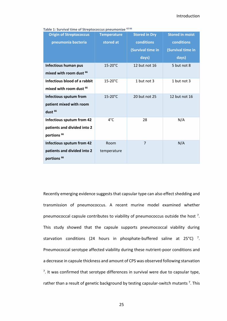
Introduction
25
Table 1: Survival time of Streptococcus pneumoniae 62 66
Origin of Streptococcus
pneumonia bacteria
Temperature
stored at
Stored in Dry
conditions
(Survival time in
days)
Stored in moist
conditions
(Survival time in
days)
Infectious human pus
mixed with room dust 66
15-20°C 12 but not 16 5 but not 8
Infectious blood of a rabbit
mixed with room dust 66
15-20°C 1 but not 3 1 but not 3
Infectious sputum from
patient mixed with room
dust 66
15-20°C 20 but not 25 12 but not 16
Infectious sputum from 42
patients and divided into 2
portions 66
4°C 28 N/A
Infectious sputum from 42
patients and divided into 2
portions 66
Room
temperature
7 N/A
Recently emerging evidence suggests that capsular type can also effect shedding and
transmission of pneumococcus. A recent murine model examined whether
pneumococcal capsule contributes to viability of pneumococcus outside the host 7.
This study showed that the capsule supports pneumococcal viability during
starvation conditions (24 hours in phosphate-buffered saline at 25°C) 7.
Pneumococcal serotype affected viability during these nutrient-poor conditions and
a decrease in capsule thickness and amount of CPS was observed following starvation
7. It was confirmed that serotype differences in survival were due to capsular type,
rather than a result of genetic background by testing capsular-switch mutants 7. This

Introduction
26
study suggests a possible new role of the pneumococcal capsule; pneumococcus may
be able to utilise their capsule as a nutrient source to maintain viability when the
organism is being transmitted between hosts. A further murine study found that
capsule type and amount affected shedding of pneumococcus during a colonisation
episode and this was associated with the rate of host-to-host transmission 69. The
authors highlighted that those specific serotypes which are strongly bound by mucin
are shed less by the host and therefore are less likely to be transmitted 69.
The results of these recent studies suggest that capsular type may lead to varying
rates of pneumococcal transmission and could explain why different serotypes have
varying propensity to cause outbreaks of disease. Serotypes which can survive for
longer in the environment could increase the likelihood of acquisition, colonisation
and potentially disease in a new human host.
Hand to nose transmission of S. pneumoniae
Hand washing interventions can reduce the transmission of disease spread by the
faecal-oral route, and is an effective and feasible means of reducing rates of
gastroenteric infections in developing countries 70. Hand washing interventions may
also be a promising intervention against acute respiratory infections. The hands can
be vectors for respiratory microorganisms which are shed from the nose and mouth
to a new host’s mucous membranes 70. A meta-analysis of 8 studies conducted in
2006 showed that hand washing interventions could reduce the risk of acute
respiratory infections (ARI’s) by around 16% (95% CI 11-21%) 70. However, the quality
of the studies examined was poor and had geographical limitations; none of the

Introduction
27
studies were conducted in developing countries and 6/8 studies were conducted in
the USA. Therefore, the generalisability r of the findings are uncertain 70.
When evaluating the possible impact of transmission of infection by self-inoculation,
it is important to consider the prevalence of face-touching behaviour. Self-
inoculation is a type of contact transmission where contaminated hands make
contact with other parts of the body such as the mouth, eyes and nostrils ,which
subsequently introduces bacteria into those sites 71. An observational study from
1973 monitored a total of 124 adults in a lecture theatre or Sunday school
environment for 30-50 minute periods 72. They observed 29 episodes of nose picking
(0.33 hr-1) and 33 episodes of eye rubbing (0.37 hr-1) 72.
Two more recent studies reported much higher rates of hand to face contact 71 73.
Nicas et al in 2008 conducted an observational study looking at the hand touching
rate of 10 volunteers. Participants sat alone at a desk for a 3-hour period performing
office-type work and the frequency of hand contact with the eyes, nostrils and lips
was examined. The average total contact rate per hour was 15.7 73. They noted that
there was significant inter-individual variability in total hand contact with facial
membranes 73. This was shown by a 35-fold difference in the range limits (lowest
total hand to face contacts 3 vs. highest 104) 73.
Kwok et al 71 observed 26 students’ face touching behaviour during two 2-hour
lectures. They found that on average each of the students touched their face 23 times
per hour; of these 44% (1024/2346) involved contact with a mucous membrane 71.

Introduction
28
Of the 1024 touches that involved mucous membranes, there was found to be a fairly
even split between the mouth (36% 327/1024), nose (31% 318/10247 and eyes (27%
273/1024) with 6% (61/1024) of touches being a combination of mucous membrane
contact 71 (Figure 3). The students involved in this study were medical students who
had previously had one 4-hour infection control lecture which involved teaching
about hand hygiene, aseptic technique and transmission-based precautions. Kwok et
al concluded that due to the high frequency of mouth and nose touching observed in
this study, hand hygiene is an essential preventative method to break colonisation
and transmission cycles 71.
Figure 3: Average number touches of mucosal surfaces observed over 1 hour period adapted from Kwok et al 71 Results from a behavioural study involving medical students in Australia. Face-touching behaviour was observed via videotape recording; frequency of hand to face contact was analysed.
NOSE 3 average touches/hour Average duration 1
MOUTH 4 average touches/hour Average duration 3 second (range 1-12)
EYE 3 average touches/hour Average duration 1

Introduction
29
The above studies support the hypothesis that hands could be an important vector
for bacterial transmission to facial mucous membranes. S. pneumoniae colonises the
upper airway; through touching of contaminated surfaces, sneezing, coughing and
rubbing/picking of the nose pneumococcus could contaminate the hands. Due to this
it is biologically plausible that hand to face contact could be a significant transmission
pathway. There is very little research investigating the link between pneumococcus
hand contamination and rates of nasal colonisation currently.
Only two relevant studies in humans were found; the first found pneumococcal nasal
colonisation in 83% (67/81) of children studied in Papua New Guinea with a
corresponding hand contamination rate of 22% (18/81) 74. They also found that 17/20
children who were found to have hand contamination with pneumococcus had the
same serotype colonising their nasopharynx 74. In conjunction with testing children,
the study also evaluated pneumococcal hand contamination of mothers; two
mothers had pneumococcus recovered from their hands but there was no
concordance with the serotype found colonising their children’s nasopharynges 74.
The second study compared the rates of pneumococcal hand contamination and
nasal colonisation in two groups of children in Australia; a remote aboriginal
community group (n=89) and an urban child care centre group (n=294) 42. They found
a two-fold increase of nasal colonisation rates in the remote group compared to the
urban group (90%, 80/89 positive for colonisation remote group vs. 43%. 125/294
urban group) using nasal swabs 42. This correlated with higher hand contamination
rates in the remote group; the remote group were >9 times more likely than the

Introduction
30
urban group to have pneumococcal hand contamination (37%, 33/89 positive hand
swab remote group vs. 4%, 13/294 urban group) 42. This suggests that pneumococcal
shedding during a colonisation episode is possible and that the hands could be a
possible vehicle for pneumococcal transmission in the community.
Rodent models investigating pneumococcal transmission are adding to the current
understanding of the extent of airborne transmission versus contact-dependent
transmission. Transmission secondary to infected secretions from a person colonised
with pneumococcus may involve direct person-to-person spread or may involve
contaminated surfaces or fomites 20. A recent study demonstrated that an
environmental reservoir of pneumococcus can facilitate transmission 50. In this study
colonised mice were kept in separate cages from un-colonised mice. Both sets of
mice had an uncolonised mother; three times per day mice were switched between
cages but were never in contact with each other 50. Over 50% transmission rate was
observed; the investigators concluded that contamination of the mother’s teats with
pneumococcus was the source of transmission between the colonised and
uncolonised mice. They confirmed that the bedding was not an environmental
source of pneumococcal transmission by doing a further experiment. When they
moved the mothers between cages of colonised and uncolonised mice transmission
was still seen suggesting that pneumococcus was likely acquired from the surface of
the mothers 50.

Introduction
31
Reduction of transmission
The benefits of hand washing for reducing the spread of respiratory bacterial
pathogens is uncertain with the evidence currently available 42. Previous intervention
studies using hand hygiene interventions to reduce viral transmission pathways
support the hypothesis that both direct and indirect contact are important in
transmission of viruses from person to person. A randomised control trial looking at
the effect of infection control measures in child care centres showed only a 5%
overall reduction of incidence of “colds” 75.
There is limited research assessing whether hand hygiene interventions and infection
control teaching can reduce upper respiratory bacterial pathogen transmission. A
cluster randomised control trial (RCT) conducted in 20 child care centres in Australia
investigated this question76. Ten centres (219 children) had one infection control
training session for staff and had regular follow up visits by researchers to implement
20 second hand washing, barrier nose-wiping, removal of contaminated toys and
non-touch sunscreen application 76. The other 10 centres (235 children) acted as
controls with no intervention given. The researchers found no reduction in
transmission of bacterial pathogens (adjusted incidence rate ratio, IIR, 0.97 95% CI
0.88, 1.08) or respiratory illness in the intervention group (adjusted IIR 1.00 95% CI
0.93, 1.01) 76. Transmission of bacterial pathogens (S. pneumoniae and H. influenza)
was assessed with nasal swabs undertaken every 2 weeks for 6 months and childhood
illness was reported by parents. However, interpretation of these findings was
limited by poor uptake of the hand hygiene practices in the intervention centres

Introduction
32
(proportion of hygiene practices consistent with guidelines; weighted mean
difference 14%, 95% CI 9, 19).
1.4 Pneumococcal disease treatment and prevention
Pathogenicity
Streptococcus pneumoniae was first described by George M Sternberg who isolated
it in 1880 from a colonised individual 77. He also described recovery of the organism
from the saliva of healthy students which established that the pneumococcus is part
of the normal human microflora 77. S. pneumoniae was first described as a cause of
lobar pneumonia in 1883 by Friedlander and Talamon. Diagnosis of pneumococcal
pneumonia improved following the development of the Gram stain as a method to
differentiate bacterial species in 1884. It is a Gram-positive bacterium which has the
potential to cause invasive infections and is considered to be an extracellular
bacterial pathogen 78. Extracellular pathogens can replicate or persist on mucosal
surfaces in the human body or in tissues outside host cells. They can also spread
quickly or establish an infection if they successfully contend with host humoral
defences and cellular immune mechanisms (e.g. T cells and phagocytes) 78. Due to
this, the ability of extracellular pathogens to evade clearance by the body’s humoral
and cellular immune defence mechanisms is an important determinant of
pathogenicity.

Introduction
33
The pneumococcus can release a pore-forming toxin called pneumolysin; which at
low levels can induce apoptosis of cells and high levels cause lysis of all cells with
cholesterol in their membrane 79. This pathogen also has pro-inflammatory cell wall
components such as C-polysaccharide and F-antigen. These release tissue damaging
enzymes such as neuraminidase and have adhesins, such as pneumococcal surface
protein A (PspA), that can bind to cell surface carbohydrates which play important
roles in combating the host’s immune responses 80.
The polysaccharide capsule surrounds the pneumococcal cell wall which is comprised
of peptidoglycan and teichoic acid. This capsule protects the bacteria from
phagocytosis by obstructing leukocyte fixation onto the cell wall 1. Pneumococcal
serotypes each have chemically distinct capsules which can affect:
• Tendency to cause outbreaks,
• Antibiotic resistance profiles,
• Likelihood of causing invasive disease (serotypes 1 and 7E are
prominent serotypes for invasive disease),
• Prevalence of mucosal disease or nasopharyngeal colonisation;
different serotypes have varying propensity to cause colonisation and
invasive disease 1 2.
Geographical variation in serotypes is often dependent on the period studied and
differences in age distribution between host’s; serotypes most commonly observed
in young children include 6B, 9V, 14, 19F and 23F 1 2 80. These differences in serotypes

Introduction
34
may be due to the quantity and degree of encapsulation of the polysaccharide
capsule. Thicker capsules protect against neutrophil-mediated killing in vitro, and
may prevent clearance of colonisation by the host 2. Other genetic factors relating to
the pneumococcus are also likely to impact on virulence such as bacterial adhesins.
Pneumococcal disease
Streptococcus pneumoniae is the most common cause of acute otitis media, sinusitis
and pneumonia worldwide and also causes a significant number of cases of
meningitis 81. Epidemiological studies have shown that there is an inverse
relationship between the frequency and severity of types of pneumococcal disease
(see Figure 4).
The archetypal presentation of a patient with pneumococcal pneumonia has been
historically described as symptoms including sudden onset of chills and pleuritic
chest pain, closely followed by fever and rusty sputum production. However, many
present with more non-specific symptoms especially in the elderly and in young
children81. Pneumococcal pneumonia can progress to pneumococcal bacteraemia in
some patients, rates of bacteraemia have been estimated at 20-25% 82 and mortality
from bacteraemia has stayed high at 20-30% despite antibiotic treatment 83.

Introduction
35
Figure 4: Burden of pneumococcal disease adapted from Edwards and Griffin 84. Estimated pneumococcal disease cases per year in adults and children in USA taken from CDC report 2002 (www.cdc.gov/nip/publications/surv-manual/chapt09_pneumo.pdf)
Pneumococcus is considered to be the most common cause of CAP worldwide and in
the UK is the most common cause of CAP in children ≤2 years of age 85. Timely and
effective management of pneumococcal pneumonia can be difficult. It is hindered by
low rates of microbiological pathogen confirmation in patients with suspected
pneumonia and lower respiratory tract infections (LRTI) 86 87. Following spread of the
pneumococcus to a sterile site it can also cause IPD which encompasses a range of
diseases including bacteraemia, meningitis, empyema and septic arthritis.
Penicillin has been the mainstay of antibiotic treatment for any pneumococcal
infection for over half a century 81. However, increasing antibiotic resistance is a
growing problem worldwide. S. pneumoniae is no exception to this and we have
Meningitis
Bacteremia
Pneumonia
Otitis Media
Invasive
Non-invasive
3,000
60,000
135,000
6,000,000

Introduction
36
observed increasing antibiotic resistance develop specifically in serotypes that have
high prevalence in children for example serotypes 6, 14, 19 and 23.
Due to these diagnostic difficulties and increasing problems with antibiotic
resistance, strategies to prevent pneumococcal disease are becoming increasingly
important.
1.4.2.1 Burden in UK and worldwide and risk factors for disease
The WHO estimates that 1.6 million deaths are caused by pneumococci annually,
with over a million of these deaths attributed to pneumococcal pneumonia in
children under the age of 5 in developing countries 81 88. The global pneumococcal
disease burden in children, especially under 5 years of age is well documented. In
2000 it was estimated that there were 13.9 million cases of pneumococcal
pneumonia per year in this age group. The burden of pneumococcal disease in adults
is less well known.
Over the last 100 years, the incidence of pneumococcal pneumonia has stayed stable
but a significant improvement in mortality rates has been observed following
widespread antibiotic use 89. In the United Kingdom (UK) and high-income countries,
the yearly incidence of community acquired pneumonia (CAP) is approximately 1%
of which half is attributable to S. pneumoniae.

Introduction
37
Overall incidence of pneumococcal pneumonia is 5 in 1000, with the incidence being
significantly higher in high risk populations such as infants and the elderly 81.
Pneumococcal pneumonia can progress to invasive pneumococcal disease (IPD) (e.g.
bacteraemia). The annual incidence of this is 10-20 cases per 100,000 individuals
annually in North America and Europe 81. The risk of IPD increases by more than 20
times this rate in young children attending day care centres 90. In Western countries,
the incidence of pneumococcal disease rises in the winter months and the increased
rates of viral respiratory infections observed during this period could be the pre-
disposing factor for this 81.
Both age and gender are found to be important risk factors for pneumococcal
pneumonia 81. The incidence of pneumococcal pneumonia is up to 50 times higher in
the elderly (over age of 65) and in the very young (under age of 2), with a male:
female ratio of approximately 1.5-2:1 81. Many underlying co-morbidities have been
found to pre-dispose patients to pneumococcal disease. A case- control study
investigated this retrospectively. They compared 63 men with culture proven
pneumococcal infections to 130 uninfected control patients and calculated relative
risks using logistic regression analysis 91. They found that the following conditions
were statistically significant independent risk factors (relative risks shown in
brackets); dementia (5.82), seizure disorders (4.38), heart failure (3.83),
cerebrovascular disease (3.82, institutionalisation (3.13) and chronic obstructive
pulmonary disease (COPD; 2.38) 91. Concurrent respiratory tract infection, especially
influenza viral infection, is also a risk factor for pneumococcal pneumonia 81.

Introduction
38
Pneumococcal disease prevention: current pneumococcal
vaccines
In the UK, there are currently three vaccine formulations licenced for the prevention
of pneumococcal infections; the pneumococcal polysaccharide vaccine (PPV23) and
two pneumococcal conjugate vaccines (PCV10 and PCV13) 92. See Table 2 for
comparisons of these vaccines.
Table 2: Comparison of currently licenced pneumococcal vaccine formulations in the UK Table reports specific aspects of the pneumococcal vaccines which have been, or currently are licenced for use in the UK. Further discussion about vaccine immunological profile and efficacy discussed in sections 1.4.3 and 1.4.4.
PCV 7 PCV13 PPV23
Serotypes
covered
1, 4, 5, 6B, 7F, 9V,
14, 18C, 19F, and
23F.
1, 3, 4, 5, 6A, 6B, 7F,
9V, 14, 18C, 19A, 19F,
and 23F.
1, 2, 3, 4, 5, 6B, 7F, 8, 9N,
9V, 10A, 11A, 12F, 14,
15B, 17F, 18C, 19A, 19F,
20, 22F, 23F and 33F.
UK Vaccination
recommendation
From 2006-2010 93:
recommended for all
children at 8 weeks
old, 16 weeks old
and 1 year old.
Since 2010 93:
recommended for all
children at 8 weeks
old, 16 weeks old and
1 year old.
Since 2003 93:
recommended for all
over 65’s and younger
adults who have chronic
illnesses that put them at
higher risk of invasive
pneumococcal disease.
Effective against
S. pneumoniae
Invasive pneumococcal disease.
Reduction in nasopharyngeal colonisation,
leading to reduction in transmission of
vaccine serotypes (in turn reduced incidence
of mucosal disease).
Moderately effective against mucosal disease
such as pneumonia and AOM 94.
Invasive pneumococcal
disease.
Non-conclusive evidence
on protection against
pneumococcal
pneumonia or other
mucosal infections92.

Introduction
39
The currently used PPV contains polysaccharide from 23 different serotypes (PPV23).
PPV23 elicits a T-cell independent, humoral immune response. Evidence suggests this
protects against IPD but there is debate about this vaccine’s effectiveness against
pneumonia 92. The immune response to PPV is not long lasting and does not show a
booster response upon challenge with native polysaccharide. Due to this, protection
induced by this vaccine is limited. A major limitation is lack of efficacy in infants under
2 years of age whose immune systems are immature 95. PPV immunisation does not
protect against nasopharyngeal colonisation by pneumococcus, therefore no herd
immunity was observed after vaccine implementation. Herd immunity is the indirect
protection against an infectious disease whereby individuals who are not immune
can benefit and which occurs when many of the population are immune following
vaccination.
A meta-analysis which evaluated 25 studies ( 18RCTs and 7 non-RCTs) found strong
evidence that PPV is effective against IPD with overall efficacy of 74% (95% CI 56% to
85%) 96. No statistical heterogeneity was found when all RCTs were included in
analysis However, there was a statistical difference between heterogeneity of studies
conduction among different populations such as healthy adults in low-income
countries (P <0.01) 96. Results of effectiveness against pneumonia were inconclusive
96. Efficacy against all-cause pneumonia in low-income countries was reported (OR
0.54, 95% CI 0.43 to 0.67) 96. However, this was not the case in high-income countries
in the general population (OR 0.71, 95% CI 0.45 to 1.12) or those with chronic disease
(OR 0.93, 95% CI 0.73 to 1.1) 96. A further meta-analysis supported this finding and
concluded that PPV did not appear to be effective against pneumonia even among

Introduction
40
elderly patients or adults with chronic disease for whom the vaccination is
recommended 97.
Some randomised control trials have found protective effect of the PPV against
pneumonia; this prospective randomised study included 1006 nursing home
residents. It reported a statistically significant reduction in pneumococcal pneumonia
cases in the vaccine arm compared to control (14/502 ,2.8% vs 37/504, 7.3%;
P<0.001) 98. Due to this specific study population (mean age of 84, nursing home
residents many with co-morbidities), it is difficult to generalise the findings to larger
population. In addition, a non-randomised observational study of 27,204 individuals
aged ≥60 years in Spain reported that recent vaccination with PPV23 (<5 years ago)
reduced risks of bacteraemic pneumococcal CAP (hazard ratio [HR], 0.38; 95%
confidence interval [CI], .09–1.68) and non-bacteraemic pneumococcal CAP (HR, 0.52;
95% CI, .29–.92)99. This was only observed following sub-analysis of patients who had
received the vaccine within the last 5 years, when evaluating the full cohort, no
protective effect was observed 99.
PVC13 is the most commonly used polysaccharide conjugate vaccine worldwide; this
vaccine covers pneumococcal 13 serotypes 100. PCV13 contains purified
polysaccharides of the capsular antigens of each of the 13 pneumococcal serotypes
covered. These are individually conjugated (coupled) with a nontoxic diphtheria toxin
(CRM197, CRM cross-reactive material). The conjugation of the capsular
polysaccharide to a carrier protein in PCV13, unlike PPV, activates a T-cell dependent
antibody response which leads to mucosal immunity and immunological memory 92

Introduction
41
101. Previous studies suggest that this leads to immunity for the individual user but
also leads to herd immunity 92. Global vaccine strategies have been very successful,
and 132 countries have introduced PCV into their national immunisation programs
(see Figure 5.
Figure 5: Worldwide current or planned implementation of PCV into national immunization schedule as of September 2016. Taken from World Health Organization, Immunization Vaccines and Biologicals Database, September 2016. http://www.who.int/immunization/monitoring_surveillance/data/en 102
Since the introduction of childhood PCV there has been a decrease in IPD and
pneumonia in children and similar reduction in rates of IPD have been observed in
older adults 92. A large randomised control trial, which included 85,000 adults aged
65 years of age or older was conducted by a PCV vaccine manufacturer 103. This study
showed 45.6% (95% confidence interval [CI]=21.8-62.5%) efficacy of PCV13 against
all vaccine-type pneumococcal pneumonia and 75% (CI=41.4-90.8%) efficacy against
vaccine-type IPD in adults ≥65 years of age 103.

Introduction
42
Rationale behind new vaccine development
Despite the success of the PCV and PPV immunisations in reducing pneumococcal
disease and mortality worldwide, the widespread use of these vaccines and addition
into public immunisation programmes has highlighted several limitations 92 101 104.
These include:
• Protective immunity is limited to the specific serotypes contained in each of
the PPV23 and PCV13 – there are currently over 90 known serotypes of S.
pneumoniae.
• ‘Serotype replacement’ is a major limitation of the PCVs. Among
asymptomatic carriers, the prevalence of non-vaccine serotypes ([NVT] non
PCV 13 serotypes) has increased significantly (serotype replacement).
However, there does not seem to be any impact of overall pneumococcal
colonisation rates 105. A review, also reported an increase in NVT
pneumococcal disease 105. The true amount of replacement may be
underestimated due to biases in the pre-vaccine colonisation data.
Surveillance systems may underestimate the prevalence of serotypes with
lower invasive potential. Because these systems monitor invasive
pneumococcal disease, serotypes which do not cause disease are likely to be
underreported. Epidemiology studies can also underestimate prevalence of
pneumococcal serotypes which have short colonisation durations. This is
because surveillance tests for colonisation often are undertaken weekly or
less frequently in these studies105. In addition, simultaneous colonisation with

Introduction
43
multiple pneumococcal serotypes has been increasingly reported with
improvements in molecular techniques for pneumococcal detection 40. Due
to this, NVT may have been more prevalent colonisers but at lower density in
a multiple serotype colonisation episode and therefore under detected.
• Limited efficacy against certain serotypes covered in PCV13 (serotype 1 and
5) 92. Clinical trials in South Africa 106 107 and The Gambia 108 109 failed to show
evidence of efficacy of PCV against serotype 1. A further study, reported
PCV13’s vaccine effectiveness at 62% and 66% against serotype 1 and 3
respectively although confidence intervals spanned zero 110. A further study
found that the vaccine effectiveness for serotype 3 was not significant (26%,
95% CI, -69%-68%) 111. In developing countries serotype 1 and 3 continue to
be prevalent serotypes causing IPD therefore the lack of efficacy is
problematic.
• Geographic variation in protection from both PCV13 and PPV23 due to
varying serotype distribution worldwide. The current vaccines favour
covering serotypes that are most prevalent in western countries such as USA
or Europe rather than the serotypes that cause most of invasive disease in
developing countries 9. This was a larger issue with the first PCV vaccine; PCV
7 than for PCV13.
• Hypo-responsiveness to the capsular polysaccharide is observed with PCV
immunisation (serotype-specific) if children had an episode of pneumococcal
nasal colonisation shortly before the first dose of PCV 112 113. This is only
partially overcome after the 2nd or 3rd dose of the vaccine. There has also been
reports that repeated doses of bacterial polysaccharides may induce a state

Introduction
44
of immune tolerance or hypo-responsiveness. All studies, except one 114,
reported that serotype-specific pneumococcal antibody concentrations were
lower after the second dose of PPV compared the first dose 115-119 .
• Complexity and high costs of protein-conjugate vaccines incurred by
pharmaceutical companies and purchasers may impact on the future
development of these vaccines to expand coverage to more serotypes 92.
• Transient efficacy in healthy elderly. Clinical effectiveness of PPV in older
adults is likely to diminish over time. The reduction in pneumococcal capsular
antibodies over time following vaccination has been shown in PPV 120.
Reduction in clinical effectiveness over time is also seen in other
unconjugated polysaccharide vaccines 121
• Lack of proven efficacy in very frail elderly or immunocompromised.
Evidence suggests that PPV23 is effective in preventing invasive
pneumococcal disease in healthy young adults and in the healthy older
population 122. However, the vaccines effectiveness in the
immunocompromised and very elderly has not been demonstrated 123 122 ,
Novel vaccine development
Limitations of the current vaccines available for protection of pneumococcal disease
have driven the development of novel vaccines some of which would be serotype-
independent. New vaccine approaches include new polysaccharide conjugate
technologies, pneumococcal whole cell vaccines and vaccines which are based on
highly-conserved noncapsular protein antigens 92 124 125. Due to the limitations

Introduction
45
discussed above, there are specific criteria any new vaccine must satisfy to make a
significant impact on disease (target product profile);
• New vaccines should be highly conserved and develop immunity to all
pneumococcal serotypes
• There should be evidence to suggest mucosal immunity and immunity against
nasopharyngeal colonisation
• The vaccines should have evidence of immunogenicity with evidence of non-
inferiority to a licensed pneumococcal vaccine
• The vaccines should induce antibody and cell mediated immune responses
• The immunity should be long-lasting
• The vaccine should be designed to prevent disease in the majority of the
population including children under 2 years of age
• The vaccine should be low cost to produce
One possible new vaccine approach is modification of the conjugate vaccine; an
international non-profit organisation (PATH) is investigating two alternatives; firstly
to develop a conserved pneumococcal protein which would be used as a carrier for
a specific number or polysaccharides which could be chosen and changed to meet
specific geographical needs 124. Secondly, they are looking at ways to accelerate the
development of vaccines that target serotypes which are more prevalent in
developing countries and are also developing new strategies to reduce the cost of
manufacturing 124.

Introduction
46
An alternative strategy is the use of broadly conserved protein combinations for
vaccines. The pneumococcus has multiple proteins exposed on its surface which
could be used as possible vaccine antigens. Well-studied examples of these are
pneumococcal surface protein A (PspA) and choline-binding protein A (PspC). While
other virulence factors that are also at the forefront of protein-based vaccine
development include choline-binding protein (PcpA), pneumolysin (Ply) and
pneumococcal histidine triad protein D (PhtD) 92 124. Research into these protein-
based serotype independent subunit vaccines have reached varying stages of
advancement. Recombinant PspA has been studied in a phase 1 clinical trial where it
was found to be safe in humans and was also immunogenic 126. This study used a
passive transfer murine challenge model as a surrogate for protection of humans 126.
Human participants were administered recombinant PspA, pre- and post-immune
serum samples were examined 126. The authors reported that human antibody to
PspA could protect mice from pneumococcal infection 126 .
Another promising area of research is whole cell pneumococcal vaccines, either in
live attenuated form or killed form, in which many pneumococcal antigens would be
present at once 92 124. Potential benefits of a killed bacteria whole-cell vaccine
development would be the very low cost of manufacture and that it could protect
against all serotypes and could lead to comprehensive mucosal and systemic
immunity 124 127.

Introduction
47
1.4.5.1 Controlled human infection studies
Controlled human infections models (CHIM) involve the experimental infection of
study participants with a pathogen. They provide the opportunity for researchers to
study organisms, their incubation periods and clinical disease. More recently these
studies have been used to accelerate the development of new drugs and vaccines for
infectious diseases by adding significant data prior to large scale efficacy trials. The
first controlled infection studies were described in the 17th century. The ethics of
these preliminary CHIM studies were questionable with the most infamous example
of Edward Jenner inoculating his gardeners son with cowpox in 1796, and following
this repeatedly with smallpox lesion material 128.
There have been significant improvements in ethical considerations of all research
and specifically CHIM studies since these preliminary studies; CHIM studies must
conform to a strict ethical framework and go through rigorous independent review
prior to starting. However, some still argue that CHIM studies are inherently
unethical due to concerns about non-maleficence 129 130. There are significant global
health benefits from these studies but to the individuals involved there is an inherent
risk of illness 128. In addition, similar to phase one studies, there are no direct benefits
to participants. Due to this, CHIM studies can only be carried out to investigate
treatable or self-limiting diseases 128.
The experimental human pneumococcal challenge (EHPC) model is different to the
majority of CHIM by having colonisation rather than infection as its primary endpoint.

Introduction
48
Due to this, from an ethical and clinical standpoint, the inherent risks and potential
for harm is much lower. The Liverpool EHPC team have inoculated over 1000
participants with pneumococcus over the last 9 years with no related significant
adverse events.
Experimental Human Pneumococcal Colonisation studies and
their use for vaccine research
A major roadblock in the process of developing new protein vaccines has been a
means of prioritising between proposed vaccine candidates 131. Nasopharyngeal
colonisation is likely the source of pneumococcal transmission into sterile sites and
therefore is likely a prerequisite for invasive disease 31 131. It has therefore been
proposed as a marker for vaccine efficacy 31 131. For new vaccines due to come to the
market, protection against colonisation may predict the overall protective effect
against mucosal or invasive disease 101. Experimental human pneumococcal
colonisation (EHPC) can be induced in humans. This was first shown by McCool et al
29 in 2002; this group successfully induced nasopharyngeal colonisation following
pneumococcal inoculation with serotypes 6B and 23F and showed that controlled
human infection with pneumococcus was safe 29 30.
Following these initial studies in the United States of America (USA), a safe and
reproducible EHPC model was establish at LSTM, which can be used to test the
protection induced by vaccination against nasopharyngeal colonisation 101.
Preliminary studies investigated what the optimal sampling method would be to
identify nasopharyngeal pneumococcal colonisation. The team compared

Introduction
49
nasopharyngeal swab (NPS) to nasal wash for the detection of potential respiratory
pathogens. This study showed that nasal wash was more comfortable for volunteers
and was significantly more likely to detect pathogens than NPS 132.
The model allows assessment of the immune responses at the mucosal surface and
systemically following inoculation with live whole bacteria. It can also be useful to
help evaluate and compare new vaccines that are possibly coming to market; it could
evaluate pneumococcal colonisation protection and any change in nasopharyngeal
flora following vaccination 101. To increase the generalisability of the model and its
use for vaccine development, it is important to test multiple serotypes of S.
pneumoniae.
It has been previously reported that pneumococcal serotype has a significant role in
determining colonisation. Some serotypes are more likely to be carried but have low
potential to cause infectious or invasive disease. Conversely others are more
commonly observed in pneumococcal disease 133. A previous human pneumococcal
challenge study performed by the Weiser group in the USA used serotype 23F P833.
They inoculated participants with either 5,000 CFU/naris, 7,000 CFU/naris or 17,000
CFU/naris to assess if there was a dose dependant relationship to colonisation
acquisition and achieved colonisation rates of 43% (6/14) with a duration of
colonisation ranging from 27-122 days 29. Following this, our team (Liverpool EHPC
team), retested the 23F serotype in addition to serotype 6B in a dose-ranging human
challenge study. Colonisation rates for 23F in this study were poor for all inoculum
doses (Figure 6) 134.

Introduction
50
Figure 6: Dose ranging curve for serotypes 6B and 23F (unpublished work from J. Gritzfeld thesis) 10 participants were inoculated with either serotype 6B (circle) or 23F (square) for each of the six doses from 1x10⁴ CFU/naris to 3.2x10⁵ CFU/naris. Y axis shows percentage of colonisation positive participants in each group defined as number of participates found to be an experimental coloniser of pneumococcus in nasal washes at any time point following inoculation.
Due to the poor colonisation rates observed above, the 23F serotype used was
sequenced to look for a cause of these results. It was discovered that this serotype
differed from the original P833 serotype tested in the USA 29. Murine models had
previously demonstrated that mutations in amiC gene could significantly affect the
ability of the pneumococcus to colonise. It was found that there was a frameshift
mutation in the amiC gene in the 23F serotype used in this initial study, which was
hypothesised to be the cause of these poor colonisation rates. However, for the ‘new
serotypes’ study (see below) the serotype used was a naturally-derived isolate of
P833; P1121. This contained a stable form of the wild-type amiC gene; so we
expected to be able to reproduce findings from the US-based group of 50%
colonisation rates at 7,000 CFU/naris and 75% at 17,000 CFU/naris 29.

Introduction
51
1.5 Project aims
This project focuses on improving knowledge about the acquisition of S. pneumoniae
colonisation using controlled human infection trials.
Aim 1: To investigate whether the hands can be a vector for transmission of S.
pneumoniae from hands into nasopharynx, leading to colonisation.
a) We will also investigate whether asymptomatic viral infection at the
time of exposure to pneumococcus affects acquisition of
pneumococcal colonisation following transmission from hands to
nose
Transmission of S. pneumoniae from person-to-person is thought to occur due to
airborne respiratory droplets. Epidemiological data provide evidence that
transmission is influenced by overcrowding and concurrent viral respiratory tract
infections 8 43. There are no published data investigating specifically how
Streptococcus pneumoniae is spread and testing different routes of transmission.
(Chapter 4: HAND TO NOSE STUDY)
Aim 2: To investigate the propensity for two different pneumococcal serotypes (23F
and 15B) to cause experimental pneumococcal colonisation in healthy adults
a) We will investigate whether exposure to pneumococcus or
pneumococcal colonisation is symptomatic in healthy adults and

Introduction
52
b) Investigate the host’s polysaccharide specific antibody (IgG) response
following experimental challenge
To improve our knowledge about nasal pneumococcal colonisation we planned to
extend the EHPC model by colonising participants’ nasopharynx with different
serotypes of S. pneumoniae (23F and a non-vaccine type [15B] were tested). Success
in this would ensure that the model is more generalisable and useful for further
development of vaccine testing studies. Specifically, the development of a
colonisation model with a non-vaccine type serotype would be an important step for
vaccine testing. New pneumococcal vaccines will need to demonstrate an impact on
reducing colonisation, and protein-based vaccines will need to show efficacy against
serotypes not covered by PCV13. (Chapter 5: NEW SEROTYPES STUDY)

Methods
53
2 Methods

Methods
54
2.1 Overview
This chapter describes the methods relevant to both studies described in this thesis.
It covers methodology used to investigate the main aims of the thesis outlined at the
end of Chapter 1. Specific methods relevant to one study are considered separately
in each chapter for ease of reading. The chapter is divided into subsections which
cover study set up, clinical procedures, methodology and laboratory processes.
2.2 Study set up: Research in the National Health Service (NHS)
Sponsorship
All clinical research undertaken in the NHS requires a sponsor. The definitions of a
sponsor and roles and responsibilities are detailed in the Research Governance
Framework, Edition 2 2005 135 (guidelines in place when New Strains study was set
up) and later by UK Policy Framework for Health and Social Care Research 2017 136
(see Table 3).The sponsor needs to be an individual, organisation or group which is a
legal entity involved in either the funding, running, hosting of the research or
employing the research staff.
Sponsorship can be sole (in the case of Hand to Nose study- single sponsorship by
LSTM) or co-sponsored by more than one organisation (in the case of New Serotypes
study- joint sponsorship between LSTM and Royal Liverpool and Broadgreen
University Hospital Trust [RLBUHT]). Co-sponsorship involves a pre-agreed division of

Methods
55
the sponsor responsibilities prior to initiation of the study. Agreement for LSTM or
RLBUHT to sponsor a research study involves the research team completing an
application to the organisation’s research governance department. Initially
sponsorship in principle is obtained. Full sponsorship is only agreed once the
organisation receives documentary evidence of ethical and relevant national
regulatory approvals. No study activities should be completed prior to full approval
and sponsorship.
Table 3: Definitions and responsibilities of a sponsor taken from Research Governance Framework 135 and UK policy for Health and Social Care Research 136
Research Governance Framework
for Health and Social Care
(Second edition, 2005) 135
UK Policy Framework for
Health and Social Care
Research (2017) 136
Definition
‘Individual, organisation or group
taking on responsibility for securing
the arrangements to initiate, manage
and finance a study. A group of
individuals and/or organisations may
take on sponsorship responsibilities
and distribute them by agreement
among the members of the group,
provided that, collectively, they make
arrangements to allocate all the
responsibilities in this research
governance framework that are
relevant to the study.’ 135
‘The sponsor is the individual,
organisation or partnership that
takes on overall responsibility
for proportionate, effective
arrangements being in place to
set up, run and report a research
project. All health and social care
research has a sponsor.’ 136
Responsibilities
Main responsibility of the sponsor is to assure the quality of the research,
including ensuring participant’s wellbeing, all legal requirements are met,
the research team are adequate to carry out the work and have resources
and arrangements in place for the study to be completed. More detailed
set of responsibilities are set out in the Research Governance Framework
for Health and Social Care, Second edition 2005 135 and the UK Policy
Framework for Health and Social Care Research 2017 136.

Methods
56
Health Research Authority (HRA)
HRA approval process for research trials was fully implemented in the UK in March
2016. This new system was designed to combine the independent ethical
opinion/approval by the Research Ethics Committee (REC) and a review of research
studies against NHS standards 137. This is an assessment of governance and legal
requirements needed for each study which is undertaken by specific HRA staff,
previously undertaken by individual NHS Research Development and Innovation
(RD&I) offices.
HRA approval was only required for the Hand to Nose study because the New
Serotypes study received ethical approval before this new system was fully
implemented. HRA application is encompassed into the Integrated Research
Application System (IRAS) form, the research team also provides the HRA with
Statement of Activities and Statement of Events forms which together aim to capture
all information about study activities and study related information at a local level.
There are many steps involved in obtaining HRA approval for a clinical trial. Figure 7
below shows the main steps in obtaining approval. The Hand to Nose study gained
HRA approval in February 2017. For the New Serotypes study which was approved
pre-implementation of HRA approval, the sole responsibility for ensuring the study
complied with all legal and governance requirements fell to RLBUHT RD&I
department, see below section 2.2.4.

Methods
57
Figure 7: HRA approval process applies to all research projects taking place in the NHS in England 112 HRA approval encompasses assessment of governance and legal compliance and independent ethical opinion by the Research Ethics Committee.
Ethics: Integrated Research Application System (IRAS) and
Research and Ethics Committee (REC)
One of the HRA’s main functions is ensuring that research undertaken in the NHS is
ethically acceptable. To do this they have multiple RECs countrywide. They aim to
protect research participants by ensuring that studies are carried out in an ethical
way and ensure that participants rights, safety, dignity and wellbeing is protected. In
addition, they aim to ensure that research being undertaking is of potential benefit
to science, society and the participants. Both studies in this thesis (Hand to Nose
[Chapter 3] and New Serotypes [Chapter 4]) required full REC approval because these
studies involved human volunteers and were carried out in NHS sites.

Methods
58
A separate IRAS form was created for each study and was the main basis of the REC
application. Following this, the research team attended a REC meeting date, this gave
the REC an opportunity to ask questions about the study and give feedback on
changes that they would like the study team to make to documents provided or IRAS
form. Ethical approvals for the studies were granted as shown in Table 4 below.
Table 4: REC study approvals and other approvals/registrations for research studies within this thesis
Study REC IRAS number
Sponsor Other approvals/registrations
Hand to Nose study
Chapter 3
Liverpool (East)
17/NW/0054
Approved: 02.02.2017
221034 LSTM ISRTN: 12909224 registered 27.06.2017
NIHR portfolio: 33503
HRA approval: 22.02.2017
RD&I: 5376 approved
17.03.2017
New Serotypes
study
Chapter 4
Liverpool (East)
15/NW/0931
Approved: 25.01.2016
193680 LSTM and RLBUHT
ISRTN: 68323432 registered 15.08.2017
NIHR portfolio: 20815
RD&I: 5124 approved
08.08.2016
Research Development and Innovation Department (RD&I)
Both studies discussed in this thesis were carried out in the Clinical Research Unit
(CRU) based in RLBUHT. As the site of these research studies RLBUHT has a legal
obligation to ensure that they are aware of all research undertaken on the site and

Methods
59
have carried out certain assessments prior to study initiation. The CRU is a Medicines
and Healthcare Products Regulatory Agency (MHRA) accredited phase one research
unit at RLBUHT. All participant visits for both studies were conducted in this unit.
Final RD&I approval was provided for both studies once all other approvals were in
place including:
• REC approval
• Finance team/ RD&I business team approval (contracts finalised)
• HRA approval (only Hand to Nose study as this step only implemented March
2016)
• Directorate managers approval
• CRU approval
• Evidence of sponsorship in place
• Evidence of liability insurance in place
• Evidence of Principle Investigator/Chief Investigator contract with RLBUHT
Patient and Public Involvement (PPI)
PPI is becoming increasingly important in research, and there are many benefits to
the research team and public with this involvement. Some of the many benefits
include:
• Supporting recruitment and consent

Methods
60
• Demonstrating to funders that the research topic is important and relevant
to the public and patients
• Demonstrating to funders that the study design and documents are
acceptable to the public
• Identifying possible ethical issues with the design of the study
The EHPC team have been studying pneumococcal colonisation in Liverpool over the
last 9 years. The clinical and lab EHPC team understand the importance of PPI in
research and we strived to ensure we had continual PPI events planned throughout
both study periods.
We attended many public engagement events; these events gave us the opportunity
to provide information about the research being undertaken and improve local
knowledge about research. This included us attending public open days, science fairs
which are open to the public, having regular display stands and face to face
engagement with the public at local gymnasiums and libraries. We also created a
newsletter which was disseminated to previous research participants, colleagues and
members of the public to which provided updates on findings of finished studies and
future studies.
We have also developed a role within the team for research ambassadors; individuals
who previously participated in one of the EHPC studies. For the studies described in
this thesis, research ambassadors were used to sense-check promotional and

Methods
61
information materials, to provide lay-input into the study protocols and to assist with
recruitment events.
A relatively new addition to our PPI work was utilising social media platforms as a
way of involving the public in the research undertaken. We used multiple platforms
including Facebook and Twitter to regularly upload information about our face-to-
face research events the public could attend and update followers about current
trials which were open to recruitment. This has impacted on the demographics of our
participants; the majority tended to be under 30 years of age who were more likely
to have an active social media connection 138.
2.3 Clinical procedures
Trial designs
The two studies in this thesis used a human challenge model study design.
Participants were challenged with live pneumococcus either intranasally (inoculated)
or on their hands (exposure). All participants were seen prior to this challenge for
screening and then were followed up for a period of up to 14 days post challenge.
Nasal wash and blood samples were obtained to assess for nasopharyngeal
pneumococcal colonisation and to investigate the immune response to challenge
with or without colonisation (sample obtained discussed further below).

Methods
62
Study specific trial design with study design flow charts are outlined in Chapters 3
and 4 (sections 3.2.2 and 4.2.2).
Ethical considerations
Autonomy
The study team ensured that participants were given sufficient information, written
and spoken, without the use of medical jargon or other language requiring specialist
knowledge. This ensured that participants could understand the research objectives
and the risks and benefits of any procedures. Participants were always given enough
time to consider the information before consenting to any involvement. We ensured
that all participants did not feel pressured or persuaded into participating in the
research study. Participants were financially compensated for their participation in
the studies. Offering this payment was not intended to unduly influence participation
but to compensate participants for their time and travel expenses incurred due to
taking part in the trial.
Participants that were recruited via the Consent4Consent database (see below
section 2.3.3) were contacted regarding the research as they had previously
expressed an interest in research. These participants had already given their consent
to be contacted about future research that they may be eligible for. Participants were
informed that they had the right to withdraw their consent and therefore withdraw
from the study at any time without giving reason.

Methods
63
Non-maleficence
The research team had a responsibility to minimise the risk of harm to the
participants. We ensured that all researchers in the team had sufficient knowledge
about the proposed interventions and potential risks. In addition, strict safety
procedures were in place for both studies described in this thesis. See sections 2.3.6
and 2.3.7 for further details on participant safety during the trial.
Beneficence
There are no direct benefits to the participants when taking part in the study.
However, participants may have benefited from a better understanding of clinical
research and from a sense of contributing to valuable medical research.
Justice
Justice is balanced with non-maleficence. We ensure that the research is open to all
individuals, but important exclusion criteria are in place, primarily to protect
individuals from undue risk.
Recruitment and advertising
Prior to the start of the study the research team ensured that there were strategies
in place to meet recruitment targets. Recruitment strategies used for both studies
described in this thesis are detailed in Table 5. Figure 8 shows a flow chart of the

Methods
64
participant recruitment process. Advertising for the studies was not aimed at any
vulnerable adults or those that may have lacked capacity to consent to take part in
research. Participants were asked to gift their samples for use in future studies and
to share with collaborators internationally. A continuous consent approach was used
throughout the study; participants were asked at each visit if they were willing to
continue.
Table 5: Recruitment strategies for both Hand to Nose study and New Serotypes study
Recruitment activity Description
Flyers and
recruitment posters
Flyers and recruitment posters were put up on notice boards,
table displays and around local public areas and local universities
Social media adverts Facebook and Twitter announcements
Electronic notice
boards
Announcements on local university’s electronic
notice/announcement boards.
Face-to-face
recruitment drives
Included: local university fresher’s fairs, local university open
days and events at local halls of residences, libraries and
gymnasiums.
Interested people were given study recruitment flyers and were
briefly spoken to about the purpose of the research and what
taking part involved.
Face to face recruitment drives were the most fruitful
recruitment strategy for our team. During the 2016 fresher’s
events at local universities we obtained contact details of over
1400 potential participants.
Consent4Consent
Database
Consent4Consent Database is a secure database of volunteers,
created by RLBUHT, who had given their permission to be
contacted about future research projects.

Methods
65
Figure 8: Flow chart of participant recruitment Following initial expression of interest in taking part in research, participants were given written information about the study and all were seen for a face-to-face discussion about the study prior to consent.
Recruitment team contacts participant if previously given
contact details at face to face event. Via email or phone.
Potential participants contact recruitment team.
Via email, phone or text.
If participants happy to consent to the research study- the consent process and eligibility check was done 1:1 with a member of the
clinical research team following this consultation
Initial recruitment activity – see Table 5 above
Email sent to potential participants with general details about the study and the study participant information leaflet (PIL). Invited to attend a face to face
consultation with the research team. Provided we had not exceeded the capacity for recruitment, participants were given an unrestricted amount of time to decide whether to participate or not.
Face-to-face consultation: participants met the research team, listen to a presentation about the study and
asked any questions about their eligibility or the study
If participants would like time to consider the study, they are given an un-restricted amount of time to consider participation and can re-contact the team if/when they are
happy to consent
Participants details were put on The Over-Volunteering Protection System (TOPS) database prior to starting the study.
This database was used to prevent volunteers from over-volunteering in trials which could be detrimental to their health.
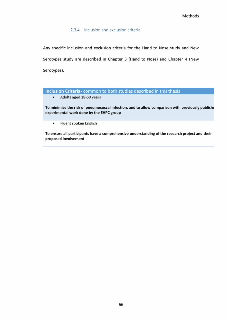
Methods
66
Inclusion and exclusion criteria
Any specific inclusion and exclusion criteria for the Hand to Nose study and New
Serotypes study are described in Chapter 3 (Hand to Nose) and Chapter 4 (New
Serotypes).
Inclusion Criteria- common to both studies described in this thesis
• Adults aged 18-50 years
To minimise the risk of pneumococcal infection, and to allow comparison with previously published experimental work done by the EHPC group
• Fluent spoken English
To ensure all participants have a comprehensive understanding of the research project and their proposed involvement

Methods
67
Exclusion Criteria- common to both studies described in this thesis
• History of major pneumococcal illness (other severe bacterial infections included if participant fully recovered)
• History of drug or alcohol abuse
• History of smoking, defined as: o Current regular smoker (smokes daily/ smokes > 5 cigarettes per week) o Recent smoker i.e. within the last 6 months o Ex-smoker with a significant smoking history (>10 pack years)
• Asthma (current and on treatment or not on treatment but symptomatic) and chronic respiratory diseases– both due to propensity to infection and due to possible confounding effect of medications such as corticosteroids.
• Any other acute or chronic medical diagnosis that is known to increase the risk of bacterial infections or could lead to hospitalisation during the study period- at the discretion of study team
• Taking daily medications that may affect the immune system such as steroids, steroid nasal spray, or retinoids
• On any courses of medications which may affect the immune system such as chemotherapy, biologics, radiotherapy
• Current illness, acute illness within 3 days prior to inoculation or antibiotic treatment within 2 weeks of inoculation- both due to propensity to infection and to minimise any effect on inoculation or transmission
• Currently pregnant
• Diabetes- type 1 and type 2 (current diagnosis)
• Involved in another clinical trial unless observational or in follow-up (non-interventional) phase- both due to propensity to infection and to minimise effect on inoculation or transmission
• Significant anaemia, thrombocytopenia, neutropenia, neutrophilia, leucocytosis- as per results of full blood count test at screening
• Previously undiagnosed abnormality found on screening clinical examination such as murmur
To minimise risk of possible pneumococcal infection following inoculation or exposure
• Allergy to penicillin/amoxicillin
For safety of the participants- pneumococcal pathogen used in the studies fully sensitive to penicillins and are therefore given to participants during the study as emergency supply if unwell or to attempt to clear carriage at the end of the study
• Close physical contact with at risk individuals (children under 5 years of age, immunosuppressed adults, elderly, chronic ill health)
To minimise pneumococcal transmission and possible subsequent infection risk to others in the Community
• Have been involved in an EHPC clinical trial involving pneumococcal inoculation in the last 3 years
• Previous pneumococcal vaccination
• Taking daily medications that may affect the inoculation such as long-term antibiotics To ensure homogeneity and interpretability of immunological endpoints of the participant population with respect to previous pneumococcal immunisation or disease

Methods
68
Study schedule
Common parts of the study schedule for the Hand to Nose study and New Serotypes
study are described below, specific details for each study are detailed in Chapter 3
for Hand to Nose Study (section3.2.5) and Chapter 4 for New Serotypes study (section
4.2.2).
Following consent, participants were given a study schedule by the research team.
This included an initial screening visit which was scheduled for the week prior to
pneumococcal inoculation or exposure (see Table 6 for screening safety
assessments). During the initial screening, additional study samples were also
obtained to allow for baseline assessment. These included nasal wash to assess for
natural pneumococcal colonisation and viral throat swab for both studies. A week
after the screening visit, all participants who had passed screening came back for day
0 which included pneumococcal challenge (inoculation or exposure).

Methods
69
Table 6: Screening safety assessments
Assessment Rationale Action taken if abnormal
Focused clinical
history with
medication review
and clinical
examination
Ensure no abnormality that
could increase risk of
pneumococcal infection or
would put participant at
increased risk in the case of
infection.
Significant abnormality: volunteer was
excluded from the study and the study
doctor arranged appropriate
investigations or treatment through
primary care.
Minor abnormality: if no potential to
increase infection risk, volunteer
included in study and study team
informed primary care for follow up.
Urinary pregnancy
test
Rule out pregnancy which
could increase risks
associated with
pneumococcal infection
Excluded from study if positive.
Full blood count
blood test
Ensure no abnormality that
could increase risk of
pneumococcal infection or
would put participant at
increased risk in the case of
infection.
No specific cut off values
were used when evaluating
full blood count results.
Results were taken into
context of clinical history
and examination.
Significant abnormality: volunteer was
excluded from the study and the study
doctor arranged appropriate
investigations or treatment through
primary care.
Minor abnormality: if no potential to
increase infection risk, volunteer
included in study and study team
informed primary care for follow up.
Safety
Pneumococcus is responsible for infections including otitis media, sinusitis,
pneumonia, bacteraemia and meningitis. Exposing patients to pneumococcus gives
a theoretical risk to participants of these infections. However, experience from
previous pneumococcal challenge studies that have been run in Liverpool, suggests
that the risk to healthy volunteers of developing an infection is very low. 10% adults
experience natural colonisation at any time; while the incidence of invasive disease

Methods
70
is 20/100,000 patient years). Nevertheless, both studies reported in this thesis were
designed to ensure any risk was minimised as follows:
• Experienced study team (in pneumococcal human challenge studies)
undertaking the work.
• Careful serotype selection and dosing. Serotypes are all fully sequenced and
have had antibiotic resistance testing by Public Health England. Dosing for
these studies was based on a previous dose ranging study undertaken by the
EHPC team 134 139. The team has experience of inoculating volunteers with
doses of serotype 6B pneumococcus between 10,000-320,000 CFU/naris 134
139. We have shown that inoculating at these doses is safe and colonisation
rates are reproducible 134 139. Further information on serotype selection and
dosing can be found in Chapter 3 and 4.
• Study design. The New Serotypes study had a dose escalation period built
into the study design. This ensured safety monitoring of at least 7 days post
exposure of the first group receiving a new dose prior to any further
inoculations (see Figure 13, Chapter 4). For both the Hand to Nose study and
the New Serotypes studies, participants were put into groups of up to 24
participants. Groups were inoculated at least a week apart to ensure that we
could minimise exposure if any safety concerns were raised.
• Participant selection and exclusion criteria. For both studies, strict exclusion
criteria must be met, and safety screen visit was carried out to ensure
participants do not have any un-diagnosed conditions which could put them

Methods
71
at increased risk (previously explained under the inclusion/exclusion
heading).
• Participant education. Participants were educated about potential risks
involved in the study and signs and symptoms to look out for during the study
at numerous times. See Appendix A for participant information leaflets.
• Rigorous safety procedures. Each volunteer received a safety information
leaflet, business card with emergency contact information, thermometer and
3-day course of amoxicillin. Participants were advised to keep this pack with
them at all times during the study. They were advised to take amoxicillin1) in
the event they were unwell and were instructed to by the research team 2) if
they were unwell and unable to contact the research team 3) if they had
carried pneumococcus during the study and did not have 2 clear nasal washes
prior to the last visit. Daily monitoring was also undertaken; participants were
required to contact the study team with their temperature reading and any
symptoms before midday for 7 days post exposure/inoculation. If the study
team had not heard from a participant by midday, the volunteer was
contacted to ensure their wellbeing. If they did not respond an allocated daily
contact person/next of kin was contacted.
• 24-hour emergency telephone contact available. A member of the clinical
team was available for participants 24 hours a day, 7 days per week. This
emergency number was covered by a research nurse during working hours
and a medical consultant out of hours. Safety queries were either dealt with
by phone advice or face to face review could be arranged.

Methods
72
• Weekly safety report. This report included study number of each participant,
bacterial dose and serotype, date of inoculation, if they were a carrier of the
bacteria post inoculation and details of any symptoms or illness that had been
reported.
Risks to researchers during the study are also very low. The main potential risks
include needle stick injury to the clinical staff during venepuncture procedures and
biological/chemical hazards within the laboratory for the lab staff. Bacterial stock
preparation is undertaken in a hood to reduce risk of staff pneumococcal
colonisation. During inoculation/exposure procedure staff also ensure that the
bacterial stock is only open to the environment for minimal amount of time to ensure
bacteria are not aerosolised and keep risk of pneumococcal colonisation of staff to a
minimum. To ensure these risks are at a minimum, only experienced staff carried out
procedures, within their competencies, in accordance with standard operating
procedures regulated by good clinical practice and national guidelines. Appropriate
risk and Control of Substances Hazardous to Health (COSHH) assessments are in place
for all laboratory procedures. All laboratory work was conducted in an appropriately
rated laboratory in line with health and safety regulations for research with human
tissues/infectious agents.
Safety monitoring
The following definitions were used in both Hand to Nose study and New Stains study
(taken from HRA):

Methods
73
• Adverse Event (AE): Any untoward medical occurrence in a participant to
whom a medicinal product has been administered, including occurrences
which are not necessarily caused by or related to that product.
• Serious Adverse Event (SAE) A serious adverse event is any untoward medical
occurrence that:
o Results in death
o Is life-threatening
o Required inpatient hospitalisation or prolongation of existing
hospitalisation
o Results in persistent or significant disability/incapacity
o Other ‘important medical events’ may also be considered serious if
they jeopardise the participant or require an intervention to prevent
one of the above consequences
There are strict guidelines for monitoring safety of participants taking part in
research studies and reporting any safety concerns; Table 7 details the procedure for
informing REC of any safety events. The Hand to Nose study and New Serotypes study
were not clinical trials of an investigational medicinal product (CTIMP). Accordingly,
the REC was only informed about SAEs that were related to the study and
unexpected. In addition to this all SAEs were recorded and reported to the DMSC and
sponsors (within 24hrs). SAE were monitored and reported until the end of the
participant’s follow-up.

Methods
74
Non-serious adverse events were collected systematically during the research and
recorded in the case report form and in the weekly safety report. Participants in the
New Serotypes study were also asked to keep a log of symptoms since new serotypes
of pneumococcus were being investigated. The results were summarised and
reported to the DMSC. Safety reporting continued until the participant’s last visit.
Table 7: Safety reporting to REC for non-CTIMP research studies; guidelines taken from the HRA 140
Who When
SAE CI or sponsor to the REC which
issued favourable opinion
Within 15 days of the CI becoming
aware of the event
Urgent safety
measures
CI or Sponsor to the REC which
issued favourable opinion
Immediately by telephone and then
in writing within 3 days
Progress reports To be submitted by sponsor,
sponsor’s legal representative
or Chief Investigator. Must
always be signed by the CI.
To the REC which issued
favourable opinion
Annually (starting 12 months after
the date of the favourable opinion)
Declaration of the
conclusion or early
termination of the
research
CI or Sponsor to the REC which
issued favourable opinion
Within 90 days (conclusion).
Within 15 days (early termination).
Summary of final
report
CI or Sponsor to the REC which
issued favourable opinion
Within one year of the conclusion of
the research.

Methods
75
Data Management and Safety Committee (DSMC)
The EHPC team has an established Data Management and Safety Committee (DMSC)
who are available for advice and support for the research team. DSMC should be an
independent group of experts which can advise the study team using their expertise
and can give recommendations. This committee was used as an additional safety
measure for our studies. It consisted of 3 members (including at least 1 statistician)
who are independent of the study team. The DMSC were supplied with a safety
report at the end of the study, in the event of an SAE, or if requested at any time by
the PI or DMSC members. Review of this safety report allows for critique of the safety
of the studies without any possible reporting bias by study staff deciding to only
escalate certain events to the committee. In the event of a SAE the DMSC and
sponsor were also informed within 24 hours of PI becoming aware.
Nasopharyngeal pneumococcal challenge
Participants in both the studies reported in this thesis were exposed to
pneumococcal bacteria (pneumococcal challenge). On the day of pneumococcal
challenge participants were specifically asked to report any upper respiratory tract
(URT) symptoms to the clinical team and an oral temperature was taken as an extra
safety check. If the participant reported any URT symptoms or had an abnormal
temperature (>38 °C) they would be reviewed by one of the study doctors who would
undertake a full clinical history and examination and decide about the safety of the
participant to undergo the pneumococcal challenge. The pneumococcal challenge
methods are described in Chapter 3 and 4, sections 3.2.7 and 4.2.1.

Methods
76
Nasal wash sampling method
Nasal wash was used for detection and quantification of pneumococcal colonisation.
For this procedure, participants were seated, with their head tilted back to 30°, and
were asked to hold their tongue at the roof of their mouths. Participants were asked
to take a deep breath in and hold this breath (to avoid lung aspiration). Following
this, 5mls of sterile 0.9% sodium chloride (saline) was instilled into one nostril, then
collected into a sterile foil bowl following leaning forward and blowing the saline out
of the nose while releasing the held breath (see Figure 9).
This same procedure was then repeated three more times, in total twice in each
nostril, with a total of 20ml of saline used. The sample was then transferred into a
50ml centrifuge tube (Falcon, Thermo Fisher scientific, USA) for transportation to the
laboratory. If less than 10ml of sample was obtained, the procedure was repeated to
obtain an adequate specimen. Up to 10mls of extra saline was used if needed.
Figure 9: Nasal wash procedure A syringe is filled with 20mls of saline, this is inserted into the nasal cavity and 5mls of saline is instilled into the nasopharynx. The participant has their head tilted back to approximately 30° while the saline is expelled and then leans forward to collect the expelled saline into a foil bowl. Picture reproduced with permission from EHPC team volunteer. Consent gained prior to photographs taken.

Methods
77
Viral swab sampling method
Swabs (flock swab, FLOQSwabs™) were taken from the palatopharyngeal arch using
a tongue depressor, then placed immediately in viral transport medium (universal
transport medium) and put on ice for transfer to the laboratory. Throat swabs
preceded nasal washes to minimise oropharyngeal contamination with nasal
pathogens (Figure 10).
Figure 10: Viral throat swab procedure. A sterile medical swab is used to sample the oropharynx. A wooden tongue depressor was used if needed. Picture was reproduced with permission from EHPC team. Volunteer consent was gained prior to photography.
Blood sampling method
Blood samples were obtained by venepuncture from the arm. The skin was cleaned
prior to the procedure (CloraPrep® chlorhexidine gluconate 20mg/ml and isopropyl
alcohol 0.7ml/ml), and samples were collected using a vacutainer system (Becton
Dickinson, Plymouth, UK) (Figure 11).
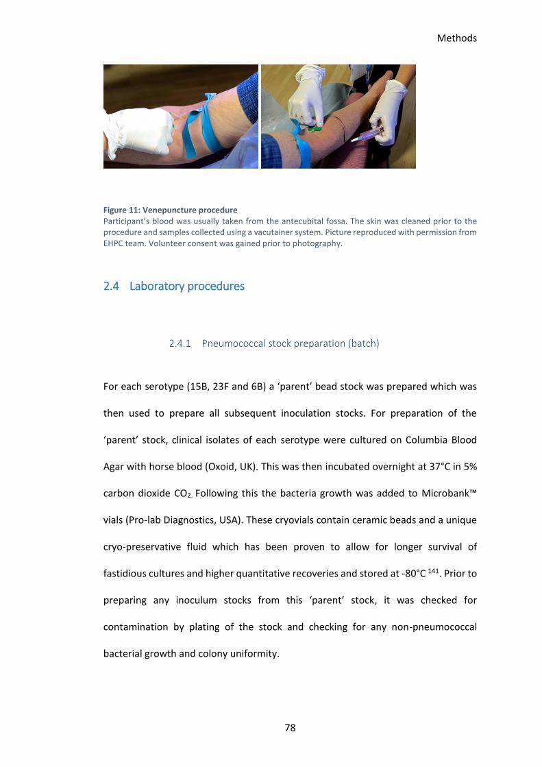
Methods
78
Figure 11: Venepuncture procedure Participant’s blood was usually taken from the antecubital fossa. The skin was cleaned prior to the procedure and samples collected using a vacutainer system. Picture reproduced with permission from EHPC team. Volunteer consent was gained prior to photography.
2.4 Laboratory procedures
Pneumococcal stock preparation (batch)
For each serotype (15B, 23F and 6B) a ‘parent’ bead stock was prepared which was
then used to prepare all subsequent inoculation stocks. For preparation of the
‘parent’ stock, clinical isolates of each serotype were cultured on Columbia Blood
Agar with horse blood (Oxoid, UK). This was then incubated overnight at 37°C in 5%
carbon dioxide CO2. Following this the bacteria growth was added to Microbank™
vials (Pro-lab Diagnostics, USA). These cryovials contain ceramic beads and a unique
cryo-preservative fluid which has been proven to allow for longer survival of
fastidious cultures and higher quantitative recoveries and stored at -80°C 141. Prior to
preparing any inoculum stocks from this ‘parent’ stock, it was checked for
contamination by plating of the stock and checking for any non-pneumococcal
bacterial growth and colony uniformity.

Methods
79
The preparation of the batch pneumococcal stock was undertaken in a fume hood
with dedicated incubator and pipettes to ensure no contamination. Using the
‘parent’ stock, each serotype was plated on blood agar and incubated overnight at
37°C in 5% CO2. Bacterial colonies which grew overnight were then mixed with
Vegitone broth (a vegetable-based growth medium, Sigma-Aldrich, UK) and grown to
mid-log phase. These were then stored in aliquots of 1ml following the addition of
10% sterile glycerol at -80°C.
The determination of colony forming units (CFU) per ml is carried out using a slight
modification of the Miles and Misra method 142. A blood agar plate was initially
divided into 6 sections and labelled as shown in Figure 12. Using a 96 well U-bottom
plate (Corning Inc, Germany), 180ul of sterile saline was mixed with 20ul of bacteria.
Serial dilutions were performed to 106. Three 10ul drops from each dilution were
placed on the corresponding section of the blood agar plate (e.g. 104 in section 4).
Following time for drying, the plate was incubated for 9-16 hours at 37°C, 5% CO2.
The following day the number of visible colonies in each section was counted.

Methods
80
Figure 12: Miles and Misra plates for determination of CFU/ml Three 10µl drops from serial dilution of inoculum were placed into the corresponding section of the blood agar plate. The plate was then inverted and incubated overnight. The following day the number of visible individual colonies in each section was counted.
Calculation of the final dose (CFU/ml) is determined using the least dilute section
where the number of individual colonies can be counted using the following formula;
Prior to human challenge, a sample of the bacterial stock was sent to an independent
reference laboratory (Public Health England, UK) to confirm bacterial stock purity,
serotype and antibiotic sensitivity.
(number of visible colonies/3) x (dilution factor of section) volume of the drop plated
CFU/ml
volume of bacteria stock used for challenge

Methods
81
Preparation of pneumococcal stock on day of challenge
Participants in the Hand to Nose study and New Serotypes studies were put into
groups. Each group was challenged on the same day in groups of up to 7 participants
per challenge. This helped to minimise variability between different bacterial
challenge stocks being used throughout the study. In addition, for all participants the
bacterial challenge stock came from the same ‘parent’ stock and same pneumococcal
batch stock made.
30 minutes prior to the scheduled pneumococcal challenge appointments the
bacterial aliquot was thawed. This was spun down at 17000g for 3 minutes, followed
by a wash step in normal saline. The bacterial pellet was re-suspended in 0.9% saline
to the desired concentration based on prior CFU quantification. Bacterial suspension
densities were prospectively quantified by Miles and Misra (M&M) serial dilution,
both from defrosted stock, and from a control sample which was transported to and
from the clinical site. This established the loss of bacterial viability related to
transport. Two independent counts were performed per sample.
Nasal wash sample processing
Nasal wash samples were transported to the laboratory and were processed within
one hour of the sample being taken to minimise loss of viability. Any large pieces of
mucus were removed (after vortexing to break up the debris or mucus) prior to
processing with as little loss of saline as possible. The nasal wash sample was then

Methods
82
centrifuged for 10 minutes at 3345g, the supernatant volume was recorded and then
stored as 1ml aliquots at -80°C.
The nasal wash pellets were mixed with 100µl of skim milk, tryptone, glucose,
glycerol medium (STGG), and the STGG plus nasal wash pellet volume was recorded.
20µl of this was then streaked onto a blood agar plate with gentamicin (4µg/ml). 10µl
was then used for M&M to assess possible colonisation density. The remaining nasal
wash pellet was diluted with 8.2ml STGG; a further 25µl was streaked onto blood
agar plates to assess for co-colonising flora. All plates were incubated overnight at
37°C in 5% CO2. The remainder of the sample was divided into 3 cryovials and stored
at -80°C.
Detection of pneumococcal colonisation by culture
Pneumococcal growth was confirmed by visual appearance of the colonies
(draughtsman-like colony morphology), presence of α-haemolysis, Gram-positive
staining, optochin sensitivity and bile solubility.
Serotype was assessed using latex agglutination (commercial kit from Statens Serum
Institute, Denmark). A participant was labelled as colonisation positive if the serotype
was proven to be the type experimentally challenged with (6B for Hand to Nose and
23F or 15B for New Serotypes). If there was growth of any other serotypes the
participant was labelled as a natural carrier of pneumococcus.

Methods
83
Detection of pneumococcal colonisation by quantitative
polymerase chain reaction (qPCR)
2.4.5.1 Bacterial DNA extraction
Stored samples were thawed and 300µl nasal wash pellet suspension was
centrifuged at 20,238g for 7 minutes. The pellet was re-suspended with lysis buffer
and protease mix (300µl; 1-part protease to 6-parts of lysis buffer [LGC Genomics
GmbH, Germany]), 100µl of zirconium beads and 300µl of phenol, and disrupted
using a TissueLyser (Qiagen, Germany), twice at maximum speed for 3 minutes (with
cooling on ice in between). Following a further centrifuge for 10 minutes at 9391g to
ensure separation of phases. The aqueous phase was mixed with binding buffer
(600µl) and magnetic beads (10µl), then vortexed and incubated for 30-90 minutes
in a gyratory rocker (Stuart™ SSM3 Gyratory Rocker) at room temperature. The
sample and magnetic beads were washed twice with 2 washer buffers (200µl each)
and bacterial DNA was eluted with 63µl of elution buffer. This buffer was the final
step which detaches the extracted DNA from the magnetic beads and makes it
soluble for recovery.
2.4.5.2 Quantification of pneumococcal DNA by qPCR
Determination of colonisation status and the density of colonisation episode by qPCR
was performed through partial amplification of the lytA gene. The master mix
included 12µl of DEEPC-treated water, 0.225µl of forward primer (5’-
Methods
84
ACGCAATCTAGCAGATGAAGCA-3’), 0.225µl of reverse primer (5’-
TCGTGCGTTTTAATTCCAGCT-3’), 0.125µl probe (5’-(FAM)-
GCCGAAAACGCTTGATACAGGGAG-(BHQ)-3’) and 12.5µl of TaqMan® Universal PCR
Master Mix (Life Technologies). In each well, 22.5µl of the master mix was mixed with
2.5µl of extracted DNA. On each plate, two negative controls used 25µl master mix
only, and each plate contained a standard curve of 10-fold dilutions of genomic DNA
extracted from S. pneumoniae (106-101). Samples were assayed as duplicates, using
thermal cycling conditions: 10 minutes at 95°C for DNA denaturation followed by 40
cycles of 15 seconds at 95°C and then finally 1 minute at 60°C. All qPCR thermal
cycling was performed in the same ABI 7500 Fast Real-Time qPCR system (Life
Technologies, UK).
The lower limit of detection was set at 40 cycles (CT). The qPCR plate was repeated
if: 1) there was DNA detected in either of the negative control wells or; 2) any of the
standards between 106-102 were not detected or; 3) both 101 standard wells were
not detected or 4) the scatter of the standard results around the fitted regression
line was poor (R2 ≤0.98, slope was not between -3.1 and -3.6) or 5) the efficiency was
not between 90% and 110%.
Detection and identification of upper respiratory tract viruses
Viral RNA was extracted from 200µl of viral transport medium (VTM), in which the
oropharyngeal swab was stored and eluted into buffer using standard protocols for
the QIAcube or QIAsymphony instruments. The samples were analysed for detection
of viral RNA using four multiplex real-time PCR assays which amplify the viral

Methods
85
genome. All PCR assays were run using the Roche LC480 Instrument (Roche
Diagnostics, UK). The four assay panels cover the detection of:
1. Influenza A and Influenza B
2. Parainfluenza 1-4, Adenovirus
3. Human Metapneumovirus (hMPV), Respiratory Syncytial Virus (RSV), Human
Rhinovirus (hRV)
4. Coronaviruses OC43, NL63, 229E, HKU1

Chapter 3: Hand to Nose Study
86
3 Hand to Nose Transmission of Streptococcus pneumoniae in
Healthy Participants – Pilot Study (Hand to Nose)

Chapter 3: Hand to Nose Study
87
This chapter was accepted for publication in a peer review journal (European
Respiratory Journal) on 1st August 2018 and is currently in press.
3.1 Introduction
In pneumococcal epidemiology, transmission routes of S. pneumoniae between
individuals remains poorly understood. It is hypothesised that transmission occurs
due to inhalation of contaminated respiratory droplets 8. It is unclear if the hands or
fomites (inanimate object capable of carrying infectious organisms) have a role in
pneumococcal transmission. If direct transmission is implicated in the spread of
pneumococcus the relative contributions of direct and indirect transmission modes
to pneumococcal colonisation and disease are unknown. Current understanding of
the transmission of this important clinical pathogen is described in Chapter 1 (section
1.3).
The hands have been identified as vehicles for the transmission of respiratory
pathogens previously. Multiple studies have identified respiratory viral pathogens on
hands and fomites 62 70 72 143 144,with persistence on dry inanimate surfaces ranging
from 3 hours for coronavirus and up to 3 months for adenovirus 62. When evaluating
bacterial respiratory pathogens, Gram-negative species such as klebsiella species and
Pseudomonas aeruginosa have been shown to survive on inanimate surfaces for
months 62. However, others only last for days such as Haemophilus influenzae (12
days) 66.

Chapter 3: Hand to Nose Study
88
Evidence is limited on the survival of pneumococcus on hands and fomites. One study
exposed the hands of 3 adult volunteers, a glass plate and a plastic ball to
pneumococcus suspended in two different media (Serum broth [10% horse serum in
Brain Heart Infusion Broth, Oxoid] and Mueller-Hinton broth [Oxoid]) 68. Persistence
of pneumococcus on the skin was reported for up to 3 hours post exposure when
suspended in Serum Broth, but with substantially lower pneumococcal counts than
the initial bacterial stock 68. Viable pneumococcus was also recovered from a glass
plate at 15 hours post contamination. For the plastic ball there was an 85% average
loss of pneumococci (266 CFU) at 4 hours but reported ongoing recoverable bacteria
8 hours post contamination.
Many epidemiological studies have examined natural pneumococcal carriage in the
nasopharynx and transmission of infection has been described in case studies of
outbreaks or epidemics of specific pneumococcal serotypes in communities 54-56.
Healthy carrier transmission is less well understood. Observational studies suggest
that person-to-person transmission occurs when in close contact with carriers such
as within family groups 52 and could be increased with overcrowding for example
during the Hajj 53.
There is emerging evidence from murine models which investigate pneumococcal
transmission from host to host. Most of these models have investigated
pneumococcal transmission during influenza A co-infection, as this enhances
pneumococcal shedding and therefore leads to higher acquisition rates by new hosts
49. More recently evidence from an infant murine model describing shedding and
Chapter 3: Hand to Nose Study
89
transmission during pneumococcal mono-infection has been published 51. Using this
model Zafar et al 51reported that bacterial shedding was highest in pups intranasally
infected with pneumococcus at age four days and that it peaks over the first four
days post inoculation. They also reported that transmission was more likely to occur
when there was a high ratio of colonisation positive pups to colonisation negative
pups 51.
We wanted to explore further possible mechanisms of human-to-human
transmission and acquisition of nasopharyngeal pneumococcal colonisation. To do
this we have developed our human pneumococcal challenge model to assess the
feasibility of self-inoculation as a possible mechanism for pneumococcal
transmission.
This chapter addresses the following question: Can the hands be vehicles for direct
transmission of pneumococcus from the hands to nostrils leading to experimental
pneumococcal colonisation?

Chapter 3: Hand to Nose Study
90
3.2 Methods
Study set up
Hand to nose study set up procedures and approvals gained can be found in Chapter
2, section 2.2.
Trial design
Non-specific EHPC trial design information is described in Chapter 2, section2.3.1,
section and ethical principles which were considered prior to the study initiation are
outlined in section 2.3.2.
The Hand to Nose study involved human participants being experimentally exposed
to Streptococcus pneumoniae on their hands and asked to facilitate transmission of
bacteria to the nasopharynx. Participants were followed up to evaluate if they had
acquired nasopharyngeal pneumococcal colonisation using nasal washes at day 2, 6
and 9. There was an assessment of two different transmission methods and the
impact of wet bacterial transmission versus transmission following drying.
The dose of pneumococcus used for the exposure procedures was 3.2 x 10⁶ CFU. In a
previous EHPC study 139 participants were intranasally inoculated with up to 3.2 x 10⁵
CFU/naris (6.4 x10⁵ CFU total) with no significant adverse events. Pre-study
experiments showed that there was a 1-2 log drop in pneumococcal counts
recovered from the hand after exposure when the bacteria were wet and

Chapter 3: Hand to Nose Study
91
immediately after drying (data not shown). Therefore, we estimated that we could
expose participants’ hands to 6.4 x 10⁶ CFU and still be confident that it would be safe
for participants. However, as this is the first study of this type, we decided to use half
this dose (3.2x10⁶ CFU) to ensure safety further.
All participants were exposed to serotype 6B Streptococcus pneumoniae. The first
forty volunteers were randomly allocated, on the day of exposure, to one of four
different transmission groups;
1. Sniffing bacterial residue after air-drying of the hands- Group A/ ‘dry
sniff’
2. Pick/poke nose with finger exposed to wet bacteria residue- Group B/
‘wet poke’
3. Sniffing wet bacterial residue- Group C/ ‘wet sniff’
4. Pick/poke nose with finger exposed to dried bacterial residue ‘dry
poke’
Randomisation was computer-generated and occurred in blocks of 6. An
independent co-ordinator from LSTM’s Tropical Clinical Trial Unit produced the
randomisation schedule. Two members of staff that were not involved in the conduct
of the study produced sealed envelopes containing the group allocations. On the day
of exposure, a clinical team member opened the envelope in from of the participant
and informed them which transmission group they would be in.

Chapter 3: Hand to Nose Study
92
Following the results from these first 4 groups, the trial steering committee (TSC),
discussed the results and decided which group would be taken on to complete a
larger number of participants (see Figure 13).

Chapter 3: Hand to Nose Study
93
Figure 13: Study Design Flow Chart The first 40 participants were randomly allocated, on the day of exposure, to one of the four transmission groups. Rates of colonisation for each of these transmission groups were discussed by the trial steering committee following the completion of the first 40 participants. One group was chosen to take forward to an extended cohort for better precision of estimated rates of colonisation.

Chapter 3: Hand to Nose Study
94
Recruitment
Recruitment strategies and where the study was advertised is outlined in Chapter 2,
section 2.3.3.
Inclusion/Exclusion criteria
Inclusion and exclusion recruitment criteria can be found in Chapter 2, section 2.3.4.
For the Hand to Nose study some additional specific inclusion criteria and exclusion
criteria are outlined below.
Table 8: Inclusion and exclusion criteria specific to Hand to Nose study
Inclusion Criteria
• Access to their own mobile telephone
To ensure safety and timely communication. Also, to ensure the participants can follow the protocol by contacting us daily with temperature and any symptoms during the first 7 days post exposure to pneumococcus
• Capacity to give informed consent
To ensure it is clear to the research ethics committee that we will only be undertaking this research in adults who have capacity to make their own decision about entering the study
Exclusion Criteria
• Any acute dermatological illness or skin injury affecting the hands or face
To ensure no confounding effects of topical medications or increased propensity to skin infections
• Natural carriers of pneumococcus as determined at screening visit
To minimise the potentially positive or negative effects on pneumococcal challenge

Chapter 3: Hand to Nose Study
95
Study schedule
Non-specific study schedule details are outlined in Chapter 2, section 2.3.5. In the
Hand to Nose study following pneumococcal exposure, participants were followed
up for 3 further visits on day 2, 6 and 9 post exposure visits. These follow up visits
entailed only a nasal wash sample. See Figure 14 for full appointment schedule for
Hand to Nose study.

Chapter 3: Hand to Nose Study
96
Figure 14: Hand to Nose study appointment schedule Following participants consent to take part in the study, appointments were arranged for all other clinical visits. The first visit is a screening appointment for baseline research samples and safety check. It also involved a nasal wash to screen for natural colonisation of pneumococcus. The week following this screening appointment participants underwent pneumococcal exposure. All participants were followed up for 9 days after exposure session. Any participant who was still colonised with pneumococcus (6B) at the end of the study was given a three-day course of amoxicillin in an attempt to clear colonisation. Clearance of colonisation was not confirmed with repeat nasal wash following antibiotics.

Chapter 3: Hand to Nose Study
97
Participant safety
Details of participant safety procedures, safety monitoring and symptom reporting
are detailed in Chapter 2, sections 2.3.6 and 2.3.7.
There is a risk of unplanned pneumococcal environmental spread during the
procedure of exposing participant’s hands to the pneumococcus and while the
participant attempts bacterial transmission into the nostrils. To reduce this risk, we
gave the participants full body gowns and eye shields to wear and used a draped area
for the exposure procedure followed by the area being decontaminated with anti-
bacterial cleaning wipes following each participant.
To mitigate any potential risk of spreading pneumococcus to vulnerable groups in the
community, we discussed these risks with our participants and excluded anyone with
close physical contact with at risk individuals (children under 5 years of age,
immunosuppressed adults, elderly, chronic ill health) during the trial period. We also
reduced pneumococcal colonisation burden in all study participants who were still
colonised with pneumococcus at the end of the study, by giving them oral amoxicillin
500mg three times daily for 3 days. Clearance of colonisation was not confirmed with
repeat nasal wash following antibiotics.
Chapter 3: Hand to Nose Study
98
Pneumococcal challenge: hand exposure and transmission
Clinical isolates of serotype 6B BHN418 (GenBank accession number
ASHP00000000.1), a gift from Professor P Hermans, Radboud University Nijmegen,
were used to create the ‘parent ‘stock for the Hand to Nose study. See Chapter 2,
section 2.4.1 for details about how the ‘parent’ stock was made and for general
aspects of pneumococcal challenge which are common to both studies in this thesis.
For the Hand to Nose study, challenge involved exposing an area of the participant’s
hand to pneumococcus, followed by the participant attempting to transmit the
bacteria into their nose using one of two transmission methods (Figure 15). The
pneumococcal stock preparation on the day of challenge and the
exposure/transmission process is outlined in Chapter 2, sections 2.3.9 and 2.4.2.

Chapter 3: Hand to Nose Study
99
Figure 15: Pneumococcal exposure and transmission process for participants using WHO hand hygiene guidelines 145

Chapter 3: Hand to Nose Study
100
Clinical sampling processes
All participant visits were carried out at the CRU in RLBUHT, UK. Samples obtained
from participants include; nasal wash, throat swab, blood sample and urine samples
(only women of child bearing age). See Chapter 2, sections 2.3.10-2.3.12 for sample
collection process. Participants’ full blood count (taken as part of safety procedure)
and viral throat swabs were analysed by RLBUHT, clinical laboratories. All other
research samples were processed and stored at LSTM.
Sample analysis
Details of nasal wash sample processing can be found in Chapter 2, section 2.4.3. The
process for detection of pneumococcal colonisation by culture and by qPCR can be
found in Chapter 2 sections 2.4.4 and 2.4.5 respectively. The process used for
detection of concurrent upper respiratory tract viral infection can be found in section
2.4.6.
Endpoints and objectives
The primary endpoint was experimental pneumococcal colonisation (detection of 6B
in nasal wash) by classical culture methods at any time point (day 2, 6 or 9) following
pneumococcal hands to nose transmission. This rate will be expressed as a
percentage of participants colonised with pneumococcus over the total number
exposed.

Chapter 3: Hand to Nose Study
101
The secondary endpoints were (1) Determination of the duration and density of
pneumococcal colonisation (2) Rates of pneumococcal nasopharyngeal colonisation
following immediate attempted transmission (3) Rates of pneumococcal
nasopharyngeal colonisation following attempted transmission following drying of
bacteria (4) The detection of pneumococcal colonisation by qPCR at any time point.
Statistical methods
As this was a pilot study, with no previously published data pertaining to
pneumococcal hand to nose transmission rates available, we used proportion
estimates to ensure the number of participants used in the study was large enough
to give a specified degree of precision.
Previous EHPC studies using nasal inoculation of S. pneumoniae bacteria found
approximately 40% colonisation rates. If our study had the similar rates, 34 people
would have given 95% confidence that the rate of colonisation lies within 18% on
either side of this rate (22-58% colonisation). However, as this study does not involve
nasal inoculation but adds a further step of hand exposure and attempted
transmission, this rate is likely to be much lower. If we estimate we will get half the
amount of colonisation observed in other studies (20%), then 34 people would give
a 95% confidence that the rate of colonisation lies within 14% on either side of this
(6-34%).
Taking into consideration the possibility of 10% of participants being natural carriers
12 and the possibility of 10% drop out/exclusion rate (taken from approximate

Chapter 3: Hand to Nose Study
102
dropout rates from previous EHPC studies), it was concluded that we would need to
recruit a maximum of 80 participants to complete 64 participants. This study was not
powered to compare rates of colonisation between the four different transmission
methods. The preliminary 40 participants were used to primarily investigate the
feasibility of the transmission methods to decide which would be taken to the full
cohort.
The Fisher’s exact test and the Chi squared test were used to analyse colonisation
rates. Graphing and statistical analysis were performed using GraphPad prism
(California, USA). All P values were two-tailed and considered significant if P≤0.05.
Differences in density were evaluated using one-way ANOVA test. Spearman’s rank
correlation coefficient was used to assess the correlation of qPCR and culture
densities.

Chapter 3: Hand to Nose Study
103
3.3 Results
Screening and recruitment
Between March and June 2017, 76 participants were recruited for the Hand to Nose
study, with 63 participants completing all the study visits. 9 participants withdrew
pre-screening due to personal reasons, 4 participants attended the screening visit; 2
were excluded due to natural carriage of pneumococcus, 1 had an abnormal full
blood count and one withdrew for personal reasons (see Figure 16). All 63
participants were challenged with pneumococcus between April and May 2017, with
the final participant’s final visit on 1st June 2017.
The mean average age of volunteers was 22.6 years (range 18-45, median 21) and
the male:female ratio was 23:40. When evaluating the first 40 participants who were
randomly allocated to 4 different transmission groups, the mean average ages were
similar (wet sniff: 23 [range19-28]; wet poke: 21.4 [range 20-24]; dry sniff: 22.5
[range 19-27]; dry poke: 23.3 [range 19-44]). There were no statistically differences
when comparing gender ratios of all 4 groups using Chi-squared (P=0.19) or when
comparing the wet and dry groups using the Fisher’s exact test (P=0.08) (male:female
ratios: wet sniff 5:5; wet poke 4:6; dry sniff 1:9; dry poke 2:8).

Chapter 3: Hand to Nose Study
104
Figure 16: Consort flow diagram for Hand to Nose study
Inoculum doses were compliant with protocol
All inoculum doses were compliant with the protocol as they fell within half and did
not exceed double the target amount (3.2x10⁶ CFU; see Table 9). The average
Completed study = (n=63)
Recruited (n=76)
Wet sniff extended group (n=23 completed)
Withdrew after consent (n=9)
Screened (n=67)
Wet sniff (n=10) Wet poke (n=10) Dry Poke (n=10) Dry sniff (n=10)
Discontinued (n=4)
• Abnormal FBC
• Natural carrier x2
• Withdrew consent
Total number of wet sniff group who completed the study (n=33)
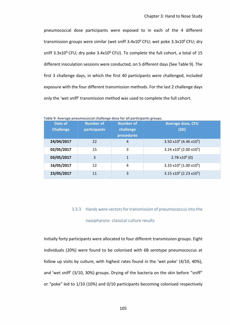
Chapter 3: Hand to Nose Study
105
pneumococcal dose participants were exposed to in each of the 4 different
transmission groups were similar (wet sniff 3.4x10⁶ CFU; wet poke 3.3x10⁶ CFU; dry
sniff 3.3x10⁶ CFU; dry poke 3.4x10⁶ CFU). To complete the full cohort, a total of 15
different inoculation sessions were conducted, on 5 different days (See Table 9). The
first 3 challenge days, in which the first 40 participants were challenged, included
exposure with the four different transmission methods. For the last 2 challenge days
only the ‘wet sniff’ transmission method was used to complete the full cohort.
Table 9: Average pneumococcal challenge dose for all participants groups.
Date of
Challenge
Number of
participants
Number of
challenge
procedures
Average dose, CFU
(SD)
24/04/2017 22 4 3.50 x10⁶ (4.46 x10⁵)
02/05/2017 15 3 3.24 x10⁶ (2.00 x10⁵)
03/05/2017 3 1 2.78 x10⁶ (0)
16/05/2017 12 4 3.33 x10⁶ (1.00 x10⁵)
23/05/2017 11 3 3.15 x10⁶ (2.23 x10⁵)
Hands were vectors for transmission of pneumococcus into the
nasopharynx- classical culture results
Initially forty participants were allocated to four different transmission groups. Eight
individuals (20%) were found to be colonised with 6B serotype pneumococcus at
follow up visits by culture, with highest rates found in the ‘wet poke’ (4/10, 40%),
and ‘wet sniff’ (3/10, 30%) groups. Drying of the bacteria on the skin before “sniff”
or “poke” led to 1/10 (10%) and 0/10 participants becoming colonised respectively

Chapter 3: Hand to Nose Study
106
(see Figure 17 ). The difference in colonisation rates between groups was not
significant (first 40 participants P=0.10; Chi-square test).
Acquisition of colonisation following attempted transmission while the bacteria were
wet was significantly higher than attempted transmission following drying of the
bacteria (7/20 vs. 1/20 respectively, two-tailed P=0.04, Fisher’s exact test). The only
group with no acquisition of colonisation following attempted transmission was the
‘dry poke’ group. No difference was observed when comparing colonisation rates in
the sniff groups versus poking groups (4/20 vs. 4/20 respectively, two-tailed P=1.00,
Fisher’s exact test).
The ‘wet sniff’ group was expanded to improve precision-estimates of rates. A total
of 33 participants completed the study in this group. Of these, 6 participants became
colonised (18.2%, 95% CI, 8.6%-34.4%). This was a lower overall colonisation rate
than we expected following a colonisation rate of 30% in the initial 10 participants
exposed. However, this fluctuation in colonisation rates week on week is not
uncommonly seen in previous EHPC studies. For serotype 6B we have often had
colonisation rates per group range from 10%-70% but with the overall study
colonisation rate consistently between 45-55%. Overall experimental pneumococcal
colonisation was found in 11/63 participants (17.5%, 95% CI, 10.0%-28.6%) at any
time point, following hand to nose transmission of the pathogen.

Chapter 3: Hand to Nose Study
107
Figure 17: Colonisation rates following classical culture of nasal wash samples at any time point after exposure in each transmission group. *No experimental carriers of serotype 6b were found in the dry poke group but 2 participants became natural carriers of pneumococci after baseline. 10 participants were exposed to pneumococcus in each of the 4 different transmission groups (dry poke, dry sniff, wet poke, wet sniff). Following discussion by the trial steering committee it was decided that the wet sniff group should be expanded to a total of 33 participants (the end graph includes participants from the initial cohort and extended cohort; 10 from initial cohort and 23 from extended). Percentage represents the number of participants in the group that were found to be experimentally colonised with pneumococcus at any time point following exposure.

Chapter 3: Hand to Nose Study
108
Natural Pneumococcal Colonisation
Overall the natural colonisation rate in our cohort was 6% (4/67). Two participants
were excluded at baseline due to natural carriage and two participants became
natural carriers during the study (Error! Reference source not found.). No
participants were co-colonised with 6B and the naturally acquired serotype when
assessed using culture. However, 6A/B specific qPCR showed that one participant
was co-colonised with naturally acquired serotype 19 and 6B at both day 2 and 6. No
baseline nasal wash samples were positive for lytA qPCR confirming that all
participants were negative for carriage at time of exposure.
Figure 18: Natural pneumococcal colonisation serotypes as found by culture in 4 individuals NVT: non-vaccine type, serotypes identified by Statens Serum latex kit; SPN: Streptococcus pneumoniae. Pool D NVT serotypes: 16(16F, 16A), 36 and 37.

Chapter 3: Hand to Nose Study
109
Pneumococcal colonisation densities were similar in each
transmission group- classical culture results
Median post-exposure colonisation densities (CFU/ml of nasal wash) of the ‘wet
sniff’, ‘wet poke’ and ‘dry sniff’ groups were 5.6x101 (range 4.3x10⁻¹-3.7x10⁶), 4.72
(range 4.5x10⁻¹-1.25x10²) and 2.42 (range 1.16-9.55) respectively. See Figure 19for
densities at each time point.
D2 D6 D9 D2 D6 D9 D2 D6 D910- 1
100
101
102
103
104
105
106
107
Wet sniff Wet poke Dry sniff
S1
S2
S3
S4
S5
S6
S7
S8
Initial 40 participants:
S9
S10
S11
Larger cohort:
Pn
eu
mo
co
ccal D
en
sit
y
CF
U/m
l o
f n
asal w
ash
Figure 19: Pneumococcal colonisation densities, using classical culture method of pneumococcal identification, at each time point post exposure to pneumococcus. Results from the first 40 participants/subjects are in filled points and participants positive from the second part of the wet sniff group are in open points. Nasal washes were performed on day 2, 6 and 9 post exposure to determine carriage status and density. Density is reported as CFU/ml of nasal wash returned.

Chapter 3: Hand to Nose Study
110
Differences in densities between groups, when comparing the colonisation positive
participants from the initial cohort (n=8), were not statistically different using a one-
way ANOVA test at any time point. Overall density over time for each participant was
calculated using area under the curve (AUC). Comparison of AUC of the different
transmission groups was also not statistically significantly different.
When the wet sniff group was extended, the AUC appeared statistically significantly
different between the wet sniff, wet poke and dry sniff groups (one-way ANOVA test)
(Table 10). However, when applying Dunn’s multiple comparison test, no significant
differences were observed (Table 11).
Table 10: Comparison of transmission groups (full cohort) colonisation densities from culture. One-way ANOVA test with a P value of <0.05 considered significant. All densities log transformed prior to analysis.
Wet Sniff
Mean Densities,
Log transformed
(Standard error)
N=6 total
Wet Poke
Mean Densities,
Log transformed
(Standard error)
N=4 total
Dry Sniff
Mean Densities,
Log transformed
N=1 total
P value
(one-way
ANOVA
test)
D2 1.96 (0.97)
N=6
0.21 (0.24)
N=3
0.98
N=1
0.23
D6 2.09 (0.67)
N=6
0.91 (0.28)
N=3
0.06
N=1
0.20
D9 1.86 (0.44)
N=6
1.05 (0.73)
N=3
0.38
N=1
0.41
AUC 14.57 (3.52)
N=6
4.62 (1.62)
N=4
4.02
N=1
0.04*

Chapter 3: Hand to Nose Study
111
Table 11: Dunn’s multiple comparison test results from AUC densities from culture of different transmission groups
Dunn's Multiple Comparison Test Difference in rank
sum
Significant? P <
0.05?
Wet Sniff AUC vs Wet Poke AUC 5.083 No
Wet Sniff AUC vs Dry Sniff AUC 5.333 No
Wet Poke AUC vs Dry Sniff AUC 0.250 No
lytA qPCR detected pneumococcal DNA in more samples than
classical culture
252 nasal wash samples were collected from the 63 participants who completed the
study; all were tested for the presence of S. pneumoniae by microbiological culture
and retrospectively using lytA qPCR. The proportion of samples positive for
colonisation by qPCR was significantly more than the proportion of samples positive
for carriage by culture (23.8% vs. 13.1%, P<0.0001, Fisher’s exact test) (Table 12).
There were no samples where pneumococcus was detected by culture but not lytA
qPCR but in 10.7% of samples pneumococcus was only detected by lytA qPCR.
Table 12: Comparison of microbiological culture and qPCR in detection of pneumococcus in nasal washes. *3 samples culture positive for natural carriage of pneumococcus after baseline included
Culture
Positive (%)
Culture
Negative (%)
Total
qPCR Positive (%) 33 * (13.1%) 27 (10.7%) 60 (23.8%)
qPCR Negative (%) 0 (0%) 192 (76.2%) 192 (76.2%)
Total 33* (13.1%) 219 (86.9%) 252(100%)

Chapter 3: Hand to Nose Study
112
qPCR detected significantly higher rates of pneumococcal
colonisation compared to classical culture
Using molecular methods (lytA qPCR) resulted in higher colonisation detection rates
compared with culture (35/63 [56%] vs 13/63 [23%] respectively, P=0.0001, Fisher’s
exact test) (Table 13).
Table 13: Comparison of microbiological culture and qPCR in detection of participants colonised with pneumococcus. * 2 participants who were natural carriers of pneumococcus after baseline included
Culture
Positive (%)
Culture
Negative (%)
Total
qPCR Positive (%) 13* (20.6%) 20 (31.7%) 33 (52.4%)
qPCR Negative (%) 0 (0%) 30 (47.6%) 30 (47.6%)
Total 13* (20.6%) 50 (79.4%) 63 (100%)
qPCR detected a larger proportion of colonisation positive participants in every
transmission group and at every time point except for day 9 in the dry sniff group
compared to culture (Figure 20). The difference was most apparent in dry poke
group, where 7/10 (70%) volunteers were experimentally colonised at any time
point, with detectable pneumococcal DNA in nasal washes at 9 days after exposure.
Of the 24 participants who were only determined as experimental colonisers of 6B
using qPCR, 5 participants were colonised for more than one time point and 8 further
participants were found to be colonisers at day 6 or day 9 nasal washes.

Chapter 3: Hand to Nose Study
113
D2
D6
D9Any
D2
D6
D9Any
D2
D6
D9Any D
2D6
D9Any
0
20
40
60
80
100
Culture of 6A/Bstrain
qPCR-lytA
Wet Sniffn = 33
Wet Poken=10
Dry Sniffn = 10
Dry Poken = 10
Co
lon
isati
on
rate
s
%
Figure 20: Comparison of culture (6A/B serotype only) and lytA qPCR results for different transmission methods Nasal washes performed on day 2, 6- and 9-days post exposure to pneumococcus to determine colonisation status. Nasal washes were plated on blood agar plates in real time and read the following day. Those participants with confirmed serotype 6B in nasal washes were deemed colonisation positive. Filled bar charts represent colonisation rates at each time point for each transmission method. At the end of the study all samples were re-run using lytA qPCR method of detecting pneumococcus. Striped bars are the additional participants found to be colonised with this molecular method. No samples were culture positive and qPCR negative for pneumococcus.
All transmission groups had similar densities of colonisation
when assessed using qPCR
Differences in densities between groups were not statistically different at any time
point or AUC analysis for either the first 40 participants or the full data set (Table 14).
Samples which were only positive with qPCR tended to have lower densities
compared to samples which were both qPCR and culture positive (Figure 21).
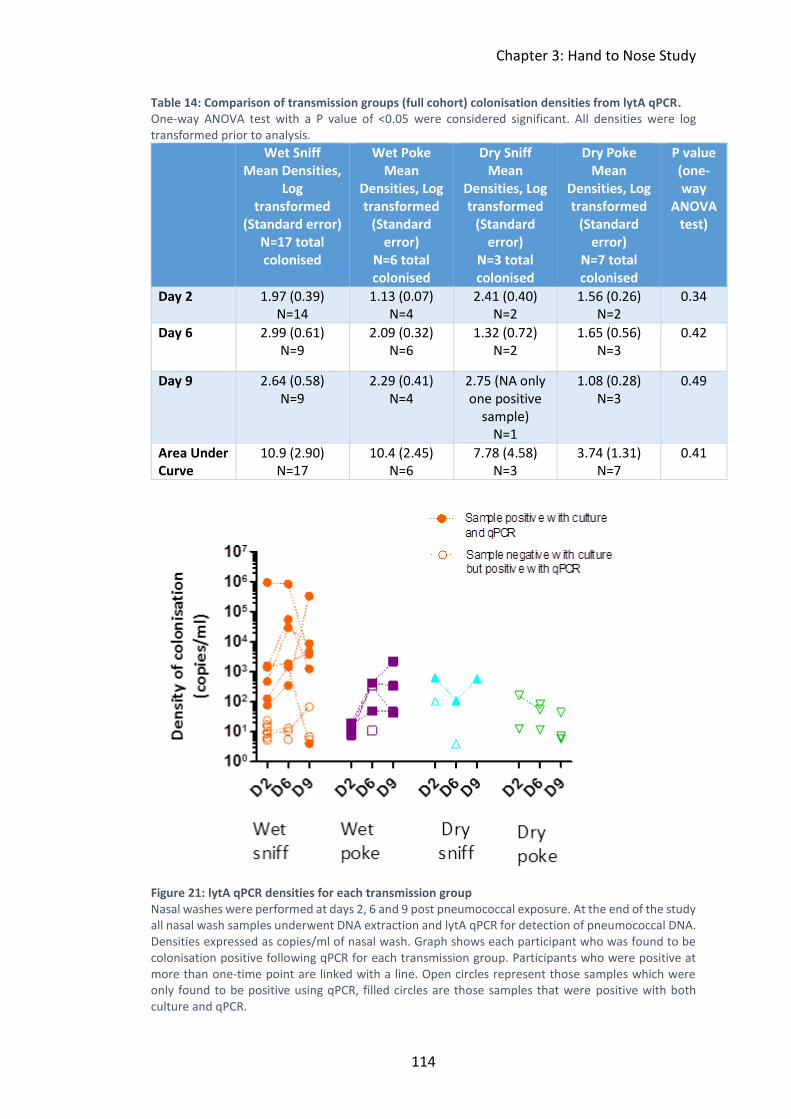
Chapter 3: Hand to Nose Study
114
Table 14: Comparison of transmission groups (full cohort) colonisation densities from lytA qPCR. One-way ANOVA test with a P value of <0.05 were considered significant. All densities were log transformed prior to analysis.
Wet Sniff Mean Densities,
Log transformed
(Standard error) N=17 total colonised
Wet Poke Mean
Densities, Log transformed
(Standard error)
N=6 total colonised
Dry Sniff Mean
Densities, Log transformed
(Standard error)
N=3 total colonised
Dry Poke Mean
Densities, Log transformed
(Standard error)
N=7 total colonised
P value (one-way
ANOVA test)
Day 2 1.97 (0.39) N=14
1.13 (0.07) N=4
2.41 (0.40) N=2
1.56 (0.26) N=2
0.34
Day 6 2.99 (0.61) N=9
2.09 (0.32) N=6
1.32 (0.72) N=2
1.65 (0.56) N=3
0.42
Day 9 2.64 (0.58) N=9
2.29 (0.41) N=4
2.75 (NA only one positive
sample) N=1
1.08 (0.28) N=3
0.49
Area Under Curve
10.9 (2.90) N=17
10.4 (2.45) N=6
7.78 (4.58) N=3
3.74 (1.31) N=7
0.41
Figure 21: lytA qPCR densities for each transmission group Nasal washes were performed at days 2, 6 and 9 post pneumococcal exposure. At the end of the study all nasal wash samples underwent DNA extraction and lytA qPCR for detection of pneumococcal DNA. Densities expressed as copies/ml of nasal wash. Graph shows each participant who was found to be colonisation positive following qPCR for each transmission group. Participants who were positive at more than one-time point are linked with a line. Open circles represent those samples which were only found to be positive using qPCR, filled circles are those samples that were positive with both culture and qPCR.

Chapter 3: Hand to Nose Study
115
When the difference between pneumococcal detection was stratified by qPCR
density it was clear that qPCR detects more carriers colonised at <101 CFU/ml density
than classical culture method (Table 15). However, at qPCR densities 103 copies/ml
and higher there was 100% concordance between qPCR and culture results.
Table 15: Detection of pneumococci in nasal wash by bacterial culture and qPCR (categorised according to qPCR density)
Density by QPCR
(copies/ml)
Number culture positive/number qPCR
positive (%)
<10 2/12 (17%)
101 8/29 (28%)
102 10/13 (77%)
103 9/9 (100%)
104 1/1 (100%)
105 3/3(100%)
Density of colonisation reported by qPCR correlated with
density reported by culture
The correlation between pneumococcal density found by microbiological culture and
qPCR was determined for 33 samples. There was a positive correlation between
density of pneumococcus by culture and qPCR (rs=0.77, P<0.0001) (Figure 22).
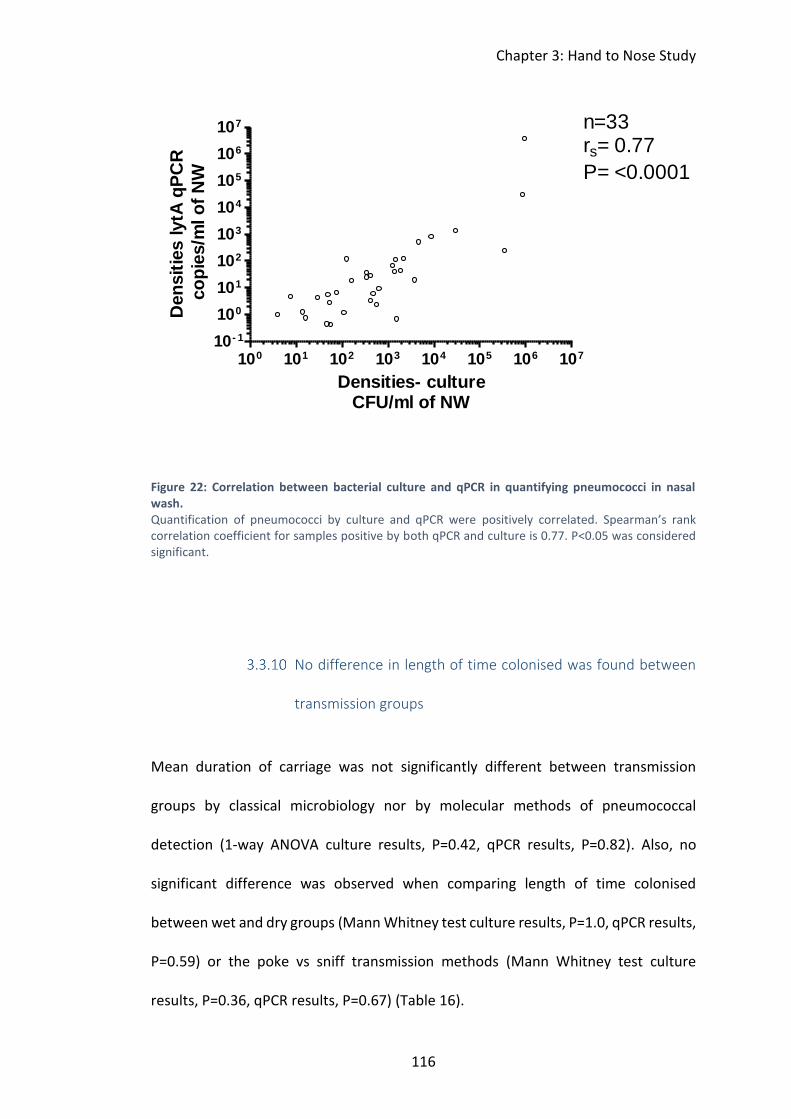
Chapter 3: Hand to Nose Study
116
100 101 102 103 104 105 106 107
10- 1
100
101
102
103
104
105
106
107 n=33rs= 0.77
P= <0.0001
Densities- cultureCFU/ml of NW
Den
sit
ies lytA
qP
CR
co
pie
s/m
l o
f N
W
Figure 22: Correlation between bacterial culture and qPCR in quantifying pneumococci in nasal wash. Quantification of pneumococci by culture and qPCR were positively correlated. Spearman’s rank correlation coefficient for samples positive by both qPCR and culture is 0.77. P<0.05 was considered significant.
No difference in length of time colonised was found between
transmission groups
Mean duration of carriage was not significantly different between transmission
groups by classical microbiology nor by molecular methods of pneumococcal
detection (1-way ANOVA culture results, P=0.42, qPCR results, P=0.82). Also, no
significant difference was observed when comparing length of time colonised
between wet and dry groups (Mann Whitney test culture results, P=1.0, qPCR results,
P=0.59) or the poke vs sniff transmission methods (Mann Whitney test culture
results, P=0.36, qPCR results, P=0.67) (Table 16).

Chapter 3: Hand to Nose Study
117
Table 16: Mean and median days of colonisation for each transmission group, detected by both culture and qPCR methods
Group (number colonised culture and qPCR)
Median – culture
(days with interquartile
range)
Median-qPCR (days with
interquartile range)
Mean- culture (days with standard
deviation)
Mean -qPCR (days with standard
deviation)
Wet sniff n=33 (culture n=6, qPCR n=17)
9.0 (9.0-9.0) 9.0 (2.0-9.0) 9.0 (0.0) 6.2 (3.3)
Wet poke (culture n=4, qPCR n=6)
9.0 (3.8-9.0) 9.0 (5.0-9.0) 7.3 (3.5) 7.3 (2.9)
Dry sniff (culture n=1, qPCR n=3)
9.0 (9.0-9.0)
6.0 (2.0-9.0) 9.0 (0.0) 5.7 (3.5)
Dry poke (culture n=0, qPCR n=7)
NA 6.0 (2.0-9.0) NA 6.1 (3.1)
Full wet group (culture n=10, qPCR n=23)
9.0 (9.0-9.0) 9.0 (2.0-9.0) 8.3 (2.2) 6.5 (3.2)
Full dry group (culture n=1, qPCR n=10)
9.0 (9.0-9.0) 6.0 (2.0-9.0) 9.0 (0.0) 6.0 (3.1)
Full sniff group (culture n= 7, qPCR n=20)
9.0 (9.0-9.0) 7.5 (2.0-9.0) 9.0 (0.0) 6.1 (3.3)
Full poke group (culture n= 4, qPCR n=15)
9.0 (5.5-9.0) 9.0 (4.0-9.0) 9.0 (3.1) 6.7 (3.0)
No participants had asymptomatic viral infection at baseline
We wanted to investigate whether the presence of asymptomatic upper respiratory
tract (URT) viral infection increases the susceptibility to experimental pneumococcal
colonisation following hand to nose transmission of the pathogen. Oropharyngeal
swabs were taken between 4-7 days prior to pneumococcal exposure for all
participants. At the end of the study we retrospectively analysed swabs from carriage

Chapter 3: Hand to Nose Study
118
positive participants and negative controls (matched by date of exposure) by PCR for
the presence of URT viruses. 22 samples were analysed in total due to funding
constraints. No viral co-infections were found in any samples (tested for Influenza A,
Influenza B, Parainfluenza 1-4, Adenovirus, Human Metapneumovirus (hMPV),
Respiratory Syncytial Virus (RSV), Human Rhinovirus (hRV), Coronaviruses OC43,
NL63, 229E, HKU1).
3.4 Discussion
This novel use of a human challenge model allowed for the study of pneumococcal
colonisation in a controlled environment evaluating transmission of pneumococcus
from hands into the nose. The study shows, for the first time, that the hands can be
a vehicle for transmission of pneumococcus leading to acquisition of nasopharyngeal
colonisation.
Success in the model expansion; hands were vehicles for
transmission of pneumococcus
We were able to successfully modify the Experimental Human Pneumococcal
Challenge model 146 to allow for the study of transmission dynamics from the hands
to the nose. Experimental pneumococcal colonisation was established in 17.4% of
participants (11/63) when assessed using microbiological culture.

Chapter 3: Hand to Nose Study
119
The EHPC model was established using nasal inoculation of the pneumococcal
bacteria. Previous EHPC studies in Liverpool showed an overall colonisation rate of
63% using this inoculation method (colonisation rates ranged from 55%-70%
depending on dose of bacterial stock inoculated with) 139. A further study found
colonisation acquisition was dose dependent with carriage rates ranging from 10%-
60% when inoculated with dose between 1x10⁴-1.6x10⁵, with carriage rates
stabilising around 50% above 4x10⁴ CFU/naris inoculation dose 10. Lower rates
observed in this study may be due to the sniffing transmission method causing
movement of the pathogen out of the nasopharynx. During the inoculation process,
the inoculum is instilled around the anterior naris and participants are instructed not
to sniff or blow. Another possible explanation for lower colonisation rates is death of
the pneumococcal bacteria during the drying process on the hands.
Epidemiological studies of nasopharyngeal colonisation rates with matched hand
contamination rates support the findings of this study and suggest that hands could
be vectors for direct and in-direct transmission of pneumococcus. One study
reported simultaneous nasopharyngeal colonisation of pneumococcus and hand
contamination rate of 22%, with serotype concordance of 85% (17/20) 74. A further
study found hand contamination with pneumococcus is an indicator for risk of otitis
media in populations at risk for tympanic membrane perforation (relative risk 8.4;
95% CI 4.6-15.2) 42.
An association between frequent sharing of drinking glasses or bottles and
pneumococcal colonisation has also been reported. A significant correlation between

Chapter 3: Hand to Nose Study
120
colonisation prevalence and self-reported frequency of sharing a drinking
glass/bottle with 33% of frequent sharing group (sharing a glass/bottle always or
usually) colonised with pneumococcus versus 16% of infrequent sharing group
(sharing a glass/bottle half of the time, occasionally or never) 44. The authors
suggested that these findings indicate pneumococcal transmission through saliva is
a highly plausible route in young adults. However, the possibility that the hands could
be vectors for this transmission was not considered or investigated. Following the
results of this study, it is plausible that hand contamination and hand-to-nose
transmission could be implicated in the higher rates of colonisation observed in the
‘frequently sharing group’.
It is widely known that hand washing is an effective means of preventing
gastroenteric infections, especially in the developing world and it is a cost effective
prevention strategy147 148 70. The effect of hand washing for preventing acute
respiratory tract infections (ARIs) is less well understood. A systematic review of 8
studies considering this question found a consistent impact of hand washing on ARIs
with reduction of risk of ARIs by 16% (95% confident interval 11-21%)70. This
reduction in risk is lower than that observed with the effect of hand washing on
gastroenteric infection (47% reduction in risk of diarrhoeal illness) 147, but supports
the results of this study that contaminated hands likely aid in the transmission of
ARIs. It supports the view that hand washing could be a cost effective and feasible
option for helping prevent ARIs and other contagious illnesses.

Chapter 3: Hand to Nose Study
121
Factors affecting transmission
When assessing colonisation rates using microbiological culture, results suggested
that transmission of pneumococcus resulting in colonisation was more likely when
the bacteria were transmitted while still wet. However, interestingly when evaluating
colonisation rates using molecular methods (qPCR) this difference disappeared with
both the wet and dry groups having approximately 50% colonisation rates.
Drying of the bacteria tended to lead to lower density of colonisation compared to
transmission while the bacteria were still wet. No significant differences were
observed between the two different transmission methods; sniffing versus poking.
Participants who attempted transmission using the sniffing technique tended to have
higher colonisation densities. This difference is likely due to the dispersion of the
bacteria into both nostrils and to a larger area of the nasopharynx compared to the
poking method.
Little is known or understood about the survival of pneumococcus on the hands and
the role of contaminated hands and fomites in pneumococcal transmission. One
study reported a significant drop in pneumococcal counts at 3 minutes post hand
exposure, but did recover some viable bacteria 3 hours post exposure 68.
Interestingly, there was significant inter-volunteer variation in pneumococcal
survival, with the greatest difference (4% recovery to 79% recovery rate at 3 minutes)
observed between two volunteers exposed to 435 CFU of pneumococcus suspended
in Serum broth (10% horse serum in Brain Heart Infusion Broth, Oxoid) 68. A limitation
of this study was that the researchers were unable to assess whether the numbers

Chapter 3: Hand to Nose Study
122
of pneumococci recovered were sufficient to lead to colonisation or infection. Our
study confirms that pneumococcus is still viable following drying and can cause
nasopharyngeal colonisation, albeit at lower densities.
Transmission models using infant mice have previously been able to give some
insights into transmission dynamics. Most transmission models have depended on
influenza A virus co-infection. This has been shown to greatly enhance the
pneumococcal shedding and leads to more acquisition of colonisation in new hosts
48 49. Direct contact between pups was not needed for transmission and acquisition
of colonisation to occur; this suggests that an environmental reservoir may be
important for the spread of this pathogen 50. When evaluating Streptococcus
pneumoniae mono-infection similar to all participants in this human challenge study,
one murine model reported bacterial shedding was highest over the first 4 days post
challenge 50. This suggests that host-to-host transmission may peak immediately
following acquisition of pneumococcal colonisation. Epidemiological studies support
this. A study investigating spread of pneumococcus within families showed rapid
spread between family members if a new serotype was introduced to the family with
17/25 transmission episodes took place within 2 weeks of a new serotype entering
the household 52.
Transmission in the same murine model was found to be increased when there was
a high ratio of colonised pups to uncolonised pups in a litter 50. This supports previous
evidence to suggest close contact increases in transmission of pneumococcus.
Pneumococcal outbreaks have been reported in nursing homes 58 and prisons 60 and

Chapter 3: Hand to Nose Study
123
colonisation rates were found to be significant higher at the end of the Hajj where
close contact and overcrowding is common 53.
Host and pathogen factors are also likely to impact transmission and the potential
for the acquisition of pneumococcal colonisation. Murine models have shown that
transmission is increased with high levels of bacterial shedding 48-50. Shedding is
increased with acute inflammation in the nasopharynx and is also observed with co-
infection with influenza A virus 48-50. It has also been shown that nasopharyngeal
inflammation in contact mice lowers their threshold for acquisition of pneumococcal
colonisation 48. High bacterial load in index mice and capsular type were also found
to affect transmission 48 69. One study used neutrophil depletion in index mice
(experimentally colonised with pneumococcus) to investigate this. They found a
small but significant increase in pneumococcal nasopharyngeal load can increase
transmission 48. A further study showed that capsular type is correlated with
transmission rates; capsule types which are better at escaping mucus entrapment
show increased transmission 69.
qPCR detected a higher rate of pneumococcal colonisation
compared with culture
The proportion of nasal wash samples positive for colonisation by qPCR was
significantly higher than the number detected by culture. This is in keeping with a
previous EHPC study which also found significantly higher colonisation rates using
qPCR (42.6% vs 27.5%, P<0.0001)149.

Chapter 3: Hand to Nose Study
124
Culture is considered the gold standard method for detection of upper respiratory
colonisation of pneumococcus and is recommended by the WHO Pneumococcal
Carriage Working Group 150. The main advantage of culture is that further tests can
determine serotype of pneumococcus recovered. Molecular methods of
pneumococcal detection are increasingly being used either to complement culture
or on their own to detect pneumococcal DNA. The WHO Pneumococcal Carriage
Working Group state that there is no current gold standard molecular method of
pneumococcal detection; they support the lytA PCR assay as widely used and species
specific. The specificity of molecular methods to detect S. pneumoniae have been
questioned due to similarities in gene profile of other streptococcal species 151-154. A
study looking at pneumococcus in cerebrospinal fluid assessed specificity of lytA
qPCR and reported a specificity of 70% 155. Many studies have showed that qPCR is
more sensitive at detecting colonisation compared to culture 155-157, with increased
sensitivity shown when an enrichment step was used 158. However, when the density
of pneumococcal carriage is of interest, as in our study, this extra step cannot be
utilised. The most thorough study methodology would utilise both of these methods;
an initial qPCR could be run with the raw samples on which density could be
measured then a second qPCR could be run following enrichment.
The main concern about using qPCR is that this technique cannot confirm the viability
of pneumococcal DNA detected; a positive PCR result which is culture-negative could
represent either live bacteria causing low-density colonisation or non-viable/dead
pneumococcal debris. It has been suggested that using an enrichment step in the
qPCR method can increase the sensitivity of the method by reducing the impact of

Chapter 3: Hand to Nose Study
125
pneumococcal DNA signal originating from possible non-viable bacteria 159. A
previous study supports our findings; they reported significantly higher rates of
pneumococcus in saliva using qPCR compared to culture (44/50 [88%] vs. 2/50 [4%]
respectively) 39. In this study, higher quantities of pneumococcal-specific genes were
detected in culture-enriched saliva samples. This study also used sham-inoculated
samples with pneumococcal DNA. These samples were found to be negative with lytA
qPCR which supports the hypothesis that qPCR detects low density colonisation of
live bacteria rather than non-viable bacterial DNA 39. A further study reported that
pneumococci were recovered following re-culturing from the majority of samples
that were qPCR positive and initially culture negative, further supporting the validity
of the method 159.
The results of this study support the hypothesis that qPCR can detect subclinical or
low-density colonisation rather than detecting non-viable pneumococcal debris. In
20% of new qPCR colonisers pneumococcus was found at more than one-time point.
In addition, 33% were found to be colonisers at day 6 or day 9 nasal washes. It is
unlikely that non-viable or dead bacterial DNA would be still in the nasopharynx
>6days following initial exposure or be detectable at multiple time points, supporting
that qPCR likely detects low density colonisation. Using a novel home sampling
method, the EHPC team have recently examined bacterial movement after
experimental pneumococcal challenge 160. In this study, the kinetics of pneumococcal
colonisation or clearance during the first 48 hours following challenge by using self-
collected saliva and nasal lining fluid samples 160. The study showed that at 48 hours
less than one fifth of non-colonised participants had detectable levels of
Chapter 3: Hand to Nose Study
126
pneumococcus from nasal lining fluid samples (4/21 (19%) 160. The results suggest
two distinct profiles of those protected against colonisation;
1. Saliva clearers; these participants demonstrated a fast-initial
movement of pneumococcus into the bacteria. With 40% having
complete clearance of pneumococcus from the nose by 8 hours
160.
2. Nasal clearers; these participants show a rapid and strong
neutrophil baseline activity 160.
This study supports the hypothesis that dead bacteria do not stay in the nasopharynx
for prolonged periods of time, with the majority of clearance happening within 24
hours of exposure or inoculation.
Culture and qPCR methods used together improved sensitivity
of pneumococcal detection
Our data suggest that using both culture and qPCR to determine pneumococcal
colonisation is beneficial. By using both methods the chance of missing
pneumococcal colonisation is likely to be reduced. Higher colonisation rates were
found using qPCR, but pneumococcal densities measured with both methods were
positively correlated. All samples that were culture-positive for pneumococcus were
also positive for lytA in qPCR.

Chapter 3: Hand to Nose Study
127
We found that culture positivity rate decreased with decreasing densities measured
by qPCR, suggesting that qPCR may be more appropriate for detecting low density
colonisation episodes. All the 27 samples that were culture negative and qPCR
positive had qPCR densities below 103 copies/ml. It has previously been shown that
culture detected fewer carriers of pneumococcus and S. aureus and H. influenzae at
bacterial densities of <105 CFU/ml (measured by qPCR) (P<0.0001) 157.
qPCR has also been shown to be superior at detecting multiple pneumococcal
serotypes at low carriage densities. One study found 28.7% of participants were co-
colonised with multiple pneumococcal serotypes when using qPCR and only 4.5% of
these were found to be co-colonised using only culture (P<0.001) 156. The majority of
additional serotypes only detected by qPCR had a density of <104 CFU/ml 156. This
correlates with the findings of this study. We found that one participant who was
determined to be colonised with only serotype 19 using culture, was co-colonised
with 6B using molecular methods of detection.
Asymptomatic viral carriage and acquisition of colonisation
We were unable to investigate the relationship between colonisation acquisition and
concurrent viral infection due to the absence of viral infection in our participants.
The lack of any concurrent viral infection is likely due to the season in which the study
was carried out (Spring; April-May)161. Previous EHPC studies, conducted during
winter months, found that asymptomatic viral co-infection increased odds of
experimental pneumococcal colonisation (15/20, 75% virus positive volunteers
became colonised vs. 37/81, 46% virus-negative, P=0.02) 162. In addition, a recent

Chapter 3: Hand to Nose Study
128
randomised control trial investigated the effect of viral upper respiratory tract
infections in pneumococcal colonisation using the live attenuated influenza vaccine
(LAIV) as a surrogate 45. No change in prevalence of colonisation was observed but
there was evidence of increasing density following vaccination 45.
Results from murine models have been able to show that viral co-infection can
increase transmission and colonisation acquisition. One study inoculated index mice
with pneumococcus and 8 days later inoculated index and contact mice with
Influenza A; 47% of the contact mice acquired colonisation at day 14. In contrast,
there were no colonisation episodes detected in any contact mice in the control
group (no influenza inoculation) 49. Epidemiological studies have also found that viral
co-infection, such as rhinovirus, facilitate acquisition and transmission of
pneumococcus between individuals 163.
Our ability to prevent pneumococcal transmission is limited by the lack of
understanding about transmission dynamics of pneumococcus and which factors
promote acquisition of nasopharyngeal colonisation. We have shown that an
experimental human carriage model can be used to investigate modes of
transmission. The study shows, for the first time, that the hands can be a vehicle for
transmission of pneumococcus and lead to acquisition of nasopharyngeal
colonisation. It also shows that pneumococcus continues to be viable following
drying. The data suggest that using both culture and qPCR methods for
pneumococcal detection is important to ensure that low density colonisation
Chapter 3: Hand to Nose Study
129
episodes are not missed. This is important for further transmission studies where
carriage density may be an endpoint.
130
4 The Effect of Different Serotypes of Pneumococcus on
Colonisation in Healthy Participants (New Serotypes)

Chapter 4: New Serotypes
131
4.1 Introduction
Epidemiological studies have shown that rates of pneumococcal colonisation and
disease are dependent on multiple factors. Many of these are host specific including
age, geographic area, socio-economic factors and viral or bacterial co-infection 14 164.
The host’s innate and adaptive immune responses have also been shown to effect
rates of colonisation and disease. Mucosal and systemic antibodies to pneumococcal
capsular polysaccharides (IgG antibodies) are thought to play a role in pneumococcal
immunity but the degree of protection against pneumococcal colonisation and
disease is unclear.
Current evidence suggests different serotypes may induce different sizes of IgG
antibody responses to the pneumococcal capsular polysaccharide. During an
outbreak of pneumococcal pneumonia caused by serotype 1 at a military training
camp, 27.8% of the men who did not develop pneumonia had serotype-specific IgG
antibodies detected in sera (systemic blood) compared to 3.6% of controls (3/83,
controls used were a comparable population of soldiers at a training base with no
outbreak of pneumococcal pneumonia) 165. A later study showed that for some
serotypes (9V, 14, 18C, 19F and 23F) a colonisation episode can lead to a significant
increase in levels of serotype-specific anticapsular IgG 12. However, serotype 6B failed
to induce the same immune response 12. For serotype 14, higher levels of
anticapsular IgG at the beginning of the study was also associated with reduced
chance of colonisation during the study 12. In contrast, in this study antibody
responses to pneumococcal proteins (PsaA, PsaA and Ply) did not correlate with

Chapter 4: New Serotypes
132
protection against colonisation. The role of antibodies against the polysaccharide
capsule and towards proteins in the protection against pneumococcus remain
unclear. The data suggest that these responses may vary depending on serotype.
However, more information is needed before this conclusion is made.
Pneumococcal serotype has also been shown to play a major role in acquisition of
colonisation or disease. There are over 90 serotypes of Streptococcus pneumoniae.
Serotypes are defined by biologically different polysaccharide capsules. Some
serotypes are more invasive and cause significant disease whereas others mainly
cause colonisation episodes and are rarely recovered in disease cases 133. A recent
pneumococcal colonisation surveillance study in the UK showed that the most
prevalent serotypes causing colonisation are 15B/C, 11A, 23B and 10A in
2015/2016southern). In addition, serotypes 8, 12F, 3 and 9N caused approximately
50% of all invasive pneumococcal disease cases in England/Wales during 2016/2017
166.
Capsular polysaccharides are understood to play a key part in bacterial physiology
and are vital in determining the host’s immune response to the pneumococcus. Due
to differences in the capsular polysaccharides between serotypes, capsule structure
has been implicated in the varying clearance and acquisition rates observed between
serotypes 167 168. Specific characteristics of the capsular polysaccharide which affect
this process are yet to be fully understood. In addition to different capsular
polysaccharide, multiple other proteins have been suggested to be important in the

Chapter 4: New Serotypes
133
colonisation process, such as PspC and ChoP 167. The relative contribution of these
factors is also unknown 167.
Better understanding of the dynamics of colonisation and systemic immunity for
different pneumococcal serotypes is vital. This study expanded the current EHPC
model by re-testing a modified 23F serotype and for the first time tested a non-
vaccine serotype 15B. Investigating the varying propensity of pneumococcal
serotypes to establish nasopharyngeal colonisation will allow for better
understanding of transmission dynamics and disease.
Serotype 6B is currently used in the EHPC model. This serotype consistently colonises
40-50% of participants. The model is currently limited in generalisability, only having
one serotype that colonised consistently at a high rate, which is already covered by
the current PCV (serotype 6B). Expanding the model to include non-vaccine
serotypes is important for vaccine testing. As colonisation is a likely a pre-requisite
for invasive infection, it could be used as a surrogate marker of disease risk.
Protection against colonisation therefore could be used in a vaccine trial to predict
the protective effect against invasive pneumococcal disease 101. The model has
previously been used to test the effectiveness of PCV13. A sample size of 100 was
needed in the study to give power to detect 50% reduction in colonisation rates
compared to control 169. New serotypes that are able to colonise at similar or higher
rates than 6B in this EHPC model would allow for further vaccine testing trials.

Chapter 4: New Serotypes
134
To develop a reproducible model of colonisation with smallest numbers necessary,
we examined colonisation rates at increasing doses until a colonisation rate of
greater than 40% was achieved. Following this, to improve the precision of our
estimates of colonisation potential, one dose was tested in a larger number of
participants.
This chapter addresses the following question: What is the capacity of 23F and 15B
serotypes to cause experimental pneumococcal colonisation in healthy adults?
Chapter 4: New Serotypes
135
4.2 Methods
Study set up
New Serotypes study set up procedures and approvals obtained can be found in
Chapter 2, section 2.2.
Trial design
Non-specific EHPC trial design information is described in Chapter 2, section 2.3.1
and ethical principles which were considered prior to the study initiation are outlined
in section 2.3.2.
The New Serotypes study experimentally exposed human participants to
Streptococcus pneumoniae:
Dose-ranging:
• Group 1: participants were entered into cohorts which were all inoculated
with the same dose of serotype 23F (P1121). Successive cohorts had
increasing does of the serotype inoculated if desired colonisation rates were
not obtained (see Figure 23;n = 33-67).
• Group 2: participants were entered into cohorts which were all inoculated
with the same dose of serotype 15B (non-vaccine serotype). Successive

Chapter 4: New Serotypes
136
cohorts had increasing does of the serotype inoculated if desired colonisation
rates were not obtained (see Figure 23;n = 33-67).
• Extended Cohort: To enhance the precision of the estimated colonisation
rate, a dose and serotype that lead to more than 40% of carriage in the dose
ranging part of the study was tested in a larger group of volunteers.
We hypothesised a dose-dependent relationship in probability of colonisation. We
used the dose escalation method to test this hypothesis, to estimate the optimum
inoculation dose for these new serotypes and to minimise unnecessary exposure of
volunteers. Also, to ensure safety of participants, by inoculating small groups we
monitored for any adverse events before continuing to higher doses and to larger
groups.

Chapter 4: New Serotypes
137
Figure 23: Dose escalation study design for New Serotypes study. Used for both 15B and 23F serotypes; methodology adapted from Waddington et al 170.

Chapter 4: New Serotypes
138
Trial procedures
General details of trial procedures common to both studies outlined in this thesis are
described in Chapter 2 Sections pertaining to different trial procedures are outlined
below in Table 17.
Table 17: Generic trial procedures outlined in Chapter 2: Methods.
Trial procedure Section
Recruitment strategies Section 2.3.3
Prevention of over-volunteering process Section 2.3.3
Inclusion and exclusion criteria Section 2.3.4
Study schedule Section 2.3.5
Participant safety procedures Sections 2.3.6, 2.3.7 and 2.3.8
In the New Serotypes study following pneumococcal inoculation, participants were
followed up for 3 further visits on day 2, 7 and 14 post inoculation. These follow up
visits involved a nasal wash sample at each visit and bloods for all participants at day
14. See Figure 24 for full appointment schedule.

Chapter 4: New Serotypes
139
Figure 24: Flow chart of New Serotypes study participant appointments Following participants’ consent to take part in the study, appointments were arranged for all other clinical visits. The first visit is a screening appointment for baseline research samples and safety check. The week following this screening appointment, participants underwent pneumococcal exposure. All participants were followed up for 14 days after exposure session. Any participants who were still colonised with pneumococcus (6B) at the end of the study were given a three-day course of amoxicillin in an attempt to clear colonisation. Longer follow up was decided for this study compared to the Hand to Nose study because we were investigating immunological responses. Previous EHPC studies found that serotype 6B induced significant immune response at day 14 post inoculation10 171.

Chapter 4: New Serotypes
140
Symptom reporting
To examine whether experimental colonisation with pneumococcus causes
symptoms, all volunteers filled out a daily symptom log on inoculation day and for
each of the following 6 days. For the 23F study a 7-point Likert-type scale was used
for symptom reporting (nasal and non-nasal symptoms) as published by Spector et al
172 (recommended to evaluate severity of rhinitis). This scoring system was slightly
modified with the removal of ‘mental function’ as a possible non-nasal symptom (see
Appendix B: Daily Symptom Logs)
For the 15B study, a different daily symptom log was used to cover a larger number
of potential non-nasal symptoms as a further safety strategy and to ensure
colonisation did not cause systemic symptoms that we previously did not enquire
about. A further benefit of this was also to move to a generic symptom log for all
EHPC studies to allow better comparison of results (see Appendix B: Daily Symptom
Logs). The numeric scoring system was replaced with descriptive options based on
participant feedback. Participants felt that the lack of explanation of what
constituted a score of 2, 4 and 6 made it difficult to use. The study team also believed
that descriptive options would reduce the subjective variation between participants
reporting on a numbered scale.

Chapter 4: New Serotypes
141
Bacterial Serotypes
4.2.2.1 23F Serotype
In 2001 a human challenge experiment was performed in the USA using serotype 23F
P833.This achieved colonisation rates of 43% (6/14 participants) 29. This serotype was
transferred to the EHPC group. A repeat study found 23F serotype had a reduced
ability to colonise humans, maximum colonisation rate of 10%. Upon discussions with
the USA team it was discovered that the 23F inoculum contained two different cell
populations, one wild-type for the amiC gene and the other contained an amiC
frameshift mutation (Dalia and Weiser, personal communication with Ferreira). It
was confirmed that the inoculum used for the EHPC study containing the amiC
frameshift mutation. Both genetic serotypes (P833 and P1123) were sequenced using
an Illumina Hi-Seq and frameshift mutation was determined by PCR amplification and
sequencing (described in J. Gritzfeld PhD thesis 173).
AmiC is a pneumococcal transmembrane protein that has been shown to play a role
in adherence and oligopeptide transport 174 175. Following this discovery, a murine
model assessed the effect of this mutation on nasopharyngeal colonisation. The 23F
serotype with amiC mutation did not establish any colonisation, while a 23F serotype
with a full length amiC gene was able to establish colonisation and is not further
considered in this thesis (unpublished work, M.de Jonge).

Chapter 4: New Serotypes
142
In this study we tested a natural derivative isolate of 23F serotype P833; P1121,
containing a wild-type amiC gene, with no frameshift mutation on amiC gene. Using
the wild type serotype, we expected to be able to reproduce findings from the USA
group which established carriage in 75% participants using 17,000 CFU/naris 29. This
penicillin-sensitive isolate was obtained from a child with otitis media by
tympanocentesis, gifted from University of Pennsylvania as described below.
4.2.2.2 Non-Vaccine Serotype
We also wanted to investigate a pneumococcal serotype not included in the PCV13
vaccine; no human infection model has tested any such serotype previously. Multiple
serotypes were assessed using laboratory testing prior to choosing the best
candidate. 15B P1262 was chosen based on superior pneumococcal survival in
presence of human blood neutrophils and adherence to human nasal cells compared
to 6B serotype (Pojar, unpublished work). This penicillin-sensitive isolate was
obtained from a child during a colonisation episode, gifted from Southampton.
Collection as described below.
Pneumococcal inoculation
Clinical isolates of serotype 23F serotype P1121 (a gift from Professor JN Weiser,
University of Pennsylvania. European Nucleotide Archive accession number:
ERS1072059) and serotype 15B serotype P1262 (a gift from Dr D. Cleary, University
of Southampton. European Nucleotide Archive accession number: ERS2632437),

Chapter 4: New Serotypes
143
were used to create two ‘parent’ stocks for the New Serotypes Study. See Chapter 2,
section 2.4.1 for details about how the ‘parent’ stock was made and for general
aspects of pneumococcal challenge which are common to both studies in this thesis.
Prior to human challenge, a sample of the bacterial stock was sent to an independent
reference laboratory (Public Health England, UK) to confirm bacterial stock purity,
serotype and antibiotic sensitivity.
Intra-nasal inoculation with either 15B or 23F serotypes of S. pneumoniae was carried
out by a research nurse or clinical fellow, trained in the inoculation procedure.
Participants were sat in a semi-recumbent position with their head tilted back. Using
a P200 micropipette 0.1ml broth containing the desired dose of pneumococcus was
inserted into each nostril Figure 25. The pipette tip never touched the nasal mucosa
during the procedure to ensure the integrity of the epithelium was not disrupted.
Following inoculation, the participant remained in this position for up to 15 minutes
without sniffing or blowing the nose.
Figure 25: Inoculation of the nasal mucosa procedure Participants sat in a semi-recumbent position. Inoculation of the nasal mucosa with pneumococcus was undertaken using a P200 pipette. 100µl of the bacterial inoculum was instilled into the anterior nasal cavity in a circular motion.

Chapter 4: New Serotypes
144
Clinical sampling processes and sample analysis
All participant visits were carried out at the Clinical Research Facility in the RLBUHT.
Samples obtained from participants include; nasal wash, blood samples and urine
sample (only women of child bearing age). Sample collection and processing can be
found in Chapter 2 (see below for sections in Table 18). Participants’ full blood count
were analysed at RLBUHT, clinical laboratories. All other research samples were
processed and stored at LSTM.
Table 18: Generic clinical sampling process and sample analysis is outlined in Chapter 2
Process Section
Nasal wash sampling method Section 2.3.10
Blood sampling method Section 2.3.12
Pneumococcal stock preparation (batch) Section 2.4.1
Preparation of pneumococcal stock on day of challenge Section 2.4.2
Nasal wash sample processing Section 2.4.3
Detection of pneumococcal colonisation by culture Section 2.4.4
Detection of pneumococcal colonisation by q PCR
Section 2.4.4
4.2.4.1 Measurement of anti-pneumococcal polysaccharide
antibodies by enzyme-linked immunosorbent assay (ELISA)
We measured the levels of serotype 15B specific polysaccharide antibody in
volunteers’ serum. Initially a 96-well plate was coated with 10µl purified 15B capsular
polysaccharide (Statens Serum Institute Diagnostica, Denmark) in 10ml of
phosphate-buffered saline (concentration 5µg/ml) then incubated at 4°C overnight.
Human anti-pneumococcal capsule reference serum lot 007sp (National Institute for

Chapter 4: New Serotypes
145
Biological Standards and Control, UK) was used to make a standard curve. The
standard was serially diluted from a concentration of 271.04ng/ml to 4.235ng/ml.
In a separate 96-well plate 2.5 µl of serum sample was mixed with 100µl of dilution
buffer (2ml of 10% heat-inactivated foetal bovine serum blocker in 20ml PBS),
10µg/ml of cell wall polysaccharide (CPWS Multi, Statens Serum Institute
Diagnostica, Denmark) was added and incubated at room temperature for 30
minutes. Following these dilution and absorption steps samples were transferred to
the pre-coated plates and incubated at room temperature for 2 hours. Bound
antibodies were detected using goat anti-human IgG (Sigma Life Sciences, UK), 2.5µl
in 10ml phosphate-buffered saline and incubated at room temperature for 1 hour 30
minutes. Finally, 100µl of p-nitrophenylphosphate (5mg added to 40ml of distilled
water) was added to plates and left to develop in the dark for 15-20 minutes,
following this absorbance was measured at 405nm using FLUOstar Omega plate
reader (BMG Labtech, Germany). All samples and standard curve were measured in
duplicate. Between all steps described above plates were washed with PBS-Tween
0.005% using a microplate washer (ELx50™, BioTek™, USA).
Endpoints and objectives
The primary endpoint was the detection of experimental pneumococcal colonisation
(serotype 23F or 15B) in nasal wash by classical culture methods at any time point
(day 2, 7 or 14) following pneumococcal challenge.

Chapter 4: New Serotypes
146
The secondary endpoints were: (1) Determination of the duration and density of
pneumococcal colonisation with 23F and 15B serotypes; (2) Rates of colonisation at
different inoculum doses for both serotypes; (3) The detection of pneumococcal
colonisation by qPCR at any time point; (4) Quantification of a systemic humoral
immune response to nasopharyngeal carriage (capsular specific IgG concentration in
serum).
Statistical methods
Previous EHPC studies using serotype 6B S. pneumoniae bacteria attained
colonisation rates of approximately 45%. We used this as the basis of our sample size
calculation; to estimate colonisation rates with 95% confidence (+/-5%) 34
participants completing the study with a single inoculation dose (2.0x10⁴, 8.0x10⁴ or
1.6x10⁵ CFU/naris), using 45% colonisation as an estimate for the ‘real rate’. To allow
for an estimated drop-out rate of 10% and natural colonisation of 10% (who will be
excluded from primary endpoint analysis) between 34 and 67 participants was
required for each serotype.
The Fisher’s exact test and Chi squared test were used to analyse colonisation rates.
We compared colonisation rates seen between serotype 23F and 15B. In addition,
comparison was made between colonisation rates found at different inoculation
doses. Differences in density was evaluated using one-way ANOVA test. Graphical
and statistical analysis were performed using GraphPad prism (GraphPad Software,
CA, USA). All P values were two-tailed and considered significant if P<0.05.

Chapter 4: New Serotypes
147
4.3 Results
Screening and recruitment
4.3.1.1 23F study
During September 2016, 18 participants were recruited for the 23F group, 16
participants completed all the study visits. This study was suspended early due to
poor results, further discussed in section 4.3.3. Two participants were excluded
following screening both due to abnormal full blood counts (see Figure 26). All 16
participants were inoculated with pneumococcus in September 2016, with final
participant, final visit on 11th October 2016. The mean average age of volunteers was
30.2 years (range 25-47, median 29) and the male:female ratio was 5:11.
Figure 26:Consort flow diagram for 23F group New Serotypes study
Recruited (n=18)
Inoculated with 20,000 CFU
N=6
Inoculated with 160,000 CFU
N=4
Inoculated with 80,000 CFU
N=6
Screened (n=18)
Discontinued (n=2) 1)Abnormal FBC n=2

Chapter 4: New Serotypes
148
4.3.1.2 15B study
Between August-November 2017, 72 participants were recruited to the 15B group, with 54 participants completing all study visits. 10 participants withdrew pre-screening, 7 excluded or withdrew post screening visit and 1 was excluded at day 1 post inoculation due to starting antibiotics for a non-study related issue (see
Figure 27). All 54 participants who completed the study were challenged with
pneumococcus between September-November 2017, with the final participants,
final visits on 13th December 2017. The mean average age of volunteers was 24.6
years (range 18-49, median 21) and the male:female ratio was 29:25.
Sequential groups were inoculated with higher doses of bacterial inoculum if optimal
rates of colonisation were not achieved by day 7 (>40% colonisation rates). This was
a safety feature to ensure that we were taking forward to an extended cohort the
lowest inoculum dose possible to achieve adequate colonisation rates. Participants
were recruited into groups depending on their availability. The inoculation dose
which achieved the optimal colonisation rates was extended to complete a full
cohort. This extended cohort would give us statistical confidence to estimate the
colonisation rate of this serotype.

Chapter 4: New Serotypes
149
Figure 27: Consort diagram for 15B group of New Serotypes study
Inoculum doses were within target range
All inoculation doses were compliant with protocol as they fell within half or double
of the target amount. The average inoculation doses per group can be seen below
(Table 19).
Table 19: Average pneumococcal challenge dose for all participant groups
Dose Group Target inoculation dose per naris
Serotype 23F Average dose (CFU/naris)
Serotype 15B Average dose (CFU/naris)
2x10⁴ 1.7x10⁴ (SD 0) 1.9x10⁴ (SD 9.9 x10²)
8x10⁴ 7.5x10⁴ (SD 0) 8.9x10⁴ (SD 1.3x10⁴)
1.6x10⁵ 1.7 x10⁵ (SD 0) 1.9x10⁵ (SD 0)
Recruited (n=72)
Inoculated with 20,000 CFU
N=10
Inoculated with 160,000 CFU
N=9
Inoculated with 80,000 CFU
N=10
Screened (n=62) Discontinued (n=7) 1)Abnormal FBC n=2 2)Participant wishes n=3 3)Health issues n=2
Discontinued (n=10) 1)Participant wishes n=9 2)Health issues n=1
Withdrew at D1 n=1 due to starting
antibiotics Extended cohort inoculated with
80,000 CFU N=26
TOTAL N=36
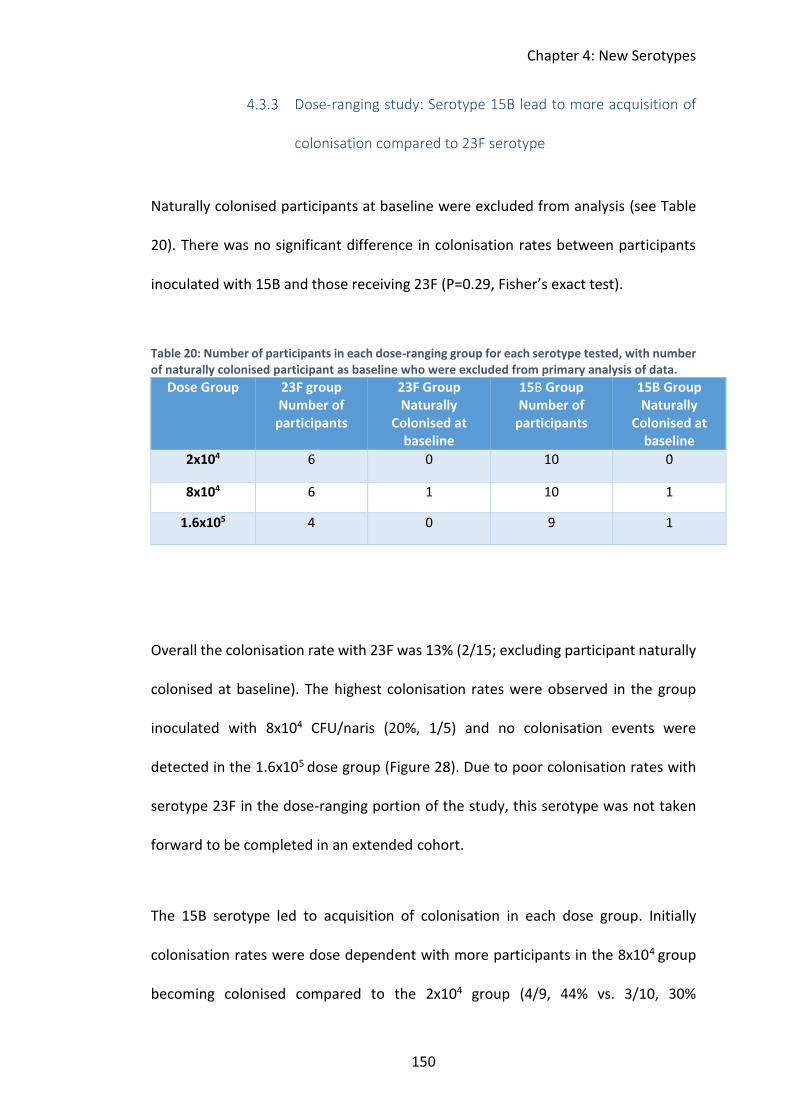
Chapter 4: New Serotypes
150
Dose-ranging study: Serotype 15B lead to more acquisition of
colonisation compared to 23F serotype
Naturally colonised participants at baseline were excluded from analysis (see Table
20). There was no significant difference in colonisation rates between participants
inoculated with 15B and those receiving 23F (P=0.29, Fisher’s exact test).
Table 20: Number of participants in each dose-ranging group for each serotype tested, with number of naturally colonised participant as baseline who were excluded from primary analysis of data.
Dose Group 23F group Number of
participants
23F Group Naturally
Colonised at baseline
15B Group Number of
participants
15B Group Naturally
Colonised at baseline
2x10⁴ 6 0 10 0
8x10⁴ 6 1 10 1
1.6x10⁵ 4 0 9 1
Overall the colonisation rate with 23F was 13% (2/15; excluding participant naturally
colonised at baseline). The highest colonisation rates were observed in the group
inoculated with 8x10⁴ CFU/naris (20%, 1/5) and no colonisation events were
detected in the 1.6x10⁵ dose group (Figure 28). Due to poor colonisation rates with
serotype 23F in the dose-ranging portion of the study, this serotype was not taken
forward to be completed in an extended cohort.
The 15B serotype led to acquisition of colonisation in each dose group. Initially
colonisation rates were dose dependent with more participants in the 8x10⁴ group
becoming colonised compared to the 2x10⁴ group (4/9, 44% vs. 3/10, 30%

Chapter 4: New Serotypes
151
respectively). However, rates fell from 44% at 8x10⁴ CFU/naris to 13% (1/8) when the
dose rose to 1.6x10⁵ CFU/naris. This difference was not statistically different (P=0.29)
(Figure 28).
Figure 28: Colonisation rates (%) for 23F and 15B during dose range portion of the study. Participants who were naturally colonised with pneumococcus at baseline were removed prior to analysis. Participants were intranasally inoculated with either serotype 23F (blue circle) or 15B (red squares) over a range of doses, starting with 2x10⁴ CFU/naris to 1.6x10⁵ CFU/naris. Y axis shows percentage of participants experimentally colonised at each dose.
Dose-ranging study: Density of colonisation and inoculum dose
Colonisation rates were not found to rise significantly with increasing inoculum dose
for either serotype. We wanted to investigate whether colonisation density may
increase in parallel with this inoculum dose increase. Figure 29 shows the
colonisation density recovered from nasal washes at each time point (day 2, 7 and
14) for both 23F and 15B dose-ranging participants. Due to low colonisation rates in
2x10⁴
8x10⁴
1.6x10⁵
0
10
20
30
40
5023F
15B
3/10
1/8
4/9
1/6
0/4
1/5
Inoculation Doses(CFU per naris)
% C
olo
nis
ati
on
po
sit
ive
pa
rtic
ipa
nts

Chapter 4: New Serotypes
152
the 23F group we cannot confidently analyse differences in colonisation density at
increasing inoculum doses.
Participants inoculated with 15B with an inoculation dose of 8x10⁴ CFU/naris had
significantly higher colonisation density at day 2 compared to those inoculated with
2x10⁴ (P=0.047, t test). This difference did not continue at later follow up time points
(day 7 and 14).
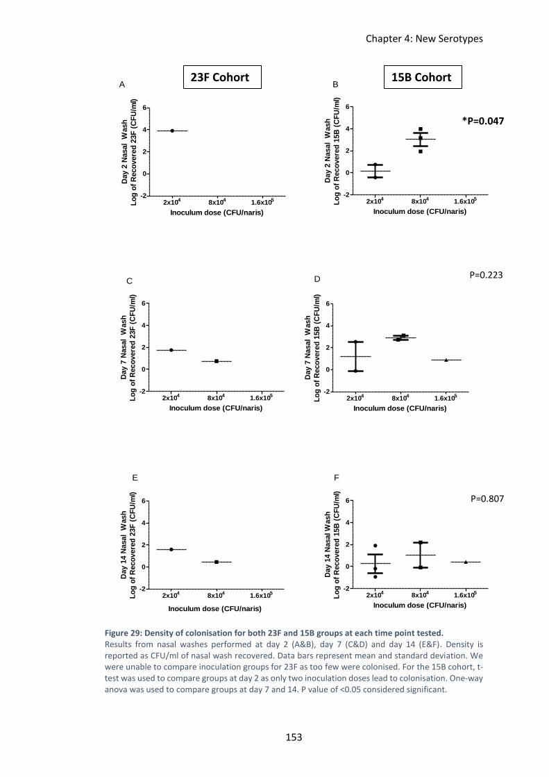
Chapter 4: New Serotypes
153
Figure 29: Density of colonisation for both 23F and 15B groups at each time point tested. Results from nasal washes performed at day 2 (A&B), day 7 (C&D) and day 14 (E&F). Density is reported as CFU/ml of nasal wash recovered. Data bars represent mean and standard deviation. We were unable to compare inoculation groups for 23F as too few were colonised. For the 15B cohort, t-test was used to compare groups at day 2 as only two inoculation doses lead to colonisation. One-way anova was used to compare groups at day 7 and 14. P value of <0.05 considered significant.
2x10⁴ 8x10⁴ 1.6x10⁵-2
0
2
4
6
A
Inoculum dose (CFU/naris)
Day 2
Nasal W
ash
Lo
g o
f R
eco
vere
d 2
3F
(C
FU
/ml)
2x10⁴
8x10⁴
1.6x10⁵
-2
0
2
4
6
B
Inoculum dose (CFU/naris)
Day 2
Nasal W
ash
Lo
g o
f R
eco
vere
d 1
5B
(C
FU
/ml)
2x10⁴
8x10⁴
1.6x10⁵
-2
0
2
4
6
C
Inoculum dose (CFU/naris)
Day 7
Nasal W
ash
Lo
g o
f R
eco
vere
d 2
3F
(C
FU
/ml)
2x10⁴
8x10⁴
1.6x10⁵
-2
0
2
4
6
D
Inoculum dose (CFU/naris)
Day 7
Nasal W
ash
Lo
g o
f R
eco
vere
d 1
5B
(C
FU
/ml)
2x10⁴
8x10⁴
1.6x10⁵
-2
0
2
4
6
E
Inoculum dose (CFU/naris)
Day 1
4 N
asal W
ash
Lo
g o
f R
eco
vere
d 2
3F
(C
FU
/ml)
2x10⁴
8x10⁴
1.6x10⁵
-2
0
2
4
6
F
Inoculum dose (CFU/naris)
Day 1
4 N
asal W
ash
Lo
g o
f R
eco
vere
d 1
5B
(C
FU
/ml)
23F Cohort 15B Cohort
*P=0.047
P=0.223
P=0.807

Chapter 4: New Serotypes
154
When evaluating the area under the curve (AUC) of densities for each serotype, no
differences were observed between 15B and 23F either when looking at all positive
participants or when this was broken down into inoculum doses (Figure 30). Overall
colonisation density during the 14 days follow up period was not a function of
inoculation dose.
2x10⁴
8x10⁴
1.6x10⁵
0
10
20
30
40
Inoculum Dose of 23F(CFU/naris)
AU
C D
en
sit
y f
ro
m N
asal W
ash
at
D2,
7 a
nd
14
2x10⁴
8x10⁴
1.6x10⁵
0
10
20
30
40
Inoculum Dose of 15B(CFU/naris)
AU
C D
en
sit
y f
ro
m N
asal W
ash
at
D2,
7 a
nd
14
Figure 30: Area under the curve densities. For 23F and 15B groups at each inoculum dose Nasal washes were performed at day 2, 7 and 14 for all participants. Pneumococcus was detected in nasal washes using classical culture techniques. Area under the curve was calculated from the density of colonisation calculated at each positive time point. Density reported as CFU/ml of nasal wash returned. Data bars represent mean ± standard deviation.
Dose-ranging study: Majority of the participants colonised up
to day 14
Both colonised participants with 23F continued to be positive to day 14. The majority
(75%, 6/8) of participants colonised with 15B during the dose-ranging portion of the
study also continued to be positive to day 14. All participants inoculated with 2x10⁴
and 1.6x10⁵ CFU/naris were colonised up to day 14, but in the 8x10⁴ CFU/naris group
50% (2/4) cleared colonisation prior to day 14. Those participants who were found to

Chapter 4: New Serotypes
155
be colonised at either day 7 or 14 received antibiotics at the end of the study to clear
colonisation.
Extended Cohort: Precision of estimated colonisation rate with
15B improved with extension of cohort to 33 participants
During the dose-ranging portion of this study, 15B was the only serotype to reach
colonisation rates above the desired 40% attack rate. Colonisation rates of 44% were
observed at the 8x10⁴ inoculation dose group; therefore, this was taken forward into
the reproducibility portion of the study.
Twenty-five further participants were inoculated with 15B at 8x10⁴ inoculation dose
in this part of the study. One participant was naturally colonised with pneumococcus
at baseline; they were removed from the population prior to analysis of the data for
the primary outcome (modified intention-to-treat). Following challenge, 29% (7/24)
of participants were experimentally colonised with 15B by microbiological culture.
Analysis including all participants who were inoculated with 8x10⁴ CFU/naris of
serotype 15B, indicated an overall colonisation rates were 33% (11/33) (See Figure
31).

Chapter 4: New Serotypes
156
2x10⁴
8x1041.6x10
58x10
48x10
40
20
40
60
80
100Postive
Negative
Dose-Ranging ExtendedCohort
Total 8x10⁴
Cohort
Inoculum doseCFU/naris
Pro
po
rtio
n o
f p
art
icip
an
ts
(%)
Figure 31: Proportion of colonisation positive and negative participants detected by classical culture Participants who were naturally colonised with pneumococcus at baseline were removed from analysis. Participants were intranasally inoculated in groups with serotype 15B over a range of doses; 2x10⁴ (10 participants), 8x10⁴ (9 participants) and 1.6x10⁵ (8 participants). We extended the 8x10⁴
group as this had the best colonisation rates. The extended cohort shows results of a further 24 participants inoculated with this dose.
Extended cohort: Half of participants had cleared colonisation
by day 14
Of the 11 participants colonised following inoculation with 8x10⁴ CFU/naris of 15B serotype, over half cleared colonisation prior to the final visit at day 14 (55%, 6/11), see

Chapter 4: New Serotypes
157
Figure 32. This was similar to results found in the dose ranging part of the study
where 50% of participants (2/4) in the 8x10⁴ CFU/naris group cleared colonisation
prior to day 14.
Figure 32: Proportion of participants colonised at each time point following inoculation for participants inoculated with 8x10⁴CFU/naris. Clearance of colonisation was considered as the first negative nasal wash found by classical culture after which all further nasal washes were also negative. All participants in this group who were colonisation positive during the study had pneumococcus detected at day 2. By the end of the study (day 14) only less than half of participants were still colonised. Any participant that did not have 2 negative nasal wash results after day 14 visit were given 3 days of amoxicillin in an attempt to clear colonisation.
Complete 15B Cohort: No difference found between
colonisation rates when using lytA qPCR compared to culture
128 nasal wash samples were collected from 32 participants in the full 15B 8x10⁴
CFU/naris inoculum group. These were tested for the presence of S. pneumoniae by
Day 2 Day 7 Day 140
20
40
60
80
100
Perc
en
tag
e o
f P
art
icip
an
ts
Co
lon
ised
at
Each
Tim
e P
oin
tP
erce
nta
ge o
f P
arti
cip
ants
Co
lon
ised

Chapter 4: New Serotypes
158
microbiological culture and retrospectively by lytA qPCR at the end of the study. For
this analysis, natural carriers (3 participants) were excluded to remove the
interaction of more than one prominent colonising serotype which might introduce
wider unexplained variation in responses.
The proportion of samples positive for carriage by qPCR was higher than by culture,
but this difference was not statistically significant (29/128 [23%] vs. 21/128 [16%]
respectively, P=0.27, Fisher’s exact test). LytA qPCR improved detection of
colonisation and therefore colonisation rate but this was also not statistically
different from the culture colonisation rate (16/32 [50%] vs. 11/32 [34%]
respectively, P=0.31, Fisher’s exact test) (Figure 33). Six participants were only
colonisation positive using qPCR. Of these 5/6 only had pneumococcus detected from
nasal wash at day 2 post inoculation (Figure 33). In 1.6% samples (2/124),
pneumococcus was detected by culture but not qPCR (one participant at 2-time
points), but in 8% of samples (10/128) pneumococci were only detected by qPCR.
Table 21 shows concordance rates between the two methods of pneumococcal

Chapter 4: New Serotypes
159
detection.
D2 D7 D14 Any 0
5
10
15Culture + Only
lytA + Only
Both Culture and lytA +
Time-points post inoculation
Nu
mb
er
of
part
icip
an
ts
Figure 33: Number of participants colonised at each time point detected by culture and lytA qPCR for 15B full cohort. For detection by classical culture techniques, nasal wash samples were plated on blood agar. For lytA qPCR detection, nasal wash was added to RNA protect and frozen on day of sample collection, DNA extraction process was completed retrospectively following completion of the study. Samples with a Ct value of <40 were considered qPCR positive. Samples from 32 participants were analysed (naturally pneumococcal colonised participants were excluded from analysis). Blocks represent participants who were culture positive only (culture +), lytA qPCR positive only (lytA +) and those that were found to be colonised by both culture and lytA qPCR. All time points post inoculation are shown and results from any time point (any). Table 21: Concordance between microbiological culture and qPCR in detection of pneumococcus in nasal washes. 3 participants who were natural carriers of pneumococcus either at baseline or during the study were excluded from this analysis (modified intention-to treat analysis). a Denominator is total number of samples or participants which are positive with either culture or lytA qPCR and numerator is number of samples positive by both methods. b Denominator is total number of samples or participants which are positive with either culture or lytA qPCR and numerator is number of samples negative by both methods.
Nasal Wash
Samples
Number of
Participants
Concordance of Positive Results 63% (19/30) a 59% (10/17) a
Concordance of Negative Results 89% (97/109) b 68% (15/22) b

Chapter 4: New Serotypes
160
Natural carriers of pneumococcus
Overall natural colonisation rates in the 23F group was 1/18 (5.5%) and 15B cohort
was 6/62 (9.6%). All serotypes found were non-vaccine serotypes (see Figure 34). No
participants were co-colonised with 23F or 15B and the naturally acquired serotype
when assessed using culture.
Figure 34: Natural pneumococcal colonisation serotypes as found by culture. NVT: non-vaccine type, serotypes identified by Statens Serum latex kit. Pool C NVT serotypes: 24 (24F, 24A, 24B), 31, 40. Pool D NVT serotypes: 16 (16F, 16A), 36, 37. Pool G NVT serotypes: 29, 34, 35 (35F, 35A, 35B, 35C), 42, 47 (47F, 47A). Pool H NVT serotypes: 13, 28 (28F, 28A).
Levels of polysaccharide 15B (PS15B) IgG in serum at baseline
were not associated with protection against colonisation
acquisition.
We measured pneumococcal antibody levels in serum samples from 34 participants
before inoculation with 8x10⁴CFU/naris of serotype 15B to assess whether levels
correlated with protection against colonisation. Levels of PS15B IgG at baseline were
similar between colonisation positive (those who acquired colonisation following
14%
29%
43%
14%

Chapter 4: New Serotypes
161
inoculation) and colonisation negative participants (those who did not acquire
colonisation) (Figure 35). Median 15B IgG levels in colonisation positive volunteers
was 3787ng/ml (interquartile range [IQR] 1607-6465) compared to 3681ng/ml (IQR
1927-6008) in colonisation negative participants (P=0.78, unpaired t test).
In addition, no association was observed between baseline PS 15B IgG levels and
colonisation density (Figure 36) (RS= -0.21 Spearman’s rank correlation coefficient,
P=0.54). Baseline levels of PS 15B IgG were therefore not predictive of colonisation
status nor associated with colonisation density.
Colonisation + Colonisation -100
1000
10000
100000 P=0.78
An
ti-P
S15B
Ig
G T
itre
(ng
/ml
in s
eru
m)
Figure 35: Baseline polysaccharide15B (PS15B) IgG levels prior to experimental human pneumococcal challenge. IgG specific ELISA’s in serum were performed against pneumococcal PS15B. levels of antibody were determined from colonisation-positive participants (colonisation+ n=11) and colonisation negative participants (colonisation – n=23). Serum samples were taken at screening visit (day -5). Horizonal bars represents geometric mean and error bars represents the 95% confident intervals. Comparison of colonisation positive and negative participants carried out using unpaired t test. P value of <0.05 where considered significant.

Chapter 4: New Serotypes
162
100 101 102 103 104 105 106 107
100
1000
10000
100000
AUC Density of Colonisation(CFU/ml nasal wash)
An
ti-P
S15B
Ig
G T
itre
(ng
/ml in
seru
m)
Figure 36: Correlation between baseline anti-PS 15B IgG levels and AUC density of colonisation positive participants. Spearman’s rank correlation coefficient is -0.21 with P value of 0.54.
4.3.10.1 PS 15B IgG levels are increased post inoculation in colonisation
positive participants
Paired baseline and final follow up (day 14) serum samples from 33/34 participants
from this cohort were evaluated. In colonisation positive participants we observed a
significant rise in levels of PS 15B IgG at day 14 post inoculation compared to baseline
(mean 4693ng/ml at baseline vs 18164ng/ml at day 14, P=0.0005 paired t-test). In
contrast, colonisation negative participants levels of PS 15B IgG at day 14 were
comparable to those at baseline (mean 4059ng/ml at baseline vs. 3866ng/ml at day
14, P= 0.31 paired t test). See Figure 37.

Chapter 4: New Serotypes
163
Pre Post Pre Post100
1000
10000
100000
Colonisation + Colonisation -
P=0.0005 P=0.3136
An
ti-P
S15B
Ig
G T
itre
(ng
/ml in
seru
m)
Pre Post Pre Post100
1000
10000
100000
Colonisation + Colonisation -
P=0.0005 P=0.3136
An
ti-P
S15B
Ig
G T
itre
(ng
/ml in
seru
m)
A)
B)
Figure 37: Levels of polysaccharide 15B (PS 15B) IgG before and after pneumococcal inoculation. IgG specific ELISA in serum were performed against pneumococcal PS15B. Levels of antibody were determined from colonisation positive participants (colonisation +) and colonisation negative participants (colonisation -). Serum samples taken at screening visit (day -5) labelled as ‘Pre’, serum samples taken on final follow up visit (day 14) labelled as ‘Post’. A) Measurement of PS 15B IgG levels was carried out in all samples as duplicate. Horizonal bars represents geometric mean and error bars represents the 95% confidence intervals. B) Pre and Post PS 15B IgG levels are linked for each subject. Comparison of antibody levels ‘Pre’ and ‘Post’ exposure was carried out using paired t test. P value of <0.05 was considered significant.
Chapter 4: New Serotypes
164
Active symptom reporting
All participants in the 23F study group completed the daily symptom logs (16/16),
and 98% (53/54) participants in the 15B study group completed the daily symptom
logs. Within the 23F study, two participants became experimentally colonised with
pneumococcus. One participant was naturally colonised with pneumococcus at
baseline and throughout follow up visits. For the 15B study, 15 participants became
experimentally colonised and a further 4 participants who were either naturally
colonised at baseline or during the study. The only participant who did not complete
the daily symptom log, was not colonised with pneumococcus throughout the study.
In both scoring systems, a score of 1-2 (no symptoms-occasional limited symptom)
was considered asymptomatic. This was to limit the impact of participants reporting
an isolated event of short duration such as one sneezing episode. Participants who
reported symptoms prior to inoculation where only considered symptomatic if they
reported a score which was higher than the baseline score.
4.3.11.1 23F group symptom analysis
There were 5 categories relating to nasal symptoms (sneezing, runny nose,
congestion/stuffiness, itchy nose and post-nasal drip) and 5 categories relating to
non-nasal symptoms (eye symptoms, throat symptoms, cough, ear symptoms and
headache) (see Appendix B: Daily Symptom Logs). Overall 19% (3/16) of participants
reported either nasal or non-nasal symptoms (score ≥3) during the 7 days following

Chapter 4: New Serotypes
165
inoculation. These symptoms consisted of one participant reporting each of the
following; sneezing, nasal congestion and throat symptoms. All were reported on day
7 post inoculation and no symptoms were scored as 4 or more (mild steady
symptoms).
Due to small number of positively colonised participants we are unable to definitively
determine whether colonisation with 23F is symptomatic. However, of the 3
participants who reported symptoms, none were experimentally colonised with 23F,
and one was a natural carrier of pneumococcus at baseline and up to day 7.
4.3.11.2 Colonisation with 15B does not increase likelihood of nasal
symptoms but may cause coughing
There were 5 categories related to nasal symptoms (sneezing, runny nose,
congestion/stuffiness, itchy nose and post-nasal drip) and 15 categories related to
non-nasal symptoms (cough, chest pain, breathlessness, coughing up phlegm,
sweating, chills, headache, nausea/vomiting, muscle pain, anorexia, trouble
concentrating, fatigue, trouble sleeping due to breathlessness, waking at night due
to breathlessness and wheeze) (see Appendix B: Daily Symptom Logs). Naturally
colonised participants were included in the colonisation positive group for this
analysis.
Overall 25% (13/53) of participants reported nasal symptoms (score ≥3) during the 7
days following inoculation and 13% (7/53) reported non-nasal symptoms. Similar

Chapter 4: New Serotypes
166
rates of colonisation positive participants reported nasal symptoms (26%, 5/19)
compared to colonisation negative participants (24% 8/34). For non-nasal symptoms
21% (4/19) of colonisation positive participant reported symptoms compared to 9%
(3/34) of negative participants. No statistically significant difference was observed
between number of participants who reported symptoms in colonisation positive or
negative groups (nasal symptoms P=1.00, non-nasal symptoms P=0.23, Fisher’s exact
test). When evaluating the number of episodes of each symptom being reported
(number of times over 7-day period), the only statistically significant difference found
was that colonisation positive participants reported more episodes of cough (9/19
colonisation positive participants reported cough vs 0/34 non-colonised, P<0.0001,
Fisher’s exact test), see Figure 38.
Sne
ezin
g
Con
gest
ion
Itchy
Nose
Run
ny N
ose
Pos
t Nas
al D
rip
Dry
Cou
ghSOB
Pro
duci
tve
Coug
h
Whe
eze
Che
st P
ain
Nau
sea
& V
omitt
ing
Hea
dache
Troub
le s
leep
ing
due to
SOB
0
10
20
30
40
50negative
postive
*
Pe
rce
nta
ge
of
vo
lun
tee
rs
wit
h s
ym
pto
ms
Figure 38: Number of symptom episodes reported over 7/7 period post inoculation. Only symptoms that have been reported as ≥3 score on the daily symptom log are shown. *Cough is the only symptom in which colonisation positive participants reported significantly more than non-colonised participants (9/19 vs 0/34 P<0.0001, Fisher’s exact test).

Chapter 4: New Serotypes
167
Five participants scored symptoms 4 or 5 (“moderately” or “quite a bit” bothered
with symptom). Three of which were colonisation positive, all from the 8x10⁴ dose
group; all complained of cough and one complained of wheeze. Of the two non-
colonised participants both from the 1.6x10⁴ dose group; one complained of sneezing
and one complained of runny nose, sneezing and congestion. During the study no
participants had any serious adverse events due to any symptoms reported. This
included no hospital admission, no extra visits for clinical review and no participants
were advised to take antibiotics due to clinical need.

Chapter 4: New Serotypes
168
4.4 Discussion
The use of the experimental human pneumococcal challenge model allowed us to
directly study the propensity of two pneumococcal serotypes to cause colonisation.
The study successfully determined which inoculation dose of 15B, a non-vaccine
serotype, achieved colonisation rates of >40% and tested the precision of estimated
colonisation rates at the chosen dose.
Success in the model expansion; experimental colonisation of
non-vaccine type was successful and reproducible
Colonisation dynamics of a non-vaccine type pneumococcal serotype (15B) was
evaluated. Experimental pneumococcal colonisation was established in 40% (4/10)
when inoculated with 8x10⁴ CFU/naris and was shown to be reproducible in the
extended cohort where colonisation rates were found to be 30% (7/23, overall
colonisation rate of 33%, 11/33).
Despite the small number inoculated, the data suggest that a non-linear increase
exists in colonisation acquisition rates for serotype 15B with increasing inoculum
doses. The EHPC model has previously used serotype 6B for experimental challenge;
with this serotype colonisation rates of 40-50% have been achieved in multiple
studies 139. We hypothesised that colonisation rates would be higher than this for the
15B serotype following promising in vivo testing which showed superior survival in
the presence of human blood neutrophils and adherence to human nasal cells

Chapter 4: New Serotypes
169
compared to the 6B serotype (not discussed further in this thesis). We expected to
see a dose dependent increase in colonisation rates; in previous EHPC studies
following inoculation with serotype 6B, colonisation rates were found to be dose
dependent between a dose of 1x10⁴-4x10⁴ CFU/naris. Following this the colonisation
rates plateaued after 4x10⁴ up to a dose of 3.2x10⁵ CFU/naris. However, in this study
following inoculation with serotype 15B, colonisation rates dropped after 8x10⁴, with
lowest colonisation rates of 13% observed with 1.6x10⁵ CFU/naris dose.
Better understanding of the dynamics of colonisation of non-vaccine pneumococcal
serotypes is of growing importance with recent epidemiologic evidence of significant
serotype replacement following the introduction of PCV13. A cross-sectional study in
the USA analysed serotype data from children under 7 who were colonised with
pneumococcus between 2004 and 2014 176. They found that 15B emerged as the
most common colonisation serotype in 2014 overtaking 19A which was the
predominant serotype in 2004 but substantially reduced following the introduction
of PCV13 176. Two UK studies further evaluated the impact of the PCV on
pneumococcal colonisation and invasive disease. A cross sectional study of
nasopharyngeal colonisation rates in children under 5 between 2006-2011 found
that the overall prevalence of pneumococcal colonisation remained stable following
introduction of PCV7 in 2006 and PCV13 in 2010 177. Vaccine efficacy was good with
a significant reduction in vaccine serotypes but with a simultaneous increase in non-
vaccine serotypes 177. A further UK observational cohort study reported a 37%
reduction in overall invasive pneumococcal disease incidence in 2016/17 compared

Chapter 4: New Serotypes
170
to pre-PCV7 incidence, however a concurrent doubling of invasive disease due to
non-PCV13 serotypes was observed with an acceleration since 2013/14 166.
No difference in colonisation rates found with lytA qPCR
compared to classical culture
The proportion of participants who were positive for colonisation and the number of
positive samples were apparently higher using lytA qPCR for pneumococcal detection
compared to culture. But these differences were not statistically significantly
different. When evaluating the whole cohort, irrespective of inoculation dose, 8
participants (8/37 previously colonisation negative participants, 22%) were found to
be new colonisers with lytA qPCR only. The majority of these were only found to be
colonised at day 2 post inoculation (7/8, 88%, one participant found to be positive
only at day 14). As previously discussed, higher rates of colonisation using qPCR
method could be explained by this method detecting more low-density colonisation
episodes.
Turner et al 178 suggested that the World Health Organisation’s suggested
methodology of culture and serotyping with the use of latex agglutination (one
colony used for latex agglutination test) can significantly underestimate multiple-
serotype colonisation. This study found that using sweep serotyping (a sweep of
colonies are taken from the culture plate and serotyped using latex agglutination)
identified 1.4 times the number of pneumococci compared to culture and S.
pneumoniae molecular-serotyping microarray found 1.6 times the number 178. qPCR
has also been identified as a method of improving pneumococcal detection during

Chapter 4: New Serotypes
171
co-colonisation episodes with one study reporting a co-colonisation rate of 28.7%
with qPCR compared to 4.5% detected by culture alone 156. It has previously been
suggested that non-vaccine serotypes such as 15B, may have been more prevalent,
prior to introduction of PCV than previously described. These non-vaccine serotypes
may be low density colonisers which were not detected if co-colonised with a vaccine
type serotype (likely high-density colonisers). Following the introduction of conjugate
vaccines, the prevalence of vaccine types reduced and true rates of non-vaccine
serotypes may have been unmasked with the possibility of no real increase in non-
vaccine type colonisation rates 105.
Baseline levels of PS 15B IgG in serum were not associated with
protection against colonisation acquisition
We evaluated levels of PS 15B IgG in serum of participants in the 15B extended cohort
who were inoculated with pneumococcus (8x10⁴ CFU/naris). All participants had
measurable levels of IgG to 15B capsular PS at screening. No relationship was found
between levels of serum IgG at screening and colonisation outcome after intranasal
inoculation. This result is in keeping with previous EHPC studies assessing
experimental colonisation with 6B serotype. Two studies found no association
between pre-exposure 6B specific polysaccharide IgG levels and protection against
colonisation acquisition 10 171.
The protective effect of pneumococcal capsular-specific antibodies against disease
has been well established. As early as 1938 published data showed that serotype-
specific anti-pneumococcal serum passively transferred to patients with

Chapter 4: New Serotypes
172
pneumococcal pneumonia could improve survival rates 179. In addition, the ability of
vaccine-induced serotype specific antibodies to protect against pneumococcal
disease has been well established. It is still unclear what amount of circulating serum
antibodies are sufficient to protect against disease. Research suggests that generally
higher levels are needed for protection against mucosal endpoints in comparison to
invasive pneumococcal disease and that there is a degree of variation between
serotypes 111 180 181. Being able to estimate the minimum antibody concentrations
needed for protection against pneumococcal disease is of critical importance for
assessing new vaccine efficacy. A meta-analysis pooled the data from three double
blinded controlled trials which evaluated the immunogenicity of the PCV
immunisation 182. The protective effect of the PCV immunisation is mediated
exclusively or primarily by antibodies, therefore, the authors sought to find the
specific concentration of antibody which is estimated to lead to protection against
pneumococcal disease. This correlate of protection level was reported as an antibody
level of 350ng/ml 107 182-185. The results of this study showed baseline levels much
higher than this; median of 3787ng/ml in participants who went on to become
colonisation positive and 3681ng/ml in those who were protected against
colonisation.
Nasopharyngeal colonisation with 15B found to boost
immunity by day 14
Murine models and experimental human challenge models have improved the
understanding of immune responses to pneumococcal colonisation episodes;

Chapter 4: New Serotypes
173
colonisation protects against re-colonisation with homologous serotypes and against
disease 26 186. The role of antibody-mediated immune responses targeting serotype-
specific pneumococcal polysaccharides has been well researched. It is believed that
protection against pneumococcal disease involves antibody mediated immune
responses which target serotype-specific capsular polysaccharides 187. We have
demonstrated that intranasal exposure to 15B serotype, without acquisition of
colonisation, did not boost serum IgG levels to the 15B capsular PS. Conversely in
participants who acquired experimental pneumococcal colonisation (with serotype
15B) during the study a significant increase in serum IgG levels to 15B capsular PS
was observed.
Our data are consistent with previous EHPC studies, in which inoculation with
serotype 6B was not associated with a change in 6B specific IgG levels in serum 24.
One study showed that nasopharyngeal pneumococcal exposure alone may be
immunising; capsular specific IgG levels were found to significantly increase in nasal
wash and broncho-alveolar lavage samples following intranasal inoculation without
colonisation 24. The EHPC team have previously demonstrated colonisation infers
100% protection against re-acquisition of a homologous serotype, up to 11 months
following the first colonisation episode 10. In this study during the initial colonisation
episode a significant rise in serum anti-6BPS IgG levels were observed which is
consistent with the finding for serotype 15B demonstrated in this thesis 10.

Chapter 4: New Serotypes
174
Low colonisation rates observed with 23F serotype
We were unsuccessful in expanding the EHPC model with a further vaccine type
serotype. Colonisation rates remained low with escalating inoculum doses between
2x10⁴-1.6x10⁵ CFU/naris, with highest colonisation rates observed of 20% in 8x10⁵
CFU/naris inoculation group. No participants became colonised following inoculation
with 1.6x10⁵ CFU/naris dose. This serotype (P833, inoculum doses ranging from
2x10⁴- 3.2x10⁵ CFU/naris) had previously been tested by our group, poor colonisation
rates were observed with a maximal colonisation rate of 10%. Following investigation
of the serotype used in this study (23F P833), an amiC frameshift mutation was found
and investigated in a murine pneumococcal challenge, results suggesting this
mutation was the cause of the poor colonisation rates.
For the current study we ensured the isolate used, 23F P1121, contained a wild-type
form of the amiC gene. This isolate change did not lead to significantly better
colonisation rates. The amiC gene may have contributed to poor colonisation rates
in the initial study but it is clear now that either another factor or multiple factors
may be implicated in a serotype’s ability to colonise the host. As previously discussed,
host factors may affect colonisation rates. Epidemiological data suggest that 23F
serotype was highly prevalent in UK prior to the introduction of PCV7 into the
childhood immunisation schedule 15. Recent studies suggest that rates of 23F
serotype have significantly fallen over the last decade 176. Previous colonisation with
this serotype may have induced immunity and impacted on colonisation rates
observed. Using the experimental human pneumococcal carriage model, it has been

Chapter 4: New Serotypes
175
shown that protection from colonisation (with 6B serotype) was associated with a
high number of circulating 6B specific IgG-secreting memory B cells at baseline 171.
Alternatively, other bacteriological differences observed between 23F and 6B
(known to have high experimental colonisation rates) may have impacted on this
serotypes ability to colonise the nasopharynx.
Poor colonisation rates with the 23F serotype are likely multifactorial and may be
due to a combination of serotype and host factors. Pneumococcus is a pathogen that
grows in chains, it has been suggested that serotypes grow in long-chain formation
may be more successful in colonising the nasopharynx 188. In vitro and in vivo analyses
have shown that pneumococcal adherence is increased proportionally to the average
size of the chain 188. The ability for serotypes or specific isolates to grow in chains and
the average length of chains may be an important predictor for determining
successful colonisation. Longer chains may improve adherence of pneumococcus to
the mucosal service and therefore give an advantage in colonisation through a larger
number of possible adhesive events per particle. Assessing chain formation of any
future serotypes prior to experimental inoculation may be beneficial for any future
experimental pneumococcal human challenge studies.
It has also been suggested that the biochemical structure of the capsular
polysaccharide of different serotypes of pneumococcus may impact on their success
at colonising the nasopharynx. In vitro assays have shown an association between
resistance to neutrophil-mediated killing and colonisation prevalence; serotypes
which were more heavily encapsulated were more resistant to this neutrophil-

Chapter 4: New Serotypes
176
mediated killing 189. Further research showed that surface charge of Streptococcus
pneumoniae may affect colonisation potential. It has been shown that serotypes
which are more negatively charged also have a higher resistance to killing by
neutrophils in vitro 190. Using previous epidemiological studies, it was also shown that
the more negatively charged serotypes were associated with higher colonisation
rates in human populations 190. Understanding better the factors which may explain
why serotypes have varying colonisation potential is important for predicting
serotype replacement and can support the identification of serotypes to be used in
future human challenge models.
Experimental colonisation does not cause nasal symptoms but
increased cough
We actively sought symptom data using a daily symptom log on the day of inoculation
and 7 days following. We were unable to compare symptoms reported in the 23F
group due to small numbers of colonisation positive participants. In the 15B group,
due to small numbers in individual inoculation dose groups, we combined all
colonisation positive and negative participants. The number of participants reporting
nasal symptoms in both groups were similar, suggesting that colonisation is an
asymptomatic process in adults. Interestingly of the non-nasal symptoms reported
cough was found to be more common in colonisation positive group, in terms of both
number of participants reporting the symptom and the frequency with which it was
reported over a 7-day period.

Chapter 4: New Serotypes
177
Nasopharyngeal colonisation is generally considered an asymptomatic event in
adults. However, there is little definitive research looking at this question. Cross-
sectional studies in children report a strong association between a history of upper
respiratory tract infection (URTI) symptoms, cough or coryza (in the weeks preceding
sampling) and the detection of nasopharyngeal pneumococcal colonisation 164 191.
Unfortunately, due to this study design the causality of symptoms is not clear.
Pneumococcal colonisation may cause URTI symptoms or coryza may increase the
rate of pneumococcal colonisation acquisition or there may be a third factor that
both colonisation and viral URTIs are independently associated with.
One study did report that for serotype 19F, colonisation was strongly associated with
children who complained of symptoms such as coryza, sneezing, cough and
expectoration 192. These children were recruited from the emergency room of a local
paediatric hospital. The study did not report on the diagnosis given to these patients
therefore an independent cause is possible for the symptoms reported 192. The same
study also recruited participants from a day care centre; for this group the results of
symptom reporting was not published 192.

Chapter 4: New Serotypes
178

179
5 General Discussion

General Discussion
180
We investigated factors affecting nasopharyngeal pneumococcal colonisation using
an experimental human carriage model.
The two main aims were:
Aim 1: To investigate if the hands can be a vector for transmission of S.
pneumoniae from hands into the nasopharynx, leading to colonisation.
Aim 2: To investigate the propensity of serotypes 23F and 15B to cause
colonisation following experimental human challenge.
These projects used the Experimental Human Pneumococcal Carriage model to
investigate these research questions. This model offers a novel and feasible method
of investigating pneumococcal transmission in a controlled environment. The
expansion of this model during this project to include a non-pneumococcal vaccine
serotype (15B), further supports the model’s potential for use in vaccine
development.
A discussion of the main findings was provided at the end of each chapter; therefore,
the aim of this general discussion is to provide an overall summary of the findings of
these research studies. It also outlines some limitations of the research studies which
may impact on interpretation of the results. Finally, it details implications of this work
and how future work could provide deeper insights.

General Discussion
181
5.1 Main findings
Chapter 3- Hands were vectors for hand to nose transmission
of pneumococcus
Chapter 3 reports the results from a pilot study we conducted to investigate
pneumococcal transmission using a human challenge model 146. It is generally
thought that transmission of S. pneumoniae occurs primarily by indirect contact via
inhalation of infected airborne droplets. Previously it has been unclear if hands or
fomites have a role in pneumococcal transmission. We found that the hands can be
vectors for transmission of pneumococcus leading to acquisition of colonisation.
When using a combination of microbiological culture and lytA qPCR for
pneumococcal detection, colonisation rates following hand to nose transmission are
comparable to rates observed following intranasal pneumococcal inoculation in
previous studies 149.
We demonstrated that pneumococcus can be transmitted leading to colonisation
even after drying on the skin. Molecular methods of detecting pneumococcus
showed similar rates of transmission when the bacteria were wet and immediately
after visual drying on skin (50%). In addition, significant direct contact with the nasal
mucosa (such as picking/poking the nose) is not needed for colonisation to occur
following pneumococcal transmission. Overall rates of colonisation following
transmission using the rubbing and sniffing method were comparable to the poking
nose method. Direct contact with the mucosa was an important factor in enhancing

General Discussion
182
acquisition of colonisation in the dry bacteria sub-groups. Dry poke group had an
overall colonisation rate of 70% (7/10) compared to 30% (3/10) in the dry sniff group.
Streptococcus pneumoniae is a leading bacterial cause of infections worldwide;
transmission, colonisation and invasion are the key stages in the pathogenesis of this
bacteria. Our understanding of the process of transmission has profited recently by
new models of transmission in rodents. In addition, experimental pneumococcal
human challenge studies have greatly improved our understanding of the
colonisation process. The Hand to Nose study described in Chapter 3 sought to build
upon previous work by using this human challenge model to study pneumococcal
transmission for the first time in humans. The results are in keeping with murine
models which suggest that an environmental reservoir of pneumococcus may be an
important source of transmission 47 50. Pneumococci have been shown to be able to
survive days outside the human host, in nutrient-sufficient conditions, and have been
cultured from common objects 193 194. It has been proposed previously that
pneumococcus could be acquired from fomites and that bio-film growth
characterises may be a key factor in length of time it remains infectious in the
environment 193. Transmission during colonisation episodes is crucial to the
infectious life cycle of pneumococcus. Future use of human infection studies
investigating mediators of pneumococcal transmission could identify interesting
targets for prevention of pneumococcal spread.
Finally, the data from this chapter suggest that using both microbiological culture and
a molecular method such as lytA qPCR for the detection of pneumococcal

General Discussion
183
colonisation can reduce the chance of missing low-density pneumococcal
colonisation episodes. We found that all culture-positive samples were also qPCR
positive. Interestingly the culture positivity rate decreased with decreasing densities
measured by qPCR. All samples that were culture negative but qPCR positive had
qPCR densities below 103 copies/ml. This suggests that qPCR may be more
appropriate for detecting low density colonisation episodes.
This study adds to the growing body of evidence suggesting that traditional culture
methods may be suboptimal for detecting low density pneumococcal colonisation
episodes. Our findings were in line with Olwagen et al 156 who found that qPCR was
more sensitive in detecting concurrent colonisation with multiple pneumococcal
serotypes. They reported that the majority of additional serotypes detected by the
molecular method had bacteria load of <104 CFU/ml156. The use of qPCR method in
addition to classical culture for detection of pneumococcal colonisation could help
improve our understanding of the process and the effect of interventions such as PCV
immunisation. Evaluation of PCV13 immunisation using the experimental human
pneumococcal challenge model suggested that the success of this vaccine lies in
reduction of colonisation acquisition 169. Using culture methods of detection, a 78%
reduction in colonisation rates was found using PCV13 compared to control 169.
Reduction of on average three logs in colonisation density in volunteers vaccinated
with PCV13 was also observed. These results suggest the efficacy of PCV vaccine may
be partially mediated by controlling pneumococcal colonisation density rather than
solely stopping acquisition of colonisation.

General Discussion
184
Chapter 4 – The EHPC model was successfully expanded to
include non-pneumococcal vaccine serotype
Chapter 4 demonstrated the propensity of serotypes 23F and 15B to cause
nasopharyngeal colonisation in health adults. Colonisation rates for serotype 23F
remained at or below 20%, regardless of dose. For serotype 15B acquisition of
colonisation was dose-dependent until 8.0x10⁴ CFU/naris, at this dose the highest
colonisation rates were observed. An extended group of participants were inoculated
with this dose to give a better precision of estimated colonisation rates (overall
colonisation rate of 33% [11/33]). At the higher dose of 1.6x10⁵ CFU/naris lower
colonisation rates were observed. A previous human challenge study using serotype
6B found that colonisation rates plateaued at inoculum doses higher than 8.0x10⁴
CFU/naris 139. This adds to previous data from human challenge studies and murine
models which have showed a lack of relationship between dose of inoculated
pneumococcus and colonisation densities 139 195.
Pneumococcal serotype affects pneumococcal pathogenesis and the bacteria’s
ability to colonise the host. It has been shown that serotypes differ in their
prevalence, tendency to cause disease or outbreaks and their age distribution 89 133
196. The New Serotypes study did not investigate possible bacterial factors which may
have impacted on colonisation potential.
This chapter also reported that nasopharyngeal colonisation does not cause nasal
symptoms when symptom data are actively sought from participants for 7 days post
inoculation. However, the data suggest that participants who are colonised are more

General Discussion
185
likely to develop a cough during a colonisation episode. Cross-sectional studies in
children have reported that upper respiratory tract symptoms can be a significant
risk factor for pneumococcal colonisation 164 191. It is unknown from this type of study
design if colonisation causes symptoms or if concurrent symptomatic viral infection
increases risk of colonisation.
Chapter 4 demonstrated that for serotype 15B pre-exposure levels of anti-capsular
antibodies were not associated with protection against colonisation acquisition. This
was in keeping with previous experimental human challenge study results where
baseline levels of 6B specific IgG levels were similar in all participants and did not
predict colonisation status following exposure to serotype 6B 10 171. The role of these
antibodies and levels needed to protect against colonisation are still unclear. Murine
models have shown that passive transfer of anti-capsular polysaccharide IgG to
contacts pups could stop acquisition of colonisation 197. Pre-exposure PS 15B IgG
levels of all participants were ten times higher than the previously established and
accepted threshold antibody level which is thought to protect against IPD of
350ng/ml. Protection against mucosal colonisation may be better associated with IgG
or IgA which are produced by B cells at the mucosal surface rather than serum levels
171.
This study corroborated observations from previous experimental human challenge
studies, demonstrating that a colonisation episode increases serum capsular-specific
antibody levels 10 171. We did not assess the function of the anti-PS responses
observed here. However, previous research found that colonisation induced a

General Discussion
186
significant increase in opsonophagocytic killing activity 10. In addition, a significant
increase in mucosal IgG to PspA levels was observed 28 days following exposure in
colonisation positive participants 10. These results suggest that colonisation may be
beneficial and could lead to immune boosting to provide protection against
subsequent pneumococcal disease and colonisation. This protection is likely to be
serotype-specific as shown by two experimental human challenge studies; one
showed 100% protection when participants were re-challenged with a homologous
serotype10. Whereas the second study demonstrated that heterologous challenge
lead to 50% colonisation rates which comparable to conisation rates observed in
previous experimental challenge studies using the same serotype (6B) 171.

General Discussion
187
5.2 Methodological criticisms
One area which could impact the generalisability of results from both studies lies
within the recruitment of individuals. All participants were healthy young adults. It is
unknown if the rates of results from the two studies described in this thesis would
be predictive of results in children, the elderly, or the immunocompromised.
Understanding of the drivers of pneumococcal colonisation in these cohorts is
important for prevention strategies as they are known to be high risk for
pneumococcal infection. Due to concerns about safety, using a human infection
model in children is likely unethical and its use in immunocompromised adults or
adults with chronic medical complaints would need significant consideration and
enhanced safety protocols. Controlled human infection model (CHIM) studies in
these groups would allow for a unique opportunity for the investigation of host-
pathogen interactions in these groups which are high risk for disease. We could also
investigate how differences in the microbiome of these groups effects the dynamics
of mucosal colonisation of pneumococcus.
In children higher rates of colonisation would be expected in human infection model
studies. Previous studies have shown that the highest rates of pneumococcal
colonisation are observed in infants and that rates of colonisation fall with increasing
age. Evidence also suggests shorter duration of colonisation episodes occur with
increasing age but that children are colonised at high densities 198. Children are also
believed to be the primary reservoirs for community pneumococcal transmission.
Therefore it could be hypothesised that the Hand to Nose study may have shown

General Discussion
188
higher rates of transmission and colonisation in a paediatric population 199. Multiple
epidemiological studies have shown that in family transmission was a key source of
new episode of nasopharyngeal colonisation; results suggest that children tend to
initiate this by bringing pneumococcus into the household 15 198 200. Higher densities
of colonisation observed in children would likely impact on transmission, this may
lessen the impact or need for direct contact for transmission to occur.
Conversely in the elderly many studies have reported low rates of pneumococcal
colonisation 201. One study that sampled over 3000 participants, with a mean age of
74, reported a colonisation rate of just 1.8% 202. Due to this we would hypothesise
that experimental colonisation would also be lower with increasing age.
Paradoxically, this reduction in colonisation coincides with an increase in the
prevalence of pneumococcal disease in this age group with causes significant
morbidity and mortality 201. The mechanisms for this are unclear. One hypothesis is
that in the elderly, increased levels of proinflammatory cytokines may lead to
clearance of colonisation before a natural boosting of pre-existing immunity can take
place. A further hypothesis is that the elderly have lower density pneumococcal
colonisation episodes and therefore colonisation is undetected but is highly
prevalent in this age group.
There are some general limitations of the model which may impact on the studies
described in this thesis. The bacteria stock used for exposure is derived from culture
media and is mid-log phase, these experimental conditions may influence
pneumococcal viability and colonisation potential. In addition, it is unknown if the

General Discussion
189
dose we expose participants to is similar to what the nasopharynx may be exposed
to naturally. Due to this our experimental carriage model may overestimate or
underestimate the potential for hand to nose transmission of pneumococcus or the
potential for 23F and 15B to colonise the nasopharynx. For both studies described in
this thesis we investigate the dynamics of mucosal colonisation with only one
serotype of pneumococcus. In the community it is likely that we are exposed to
multiple pneumococcal serotypes simultaneously and there is evidence to suggest
that pneumococcal co-colonisation is not uncommon. One study found that 28.7% of
participants who were known to be colonised with pneumococcus using classical
culture, were co-colonised with multiple pneumococcal serotypes when using qPCR
to detect the serotype 156.
One possible methodological criticism could be the use of nasal washes for detection
of nasopharyngeal colonisation. This method has been validated by our team
previously; it was found that nasal wash detected significantly more pathogens
compared to nasopharyngeal swab and was more comfortable for volunteers 132.
More recently, it has been suggested that for adults trans-oral sampling may be
superior for detecting colonisation 159. No trans-oral samples were taken in either
study described in this thesis, using a combination of nasal wash and oropharyngeal
swab may have increased detection of pneumococcal colonisation further 159.
Another possible methodological criticism may be the cycle threshold (CT) value of
40 that was used for the qPCR experiments. Many of the culture-negative samples,
that were found to be positive for pneumococcal colonisation using qPCR, had an

General Discussion
190
average Ct value >38, which is very close to the assay limit of 40. This amplification
of samples that are culture negative has also been reported previously 155 203-205.
There is no consensus about these borderline positive results; we could attribute
these to the superior sensitivity of PCR over culture or it could be attributed to
detection bacterial DNA from dead organisms or contamination 151 203-205. In these
studies, our strict laboratory procedures regarding qPCR reduces the possibility that
these results are due to contamination by PCR amplicons. In addition, as our samples
are from non-sterile sites and the known difficulties of pneumococcal culture, the
increased rates of pneumococcal colonisation using qPCR is not unexpected and have
been seen in previous studies 149 151.
A further methodological criticism may be that patients were followed up for a
maximum of 14 days post pneumococcal exposure and colonisation positive
participants completed a course of antibiotics after this final visit to clear
colonisation. This limits the ability for us to comment on duration of colonisation
episodes in either study. The short follow up period of participants limits our ability
to assess how duration of colonisation and different transmission methods affect
longevity of colonisation. To confidently answer these questions participants would
need to be followed up at regular intervals until they naturally cleared the
pneumococcus.
For the Hand to Nose study one possible criticism may be the investigation of only
potential transmission of pneumococcus by self-inoculation. There was also no
investigation of pneumococcal shedding from a colonisation episode. Recent murine

General Discussion
191
models have highlighted the importance of understanding pneumococcal shedding
to understand transmission, we need to better understand the process of
pneumococcal exit from the colonised host 20 49. Examining the relative risk of
pneumococcal spread via aerosolised bacteria, direct contact or indirect contact
would be beneficial similar to previous studies investigating transmission of
respiratory viruses 206-209. One study described multiple experiments to investigate
the transmission of rhinovirus using laboratory infected participants (donors) and
susceptible participants (recipients) 206. These experiments included:
• Investigation of spread by aerosol. The infection rate of recipients who were
not allowed to touch their faces during contact with donors was analysed.
• Investigation of a combination of spread by aerosol, indirect contact and
direct contact. Infection rate of recipients who were in contact with donors
with no restrictions.
• Investigation of possible transmission through infected fomites. The only
contact between donors and recipients in this experiment was potential
fomites/objects which had been heavily used by donors for 12 hours.
Finally, neither study investigated the potential effects of the URT microbiome on
pneumococcal colonisation. To successfully colonise the nasopharynx, the
pneumococcus must compete with many other micro-organisms including
Staphylococcus aureus and coagulase-negative staphylococci. It is still unclear why
some individuals are at lower risk of pneumococcal colonisation and subsequent
disease. It is hypothesised that competition and co-operation between the

General Discussion
192
pneumococcus and other bacteria which colonise the nasopharynx likely influences
the incidence of colonisation episodes 210 211. Investigating and comparing the
microbiome of participants in our model who become colonised and those who do
not could provide potential new strategies to limit pathogenesis.

General Discussion
193
5.3 Implications and future work
Chapter 3: Hand to Nose
The hands have been implicated as vehicles for transmission of many pathogens and
viruses. The results from Chapter 3 suggest that pneumococci can be transmitted by
a similar process. Due to the high burden of disease caused by pneumococcus and
the rise in antibiotic resistance, strategies to prevent pneumococcal infection are
more important than ever. Better understanding of how pneumococcus is
transmitted from person-person is crucial and can inform more effective prevention
strategies. There is clear evidence that handwashing reduces transmission of many
bacterial and viral pathogens and can save lives. This study adds to this knowledge
base by specifically looking at hand transmission of pneumococcus. Unlike many
other pathogens, the person colonised with pneumococcus is generally clinically well
and asymptomatic which often relates to lower rates of hand washing. Our results
suggest that regular hand washing, and cleaning of potential fomites could be an
important approach to reducing the transmission of pneumococci via these routes.
This is particularly important in care setting such as child care centres and hospitals
where transmission and colonisation have a higher risk of leading to disease. These
findings can be useful in health education campaigns and in the event of further
epidemics of pneumococcal disease can inform containment strategies. Gavi, the
Vaccine Alliance, in 2017, partnered with Unilever to promote handwashing with
soap and immunisation together as they believe that promoting these two cost-
effective child survival interventions together could save many lives 212.

General Discussion
194
We have developed a human infection model which can investigate transmission
dynamics in a controlled environment. Following this pilot study, the international
consumer goods company Unilever have funded the use of this modification of the
EHPC model. Our group has recently concluded a randomised control trial to
investigate the effectiveness of their anti-bacterial soap, Lifebuoy. In future the
model could be in the product development phase of new hand cleaning products to
ensure reduction in transmission of this important bacterial pathogen. On the other
hand, future work could show that soap universally stops pneumococcal
transmission, irrespective of formulation which could advise public health
interventions in the developing world.
This chapter’s results also add to the growing amount of research suggesting that
molecular methods of pneumococcal detection have an important role to play in the
detection of colonisation episodes. Culture is considered the gold standard method
for detection of upper respiratory colonisation of pneumococcus and is
recommended by the WHO Pneumococcal Carriage Working Group 150. We have
demonstrated that using microbiological culture and lytA qPCR methods are
complementary. Simultaneous use improves detection rates of pneumococcus,
especially in low density colonisation episodes and improves detection of
simultaneous colonisation with multiple pneumococcal serotypes. This is important
for any future transmission studies and for vaccine efficacy studies using
experimental colonisation as a primary endpoint. If the endpoint used in future
studies is a reduction in colonisation rates or density, the sensitivity of methods used
to accurately detect these changes becomes increasingly important. The results from

General Discussion
195
our study suggest that using culture only may overestimate or underestimate the
effect of any intervention if low density colonisation episodes are missed. From the
current research, the significance and natural history of low density colonisation
episodes is unclear. Future research investigating the dynamics of mucosal
colonisation in participants who are qPCR positive culture negative would be
beneficial to further inform this issue.
One way in which this research could be taken forward is to investigate how long
pneumococcus continues to be viable at high enough numbers to cause colonisation
following drying on the hands. In addition, better understanding of pneumococcal
survival duration in nasal secretions would help to understand the dynamics of
pneumococcal transmission. Pneumococcal shedding in humans during a
colonisation episode has yet to be researched. Understanding how the
pneumococcus exits the colonised host and which factors increase this process is also
important for prevention of transmission.
Chapter 4: New Serotypes
Chapter 4 increases the scope of the EHPC model, this study showed the success of
a non-pneumococcal vaccine type serotype for the first time in a human infection
model. The results of this study improves current understanding of colonisation
dynamics of serotype 15B, which is one of the most prevalent serotypes causing
invasive pneumococcal disease currently in England and Wales 166. Understanding of
non- vaccine serotypes is increasingly important. The widespread use of
pneumococcal conjugate vaccines has significantly reduced invasive pneumococcal

General Discussion
196
disease caused by serotypes covered by the vaccine. It has been estimated that
nearly 40,000 invasive pneumococcal disease cases have been prevented in England
and Wales since PCV was introduced 166. However, the rapid increase in some non-
vaccine serotypes highlights the importance of improved understanding of these
non-vaccine serotypes. Using the EHPC model we can investigate colonisation
dynamics of different pneumococcal serotypes which represent a variety of capsular
types and protein expression profiles. Evaluating how serotype specific factors effect
colonisation and immune responses, especially for serotypes not included currently
in vaccines, may help aid in the development of new vaccines. This MD project
focuses predominantly on the clinical aspects of the model, a number of other
analyses and mechanistic work were undertaken by the wider EHPC team but are not
discussed in this thesis.
The success in developing the EHPC model to include this non-vaccine serotype
further supports its use for the testing of novel vaccines. Colonisation is a
prerequisite for invasive disease and is the primary reservoir for pneumococcal
transmission in the community. Herd immunity has been observed following the
introduction of PCV into childhood immunisation programmes; control of
nasopharyngeal colonisation has been suggested as the mechanism for this
phenomenon. Due to this, it has been suggested that the EHPC model can use
prevention of colonisation, or reduction in colonisation density as a surrogate of
vaccine-induced immunity and potential for herd immunity. If this is true, it would
allow for a cost-effective method of down-selecting pneumococcal vaccine
candidates early in development. Using the controlled human infection model

General Discussion
197
(CHIM) early in vaccine development timeline, allows for preliminary efficacy testing
to be carried out in a small number of participants. Early results suggesting poor
efficacy can minimise the risks of a late clinical failure in following expensive phase 2
and 3 clinical trials 128.
Extending this model to investigate other serotypes and testing of a mixed serotype
inoculum may provide important information about interspecies competition and
how different serotypes with different capsules and expressed proteins effect
colonisation. A recent study which used both standard culture methods and qPCR for
pneumococcal detection found that culture alone significantly underestimated the
rate of simultaneous colonisation of multiple serotypes 156. This study highlighted the
need for better understanding of the dynamics of interspecies competition and
simultaneous colonisation. This may help to identify steps during transmission,
colonisation or infection, where pneumococci must pass through a microbiological
bottleneck and may help understanding of vaccine escape mechanisms. A study in
which healthy adult participants are inoculated with a mixed inoculum could
investigate this, either multiple serotypes, or similar serotypes with different gene
expression, genome sequencing could be used to support identification of serotype
recovery.
5.4 Overall considerations
The work conducted for this thesis adds to the current understanding of what drives
nasopharyngeal pneumococcal colonisation. Results from the Hand to Nose project

General Discussion
198
uncovered the role of the hands as vehicles for the transmission of pneumococcus
and acquisition of colonisation. This has never been shown in humans before, this
modification of the human challenge model can in the future build upon recently
developed murine models investigating pneumococcal transmission dynamics.
Future studies should aim to investigate bacterial shedding during colonisation
episodes and identify potential factors that promote shedding. Identification of
factors that must be inhibited in order to effectively reduce pneumococcal
transmission is key in prevention of pneumococcal disease.
The growing prevalence of serotypes not included in the PCV immunisation is a major
concern. In children serotype 15B/C was a common coloniser prior to the
introduction of PCV (2001) and its prevalence has continued to expand since the
vaccine was introduced into many childhood immunisation programmes worldwide
176. The use of the novel human challenge model used in this project has allowed for
better understanding of this serotype and how host and bacterial factors can affect
pneumococcal colonisation. A major challenge of new potential vaccine candidates
is the huge costs involved in large scale phase II and III studies. The successful
development of the EHPC model to include serotype 15B, which PCV 13
immunisation does not protect against, would support the models use in future
vaccine testing, aiming at down-selecting candidates that failure to show an impact
on colonisation. An area which needs further study prior to this is the question of
how we measure vaccine efficacy with colonisation; what level reduction in
acquisition is needed for protection, or can a reduction in colonisation density suffice
to infer protection and herd immunity by reducing transmission?

References
199

References
200
6 References:
1. Henriques-Normark B, Tuomanen EI. The pneumococcus: epidemiology, microbiology, and pathogenesis. Cold Spring Harbor perspectives in medicine 2013;3(7) doi: 10.1101/cshperspect.a010215 [published Online First: 2013/07/03]
2. Weinberger DM, Trzci¨½ski K, Lu Y-J, et al. Pneumococcal Capsular Polysaccharide Structure Predicts Serotype Prevalence. PLOS Pathogens 2009 doi: 10.1371/journal.ppat.1000476
3. Book CP. Pneumococcal Disease. Chapter 2015;15:217-29.
4. Yother J. Capsules of Streptococcus pneumoniae and other bacteria: paradigms for polysaccharide biosynthesis and regulation. Annual review of microbiology 2011;65:563-81.
5. Siegel SJ, Weiser JN. Mechanisms of Bacterial Colonization of the Respiratory Tract. Annual review of microbiology 2015;69:425-44. doi: 10.1146/annurev-micro-091014-104209 [published Online First: 2015/10/22]
6. Brooks LR, Mias GI. Streptococcus pneumoniae’s virulence and host immunity: aging, diagnostics and prevention. Frontiers in immunology 2018;9:1366.
7. Hamaguchi S, Zafar MA, Cammer M, et al. Capsule prolongs survival of Streptococcus pneumoniae during starvation. Infection and immunity 2018 doi: 10.1128/iai.00802-17 [published Online First: 2018/01/10]
8. Musher DM. How contagious are common respiratory tract infections? The New England journal of medicine 2003;348(13):1256-66. doi: 10.1056/NEJMra021771 [published Online First: 2003/03/28]
9. Bogaert D, De Groot R, Hermans PW. Streptococcus pneumoniae colonisation: the key to pneumococcal disease. The Lancet Infectious diseases 2004;4(3):144-54. doi: 10.1016/s1473-3099(04)00938-7 [published Online First: 2004/03/05]
10. Ferreira DM, Neill DR, Bangert M, et al. Controlled human infection and rechallenge with Streptococcus pneumoniae reveals the protective efficacy of carriage in healthy adults. American journal of respiratory and critical care medicine 2013;187(8):855-64. doi: 10.1164/rccm.201212-2277OC [published Online First: 2013/02/02]
11. Regev-Yochay G, Dagan R, Raz M, et al. Association between carriage of Streptococcus pneumoniae and Staphylococcus aureus in children. Jama 2004;292(6):716-20.
12. Goldblatt D, Hussain M, Andrews N, et al. Antibody responses to nasopharyngeal carriage of Streptococcus pneumoniae in adults: a longitudinal household

References
201
study. The Journal of infectious diseases 2005;192(3):387-93. doi: 10.1086/431524 [published Online First: 2005/07/05]
13. Darboe MK, Fulford AJ, Secka O, et al. The dynamics of nasopharyngeal streptococcus pneumoniae carriage among rural Gambian mother-infant pairs. BMC infectious diseases 2010;10:195. doi: 10.1186/1471-2334-10-195 [published Online First: 2010/07/07]
14. Adetifa IM, Antonio M, Okoromah CA, et al. Pre-vaccination nasopharyngeal pneumococcal carriage in a Nigerian population: epidemiology and population biology. PloS one 2012;7(1):e30548.
15. Hussain M, Melegaro A, Pebody R, et al. A longitudinal household study of Streptococcus pneumoniae nasopharyngeal carriage in a UK setting. Epidemiology & Infection 2005;133(5):891-98.
16. Bogaert D, van Belkum A, Sluijter M, et al. Colonisation by Streptococcus pneumoniae and Staphylococcus aureus in healthy children. The Lancet 2004;363(9424):1871-72.
17. Cobey S, Lipsitch M. Niche and neutral effects of acquired immunity permit coexistence of pneumococcal serotypes. Science (New York, NY) 2012;335(6074):1376-80. doi: 10.1126/science.1215947 [published Online First: 2012/03/03]
18. Jochems SP, Weiser JN, Malley R, et al. The immunological mechanisms that control pneumococcal carriage. PLoS Pathog 2017;13(12):e1006665. doi: 10.1371/journal.ppat.1006665 [published Online First: 2017/12/22]
19. Ghaffar F, Friedland IR, McCracken GHJ. Dynamics of nasopharyngeal colonization by Streptococcus pneumoniae. The Pediatric infectious disease journal 1999;18(7):638-46.
20. Weiser JN, Ferreira DM, Paton JC. Streptococcus pneumoniae: transmission, colonization and invasion. Nature Reviews Microbiology 2018:1.
21. Briones ML, Blanquer J, Ferrando D, et al. Assessment of analysis of urinary pneumococcal antigen by immunochromatography for etiologic diagnosis of community-acquired pneumonia in adults. Clinical and Vaccine Immunology 2006;13(10):1092-97.
22. Weinberger DM, Dagan R, Givon-Lavi N, et al. Epidemiologic evidence for serotype-specific acquired immunity to pneumococcal carriage. The Journal of infectious diseases 2008;197(11):1511-8. doi: 10.1086/587941 [published Online First: 2008/05/13]
23. Granat SM, Ollgren J, Herva E, et al. Epidemiological Evidence for Serotype‐Independent Acquired Immunity to Pneumococcal Carriage. The Journal of infectious diseases 2009;200(1):99-106.
24. Wright AK, Ferreira DM, Gritzfeld JF, et al. Human nasal challenge with Streptococcus pneumoniae is immunising in the absence of carriage. PLoS
References
202
Pathog 2012;8(4):e1002622. doi: 10.1371/journal.ppat.1002622 [published Online First: 2012/04/13]
25. Zhang Z, Clarke TB, Weiser JN. Cellular effectors mediating Th17-dependent clearance of pneumococcal colonization in mice. The Journal of clinical investigation 2009;119(7):1899-909. doi: 10.1172/jci36731 [published Online First: 2009/06/11]
26. Richards L, Ferreira DM, Miyaji EN, et al. The immunising effect of pneumococcal nasopharyngeal colonisation; protection against future colonisation and fatal invasive disease. Immunobiology 2010;215(4):251-63. doi: 10.1016/j.imbio.2009.12.004 [published Online First: 2010/01/15]
27. Malley R, Trzcinski K, Srivastava A, et al. CD4+ T cells mediate antibody-independent acquired immunity to pneumococcal colonization. Proceedings of the National Academy of Sciences of the United States of America 2005;102(13):4848-53. doi: 10.1073/pnas.0501254102 [published Online First: 2005/03/23]
28. Cohen JM, Khandavilli S, Camberlein E, et al. Protective contributions against invasive Streptococcus pneumoniae pneumonia of antibody and Th17-cell responses to nasopharyngeal colonisation. PloS one 2011;6(10):e25558. doi: 10.1371/journal.pone.0025558 [published Online First: 2011/10/18]
29. McCool TL, Cate TR, Moy G, et al. The immune response to pneumococcal proteins during experimental human carriage. The Journal of experimental medicine 2002;195(3):359-65. [published Online First: 2002/02/06]
30. McCool TL, Cate TR, Tuomanen EI, et al. Serum immunoglobulin G response to candidate vaccine antigens during experimental human pneumococcal colonization. Infection and immunity 2003;71(10):5724-32.
31. Simell B, Auranen K, Kayhty H, et al. The fundamental link between pneumococcal carriage and disease. Expert review of vaccines 2012;11(7):841-55. doi: 10.1586/erv.12.53 [published Online First: 2012/08/24]
32. Kadioglu A, Weiser JN, Paton JC, et al. The role of Streptococcus pneumoniae virulence factors in host respiratory colonization and disease. Nature Reviews Microbiology 2008;6(4):288.
33. Balachandran P, Brooks-Walter A, Virolainen-Julkunen A, et al. Role of pneumococcal surface protein C in nasopharyngeal carriage and pneumonia and its ability to elicit protection against carriage of Streptococcus pneumoniae. Infection and immunity 2002;70(5):2526-34.
34. Mastro TD, Nomani NK, Ishaq Z, et al. Use of nasopharyngeal isolates of Streptococcus pneumoniae and Haemophilus influenzae from children in Pakistan for surveillance for antimicrobial resistance. The Pediatric infectious disease journal 1993;12(10):824-30. [published Online First: 1993/10/01]

References
203
35. Lloyd-Evans N, O'Dempsey TJ, Baldeh I, et al. Nasopharyngeal carriage of pneumococci in Gambian children and in their families. The Pediatric infectious disease journal 1996;15(10):866-71. [published Online First: 1996/10/01]
36. Gray BM, Converse GM, 3rd, Dillon HC, Jr. Epidemiologic studies of Streptococcus pneumoniae in infants: acquisition, carriage, and infection during the first 24 months of life. The Journal of infectious diseases 1980;142(6):923-33. [published Online First: 1980/12/01]
37. Brueggemann AB, Peto TE, Crook DW, et al. Temporal and geographic stability of the serogroup-specific invasive disease potential of Streptococcus pneumoniae in children. The Journal of infectious diseases 2004;190(7):1203-11.
38. Albrich WC, Madhi SA, Adrian PV, et al. Use of a rapid test of pneumococcal colonization density to diagnose pneumococcal pneumonia. Clinical infectious diseases : an official publication of the Infectious Diseases Society of America 2012;54(5):601-9. doi: 10.1093/cid/cir859 [published Online First: 2011/12/14]
39. Wyllie AL, Chu MLJ, Schellens MH, et al. Streptococcus pneumoniae in saliva of Dutch primary school children. PloS one 2014;9(7):e102045.
40. Rodrigues F, Danon L, Morales-Aza B, et al. Pneumococcal Serotypes Colonise the Nasopharynx in Children at Different Densities. PloS one 2016;11(9):e0163435. doi: 10.1371/journal.pone.0163435 [published Online First: 2016/09/30]
41. Wolter N, Tempia S, Cohen C, et al. High nasopharyngeal pneumococcal density, increased by viral coinfection, is associated with invasive pneumococcal pneumonia. The Journal of infectious diseases 2014;210(10):1649-57.
42. Stubbs E, Hare K, Wilson C, et al. Streptococcus pneumoniae and noncapsular Haemophilus influenzae nasal carriage and hand contamination in children: a comparison of two populations at risk of otitis media. The Pediatric infectious disease journal 2005;24(5):423-28.
43. Hodges RG, Mac LC. Epidemic pneumococcal pneumonia; the influence of population characteristics and environment. American journal of hygiene 1946;44(2):193-206. [published Online First: 1946/09/01]
44. Levine H, Zarka S, Dagan R, et al. Transmission of Streptococcus pneumoniae in adults may occur through saliva. Epidemiology and Infection 2011;140(3):561-65. doi: 10.1017/S0950268811000884 [published Online First: 05/19]
45. Thors V, Christensen H, Morales-Aza B, et al. The Effects of Live Attenuated Influenza Vaccine on Nasopharyngeal Bacteria in Healthy 2 to 4 Year Olds. A Randomized Controlled Trial. American journal of respiratory and critical care medicine 2016;193(12):1401-9. doi: 10.1164/rccm.201510-2000OC [published Online First: 2016/01/08]

References
204
46. McCullers JA, McAuley JL, Browall S, et al. Influenza enhances susceptibility to natural acquisition of and disease due to Streptococcus pneumoniae in ferrets. The Journal of infectious diseases 2010;202(8):1287-95.
47. Diavatopoulos DA, Short KR, Price JT, et al. Influenza A virus facilitates Streptococcus pneumoniae transmission and disease. The FASEB Journal 2010;24(6):1789-98.
48. Short KR, Reading PC, Wang N, et al. Increased nasopharyngeal bacterial titers and local inflammation facilitate transmission of Streptococcus pneumoniae. MBio 2012;3(5) doi: 10.1128/mBio.00255-12 [published Online First: 2012/09/28]
49. Richard AL, Siegel SJ, Erikson J, et al. TLR2 signaling decreases transmission of Streptococcus pneumoniae by limiting bacterial shedding in an infant mouse Influenza A co-infection model. PLoS Pathog 2014;10(8):e1004339. doi: 10.1371/journal.ppat.1004339 [published Online First: 2014/08/29]
50. Zafar MA, Wang Y, Hamaguchi S, et al. Host-to-Host Transmission of Streptococcus pneumoniae Is Driven by Its Inflammatory Toxin, Pneumolysin. Cell host & microbe 2017;21(1):73-83. doi: 10.1016/j.chom.2016.12.005 [published Online First: 2017/01/13]
51. Zafar MA, Kono M, Wang Y, et al. Infant Mouse Model for the Study of Shedding and Transmission during Streptococcus pneumoniae Monoinfection. Infection and immunity 2016;84(9):2714-22. doi: 10.1128/iai.00416-16 [published Online First: 2016/07/13]
52. Hendley JO, Sande MA, Stewart PM, et al. Spread of Streptococcus pneumoniae in families. I. Carriage rates and distribution of types. The Journal of infectious diseases 1975;132(1):55-61. [published Online First: 1975/07/11]
53. Memish ZA, Assiri A, Almasri M, et al. Impact of the Hajj on pneumococcal transmission. Clinical microbiology and infection : the official publication of the European Society of Clinical Microbiology and Infectious Diseases 2015;21(1):77.e11-8. doi: 10.1016/j.cmi.2014.07.005 [published Online First: 2015/02/01]
54. Proulx JF, Dery S, Jette LP, et al. Pneumonia epidemic caused by a virulent strain of Streptococcus pneumoniae serotype 1 in Nunavik, Quebec. Canada communicable disease report = Releve des maladies transmissibles au Canada 2002;28(16):129-31. [published Online First: 2002/10/22]
55. Leimkugel J, Adams Forgor A, Gagneux S, et al. An outbreak of serotype 1 Streptococcus pneumoniae meningitis in northern Ghana with features that are characteristic of Neisseria meningitidis meningitis epidemics. The Journal of infectious diseases 2005;192(2):192-9. doi: 10.1086/431151 [published Online First: 2005/06/18]
56. Yaro S, Lourd M, Traore Y, et al. Epidemiological and molecular characteristics of a highly lethal pneumococcal meningitis epidemic in Burkina Faso. Clinical infectious diseases : an official publication of the Infectious Diseases Society

References
205
of America 2006;43(6):693-700. doi: 10.1086/506940 [published Online First: 2006/08/17]
57. Gupta A, Khaw FM, Stokle EL, et al. Outbreak of Streptococcus pneumoniae serotype 1 pneumonia in a United Kingdom school. BMJ (Clinical research ed) 2008;337:a2964. doi: 10.1136/bmj.a2964 [published Online First: 2009/01/02]
58. Nuorti JP, Butler JC, Crutcher JM, et al. An outbreak of multidrug-resistant pneumococcal pneumonia and bacteremia among unvaccinated nursing home residents. The New England journal of medicine 1998;338(26):1861-8. doi: 10.1056/nejm199806253382601 [published Online First: 1998/06/25]
59. Rauch AM, O'Ryan M, Van R, et al. Invasive disease due to multiply resistant Streptococcus pneumoniae in a Houston, Tex, day-care center. American journal of diseases of children (1960) 1990;144(8):923-7. [published Online First: 1990/08/01]
60. Hoge CW, Reichler MR, Dominguez EA, et al. An epidemic of pneumococcal disease in an overcrowded, inadequately ventilated jail. The New England journal of medicine 1994;331(10):643-8. doi: 10.1056/nejm199409083311004 [published Online First: 1994/09/08]
61. Mercat A, Nguyen J, Dautzenberg B. An outbreak of pneumococcal pneumonia in two men's shelters. Chest 1991;99(1):147-51. [published Online First: 1991/01/01]
62. Kramer A, Schwebke I, Kampf G. How long do nosocomial pathogens persist on inanimate surfaces? A systematic review. BMC infectious diseases 2006;6:130. doi: 10.1186/1471-2334-6-130 [published Online First: 2006/08/18]
63. Bures S, Fishbain JT, Uyehara CF, et al. Computer keyboards and faucet handles as reservoirs of nosocomial pathogens in the intensive care unit. American journal of infection control 2000;28(6):465-71. doi: 10.1067/mic.2000.107267 [published Online First: 2000/12/15]
64. Boyce JM, Potter-Bynoe G, Chenevert C, et al. Environmental contamination due to methicillin-resistant Staphylococcus aureus: possible infection control implications. Infection control and hospital epidemiology 1997;18(9):622-7. [published Online First: 1997/10/06]
65. Scott E, Bloomfield SF. The survival and transfer of microbial contamination via cloths, hands and utensils. The Journal of applied bacteriology 1990;68(3):271-8. [published Online First: 1990/03/01]
66. Mitscerlich EM, EH. Microbial survival in the environment / bacteria and rickettsiae important in human and animal health. Berlin: Springer-Verlag 1984.
67. Walsh RL, Camilli A. Streptococcus pneumoniae is desiccation tolerant and infectious upon rehydration. MBio 2011;2(3):e00092-11.

References
206
68. Smith-Vaughan H, Crichton F, Beissbarth J, et al. Survival of pneumococcus on hands and fomites. BMC research notes 2008;1(1):112.
69. Zafar MA, Hamaguchi S, Zangari T, et al. Capsule Type and Amount Affect Shedding and Transmission of Streptococcus pneumoniae. mBio 2017;8(4):e00989-17. doi: 10.1128/mBio.00989-17
70. Rabie T, Curtis V. Handwashing and risk of respiratory infections: a quantitative systematic review. Tropical medicine & international health : TM & IH 2006;11(3):258-67. doi: 10.1111/j.1365-3156.2006.01568.x [published Online First: 2006/03/24]
71. Kwok YL, Gralton J, McLaws ML. Face touching: a frequent habit that has implications for hand hygiene. American journal of infection control 2015;43(2):112-4. doi: 10.1016/j.ajic.2014.10.015 [published Online First: 2015/02/01]
72. Hendley JO, Wenzel RP, Gwaltney JM, Jr. Transmission of rhinovirus colds by self-inoculation. The New England journal of medicine 1973;288(26):1361-4. doi: 10.1056/nejm197306282882601 [published Online First: 1973/06/28]
73. Nicas M, Best D. A study quantifying the hand-to-face contact rate and its potential application to predicting respiratory tract infection. Journal of occupational and environmental hygiene 2008;5(6):347-52. doi: 10.1080/15459620802003896 [published Online First: 2008/03/22]
74. Pickering H, Rose G. Nasal and hand carriage of Streptococcus pneumoniae in children and mothers in the Tari Basin of Papua New Guinea. Transactions of the Royal Society of Tropical Medicine and Hygiene 1988;82(6):911-3. [published Online First: 1988/01/01]
75. Roberts L, Smith W, Jorm L, et al. Effect of infection control measures on the frequency of upper respiratory infection in child care: a randomized, controlled trial. Pediatrics 2000;105(4 Pt 1):738-42. [published Online First: 2000/04/01]
76. Bailie R, Morris P, Leach A, et al. The effect of additional training in recommended hygiene practices on rates of bacterial cross-infection and respiratory illness in Australian child care centers:randomized controlled trial:ISEE-30 Epidemiology 2003;14(5):S15.
77. Austrian R. Some aspects of the pneumococcal carrier state. The Journal of antimicrobial chemotherapy 1986;18 Suppl A:35-45. [published Online First: 1986/07/01]
78. Autenrieth IB, Hein J, Schulte R. Immune Mechanisms Against Extracellular Pathogens. eLS: John Wiley & Sons, Ltd 2001.
79. Hirst RA, Kadioglu A, O'Callaghan C, et al. The role of pneumolysin in pneumococcal pneumonia and meningitis. Clinical & Experimental Immunology 2004;138(2):195-201. doi: 10.1111/j.1365-2249.2004.02611.x
References
207
80. Gillespie SH, Bamford KB. Medical microbiology and infection at a glance. Chichester, West Sussex; Hoboken, NJ: Wiley-Blackwell 2012.
81. Streptococcus pneumoniae: epidemiology, risk factors, and clinical features. Seminars in respiratory and critical care medicine; 2005. Copyright© 2005 by Thieme Medical Publishers, Inc., 333 Seventh Avenue, New York, NY 10001, USA.
82. Mufson MA. Pneumococcal infections. Jama 1981;246(17):1942-8. [published Online First: 1981/10/23]
83. Mufson MA, Kruss DM, Wasil RE, et al. Capsular types and outcome of bacteremic pneumococcal disease in the antibiotic era. Archives of Internal Medicine 1974;134(3):505-10. doi: 10.1001/archinte.1974.00320210115016
84. Edwards KM, Griffin MR. Great expectations for a new vaccine. The New England journal of medicine 2003;349(14):1312-4. doi: 10.1056/NEJMp038102 [published Online First: 2003/10/03]
85. Said MA, Johnson HL, Nonyane BAS, et al. Estimating the Burden of Pneumococcal Pneumonia among Adults: A Systematic Review and Meta-Analysis of Diagnostic Techniques. PloS one 2013;8(4):e60273. doi: 10.1371/journal.pone.0060273
86. Howard L, Sillis M, Pasteur M, et al. Microbiological profile of community-acquired pneumonia in adults over the last 20 years. Journal of Infection 2005;50(2):107-13.
87. Ingarfield S, Celenza A, Jacobs I, et al. The bacteriology of pneumonia diagnosed in Western Australian emergency departments. Epidemiology & Infection 2007;135(8):1376-83.
88. organisation Wh. WHO Guidelines Approved by the Guidelines Review Committee. WHO Guidelines on Hand Hygiene in Health Care: First Global Patient Safety Challenge Clean Care Is Safer Care. Geneva: World Health Organization
World Health Organization. 2009.
89. Austrian R. Some observations on the pneumococcus and on the current status of pneumococcal disease and its prevention. Reviews of infectious diseases 1981;3 Suppl:S1-17. [published Online First: 1981/03/01]
90. Marrie TJ. Pneumococcal pneumonia: epidemiology and clinical features. Seminars in respiratory infections 1999;14(3):227-36. [published Online First: 1999/09/29]
91. Lipsky BA, Boyko EJ, Inui TS, et al. Risk factors for acquiring pneumococcal infections. Archives of Internal Medicine 1986;146(11):2179-85.

References
208
92. Feldman C, Anderson R. Review: current and new generation pneumococcal vaccines. The Journal of infection 2014;69(4):309-25. doi: 10.1016/j.jinf.2014.06.006 [published Online First: 2014/06/27]
93. England PH. Pneumococcal: The green book, chapter 25. 2013
94. Whitney CG, Pickering LK. The potential of pneumococcal conjugate vaccines for children. The Pediatric infectious disease journal 2002;21(10):961-70. doi: 10.1097/01.inf.0000034249.50416.34 [published Online First: 2002/10/24]
95. Pilishvili T, Bennett NM. Pneumococcal Disease Prevention Among Adults: Strategies for the Use of Pneumococcal Vaccines. American journal of preventive medicine 2015;49(6 Suppl 4):S383-90. doi: 10.1016/j.amepre.2015.09.008 [published Online First: 2015/11/23]
96. Moberley SA, Holden J, Tatham DP, et al. Vaccines for preventing pneumococcal infection in adults. The Cochrane database of systematic reviews 2008(1):Cd000422. doi: 10.1002/14651858.CD000422.pub2 [published Online First: 2008/02/07]
97. Huss A, Scott P, Stuck AE, et al. Efficacy of pneumococcal vaccination in adults: a meta-analysis. CMAJ : Canadian Medical Association journal = journal de l'Association medicale canadienne 2009;180(1):48-58. doi: 10.1503/cmaj.080734 [published Online First: 2009/01/07]
98. Maruyama T, Taguchi O, Niederman MS, et al. Efficacy of 23-valent pneumococcal vaccine in preventing pneumonia and improving survival in nursing home residents: double blind, randomised and placebo controlled trial. BMJ (Clinical research ed) 2010;340:c1004.
99. Ochoa-Gondar O, Vila-Corcoles A, Rodriguez-Blanco T, et al. Effectiveness of the 23-valent pneumococcal polysaccharide vaccine against community-acquired pneumonia in the general population aged >/= 60 years: 3 years of follow-up in the CAPAMIS study. Clinical infectious diseases : an official publication of the Infectious Diseases Society of America 2014;58(7):909-17. doi: 10.1093/cid/ciu002 [published Online First: 2014/02/18]
100. Randle E, Ninis N, Inwald D. Invasive pneumococcal disease. Archives of disease in childhood - Education & practice edition 2011;96(5):183-90. doi: 10.1136/adc.2010.191718
101. Ferreira DM, Jambo KC, Gordon SB. Experimental human pneumococcal carriage models for vaccine research. Trends in microbiology 2011;19(9):464-70. doi: 10.1016/j.tim.2011.06.003 [published Online First: 2011/07/26]
102. Organization WH. Immunization, Vaccines and Biologicals Geneva, SwitzerlandSeptember 2016 [Available from: http://www.who.int/immunization/monitoring_surveillance/data/en/.
103. Hak E, Grobbee DE, Sanders EA, et al. Rationale and design of CAPITA: a RCT of 13-valent conjugated pneumococcal vaccine efficacy among older adults.

References
209
The Netherlands journal of medicine 2008;66(9):378-83. [published Online First: 2008/11/08]
104. Miyaji EN, Oliveira ML, Carvalho E, et al. Serotype-independent pneumococcal vaccines. Cellular and molecular life sciences : CMLS 2013;70(18):3303-26. doi: 10.1007/s00018-012-1234-8 [published Online First: 2012/12/28]
105. Weinberger DM, Malley R, Lipsitch M. Serotype replacement in disease after pneumococcal vaccination. The Lancet 2011;378(9807):1962-73.
106. Madhi SA, Adrian P, Kuwanda L, et al. Long-term immunogenicity and efficacy of a 9-valent conjugate pneumococcal vaccine in human immunodeficient virus infected and non-infected children in the absence of a booster dose of vaccine. Vaccine 2007;25(13):2451-57.
107. Klugman KP, Madhi SA, Huebner RE, et al. A trial of a 9-valent pneumococcal conjugate vaccine in children with and those without HIV infection. New England Journal of Medicine 2003;349(14):1341-48.
108. Cutts F, Zaman S, Enwere Gy, et al. Efficacy of nine-valent pneumococcal conjugate vaccine against pneumonia and invasive pneumococcal disease in The Gambia: randomised, double-blind, placebo-controlled trial. The Lancet 2005;365(9465):1139-46.
109. Saaka M, Okoko B, Kohberger R, et al. Immunogenicity and serotype-specific efficacy of a 9-valent pneumococcal conjugate vaccine (PCV-9) determined during an efficacy trial in The Gambia. Vaccine 2008;26(29):3719-26.
110. Miller E, Andrews NJ, Waight PA, et al. Effectiveness of the new serotypes in the 13-valent pneumococcal conjugate vaccine. Vaccine 2011;29(49):9127-31.
111. Andrews NJ, Waight PA, Burbidge P, et al. Serotype-specific effectiveness and correlates of protection for the 13-valent pneumococcal conjugate vaccine: a postlicensure indirect cohort study. The Lancet infectious diseases 2014;14(9):839-46.
112. Dagan R, Givon-Lavi N, Greenberg D, et al. Nasopharyngeal carriage of Streptococcus pneumoniae shortly before vaccination with a pneumococcal conjugate vaccine causes serotype-specific hyporesponsiveness in early infancy. The Journal of infectious diseases 2010;201(10):1570-79.
113. Väkeväinen M, Soininen A, Lucero M, et al. Serotype-specific hyporesponsiveness to pneumococcal conjugate vaccine in infants carrying pneumococcus at the time of vaccination. The Journal of pediatrics 2010;157(5):778-83. e1.
114. Davidson M, Bulkow LR, Grabman J, et al. Immunogenicity of pneumococcal revaccination in patients with chronic disease. Archives of internal medicine 1994;154(19):2209-14.

References
210
115. Törling J, Hedlund J, Konradsen HB, et al. Revaccination with the 23-valent pneumococcal polysaccharide vaccine in middle-aged and elderly persons previously treated for pneumonia. Vaccine 2003;22(1):96-103.
116. Jackson LA, Benson P, Sneller V-P, et al. Safety of revaccination with pneumococcal polysaccharide vaccine. Jama 1999;281(3):243-48.
117. Mufson MA, Hughey DF, Turner CE, et al. Revaccination with pneumococcal vaccine of elderly persons 6 years after primary vaccination. Vaccine 1991;9(6):403-07.
118. Musher DM, Groover JE, Rowland JM, et al. Antibody to capsular polysaccharides of Streptococcus pneumoniae: prevalence, persistence, and response to revaccination. Clinical Infectious Diseases 1993;17(1):66-73.
119. Borgoño JM, McLean AA, Vella PP, et al. Vaccination and revaccination with polyvalent pneumococcal polysaccharide vaccines in adults and infants. Proceedings of the Society for Experimental Biology and Medicine 1978;157(1):148-54.
120. Shapiro ED, Berg AT, Austrian R, et al. The protective efficacy of polyvalent pneumococcal polysaccharide vaccine. New England Journal of Medicine 1991;325(21):1453-60.
121. Plotkin S, Jackson LA, Janoff EN. Pneumococcal Vaccination of Elderly Adults: New Paradigms for Protection. Clinical Infectious Diseases 2008;47(10):1328-38. doi: 10.1086/592691
122. Control CfD, Prevention. Use of 13-valent pneumococcal conjugate vaccine and 23-valent pneumococcal polysaccharide vaccine for adults with immunocompromising conditions: recommendations of the Advisory Committee on Immunization Practices (ACIP). MMWR Morbidity and mortality weekly report 2012;61(40):816.
123. Kyaw MH, Rose Jr CE, Fry AM, et al. The influence of chronic illnesses on the incidence of invasive pneumococcal disease in adults. The Journal of infectious diseases 2005;192(3):377-86.
124. Moffitt KL, Malley R. Next generation pneumococcal vaccines. Current opinion in immunology 2011;23(3):407-13.
125. Organisation WH. Target Product Profile for the Advance Market Commitment for Pneumococcal Conjugate Vaccines. In: Organisation WH, ed., 2008.
126. Briles DE, Hollingshead SK, King J, et al. Immunization of humans with recombinant pneumococcal surface protein A (rPspA) elicits antibodies that passively protect mice from fatal infection with Streptococcus pneumoniae bearing heterologous PspA. The Journal of infectious diseases 2000;182(6):1694-701.

References
211
127. Mohammadzadeh M, Mamishi S, Pourakbari B, et al. Recent approaches in whole cell pneumococcal vaccine development: a review study. Iranian journal of microbiology 2017;9(6):381.
128. Roestenberg M, Hoogerwerf M-A, Ferreira DM, et al. Experimental infection of human volunteers. The Lancet Infectious Diseases 2018
129. Hope T, McMillan J. Challenge studies of human volunteers: ethical issues. Journal of medical ethics 2004;30(1):110-16.
130. Franklin GM, Grady C. The ethical challenge of infection-inducing challenge experiments. Clinical Infectious Diseases 2001;33(7):1028-33.
131. Goldblatt D, Ramakrishnan M, O'Brien K. Using the impact of pneumococcal vaccines on nasopharyngeal carriage to aid licensing and vaccine implementation; a PneumoCarr meeting report March 27-28, 2012, Geneva. Vaccine 2013;32(1):146-52. doi: 10.1016/j.vaccine.2013.06.040 [published Online First: 2013/08/13]
132. Gritzfeld JF, Roberts P, Roche L, et al. Comparison between nasopharyngeal swab and nasal wash, using culture and PCR, in the detection of potential respiratory pathogens. BMC research notes 2011;4:122. doi: 10.1186/1756-0500-4-122 [published Online First: 2011/04/15]
133. Brueggemann AB, Griffiths DT, Meats E, et al. Clonal relationships between invasive and carriage Streptococcus pneumoniae and serotype- and clone-specific differences in invasive disease potential. The Journal of infectious diseases 2003;187(9):1424-32. doi: 10.1086/374624 [published Online First: 2003/04/30]
134. Gritzfeld JF. Experimental human pneumococcal carriage. University of Liverpool, 2015.
135. Health Do. Research governance framework for health and social care Second ed: Department of Health, 2005.
136. Authority HR. UK policy framework for health and social care research. In: Health Do, ed. First ed, 2017.
137. Authority HR. HRA approval 2017 [Available from: https://www.hra.nhs.uk/approvals-amendments/what-approvals-do-i-need/2017.
138. Perrin A. Social media usage. Pew research center 2015:52-68.
139. Shak JR, Cremers AJ, Gritzfeld JF, et al. Impact of experimental human pneumococcal carriage on nasopharyngeal bacterial densities in healthy adults. PloS one 2014;9(6):e98829. doi: 10.1371/journal.pone.0098829 [published Online First: 2014/06/11]

References
212
140. Authority HR. Safety and Progress Reports Table (non-CTIMPs) for UK health departments’ RES 2015 [Available from: https://www.hra.nhs.uk/approvals-amendments/managing-your-approval/safety-reporting/2017.
141. Espinel-Ingroff A, Montero D, Martin-Mazuelos E. Long-term preservation of fungal isolates in commercially prepared cryogenic Microbank vials. Journal of clinical microbiology 2004;42(3):1257-59.
142. Miles AA, Misra SS, Irwin JO. The estimation of the bactericidal power of the blood. The Journal of hygiene 1938;38(6):732-49. [published Online First: 1938/11/01]
143. Mahl MC, Sadler C. Virus survival on inanimate surfaces. Canadian journal of microbiology 1975;21(6):819-23. [published Online First: 1975/06/01]
144. Ansari SA, Springthorpe VS, Sattar SA, et al. Potential role of hands in the spread of respiratory viral infections: studies with human parainfluenza virus 3 and rhinovirus 14. Journal of clinical microbiology 1991;29(10):2115-9. [published Online First: 1991/10/01]
145. Safety WP, Organization WH. WHO Guidelines on Hand Hygiene in Health Care. 2009
146. Gritzfeld JF, Wright AD, Collins AM, et al. Experimental human pneumococcal carriage. Journal of visualized experiments: JoVE 2013(72)
147. Curtis V, Cairncross S. Effect of washing hands with soap on diarrhoea risk in the community: a systematic review. The Lancet Infectious diseases 2003;3(5):275-81. [published Online First: 2003/05/03]
148. Borghi J, Guinness L, Ouedraogo J, et al. Is hygiene promotion cost‐effective? A case study in Burkina Faso. Tropical Medicine & International Health 2002;7(11):960-69.
149. Gritzfeld JF, Cremers AJ, Ferwerda G, et al. Density and duration of experimental human pneumococcal carriage. Clinical microbiology and infection : the official publication of the European Society of Clinical Microbiology and Infectious Diseases 2014;20(12):O1145-51. doi: 10.1111/1469-0691.12752 [published Online First: 2014/07/06]
150. Satzke C, Turner P, Virolainen-Julkunen A, et al. Standard method for detecting upper respiratory carriage of Streptococcus pneumoniae: updated recommendations from the World Health Organization Pneumococcal Carriage Working Group. Vaccine 2013;32(1):165-79.
151. Carvalho Mda G, Pimenta FC, Moura I, et al. Non-pneumococcal mitis-group streptococci confound detection of pneumococcal capsular serotype-specific loci in upper respiratory tract. PeerJ 2013;1:e97. doi: 10.7717/peerj.97 [published Online First: 2013/07/05]
152. Donati C, Hiller NL, Tettelin H, et al. Structure and dynamics of the pan-genome of Streptococcus pneumoniae and closely related species. Genome biology

References
213
2010;11(10):R107. doi: 10.1186/gb-2010-11-10-r107 [published Online First: 2010/11/03]
153. Kilian M, Poulsen K, Blomqvist T, et al. Evolution of Streptococcus pneumoniae and its close commensal relatives. PloS one 2008;3(7):e2683. doi: 10.1371/journal.pone.0002683 [published Online First: 2008/07/17]
154. Whatmore AM, Efstratiou A, Pickerill AP, et al. Genetic relationships between clinical isolates of Streptococcus pneumoniae, Streptococcus oralis, and Streptococcus mitis: characterization of "Atypical" pneumococci and organisms allied to S. mitis harboring S. pneumoniae virulence factor-encoding genes. Infection and immunity 2000;68(3):1374-82. [published Online First: 2000/02/26]
155. Maria da Gloria SC, Tondella ML, McCaustland K, et al. Evaluation and improvement of real-time PCR assays targeting lytA, ply, and psaA genes for detection of pneumococcal DNA. Journal of clinical microbiology 2007;45(8):2460-66.
156. Olwagen CP, Adrian PV, Madhi SA. Comparison of traditional culture and molecular qPCR for detection of simultaneous carriage of multiple pneumococcal serotypes in African children. Scientific reports 2017;7(1):4628.
157. Chien Y-W, Vidal JE, Grijalva CG, et al. Density interactions between Streptococcus pneumoniae, Haemophilus influenzae and Staphylococcus aureus in the nasopharynx of young Peruvian children. The Pediatric infectious disease journal 2013;32(1):72.
158. da Gloria Carvalho M, Pimenta FC, Jackson D, et al. Revisiting pneumococcal carriage by use of broth enrichment and PCR techniques for enhanced detection of carriage and serotypes. Journal of clinical microbiology 2010;48(5):1611-18.
159. Trzciński K, Bogaert D, Wyllie A, et al. Superiority of trans-oral over trans-nasal sampling in detecting Streptococcus pneumoniae colonization in adults. PloS one 2013;8(3):e60520.
160. Nikolaou E, Jochems SP, Mitsi E, et al. Experimental Human Challenge Reveals Distinct Mechanisms of Acquisition or Protection Against Pneumococcal Colonization. bioRxiv 2018:459495.
161. Eccles R. An explanation for the seasonality of acute upper respiratory tract viral infections. Acta oto-laryngologica 2002;122(2):183-91.
162. Glennie S, Gritzfeld JF, Pennington SH, et al. Modulation of nasopharyngeal innate defenses by viral coinfection predisposes individuals to experimental pneumococcal carriage. Mucosal immunology 2016;9(1):56-67. doi: 10.1038/mi.2015.35 [published Online First: 2015/04/30]
163. Karppinen S, Terasjarvi J, Auranen K, et al. Acquisition and Transmission of Streptococcus pneumoniae Are Facilitated during Rhinovirus Infection in

References
214
Families with Children. American journal of respiratory and critical care medicine 2017;196(9):1172-80. doi: 10.1164/rccm.201702-0357OC [published Online First: 2017/05/11]
164. Abdullahi O, Nyiro J, Lewa P, et al. The descriptive epidemiology of Streptococcus pneumoniae and Haemophilus influenzae nasopharyngeal carriage in children and adults in Kilifi district, Kenya. The Pediatric infectious disease journal 2008;27(1):59.
165. Musher DM, Groover JE, Reichler MR, et al. Emergence of antibody to capsular polysaccharides of Streptococcus pneumoniae during outbreaks of pneumonia: association with nasopharyngeal colonization. Clinical infectious diseases 1997;24(3):441-46.
166. Ladhani SN, Collins S, Djennad A, et al. Rapid increase in non-vaccine serotypes causing invasive pneumococcal disease in England and Wales, 2000–17: a prospective national observational cohort study. The Lancet Infectious Diseases 2018
167. Lees JA, Croucher NJ, Goldblatt D, et al. Genome-wide identification of lineage and locus specific variation associated with pneumococcal carriage duration. Elife 2017;6
168. Abdullahi O, Karani A, Tigoi CC, et al. Rates of acquisition and clearance of pneumococcal serotypes in the nasopharynges of children in Kilifi District, Kenya. The Journal of infectious diseases 2012;206(7):1020-9. doi: 10.1093/infdis/jis447 [published Online First: 2012/07/26]
169. Collins AM, Wright AD, Mitsi E, et al. First human challenge testing of a pneumococcal vaccine. Double-blind randomized controlled trial. American journal of respiratory and critical care medicine 2015;192(7):853-58.
170. Waddington CS, Darton TC, Jones C, et al. An outpatient, ambulant-design, controlled human infection model using escalating doses of Salmonella Typhi challenge delivered in sodium bicarbonate solution. Clinical infectious diseases : an official publication of the Infectious Diseases Society of America 2014;58(9):1230-40. doi: 10.1093/cid/ciu078 [published Online First: 2014/02/13]
171. Pennington SH, Pojar S, Mitsi E, et al. Polysaccharide-Specific Memory B Cells Predict Protection against Experimental Human Pneumococcal Carriage. American journal of respiratory and critical care medicine 2016;194(12):1523-31. doi: 10.1164/rccm.201512-2467OC [published Online First: 2016/07/13]
172. Spector SL, Nicklas RA, Chapman JA, et al. Symptom severity assessment of allergic rhinitis: part 1. Annals of Allergy, Asthma & Immunology 2003;91(2):105-14.
173. J.F.Gritzfeld. Experimental human pneumococcal carriage. University of Liverpool, 2015.

References
215
174. Alloing G, de Philip P, Claverys J-P. Three highly homologous membrane-bound lipoproteins participate in oligopeptide transport by the Ami system of the gram-positive Streptococcus pneumoniae. Journal of molecular biology 1994;241(1):44-58.
175. Cundell D, Pearce B, Sandros J, et al. Peptide permeases from Streptococcus pneumoniae affect adherence to eucaryotic cells. Infection and immunity 1995;63(7):2493-98.
176. Lee GM, Kleinman K, Pelton S, et al. Immunization, antibiotic use, and pneumococcal colonization over a 15-year period. Pediatrics 2017;140(5):e20170001.
177. Gladstone RA, Jefferies JM, Tocheva AS, et al. Five winters of pneumococcal serotype replacement in UK carriage following PCV introduction. Vaccine 2015;33(17)
178. Turner P, Hinds J, Turner C, et al. Improved detection of nasopharyngeal cocolonization by multiple pneumococcal serotypes by use of latex agglutination or molecular serotyping by microarray. Journal of clinical microbiology 2011;49(5):1784-89.
179. Lord FT, Heffron R. Pneumonia and serum therapy: The Commonwealth Fund 1938.
180. Dagan R, O'brien KL. Modeling the association between pneumococcal carriage and child-care center attendance: The University of Chicago Press, 2005.
181. Dagan R, Juergens C, Trammel J, et al. Modeling pneumococcal nasopharyngeal acquisition as a function of anticapsular serum antibody concentrations after pneumococcal conjugate vaccine administration. Vaccine 2016;34(36):4313-20.
182. Siber GR, Chang I, Baker S, et al. Estimating the protective concentration of anti-pneumococcal capsular polysaccharide antibodies. Vaccine 2007;25(19):3816-26.
183. Voysey M, Fanshawe TR, Kelly DF, et al. Serotype-Specific Correlates of Protection for Pneumococcal Carriage: An Analysis of Immunity in 19 Countries. Clinical Infectious Diseases 2018;66(6):913-20. doi: 10.1093/cid/cix895
184. Black S, Shinefield H, Fireman B, et al. Efficacy, safety and immunogenicity of heptavalent pneumococcal conjugate vaccine in children. The Pediatric infectious disease journal 2000;19(3):187-95.
185. O'Brien KL, Moulton LH, Reid R, et al. Efficacy and safety of seven-valent conjugate pneumococcal vaccine in American Indian children: group randomised trial. The Lancet 2003;362(9381):355-61.
186. Cohen JM, Wilson R, Shah P, et al. Lack of cross-protection against invasive pneumonia caused by heterologous strains following murine Streptococcus

References
216
pneumoniae nasopharyngeal colonisation despite whole cell ELISAs showing significant cross-reactive IgG. Vaccine 2013;31(19):2328-32.
187. AlonsoDeVelasco E, Verheul A, Verhoef J, et al. Streptococcus pneumoniae: virulence factors, pathogenesis, and vaccines. Microbiological reviews 1995;59(4):591-603.
188. Rodriguez JL, Dalia AB, Weiser JN. Increased chain length promotes pneumococcal adherence and colonization. Infection and immunity 2012;80(10):3454-59.
189. Weinberger DM, Trzciński K, Lu Y-J, et al. Pneumococcal Capsular Polysaccharide Structure Predicts Serotype Prevalence. PLoS Pathogens 2009;5(6):e1000476. doi: 10.1371/journal.ppat.1000476
190. Li Y, Weinberger DM, Thompson CM, et al. Surface Charge of Streptococcus pneumoniae Predicts Serotype Distribution. Infection and immunity 2013;81(12):4519-24. doi: 10.1128/IAI.00724-13
191. Abdullahi O, Karani A, Tigoi CC, et al. The prevalence and risk factors for pneumococcal colonization of the nasopharynx among children in Kilifi District, Kenya. PloS one 2012;7(2):e30787.
192. Neves FP, Pinto TC, Correa MA, et al. Nasopharyngeal carriage, serotype distribution and antimicrobial resistance of Streptococcus pneumoniae among children from Brazil before the introduction of the 10-valent conjugate vaccine. BMC infectious diseases 2013;13:318. doi: 10.1186/1471-2334-13-318 [published Online First: 2013/07/16]
193. Marks LR, Reddinger RM, Hakansson AP. Biofilm formation enhances fomite survival of Streptococcus pneumoniae and Streptococcus pyogenes. Infection and immunity 2014;82(3):1141-46.
194. Verhagen LM, de Jonge MI, Burghout P, et al. Genome-wide identification of genes essential for the survival of Streptococcus pneumoniae in human saliva. PloS one 2014;9(2):e89541.
195. Wu H-Y, Virolainen A, Mathews B, et al. Establishment of aStreptococcus pneumoniaenasopharyngeal colonization model in adult mice. Microbial pathogenesis 1997;23(3):127-37.
196. Hausdorff WP, Feikin DR, Klugman KP. Epidemiological differences among pneumococcal serotypes. The Lancet Infectious diseases 2005;5(2):83-93. doi: 10.1016/s1473-3099(05)01280-6 [published Online First: 2005/02/01]
197. Roche A, Richard A, Rahkola J, et al. Antibody blocks acquisition of bacterial colonization through agglutination. Mucosal immunology 2015;8(1):176.
198. Hill PC, Townend J, Antonio M, et al. Transmission of Streptococcus pneumoniae in rural Gambian villages: a longitudinal study. Clinical infectious diseases : an official publication of the Infectious Diseases Society of America

References
217
2010;50(11):1468-76. doi: 10.1086/652443 [published Online First: 2010/04/28]
199. van der Poll T, Opal SM. Pathogenesis, treatment, and prevention of pneumococcal pneumonia. The Lancet 2009;374(9700):1543-56.
200. Melegaro A, Choi Y, Pebody R, et al. Pneumococcal carriage in United Kingdom families: estimating serotype-specific transmission parameters from longitudinal data. American journal of epidemiology 2007;166(2):228-35.
201. Adler H, Ferreira DM, Gordon SB, et al. Pneumococcal capsular polysaccharide immunity in the elderly. Clinical and Vaccine Immunology 2017;24(6):e00004-17.
202. Almeida ST, Nunes S, Paulo ACS, et al. Low prevalence of pneumococcal carriage and high serotype and genotype diversity among adults over 60 years of age living in Portugal. PloS one 2014;9(3):e90974.
203. Keith ER, Podmore RG, Anderson TP, et al. Characteristics of Streptococcus pseudopneumoniae isolated from purulent sputum samples. Journal of clinical microbiology 2006;44(3):923-27.
204. Greiner O, Day PJ, Bosshard PP, et al. Quantitative detection of Streptococcus pneumoniae in nasopharyngeal secretions by real-time PCR. Journal of clinical microbiology 2001;39(9):3129-34.
205. Kaijalainen T, Rintamäki S, Herva E, et al. Evaluation of gene-technological and conventional methods in the identification of Streptococcus pneumoniae. Journal of microbiological methods 2002;51(1):111-18.
206. Dick EC, Jennings LC, Mink KA, et al. Aerosol transmission of rhinovirus colds. Journal of Infectious Diseases 1987;156(3):442-48.
207. Gwaltney JM, Moskalski PB, Hendley JO. Hand-to-hand transmission of rhinovirus colds. Annals of Internal Medicine 1978;88(4):463-67.
208. Gwaltney Jr JM, Moskalski PB, Hendley JO. Interruption of experimental rhinovirus transmission. Journal of Infectious Diseases 1980;142(6):811-15.
209. Gwaltney Jr JM, Hendley JO. Transmission of experimental rhinovirus infection by contaminated surfaces. American journal of epidemiology 1982;116(5):828-33.
210. Biesbroek G, Tsivtsivadze E, Sanders EA, et al. Early respiratory microbiota composition determines bacterial succession patterns and respiratory health in children. American journal of respiratory and critical care medicine 2014;190(11):1283-92.
211. Biesbroek G, Wang X, Keijser BJF, et al. Seven-Valent Pneumococcal Conjugate Vaccine and Nasopharyngeal Microbiota in Healthy Children.

References
218
Emerging Infectious Diseases 2014;20(2):201-10. doi: 10.3201/eid2002.131220
212. Alliance GTV. Gavi and Unilever's Lifebuoy join forces to tackle preventable diseases and save children's lives. Ground-breaking partnership will raise awareness of the power of immunisation and of handwashing with soap to accelerate child survival efforts. Davos, Swizerland: GAVI, 2017.

Appendices
219
7 Appendices
7.1 Appendix A: Safety information leaflets
Hand to nose transmission of streptococcus pneumoniae in healthy participants – pilot study
Safety Information Sheet
If you are very unwell
Contact your GP or Emergency Department Simultaneously inform the research team tel: 07912 053 981
If you have mild or moderate symptoms
Contact the Respiratory Research team Land line: 0151 706 3381 Mobile: 07912 053 981 Royal Liverpool Hospital 0151 706 2000
If you are well For safety monitoring we ask you to text us daily for the first 7 days before 12.00 (inc weekends) Tel: 07912 053 981
If you are unwell: Pneumococcus bacteria may cause infection. Although this is very unlikely we ask you to familiarise yourself with early symptoms or signs that may indicate infection to make sure they are recognised and treated. Please contact the research team who are available day and night if you have any symptoms as follows:
If you have these symptoms Contact the research team:
Fever (temp>37.5 ˚C) (if below 36 ˚C please recheck) Shivering Headache New rash Drowsiness Cough Earache Signs of skin infection: pimples, blisters or raised red itchy bumps with redness or swelling

Appendices
220
If you are very unwell or concerned about your health Caution: it is possible that you may be unwell for another reason not related to these bacteria.
Seek urgent care from your GP or hospital Start taking the antibiotic (one tablet of amoxicillin three times a day) Tell the doctor: You were exposed to live Streptococcus pneumoniae on your hand and attempted to transmit this into your nose. It is sensitive to amoxicillin Your GP records say you have no history of allergy to this antibiotic.
What if I am not near a phone? If you are unwell and you are unable to make contact with the research team, we would advise that you start taking your antibiotics straight away. If you have any concerns we recommend you attend your nearest GP, Walk in centre or Emergency Department. What do I tell the doctor? If, for any reason you have to attend your doctor or the hospital you need to inform them that you have had live Streptococcus pneumoniae inoculated into your nose which is can be treated by amoxicillin as it is sensitive to this antibiotic). Please contact us as soon as you can. What if I have a general cough or cold? Please contact us and we can advise you whether we would like to assess you in the clinic. In some cases, we may ask you to have a throat swab to confirm if you have an infection or advise you to take the amoxicillin antibiotic or advise you to see your GP. Safety monitoring

Appendices
221
To ensure you are not experiencing any problems we ask you to contact us daily after we have put bacteria in your nose for week and to inform us at any time in the study if you are unwell for any reason.
For the first 7 days please: Set an alarm on your phone Take your temperature each morning Text your temperature and if you are well any time before 12.00 If we do not hear from you, we will text then phone you. Provide contact details of someone that can contact you if we have not heard from you (housemate etc) Explain to them that you are in this research and that we may contact them if we are unable to contact you... Record symptoms daily on the diary provided and return this at your clinic appointment Keep your antibiotics and our contact details with you at all times during the study (return the antibiotic if unused on your last visit). At any time in the study: If you feel unwell, please contact us to let us know we are available day and night

Appendices
222
Do you have any of the following?
Fever (>37.5 ˚C)
Headache
Rash
Drowsiness
Cough
Earache
Generally Unwell
YES NO
Contact Research Team by 12 noon every day for first 7 days on 07912 053 981
Call the Emergency Research Team on 07912 053 981 24 hours/ day Or Call 0151 706 2000 and Ask for Respiratory Research
If you are unable to get to a telephone
start taking your antibiotics
immediately
Inoculation LEAFLET Experimental Human Pneumococcal
Carriage (EHPC) Model Using New Serotypes
Information Sheet
EMERGENCY RESEARCH TEAM 07912053981 24 hours/ day
OR CALL 0151 706 2000
Hospital Switchboard Ask for Respiratory Research

Appendices
223
Do I need to do anything if I feel well? We ask that for the first 7 days you text or phone the research nurse by 12noon every day on the following number: 07912 053 981 This is to ensure that you are not experiencing any problems. If we do not hear from you by 12noon we will contact you to make sure you are not experiencing any problems. In the event that we cannot contact you, your next of kin will be contacted. Things you should know........Following inoculation with pneumococcus After the pneumococcus is put into your nose it is possible that it may cause an infection. Although this is very unlikely it is sensible that you familiarise yourself with symptoms or signs that may indicate infection to make sure they are recognised and treated early. Keep your thermometer, antibiotics and contact numbers with you at all times during the study. WHAT SHOULD I LOOK OUT FOR? If you feel generally unwell or have any of the following: Fever (temp>37.5 ˚C) Shivering Headache New rash Drowsiness Cough Earache If you have any of the symptoms or signs marked in bold please call the emergency number immediately. 07912 053 981 24 hours/ Day OR Phone 0151 706 2000 and ask for Respiratory Research
What should I do? If you have any of the above symptoms we would ask that you should contact the research team on the following numbers without delay 07912 053 981 24 Hours/ Day 0151 706 2000 Hospital switchboard - ask for Respiratory Research. The Consultant will be available by telephone 24 hours a day for advice. What if I feel very unwell? In the unlikely event you feel very unwell we advise you to start taking the antibiotics immediately and phone the emergency research team (xxxxx xxx xxx). In the unlikely situation that you are unable to make contact with the team we recommend you attend your nearest Emergency department. What if I am not near a phone? If for any reason you are unable to make contact with the emergency research team, we would advise that you start taking your antibiotics straight away. This is one tablet (500mg) of AMOXICILLIN to be taken three times per day. If you have any concerns we recommend you attend your nearest Emergency Department. What do I tell the doctor? If, for any reason you have to attend your doctor or the hospital you need to inform them that: You have had live Streptococcus pneumoniae type _______ inoculated into your nose on ___/___/_______ as part of a randomised control trial into nasal carriage and vaccination. The bacteria you carry are fully sensitive to amoxicillin and you have no history of allergy to this antibiotic.

224
7.2 Appendix B: Daily Symptom Logs
Daily symptom log used for all participants inoculated with 23F serotype. Adapted
from modified Likert Score from Spector et al 172.

225
Daily symptom log for all participants inoculated with 15B serotype