Embed Size (px)
Citation preview

Dr.Fatma AL Dammas
Epidural Analgesia

OBJECTIVES
Identify the anatomy and physiology of the spinal column in relation to the placement of an epidural catheter.
Identify the nursing responsibilities in caring for a patient receiving epidural analgesia.

DEFINITIONS
EPIDURAL=administration of medication into epidural space
INTRATHECAL=administration of medication into subarachnoid space

OVERVIEW
OF THE
SPINAL ANATOMY

SPINAL CORD
Located and protected within vertebral column
Extends from the foramen magnum to lower border 1st L1 (adult) S2 (kids)
SC taper to a fibrous band - conus medullaris
Nerve root continue beyond the conus- cauda equina
Surrounded by the meninges,(dura,arachnoid &pia mater.)


VERTEBRAL COLUMN
Vertebral columnProtects the spinal cord & consists of 7٭ cervical12٭ thoracic5٭ lumbar5٭ caudal or sacral fused into one5-4٭ coccygeal fused one bone coccyx


The ligaments1c.supraspinous ligament2b.Interspinousligament3a.ligamentum flavum

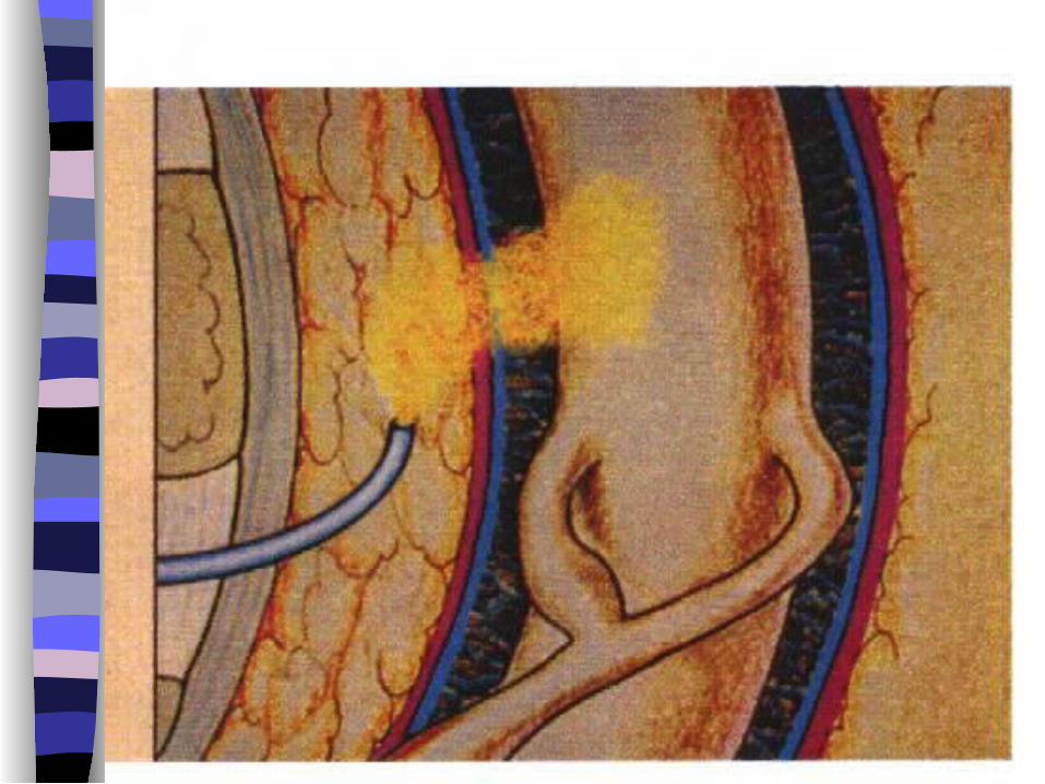
EPIDURAL SPACE
Potential space Between the dura mater,luigamentum
flavum Made up of vasculature, nerves, fat and
lymphatic Extends from foramen magnum to the
sacrococcygeal ligament

INDICATIONS
The objective of epidural analgesia is to relieve pain.
Major surgery
Trauma (# ribs)
Palliative care (intractable pain)
Labour and Delivery

CONTRAINDICATIONS
Patient refusal Known allergy to opioid or local
anesthetic Infection/abscess near the proposed
injection site Sepsis Coagulation disorder Hypotension / hypovolemia Spinal deformity/increased ICP


Patient assume a sitting or side-lying position with the back arched toward the physician.Help to spread the vertebrae apart

Height of sensory blockLumbar-T4Thoracic-T2


Patients with epidural catheters may shower, as long as the dressing is intact.

Epidural Analgesia

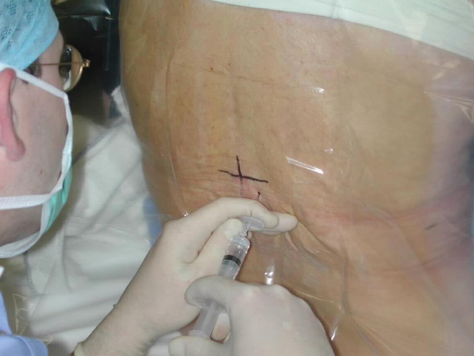
INSERTION OF EPIDURAL CATHETER
Positioning of patient The site is dependent upon the area of
pain Fixing the catheter
Incision LevelThoracic T4-T6
Upper abdo T6-T8
Lower abdo T8-T10
Pelvic T8-T10
Lower extremity L1-L4

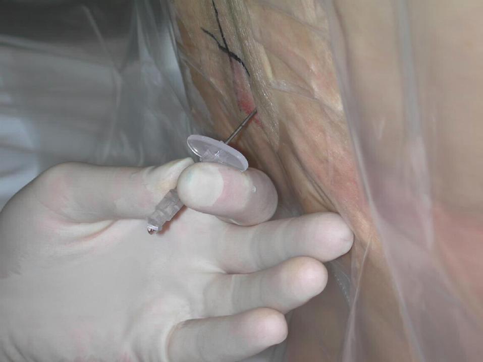
EPIDURAL CATHETERS Ideal Placement (adult) 10-12 cm at the
skin
Epidural catheters have markings that indicate their length.
= there is a mark at the tip of the catheter = the 1st single mark up the catheter is 5cm = double mark up the catheter is 10 cm = triple mark on the catheter is 15 cm = four mark together indicate 20cm
A change in depth of the catheter indicates migration either into or out of the epidural space.

CATHETER MIGRATION
Catheter migration into a blood vessel in the epidural space or subarachnoid space
rapid onset LOC Decrease loss of sensory or motor loss
(marcain) Toxicity Profound hypotension

CATHETER MIGRATION
Out of the epidural space ineffective analgesia no analgesia drugs deposited into soft tissue.

DRUGS
One of the most important factors influencing drug absorption and bioavailability is the drug SOLUBILITY
The more lipid soluble rapid onset & shorter duration

MEDICATION COMMONLY USED
OPIOIDS-Fentanyl +Morphine
(affect the pain transmission at the
opioid receptors)
L.A.-Bupivacaine(marcaine)
(inhibits the pain impulse
transmission in the nerves with
which it comes in contact)

METHODS OF ADMINISTRATION
BOLUS (FENTANYL, DURAMORPH)
CONTINUOUS INFUSION(MARCAINE+FENTANYL)
All drugs administered epidural should be preservative free.
All epidural opioids should be diluted with normal saline prior to intermittent bolus administration.
EPIDURAL LOCAL
Bupivacaine (marcaine)

Mechanism of Action
Bupivacaine (marcaine) - local anaesthetic works as an analgesic (subanesthetic dose) - inhibiting impulse transmission in the nerve fibers - sensory nerves are blocked first before the motor fibers - sensory fibers carrying the pain is blocked before those carrying heat cold touch and pressure.

EPIDURAL LOCAL
ANESTHETIC(MARCAINE)
Onset 10-15 minutes Duration- 4 hrs+ after a bolus or after
infusion is stopped Marcaine(0.0625%-0.125%-0.25%) Extend of spread influenced by volume
and position of patient

OPIOIDS Mechanism of action-distribution Vascular uptake by blood vessels in the
epidural space
Diffusion through dura into CSF to spinal cord to the site of action.
Uptake by the fat in the epidural space.

Morphine (Duramorph/Astramorph) Hydrophilic(water soluble) Slow to diffuse across the dura on to the
spinal cord Can cause late respiratory depression Monitor respiratory status for 12 hrs after
the last dose of duramorph Duration 6 hrs+ Broad spread

Fentanyl (preservativefree)
Lipophilic(fat soluble) Crossess the dura rapidly Rapid onset of action(segmental) Decreased risk of late respiratory
depression Onset 5-20 mins Duration 2-4hrs Excellent for breakthrough pain

Opioid pharmacology
•Peak plasma concentration after po = 1 hourSC,IM = 30MINS IV = 6MIN
•Half- life at steady state PO,PR,SC,IM,IV =3-4 H.
Opioid pharmacology

Adverse Effects -Opioids
Sedation and resp.depression- IV narcan N/V-Opioids stimulate the chemoreceptor trigger zone
primperan Pruritus- diphenhydramine or narcan (low dose) Urinary retention- low dose narcan and /or
catheterization Slowing of GI motility Hypotension
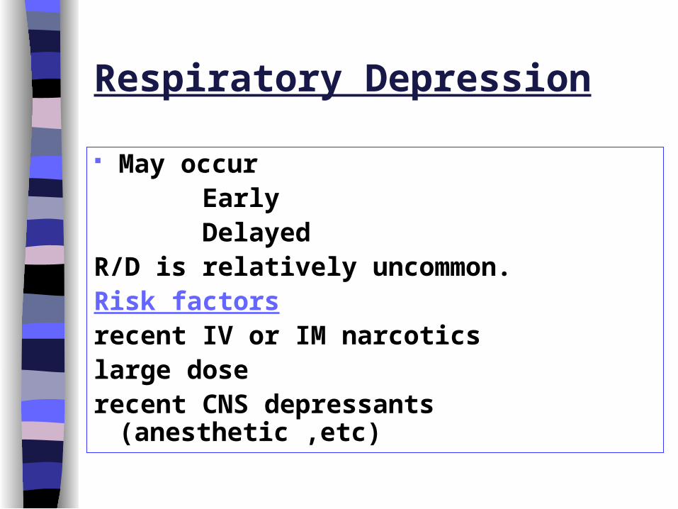
Respiratory Depression
May occur Early DelayedR/D is relatively uncommon.Risk factorsrecent IV or IM narcoticslarge doserecent CNS depressants (anesthetic ,etc)
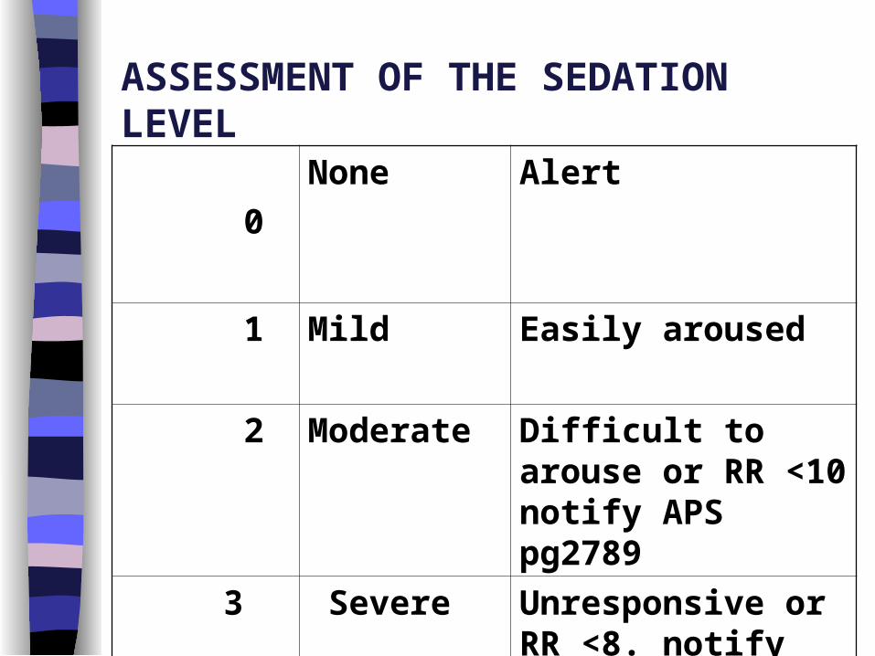
ASSESSMENT OF THE SEDATION LEVEL
0
None Alert
1 Mild Easily aroused
2 Moderate Difficult to arouse or RR <10 notify APS pg2789
3 Severe Unresponsive or RR <8. notify APS2789

Motor and Sensory Assessment
Motor assessment Sensory assessment: * Use ice in the tip of a glove * Start in upper neck and move down thorax bilaterally assessing all potential dermatomes * Level of block is where intensity of cold changes or the cold sensation is absent * assess the dermatomes below the pelvis

Assessment of motor block
Bromage Score

Motor and Sensory Assessment
Motor assessment Sensory assessment: * Use ice in the tip of a glove * Start in upper neck and move down thorax bilaterally assessing all potential dermatomes * Level of block is where intensity of cold changes or the cold sensation is absent * assess the dermatomes below the pelvis

Sensory assessment


Adverse Effects L.A
Hypotension- -assess intravascular
volume status
-no trendelenberg positioning
Teach patient to move slowly from a lying position to sitting to standing position.
Treatment fluids

Cont. Temporary lower-
extremity motor or sensory deficits.
Tx: lower the rate or
concentration.
Urine retention
Tx: catheter
Local anesthetic toxicity (neurotoxicity)
Tx: stop infusion.
Resp. insufficiency
Tx:stop infusion
- ABC(100% o2
call for help)
- Assess spread
and
height of block
- Alt.analgesia

OTHER COMPLICATIONS
Headache (dural puncture)
Tx: symptomatic treatment Autologous blood patch
Infection nausea and
vomiting.
Intravenous placement of catheter
Subdural placement of catheter
Haematoma
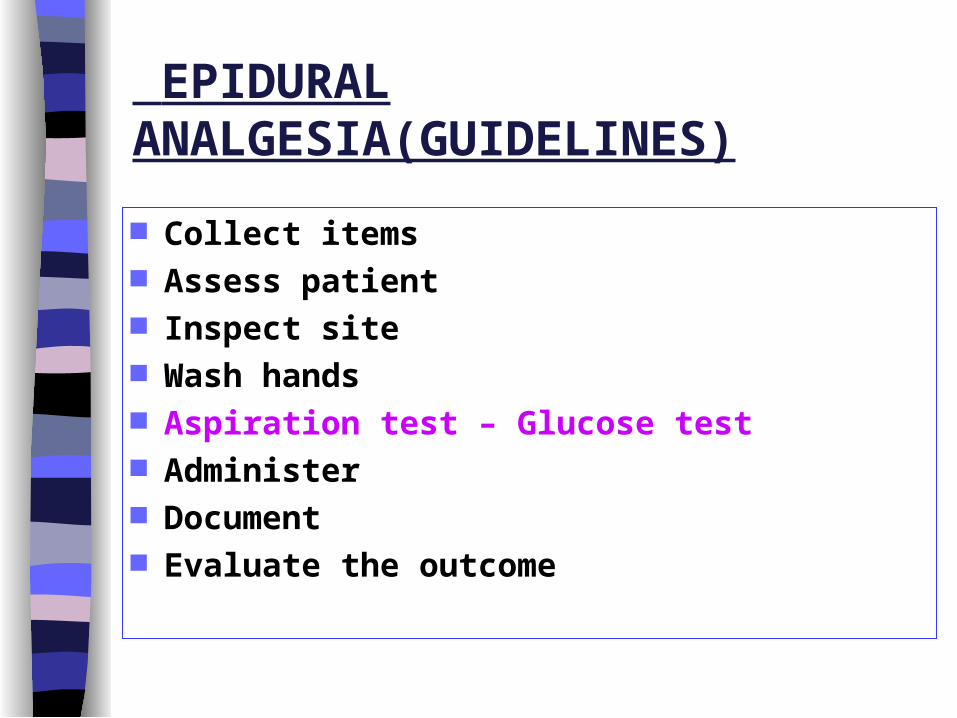
EPIDURAL ANALGESIA(GUIDELINES)
Collect items Assess patient Inspect site Wash hands Aspiration test – Glucose test Administer Document Evaluate the outcome

POLICIES
1. Placement of epidural catheters is performed by the anesthetist in the Operating Room .
2. All patients must have a patent IV access for the duration of epidural therapy and for 12 hours after the catheter is removed.
3. The Acute Pain Service (APS) / Anesthesiologist will be responsible for ordering all epidural analgesia.

4. When the nurse receives a patient with a continuous
epidural infusion, the RN must follow every order on the order sheet.
4.1. If there is any query about the orders, if any of the orders are not filled in and/or if there is no date and time and/or signature, contact the anesthetist who wrote the order.

6. Epidural Medications
6.1. No medications, other than those offered by the Acute Pain Service (APS), are to be administered into the epidural line
6.2. Do not use agents from a multiple dose vial. Most multi-dose vial medications contain preservatives, which can cause intra-spinal neurotoxicity
6.3. Do not use alcohol or alcohol based products near the epidural catheter. Alcohol is neurotoxic and can damage the nerves.
6.4. Return any medications that are unclearly labeled, cloudy or contain particulate matter to pharmacy.
6.5. Return any unused used syringes and cassettes to pharmacy

7. Local anesthetic must not be given as a bolus via the epidural catheter by the nurse.
8. The APS / anaesthetist must be notified if pain is not well
controlled by the epidural infusion.
9. No other narcotic drug will be administered to the patient by any other route unless ordered by the anesthetist / APS .
10. All monitoring information and assessments must be documented on the Continuous Epidural Infusion Flowsheet

11. Patient monitoring will be done as outlined for 12 hours after the discontinuation of the epidural infusion and after removal of epidural catheter, whether it is opioids or local anesthetic which has been infused.
11.1.Assess pain scores, blood pressure, heart rate, sedation level, respiratory and sensory levels, and motor function every 15 mins x1h, every 1 hour x 4 hours and then every 4 hours until 12 hours after the infusion is discontinued.

12. Motor functions of patients receiving local anesthetic via epidural catheter must be assessed by an RN.
13. To assess the height of sensory block, use the ice technique.
14. All epidural catheters must be identified with the label “Epidural Catheter” at the access hub, to prevent inappropriate use of the catheter.


15. Dressing changes are done only when necessary, by nurses
who have been trained in proper techniques of dressing change in present of APS .
16. The administration set (including the 0.22 micron filter) will be changed every 72 hours and the tubing labeled with the date and time of change.

Dressing Changes and Removal of Epidural Catheters
EQUIPMENT/MATERIALS
Dressing Change
Sterile 4 x 4 gauze x 2 packages
Povidone-lodine swabs sticks x 3 packages
Large Transparent dressing x 1
Sterile gloves x 2 pair
Transparent tape

Dressing Changes:
Dressings should not be changed unless it is absolutely necessary, however, they may be changed if:
1. the dressing is wet due to oozing from the puncture site
2. the dressing has become loose
Dressing Changes:
PROCEDURE
1. Gather all equipment and supplies.
2. Explain the procedure to the patient.
3. Position patient on bed.
4. Wash hands.
Open the sterile gloves, transparent dressing
4x 4 gauze package.

Dressing Changes:
5. Put on sterile gloves. With a finger tip, applygentle pressure over the catheter insertion siteand slowly peel back the opsite dressing using extreme care.6. Remove gloves and dispose soiled dressing andgloves into garbage bin.7. Wash hands and put on second pair of sterile gloves.8. Supporting the catheter with one hand, clean theInsertion site with povidone-iodine swabs, movingfrom center to periphery of site. Allow to dry.

Dressing Changes:
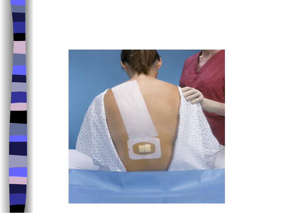
9. Loop the catheter, and fix using transparent dressing.
10. Gently run finger over catheter and dressing.
11. Fix catheter along back and over shoulder .
12. Document dressing change and observations on Nursing Record.


Epidural Catheter Removal:
1. Removal of the epidural catheter must be ordered by
the APS physician.2. Low Molecular Weight Heparin (LMWH): Catheter
removal should be delayed until 12 hours after a dose of LMWH. If LMWH is to be continued, it should not be resumed until at least 4 hours after catheter removal.
3. Standard Heparin Therapy: Catheter removal should be delayed until 6 hours after a dose of standard heparin. If standard heparin is to be continued, it should not be resumed until 2 at least 2 hours after catheter removal.

Removal of the epidural catheter
4. Heparin Infusion: The coagulation status of the patient receiving heparin infusion should be assessed. The heparin infusion shall be stopped for 6 hours prior to catheter removal, and not resumed for at least 2 hours after the catheter is removed.

PROCEDURE
1. The APS staff should be notified if the patient is receiving anticoagulant.2, Gather all equipment and supplies.3. Explain the procedure to the patient. 4. Position the patient side-lying or sitting with the backexposed and arched out. Flexion of the back widens the vertebral space,allowing for easy withdrawal of the catheter
5. Stop the epidural pump.

6. Wash hands. Put on sterile gloves.7. Gently remove tape and dressing.8. inspect catheter site for redness, swelling or
drainage. If the area is reddened or if there is drainage, notify theAPS physician. Collect surface swabs and catheter tipfor cultures and sensitivity (per APS physician) usingaseptic technique.9. Clean the catheter insertion site, from center
outwards,and allow to air dry.

10. Apply steady traction to remove catheter. Do not pull vigorously.
If resistance is met, ask the patient to flex or arch hisback more. If resistance remains, stop and notify APS.11. When catheter is removed, check that tip is intact. Ifnot, notify APS immediately.
12. Apply band-aid to the site.13. Instructions to patients should include: A. report any pain at the insertion

14. Maintain IV access for 12 hours after the last dose of opioid is given15. Document epidural catheter removed in the nursing record.
Include date, time, condition of catheter, and site andpatient’s tolerance of the procedure.
16. Obtain co signature of a second nurse to witnesswaste of narcotic (if required)
17. Clean pump thoroughly and return to Recovery Room Level 2.


1.Measure the length of the epidural catheter from epidural space to the skin.

1.Measure the length of the epidural catheter from epidural space to the skin.
2.What will you do to check the CSF and Blood- tinged fluid.

1.Measure the length of the epidural catheter from epidural space to the skin.
2.What will you do to check the CSF and Blood- tinged fluid.3.How you perform a SENSORY level assessment using an ice
method.

1.Measure the length of the epidural catheter from epidural space to the skin.
2.What will you do to check the CSF and Blood- tinged fluid.3.How you perform a SENSORY level assessment using an ice
method.4.How you perform a MOTOR level.

1.Measure the length of the epidural catheter from epidural space to the skin.
2.What will you do to check the CSF and Blood- tinged fluid.3.How you perform a SENSORY level assessment using an ice
method.4.How you perform a MOTOR level.5.What will you check for the site.

1.Measure the length of the epidural catheter from epidural space to the skin.
2.What will you do to check the CSF and Blood- tinged fluid.3.How you perform a SENSORY level assessment using an ice
method.4.How you perform a MOTOR level.5.What will you check for the site.6.Guidelines for removing epidural catheter for those patients who
receives anticoagulant.
LOW MOLECULAR WEIGHT HEPARINSTANDARD HEPARIN THERAPYHEPARIN INFUSION

1.Measure the length of the epidural catheter from epidural space to the skin.
2.What will you do to check the CSF and Blood- tinged fluid.3.How you perform a SENSORY level assessment using an ice
method.4.How you perform a MOTOR level.5.What will you check for the site.6.Guidelines for removing epidural catheter for those patients who
receives anticoagulant. 7.Based on your knowledge and understanding how you will do the
epidural dressing after preparing all the needed materials.

1.Measure the length of the epidural catheter from epidural space to the skin.
2.What will you do to check the CSF and Blood- tinged fluid.3.How you perform a SENSORY level assessment using an ice
method.4.How you perform a MOTOR level.5.What will you check for the site.6.Guidelines for removing epidural catheter for those patients who
receives anticoagulant.7.Based on your knowledge and understanding how you will do the
epidural dressing after preparing all the needed materials.8. How you will remove the epidural catheter.

REMMEMEBER

STAFF NURSE RESPONSIBILITIES
1. Upon receiving patient check for:
1.1. IV cannula
1.2. urinary catheter
1.3. epidural catheter length (if visible)
1.4. if dressing is intact
1.5. doctors order
1.6. ongoing epidural infusion bag

STAFF NURSE RESPONSIBILITIES
2. Assess and monitor as indicated on epidural flowsheet.
3. Notify APS or on call anaesthetist any untoward complications, emergency , side effects or inadequate relief related to therapy.
3.1 pager 2113 aps anaesthetist weekdays 0730 – 1600
3.2 pager 2789 aps nurse weekdays 0730 – 1600 3.3 pager 3540 maternity on call anaesthetist
1630 – 0730 daily and weekends

STAFF NURSE RESPONSIBILITIES
4. Certified nurse should connect the new bag.5. CN are allowed to increase or decrease the infusion rate based
on the rate ordered or patients pain response from ongoing infusion.
6. Infusions: 6.1. if used : discard with the presence of other witness or
staff. 6.2. if not used: Incident report and send back to pharmacy.7. Keep patent IV access and continue to monitor for 12 hours
after removing the epidural catheter. 8. Inform APS team or on call anesthetist if patients is on
anticoagulant.

You’re!!! What did you learn?