Embed Size (px)
Citation preview

Rob Webster RCI Sheffield
Dr Rob WebsterNHSBT Sheffield

Rob Webster RCI Sheffield
1932 Diamond : erythroblastosis fetalishydrops fetalis/kernicterus-part of same disordercharacteristics
fetal red cell haemolysisextramedullary haemopoiesisnucleated red cells in excess in circulation

Rob Webster RCI Sheffield
Cause unknown1940 Landsteiner & Weiner
discovery of Rh blood group system1941 Levine
HDN due to development of anti-D in D negative woman following exposure to D positive red cellsantibody crosses placenta, coats D positive cells

Rob Webster RCI Sheffield
1948 WeinerRh alloimmunisation caused by transplacentalpassage of fetal red cells into maternal circulation (pregnancy/delivery)
subsequently other antibodies implicatedA,B,ABRh : C,c,E,enon Rh : e.g. K, Duffy, Kiddmost significant are D, c, K

Rob Webster RCI Sheffield

Rob Webster RCI Sheffield
-Clinical Features
Hydrops : primarily hepatic in originhepatosplenomegaly, portal hypertension, hypoalbuminaemia, anasarca
Kernicterus :deposition of bilirubin in basal ganglia
neurosensory deafnessspastic choreoathetosismental retardation

Rob Webster RCI Sheffield
Fetal oedemaFirst described by Ballantyne in 1892But recognised for over 200 years

Rob Webster RCI Sheffield
Yellow KernKern consists of basal ganglia, hippocampus, geniculate bodies and cranial nerve nuclei especially occulomotor, vestibular and cochlear

Rob Webster RCI Sheffield
Opisthotonus

Rob Webster RCI Sheffield
1.Transfusion of red cells to mother2.Fetomaternal haemorrhage (FMH) during
pregnancy

Rob Webster RCI Sheffield
Before modern therapy: 1% of all pregnant women developed Rh alloimmunisationWith anti D prophylaxis incidence now 10.2 per 10,000 live births with <10% requiring intrauterine transfusionOf those requiring IUT:
85% anti-D10% anti-Kell (causes hypoproliferative anaemia, with less hyperbilirubinaemia3.5% anti-c
ABO incompatibility can occur in <1% live births associated with significant haemolysis (mother usually Group O)

Rob Webster RCI Sheffield
Common Rare Never
Anti- D Anti Fya Anti Lea
Anti D+C Anti s Anti Leb
Anti- D+E Anti M Anti I
Anti C Anti N Anti IH
Anti E Anti S Anti P1
Anti c Anti - Jka
Anti e
Anti - K

Rob Webster RCI Sheffield
delivery (including CS)abortionantepartum haemorrhage (APH)external version of fetusclosed abdominal injuryectopic pregnancyintrauterine death (IUD)stillbirth

Rob Webster RCI Sheffield
invasive prenatal diagnosis:amniocentesischorionic villus sampling (CVS)fetal blood sampling (FBS)
other intrauterine proceduresinsertion of shuntsembryo reduction

Rob Webster RCI Sheffield
Events likely to be associated with large FMH:- traumatic deliveries including Caesarean
section- manual removal of placenta- stillbirths- intrauterine deaths- abdominal trauma during 3rd trimester- delivery of twin pregnancy
- unexplained hydrops

Rob Webster RCI Sheffield
antenatal screening and monitoringtreatment -antenatal/post natalprevention (anti-D HDN)

Rob Webster RCI Sheffield
ABO Rh(D) group and antibody screen at bookingIf screen positive
antibody identificationantibody quantitationtesting protocolpaternal screeningfetal genotypingpost-delivery ABO/D group / Hb /DAT/ Bilirubin

Rob Webster RCI Sheffield
BCSH guidelines 2006
- anti-D, c, K : 4 weekly to 28/40 then2 weekly to gestation
-others : booking and 28 weeks

Rob Webster RCI Sheffield

Rob Webster RCI Sheffield
Fetal Blood Typing First StepPaternal Rh genotyping for Rh DCE / implicated antigenFather heterozygous / homozygousSerologic analysis most probable genotype by haplotypetablesPCR analysis
Fetal Blood Typing Second StepOnly if fetus at risk of inheriting antigenMore recently from fetal DNA in maternal plasma

Rob Webster RCI Sheffield
Antibody titresQuantitation of antibodiesMiddle Cerebral Artery Peak Systolic Velocity Foetal UltrasonographyInvasive monitoring with umbilical blood samplingFetal blood typing

Rob Webster RCI Sheffield
Antibody titresAssist clinicians to determine when more invasive tests neededMethods:
Saline tube techniques onlyGel technology to be avoidedPrevious samples run in parallel
Every 4 weeks till 28 weeks, then once very 2 weeksCritical titres 1:32 Once critical levels reached, no further tires should be doneOnly of value in first affected pregnancy, subsequent pregnancies require more aggressive monitoring

Rob Webster RCI Sheffield
Assessing the severity of HDFNQuantitation of antibodies:
Anti- DMeasured in IU/mlDone using NIBSC standard anti-DLess than 4 IU/ml HDN unlikely4- 15 IU/ml Moderate risk of HDNMore than 15 IU/ml High risk of Hydrops fetalis
Anti c Less than 7.5 IU/ml Continue to monitor7.5 to 20 IU/ml Risk of moderate HDNMore than 20 IU/ml Risk of severe HDN
others titres > 32 significant risk (??anti-K)

Rob Webster RCI Sheffield
Is there an antibody?is it capable of causing fetal haemolysis / anaemia?
what antibody?level?
how frequently to test?refer to FMU?early delivery?blood for fetus/infant/mother?

Rob Webster RCI Sheffield
Indicators of high fetal risk, ie refer to FMU
History of non-ABO HDFN requiring transfusion, irrespective of antibodyanti-D >10iu/mlanti-K
untransfused womenpartner K+
rising anti-c (>20iu)

Rob Webster RCI Sheffield
Doppler measurement of peak velocity of systolic blood flow in the Middle Cerebral Artery best non-invasive test to diagnose mild-moderate fetal anaemia.
(false +ves in cardiac anomalies and non-allo immune anaemia )
Mari et al NEJM 2000, 342:9-14

Rob Webster RCI Sheffield
Middle cerebral artery peak systolic velocity & Fetal ultrasonography
Accurate test for detecting fetal anaemiaNon invasive no fetal risk of miscarriage / preterm labourReciprocal relationship between fetal Hb & Velocity of cerebral flowCan be used for all alloimmunised pregnancies100% sensitivity for moderate / severe anaemia-Threshold value of peak systolic x 1.5 multiples of the median

Rob Webster RCI Sheffield

Rob Webster RCI SheffieldCopyright © 2007 by the American Roentgen Ray Society
McCarville, M. B. et al. Am. J. Roentgenol. 2004;183:1117-1122
--Typical transcranial Doppler sonographic recording from middle cerebral artery

Rob Webster RCI Sheffield
Flow velocity wave form in the fetal middle cerebral artery in a severely anaemic fetus at 22 weeks (left) and in a normal fetus (right). In fetal anaemia, blood velocity is increased

Rob Webster RCI Sheffield
Tests used by obstetricians to predict fetal anaemia
Ultrasound
placental thickness
umbilical vein diameter
liver length
spleen perimeter
Doppler
measure fetal blood flow
useful to assess fetal maturity
does not identify early fetal disease -
changes visible only once hydrops
has occurred
weak correlation with fetal Hct/Hb
detects early fetal anaemia

Rob Webster RCI Sheffield

Rob Webster RCI Sheffield
when any IAT-reactive antibody is present in pregnancy, a cord DAT must be done ASAP
if DAT is +ve, check Hb and bilirubin to diagnose HDN (NB. RAADP)
if DAT is -ve, no risk of HDN (except ABO)

Rob Webster RCI Sheffield
Intrauterine red cell transfusionsIVIgGPremature delivery

Rob Webster RCI Sheffield
Performed by direct infusion of allogeneic blood into:Umbilical cordIntrahepatic portion of hepatic veinIntraperitoneally

Rob Webster RCI Sheffield
Red cells selected are :Group O Rh D Neg (unless anti c), Kell Neg, High Titre NegHct 0.7-0.85CMV Neg, irradiated< 72 hrs old (<24 hours following irradiation)Hb S-NegNegative for the offending antigenCrossmatch-compatible with maternal plasma

Rob Webster RCI Sheffield
Maternal blood used in case of rare antibodiesIUT is only possible from 20 weeks gestationIUT also useful for
PRCA from Parvovirus B19Fetomaternal haemorrhageTwin-to-twin transfusion

Rob Webster RCI Sheffield
Amount of blood to be transfused Desired PCV Fetal PCV x Fetoplacental blood volumeDonor PCV Desired PCV
Final Hct ~ 40%Transfusions every 14 days until 35 weeks

Rob Webster RCI Sheffield
Fetal outcomes superior with IUT:Mortality before Hydrops 2 to 8%Mortality after Hydrops 22 to 30%
IUT suppress erythropoiesis causing hypoproliferative anaemia in the neonatal period may need top upsDespite severe fetal haemolytic disease, normal developmental outcomes can be expected from children treated with IUT

Rob Webster RCI Sheffield
Van Kamp et al; AJOG, 2005:254 foetus with 740 IUTs treated in between 1988 2001Death from procedure related complications 1.6%Overall procedure-related complication rate 3.1%:
0.1% PROM0.3% Infection2.0% Emergency Ceasarean Section0.9% Fetal death0.7 % Neonatal death
Intrauterine Red Cell Transfusion - outcomes

Rob Webster RCI Sheffield
Prenatal use Efficacy unknownNo RCTOnly Case reports & Case series
Postnatal use 1g/kg within 12 hoursEffective in reducing the hospital stayReducing the duration of phototherapyNeed for exchange transfusion

Rob Webster RCI Sheffield
Early delivery after 35 weeks performed in all severe cases of HDFNHave blood on standby for exchange:
Group O Rh D Neg (unless anti c), Kell Neg, High Titre NegHct 0.5-0.6CMV Neg, irradiated< 72 hrs old (<24 hours following irradiation)Hb S-NegNegative for the offending antigen

Rob Webster RCI Sheffield
Samples after deliveryExchange transfusionsPhototherapyTop Up transfusions

Rob Webster RCI Sheffield
Indicated if:cord Hb <8 g/dLcord bilirubin > 100 µmol/L, andrapidly rising bilirubin
Double volume exchange removes 85 - 90% of infant s cellsUse plasma-reduced blood (Hct 0.5 - 0.6)Check Hb 2-weekly until 3 months may have hypoproliferative anaemia

Rob Webster RCI Sheffield
Exchange transfusion: Two-vessel technique

Rob Webster RCI Sheffield
10 - 15 ml/kgUse multi-doses from paedipack if possibleSAG-M acceptableCMV negHT anti-A/B negExtended Ab screen negSickle negIrradiated if previous IUT

Rob Webster RCI Sheffield
Usually group O mother with Group A (or B) babyIgG anti A (or B)Expression of A and B antigens much weaker on neonatal cells haemolysis often not significantDAT may or may not be positiveHyperbilirubinaemia more common than anaemia

Rob Webster RCI Sheffield

Rob Webster RCI Sheffield
We wanted in the initial analysis to identify how often fetal treatment was required for non-D, c or K antibodies

Rob Webster RCI Sheffield
A form was sent to hospitals around the time of delivery, for women found by RCI Labs in England, to have antibodies with a titre 16. Information requested included any need for intrauterine transfusion (IUT) phototherapy, exchange or top-up transfusion, or deathData from 2006-11 were analysed. Whilst
NHSBT tests most antenatal women in England with antibodies, we also cross-checked against IUT blood issued in 2010 and 2011, to ensure no cases of HDF were missed

Rob Webster RCI Sheffield
Dear Colleague
Re: Collection of data on Haemolytic Disease of the Newborn
Thank you for supporting the National Health Service Blood and Transplant (NHSBT) programme of data collection on the outcome of pregnancies of women with red cell antibodies. By recording the data collected in this exercise we hope to accumulate a body of evidence for the effect of specific antibodies of known strength on the fetus/newborn. We hope that you will be willing to help by completing the attached questionnaire.
Any information you can provide will be useful, and we hope that you will be able to complete the forms as fully as possible. If necessary, please contact the appropriate clinician or midwife so that all the relevant clinical information can be captured.
Please return the completed form in the envelope provided, to the address below.
Data ProtectionPatients are informed that we collect information about their baby in the patient information leaflet entitled Blood Groups and Red Cell Antibodies in Pregnancy produced by the NBS and distributed to all hospital clinics and GPs for whom we provide a screening service.Please be assured that all patient data will be held securely, and in accordance with the patient s rights, under the Data Protection Act (1998).
Thank you in anticipation of your help. If you have any queries please contact your local NHSBT Red Cell Immunohaematology laboratory or myself.
Liz PepperellRCI Project ScientistNHSBT CambridgeLong RoadCambridgeCB2 0PT
Tel. 01223 588132e-mail: [email protected]

Rob Webster RCI Sheffield
This data is being collected by the NBS in order to establish the outcomes of pregnancies with antibodies. Expectant mothers are informed via the antenatal information leaflet Blood Groups and Red Cell Antibodies in Pregnancy and NBS antibody card. Data Protection issues are covered by these documents and the Service Level Agreement between the NBS and your Trust. Mother s details: [to be completed by NBS]Surname: Forename[s]:DoB:Hospital/GP?NHS Number: Hospital Reference No:NBS Ref No:E.DD.Maternal antibody specificity Father Rh phenotype: Other relevant positive antigens
Rob Webster RCI Sheffield
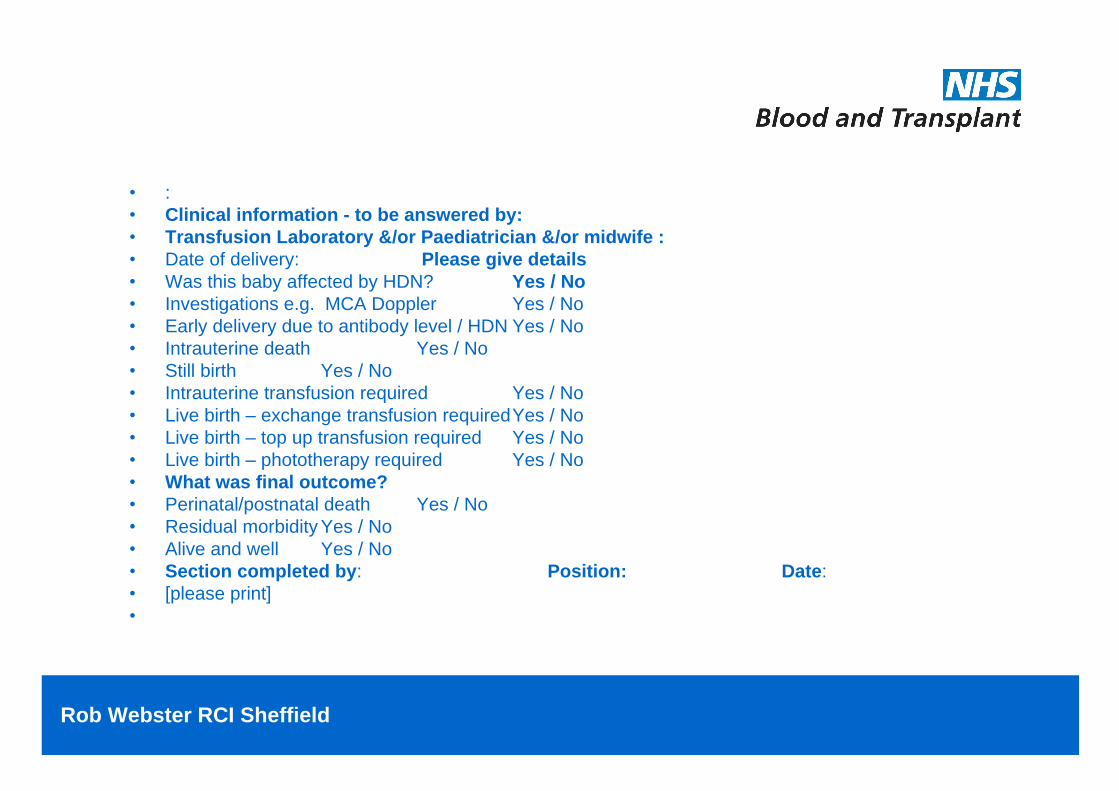
:Clinical information - to be answered by:Transfusion Laboratory &/or Paediatrician &/or midwife :Date of delivery: Please give details Was this baby affected by HDN? Yes / NoInvestigations e.g. MCA Doppler Yes / NoEarly delivery due to antibody level / HDN Yes / NoIntrauterine death Yes / NoStill birth Yes / NoIntrauterine transfusion required Yes / NoLive birth exchange transfusion requiredYes / NoLive birth top up transfusion required Yes / NoLive birth phototherapy required Yes / NoWhat was final outcome?Perinatal/postnatal death Yes / NoResidual morbidity Yes / NoAlive and well Yes / NoSection completed by: Position: Date:[please print]

Rob Webster RCI Sheffield
Laboratory information on baby:Blood group [ABO + RhD]Relevant positive antigen(s) Cord DAT scoreHaemoglobin at delivery Bilirubin at deliveryLowest haemoglobin Maximum serum bilirubinRecords of transfusion - please complete details (dates/volumes) below:Intrauterine transfusion Exchange transfusionTop up transfusionCompleted by: Position: Date:[please print]

Rob Webster RCI Sheffield
Year2006-7 633,0002007-8 653,0002008-9 656,0002009-10 656,0002010-11 672,0002011-12 673,000

Rob Webster RCI Sheffield
Replies were returned on 3787 cases (67% response rate).Total number of cases with red cell antibodies, but excluding anti-D, c or K = 1454 (figure 2[f1] ). Only 4 required IUT: 1 with anti E titre of 512; 3 with anti-Fya titre 512[p2] .Of 485 cases of anti-E alone, 132 (27%) babies were E neg; 102 (21%) were E pos and 251 were unknown (but as 30% of population are expected to be E pos, about 75 of these may be E pos). So, 1/177 = 0.6% needed IUT (see figure 3).Of 137 cases with anti-Fya alone, 20 (15%) babies were Fya neg; 26 (19%) were pos and 91 were unknown (but as 60% of population are Fya pos, about 54 of these may be Fya pos). So, 3/80 = 4% needed IUT. There were no deaths due to HDFN.Of note, while IUT was required in 4 cases with non-D, c or K antibodies, the number of cases requiring IUT for anti-D = 73; anti-c = 10 and anti-K = 8.

Rob Webster RCI Sheffield
F ig u r e 3 - N u m b e r o f w o m e n w it h a n t ib o d ie s a n d n u m b e r o f IU T s
a n t i - E a n t i - F y a
1 3 22 0
2 5 19 1
1 0 1 2 3
1 3
n = 4 8 5 n = 1 3 7
Nu
mb
er o
f ca
ses
n e e d e d IU T
a n t ig e n p o s b a b y
a n t ig e n s ta tu su n kn o w n
a n t ig e n n e g b a b y

Rob Webster RCI Sheffield
While predictors of HDFN include a previous history of HDFN, antibody titre >32 or a rising titrePractice in the frequency of monitoring non-D, c or K antibodies after 28 weeks varies from doing nothing until testing cord blood at delivery (for Hb and bilirubin to monitor for haemolysis), to fortnightly monitoring of antibody titres +/- MCA Doppler monitoring for fetalanaemia. This has associated implications for resources involved and the inconvenience to mothers of extra hospital attendances and potential for the 5% false positive rate of MCA Doppler testing to result in unnecessary fetal blood sampling ± IUT, with a 1-2% rate of miscarriage.

Rob Webster RCI Sheffield
In light of our findings, together with previous literature, discussion is needed with the Royal College of Obstetricians and Gynaecologists to rationalise monitoring for optimal patient care, without overuse of resources. Monitoring could be tailored differently for some antibodies.