Embed Size (px)
Citation preview
Table of Contents
Introduction
Red Blood Cells
Platelets
Whole Blood
Plasma
Cryoprecipitated AHF
Blood Component Modifications
Hospital Transfusion Committee
Patient Blood Management
Appendices
Introduction
Enriching a long tradition in blood banking, the American Red Cross is committed to the ongoing education of healthcareprofessionals who prescribe and transfuse blood. A 1958 editorial published in Blood voiced a concern about transfusionpractices: “The reason for misuse of blood transfusions is that we are not sufficiently aware of what are valid indications for this procedure. A surgical operation is not an indication for blood transfusion. Uterine bleeding is not an indication.Neither is a low hematocrit. Blood is not a tonic. It is not a placebo. It does not improve wound healing. Neither is it a substitute for careful consideration of the patient and his problem.”(1) Four years later, the same writer endorsed the concept of a “Hospital Transfusion Board” to educate physicians about indications for blood, establish policies and rules for the transfusion service, investigate severe reactions, and periodically review “all aspects of the transfusion service.”(2)
In the decades that followed, a trickle and then a torrent of well-designed robust clinical trials gave blood bankers and physicians of all specialties an impressive body of scientific literature for the development of evidence-based transfusion practices. Guidelines from professional societies and accrediting organizations such as AABB are beingintegrated into day-to-day practice and hospital transfusion protocols.
To affirm and advance its mission of educating transfusion and medical communities, the American Red Cross presents the Third Edition of the Compendium of Transfusion Practice Guidelines (‘Compendium’). American Red Cross and other physicians and scientists have updated all chapters, incorporating recent peer-reviewed publications on blood component therapy, transfusion strategies and alternatives, the transfusion committee, transfusion complications, and infectious disease testing. In light of the fact that new pathogens and infectious diseases may present challenges to the safety of the blood supply, this edition includes an appendix on Zika virus.
A Compendium of Transfusion Practice GuidelinesEdition 3a
The current generation of blood bankers has essentially created a new subspecialty —patient blood management—that includes best practices and innovative approaches to safer transfusion, and embraces information technology to guide and monitor transfusion therapy. Consequently, this edition of the Compendium introduces a new chapter titled “Patient Blood Management” which describes the elements of a successful Patient Blood Management program, explores the use of information technology to influence transfusion practices, and details blood management in the perioperative period.
To further the dissemination of transfusion and blood safety information, an app of the second edition of the Compendium was first launched in 2015, and thisnew app is now available for the Third Edition. This complements the print edition to increase accessibility to physicians, physicians-in-training, nurses, medical technologists, and other medical professionals (redcross.org/mobileapp). The Compendium is one element of the Red Cross educational program which also encompasses the continuing education program, SUCCESS (success.redcross.org), the journal Immunohematology and the support of more than 30 Red Cross physicians.
The Compendium is intended as a reference work for practitioners of transfusion medicine. Guidelines reflect the authors’ understanding of relevant literature and other publications such as the Circular of Information (‘Circular’). As in all areas of clinical practice, transfusion medicine is constantly evolving; as our understanding grows, there is an unavoidable risk of any publication on this topic becoming outdated. The guidelines are intended to be exactly that and are not prescriptive. Each clinical situation should be evaluated independently and treatment tailored accordingly. As stated in the Circular: “Blood banks and transfusion services are referred to the AABB Standards for Blood Banks and Transfusion Services for additional information and policies, especially in the areas of recipient sample identification, compatibility testing, issue and transfusion of blood and blood components, investigation of transfusion reactions, and proper record-keeping practices. Transfusionists are referred to the AABB Technical Manual for applicable chapters on adult and pediatric transfusion.”(3)
We hope that the Compendium becomes an essential educational resource and reference guide for transfusion management of your patients.
Joy L. Fridey MD, MBA Editor in ChiefLiz Marcus BSc, PMP Production Editor
References1. Crosby WH. Misuse of blood transfusion. Blood.1958;13:1198-1200.
2. Crosby WH. The Hospital Transfusion Board. Transfusion. 1962;2(1):1-2.
3. AABB, American Red Cross, America’s Blood Centers, Armed Services Blood Program. Circular of Information for the Use of Human Blood and Blood Components. Revised November 2013. Available at AABB.org
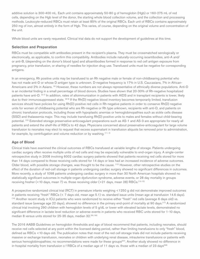
Red Blood Cells
Components
Approved name: Red Blood Cells
Commonly used names: • Packed cells• Red cells• Packed red blood cells• RBCs
Description (1-3)
Red Blood Cells (RBCs) consist of erythrocytes concentrated from whole blood donation or collected by apheresis. They contain citrate anticoagulant and usually one of several types of preservative solutions. Depending on the preservative-anticoagulant system, the hematocrit (Hct) of RBCs is about 55-65% for additive solutions (AS), AS-1, AS-3, AS-5, AS-7 and about 65-80% for citrate-phosphate-dextrose-adenine solutions, CPDA-1, CPD, CP2D. RBCs contain 20-100 mL of donor plasma, usually <50 mL, in addition to preservative and anticoagulant. The typical volume of AS RBCs including
additive solution is 300-400 mL. Each unit contains approximately 50-80 g of hemoglobin (Hgb) or 160-275 mL of red cells, depending on the Hgb level of the donor, the starting whole blood collection volume, and the collection and processing methods. Leukocyte-reduced RBCs must retain at least 85% of the original RBCs. Each unit of RBCs contains approximately 250 mg of iron, almost entirely in the form of Hgb. This varies, of course, depending on the original volume and concentration of the unit.
Whole blood units are rarely requested. Clinical trial data do not support the development of guidelines at this time.
Selection and Preparation
RBCs must be compatible with antibodies present in the recipient’s plasma. They must be crossmatched serologically or electronically, as applicable, to confirm this compatibility. Antibodies include naturally-occurring isoantibodies, anti-A and/or anti-B, (depending on the donor’s blood type) and alloantibodies formed in response to red cell antigen exposure from pregnancy, prior transfusion, or sharing of needles for injection drug use. Transfused units must be negative for corresponding antigens.
In an emergency, Rh positive units may be transfused to an Rh negative male or female of non-childbearing potential who has not made anti-D or whose D antigen type is unknown. D-negative frequency is 17% in U.S. Caucasians, 7% in African-Americans and 2% in Asians. (4) However, these numbers are not always representative of ethnically diverse populations. Anti-D is an incidental finding in a small percentage of blood donors. Studies have shown that 20-30% of Rh-negative hospitalized patients have anti-D. (5-7) In addition, rates of alloimmunization in patients with AIDS and in transplant recipients is lower, likely due to their immunosuppressed state. (8-9) If the Rh(D) negative blood inventory becomes temporarily limited, transfusion services should have policies for using Rh(D) positive red cells in Rh-negative patients in order to conserve Rh(D) negative units for women of childbearing potential who are Rh-negative or Rh type unknown, recipients with anti-D, and patients on chronic transfusion protocols, including those with hypoplastic anemias or hemoglobinopathies such as sickle cells disease (SSD) and thalassemia major. This may include transfusing Rh(D) positive units to males and females without child-bearing potential. (10) Extended storage preservative-anticoagulant preparations such as AS-1 and AS-3 are appropriate for nearly all patients and extend the shelf-life of RBCs to 42 days. Physicians concerned about preservative-anticoagulant for large volume transfusion to neonates may elect to request that excess supernatant in transfusion aliquots be removed prior to administration, for example, by centrifugation and volume reduction or by washing. (11, 2)
Age of Blood
Clinical trials have examined the clinical outcomes of RBCs transfused at variable lengths of storage. Patients undergoing cardiac surgery often receive multiple units of red cells and may be especially vulnerable to end-organ injury. A single-center retrospective study in 2008 involving 6002 cardiac surgery patients showed that patients receiving red cells stored for more than 14 days compared to those receiving cells stored for 14 days or less had an increased incidence of adverse outcomes. Older blood, with possible storage changes, was thought to be the cause. (12) However, other retrospective studies on the effect of the duration of red-cell storage in patients undergoing cardiac surgery showed no significant differences in outcomes. More recently, a study of 1098 patients undergoing cardiac surgery in more than 30 North American hospitals showed no statistically significant outcomes in multiple-organ dysfunction syndrome, adverse events, or 28 day mortality in groups receiving fresher (<10 days, mean 7) vs. those receiving older (>21 days, mean 28) RBCs.(13-15)
A prospective randomized clinical trial (RCT) in premature infants weighing <1250 g did not demonstrate improved outcomes in patients receiving “fresh” RBCs (< 7 days old, mean age 5.1) vs. standard issue units (mean age at transfusion 14.6 days). (16) Another recent study in ICU patients who were randomized to receive either “fresh” red cells (average 6 days old) vs. standard issue (average age 22 days), showed no difference in the primary end-point of mortality at 90 days.(17) A randomized clinical trial involving 290 children with hemoglobin levels of 5 g/dL or lower with elevated lactate levels, demonstrated no significant difference in lactate level reduction or adverse events in patients who received RBC units stored for 1-10 days, median 8 versus units stored for 25-35 days, median 32.(55, 56)
The 2016 AABB Guidelines on hemoglobin thresholds and age of blood recommend that patients, including neonates, should receive red cells selected at any point within the licensed dating period, rather than limiting transfusions to only “fresh” blood, defined as RBCs <10 days old. The publication notes that most of the red cell storage trials did not include patients receiving massive or exchange transfusion, neonates or children with underlying renal disease, intrauterine transfusions or patients with serious hemoglobinopathies; no recommendations were made for these groups(57). Another study showed no difference in in-hospital mortality from transfusion o f RBCs of a median age of 11 days vs. those with a median of 23 days(58)
General Information
Dosing
RBCs should be transfused according to clinical need, including signs and symptoms, Hgb level, and the results of hematologic assays. In the absence of acute hemorrhage, RBCs should be given as single units followed by appropriate evaluation to justify additional units.(10, 17) Transfusion of an RBC unit should be completed within four hours. If more time is required, smaller aliquots can be prepared and transfused sequentially.
Response
In a non-bleeding, non-hemolysing adult transfused with compatible RBCs, the hemoglobin level should equilibrate within 15 minutes of transfusion. One unit should increase the Hgb in an average-size patient (70-80 kg) by approximately one g/dL and the hematocrit by 3%.(18)
In neonates, a dose of 10-15 mL/kg is generally given, and additive solution red cells with a Hct of approximately 60% will increase the Hgb by about 3 g/dL.
Transfused red cells have a half-life of approximately 30 days in the absence of blood loss, hemolysis, or other processes that might affect in vivo survival. Seriously ill adult or pediatric patients may lose significant amounts of blood from phlebotomy for laboratory analysis.(19) In addition, when active bleeding is taking place, the anticipated post-transfusion hemoglobin level may be impacted by the dilutional effect of volume replacement with crystalloid or colloid.
Indications and Contraindications
RBCs are indicated for patients with symptomatic deficiency of oxygen-carrying capacity or tissue hypoxia due to inadequate circulating red cell mass. They are also indicated for exchange transfusion (for example, hemolytic disease of the fetus or newborn) and red cell exchange for acute chest syndrome in sickle cell anemia. Patients must be evaluated individually to determine the proper transfusion therapy, with care to avoid under – or over – transfusion. Transfusion decisions should be based on careful clinical assessment as well as Hgb levels.(20)
RBCs may be used for patients with acute blood loss whose symptoms or conditions may not improve with administration of crystalloid solutions. RBCs should not be used to treat anemia that can be corrected with therapies other than transfusion, such as pharmacolog agents. RBCs should also not be used as a means of increasing blood volume, to increase oncotic pressure, improve wound healing, or improve a patient’s sense of well-being.
For side effects and hazards, please see the Appendices.
Utilization Guidelines
Perioperative/Periprocedural
The function of an RBC transfusion is to augment O2 delivery to the tissues. Hemoglobin levels during bleeding are imprecise measures of tissue oxygenation. Intravenous fluid resuscitation and the time needed for equilibration can significantly alter the Hgb concentration.(21) A number of factors beside blood Hgb level must be considered, such as pulmonary oxygenation, blood flow, Hgb O2 affinity and tissue demands for O2.(21–23) The Hgb level and clinical status of the patient should both be considered in assessing the need for RBC transfusion.
The adequacy of oxygen delivery must be assessed in individual patients, particularly in patients with limited cardiac reserve or significant atherosclerotic vascular disease. If available, mixed venous O2 levels, O2 extraction ratios, or changes in oxygen consumption may be helpful in determining tissue oxygenation. Other factors to consider include anticipated degree and rate of blood loss and the effect of body temperature or drugs and anesthetics on oxygen consumption. The American Society of Anesthesiologists Task Force (ASATF) recommends that RBCs should usually be administered when the Hgb concentration is low (for example, <6g/dL in a young healthy patient), especially when the anemia is acute. Further, the ASATF also states that RBCs are usually unnecessary when the Hgb concentration is >10 g/dL. These guidelines may be altered in cases of anticipated blood loss.(23)
The decision to transfuse should be based on any indication of organ ischemia, potential bleeding or the rate and magnitude of actual ongoing bleeding, the patient’s intravascular volume status, and the risk factors for complications of inadequate
oxygenation. These risk factors include a low cardiopulmonary reserve and high oxygen consumption. AABB clinical practice guidelines recommend considering transfusion in post-operative surgical patients for Hgb <8 g/dL or when clinically significant symptoms of anemia are present (for example, tachycardia unresponsive to fluid resuscitation).(21)
Preoperative assessment and efforts to reduce RBC transfusion requirements in the perioperative period include the evaluation and treatment of anemia prior to surgery.(10) The use of alternative measures to reduce allogeneic red blood cell use should be considered. These include acute normovolemic hemodilution, intraoperative and postoperative autologous blood recovery, and operative and pharmacologic measures that reduce blood loss.
The Society of Thoracic Surgeons and the Society of Cardiovascular Anesthesiologists blood conservation clinical practice guidelines for patients undergoing cardiothoracic surgery recommend the following:
• preoperative assessment to identify patients at elevated risk of bleeding and subsequent blood transfusions (advanced age, decreased preoperative red blood cell volume, and emergent or complex procedures)
• effective treatment of preoperative anemia and minimizing hemodilution during cardiopulmonary bypass (CPB) to preserve red blood cell volume.
• appropriate management of preoperative antiplatelet and anticoagulant drug therapy, and the use of anti-fibrinolytic agents such as epsilon-aminocaproic acid or tranexamic acid to reduce total blood loss.(32)
Refer also to the Patient Blood Management chapter Liberal vs. Restrictive Transfusion Thresholds
Publications from 2013 include: a meta-analysis of studies in which higher vs. lower Hgb levels used as transfusion thresholds (10 g vs. 7 g) led to fewer adverse reactions, including death, and decreased costs in the lower threshold patients(24); and, greater survival percentages in patients with acute upper GI bleeding receiving fewer RBC transfusions.(25) Other prospective RCTs examining various patient populations are ongoing. This question has also been investigated with regard to cardiac patients.
Cardiac Surgery
Cardiac surgery transfusion thresholds have been evaluated in randomized trials(26, 27) and suggest a Hgb threshold of 7.5 g/dL – 8 g/dL. The Transfusion Indication Threshold reduction (TITRe2) trial randomized patients with underlying cardiac disease undergoing coronary artery bypass graft (CABG), valve surgery, or both, to a post-operative transfusion threshold of <7.5 g/dL versus <9 g/dL. The trial demonstrated that the composite endpoint of infection or an ischemic event was similar in both groups and that more deaths were noted in the restrictive group at 90 days, although the 30 day mortality rate was similar in both cohorts.(26)
General Critical Care
Individualization of red cell transfusion applies to critical care patients as well as perioperative patients. To the degree possible, the effects of anemia should be differentiated from those of hypovolemia, although both can impede tissue O2 delivery. Blood loss greater than 30% of blood volume generally causes significant clinical symptoms and signs, but in younger healthy patients, resuscitation with crystalloids alone may be successful with blood loss of up to 40% of volume (approximately 2 liters of blood loss in an average adult male). Ultimately, the need for adequate intravascular volume begins to outweigh the type of resuscitative fluids that are administered.. Beyond that level of acute blood loss, even with adequate volume replacement, normovolemic anemia will exist.
In otherwise healthy adults, adequate O2 delivery is maintained at Hgb levels of 6-7 g/dL.(23) RBC transfusion should be strongly considered in critically ill trauma patients if, after adequate fluid replacement, the Hgb is < 7g/dL.(20) Tranexamic acid, an antifibrinolytic agent, may be helpful in trauma or surgical patients whose anemia is related to ongoing blood loss.(28, 29) RBC transfusion is indicated in patients with hemorrhagic shock and should be considered in patients with a Hgb < 7 g/dL who are on mechanical ventilation.(20)
A restrictive RBC transfusion strategy (Hgb < 7-8 g/dL transfusion trigger) is recommended for stable hospitalized patients.(21)Several prospective studies demonstrated a higher mortality rate in patients receiving RBCs than in those not receiving them.(30) The TRICC (Transfusion Requirements in Critical Care) trial, a multicenter, randomized, controlled trial, compared a
transfusion trigger of 7g/dL with one of 9 g/dL in normovolemic critically ill patients.(31) Overall, 30 day mortality was similar in the two groups and in the subsets of more seriously ill patients, but the restrictive group received significantly fewer RBCs. For younger patients or those with a lower acuity level,, the restrictive strategy resulted in lower 30 day mortality while decreasing RBC transfusions. The Transfusion Requirements in Septic Shock (TRISS) trial demonstrated that a Hgb threshold of 7g/dL was safe in patients with septic shock.(33)
Cardiovascular Disease
Clinical data are limited for determining optimal Hgb levels in patients with or having significant risk for underlying cardiovascular disease. The 2012 AABB Clinical Practice Guideline suggests a restrictive transfusion strategy for hospitalized patients with underlying cardiovascular disease, with transfusion considered at a Hgb level < 8g/dL, or when clinically significant symptomatic anemia is present.(21) The Transfusion Trigger Trial for Functional Outcomes in Cardiovascular Patients Undergoing Surgical Hip Fracture Repair (FOCUS) trial demonstrated that in randomized patients with pre-existing cardiac disease or cardiovascular risk factors, a restrictive transfusion strategy (defined as a transfusion threshold of 8 g/dL or cardiac symptoms) was not associated with worse outcomes compared to a liberal transfusion strategy using a threshold of 10 g/dL after hip repair surgery.(34) There was a slight increase in myocardial infarction in the restrictive group (3.8% vs. 2.3%) compared to the liberal group, but a small increase of in-hospital death in the liberal group (2.0% vs. 1.4%). A Cochrane systemic review of the FOCUS trial and five other trials demonstrated the safety of a restrictive transfusion strategy.(35)
Patients with acute coronary syndrome (ACS) may also present with varying degrees of anemia. In general, RBC transfusions may be beneficial in patients with ACS which is defined as unstable angina, non-ST segment elevation myocardial infarction, and ST segment elevation myocardial infarction). However, there are few data evaluating the appropriate Hgb level in patients with ACS, and the AABB Clinical Practice Guidelines could not recommend for or against a liberal or restrictive transfusion threshold in this population. Similarly, a systematic review in 2013 of 6 clinical trials and 26 observational studies concerning treatment of anemia in a variety of coronary patients could not find convincing evidence for restrictive or liberal transfusion policies and strongly advocated for additional investigation.(36)
In the setting of acute upper gastrointestinal (UGI) bleeding, a prospective, randomized, controlled trial comparing a liberal transfusion threshold (Hgb <9 g/dL) to a more restrictive one (<7 g/dL), clearly demonstrated reduced mortality at 45 days and a decreased rate of further bleeding in the restrictive threshold group, predominantly in patients with cirrhosis and Child-Pugh class A or B liver disease.(25) The risk of bleeding was attributed to increases in splanchnic pressure caused by the transfused blood.
Pediatric Critical Care
Infants may require simple or exchange transfusion for hemolytic disease of the fetus and newborn (HDFN), or for symptomatic anemia in the first months of life. The American Academy of Pediatrics has published guidance on specific indications for exchange transfusion for newborn infants at 35 or more weeks of gestation with hyperbilirubinemia, including that caused by HDFN.(37) Infants with jaundice caused by HDFN are at greater risk of bilirubin-related encephalopathy and are treated more intensively than infants with physiologic jaundice at any given unconjugated bilirubin level.
Apart from HDFN, neonatal anemia occurs mainly in preterm infants because of iatrogenic blood loss for laboratory testing, concurrent infection or illness, and inadequate hematopoiesis in the first weeks of life. Transfusion thresholds for preterm infants and critically ill children have been widely debated for years, but recent randomized studies support the use of a restrictive strategy, e.g., transfusion at lower, rather than higher, Hgb thresholds.(38–40) In the multicenter PINT (Premature Infants in Need of Transfusion) study, 451 very low birth-weight infants were assigned to receive red cell transfusions using either restrictive or liberal criteria. Infants in the restrictive group had lower mean Hgb levels than those in the liberal group, and more infants avoided transfusion completely in the restrictive group (11%) compared to the liberal group (5%).(40) There was no difference between the two groups in composite outcomes of death, severe retinopathy, bronchopulmonary dysplasia, and brain injury, supporting the use of restrictive transfusion criteria. In a smaller, single center trial, Bell et al., randomized 100 preterm infants to either restrictive or liberal transfusion criteria and found a reduction in the number of transfusions in the restrictive group.(38) However, infants in the restrictive group were found to have more episodes of apnea and neurologic events than infants in the liberal group. A comparison of these studies suggests that the documented benefits of a restrictive transfusion practice are a decrease in the number of transfusions and exposure to fewer RBC donors. It is possible that the higher Hgb values maintained in the liberal transfusion group in Bell’s study compared to the similar group in the PINT study(40) may have decreased the risk of apnea and brain injury.
A more recent meta-analysis of clinical trials comparing outcomes between restrictive vs. liberal hematocrit thresholds in neonates suggested that transfusion thresholds could be lowered, but identified the need for additional clinical studies to clarify the impact of this practice on long-term outcome.(41) As for all pediatric patients, transfusion must take into consideration an infant’s cardiorespiratory status, and transfusion decisions individualized for each patient.
General Guidelines for Small-Volume (10-15 mL/kg) Transfusion in Infants(42)
• Severe cardiopulmonary disease with, e.g., mechanical ventilation with FiO2 > 0.35: Hct < 40-45% (must be defined by institution)
• Moderate cardiopulmonary disease, e.g., less intensive assisted ventilation, such as nasal CPAP or supplemental O2: Hct < 30-35%.
• Major surgery: Hct < 30-35%.
• Stable anemia, especially if unexplained poor growth or unexplained breathing disorder: Hct < 20-30%.
2016 AABB Guidelines
In October, 2016, AABB published consensus guidelines addressing two major issues in red blood cell transfusion: hemoglobin thresholds and red blood cell storage age 57. Review of articles published between 1950 and May 2016 was performed. Only randomized, controlled trials (RCTs) were included. Summary estimates across 31 of these RCTs that cumulatively included 12,587 patients demonstrated that restrictive thresholds of 7-8 g/dL compared to 9-10 g/dL were not associated with higher rates of adverse outcomes. In some studies, fewer adverse events were reported when transfusion was administered at lower thresholds.
AABB guidelines recommend a restrictive red cell transfusion threshold of 7 g/dL for hospitalized adults that are hemodynamically stable, including critically ill, i.e., ICU patients, rather than a liberal threshold of 10g/dL. For those patients undergoing orthopedic surgery, cardiac surgery, and patients with pre-existing cardiac disease, a restrictive threshold of 8 g/dL is recommended. Citing lack of sufficient evidence, no recommendations for a restrictive threshold were advocated for patients with acute coronary syndrome, severe thrombocytopenia (primarily chemotherapy-treatment patients) or those with chronic, transfusion-dependent anemia.
Regarding length of storage, the guidelines recommend that patients, including neonates, should receive red cells selected at any point within the licensed dating period, rather than limiting transfusions to only “fresh” blood, defined as RBCs <10 days old. The publication notes that most of the red cell storage trials did not include patients receiving massive or exchange transfusion, neonates, or children with underlying renal disease, intrauterine transfusion or patients with serious hemoglobinopathies; no recommendations were made for these groups.
Chronic Anemia
Asymptomatic Chronic Anemia
Based on the specific diagnosis, treat with pharmacologic agents, for example, vitamin B12, folic acid, erythropoietin, iron.
Symptomatic Chronic Anemia
Transfuse to minimize symptoms and risks associated with anemia. Transfusion is usually required when the Hgb is <6 g/dL, but this lower level is appropriate only for the healthiest and most stable of patients able to tolerate such a low red cell mass.
Anemia in Patients Receiving or Awaiting Chemo – or Radiotherapy
A large proportion – 30-90% – of all cancer patients experience anemia associated either with the disease itself or the treatment regimen.(43) Anemia has been shown to have an effect on tumor hypoxemia and thus on tumor response to chemo – or radiotherapy, as well as on the quality of life. When Hgb levels are >12 g/dL due to administration of erythropoietin stimulating agents (ESAs), morbidity and mortality may be greater and could be associated with the use of ESAs. Use of ESAs is not advised for cancer patients receiving myelosuppressive agents for treatment of hematologic or lymphoid malignancies. Meta-analyses of recent clinical studies indicate that transfusion triggers differ depending on the type of cancer, underlying causes of anemia, and type of treatment. Generalization is difficult, but using a modest trigger of < 10 g/dL for transfusion in such patients is generally accepted.(44) Guidelines for oncology patients may differ across institutions, but each patient’s specific needs should be taken into account.
Sickle Cell Disease (SCD)
Evidence-based clinical guidelines and consensus statements have provided indications for transfusion in SCD. As with most patients, especially those who will need chronic transfusion therapy, SCD patients should receive leukoreduced red blood cells. In addition, the patient’s RBC antigen phenotypeshould be determined in some or all patients older than 6 months, including ABO, Rh, Kell, Duffy, Kidd, Lutheran, Lewis, P and MNS groups. Rh (E, C) and Kell are particularly antigenic and warrant increased attention. Alloimmunization and potential hemolytic transfusion reactions can be reduced by performing antigen typing on the patient and prophylactic selection of antigen-negative RBCs, particularly those of the Rh E and C and Kell phenotypes. As patients are exposed over time, other antibodies may appear, and more extensive matching will become necessary.(45) Increasingly, pediatric medical centers are performing or obtaining molecular RBC antigen typing for patients with SCDis increasingly dependent on special repositories and access to donors who can supply antigen negative units.
The choice between simple RBC transfusion and exchange transfusion is generally based on clinical judgment and available resources. The National Heart, Lung, and Blood Institute (NHLBI) 2014 Evidence Based Management of Sickle Cell Disease Guidelines (46) recommend exchange transfusion for symptomatic severe acute chest syndrome (ACS) in patients with SCD. Acute chest syndrome is defined as O2 saturation < 90% despite supplemental O2. For other conditions such as acute splenic sequestration with severe anemia, aplastic crisis, and simple anemia, “simple” transfusion is recommended.
In preparation for surgery requiring general anesthesia, simple transfusion to increase the Hgb to 10 g/dL was as effective as exchange transfusion in preventing complications, and resulted in lower blood usage and a lower rate of red cell alloimmunization. (45) A regimen of prophylactic transfusion therapy to maintain a Hgb S level below 30% of the total Hgb prevents stroke in high risk children with abnormal transcranial Doppler studies and prevents recurrent stroke in those with a history of infarctive stroke. (47, 48)
In a recent multi-center clinical trial, authors from several major institutions published a large study on the preventive role of red cell transfusions in children with SCD and cerebral infarcts. Silent cerebral infarcts are the most common neurologic injury in such children and are associated with clinical stroke. In this three year study, the investigators compared children receiving “standard therapy” to those receiving a regular transfusion, maintaining a Hgb S level < 30%. The standard group received neither blood nor hydroxyurea therapy for silent infarcts. The transfusion group received a monthly transfusion to keep the Hgb > 9 g/dL and Hgb S < 30%. Transfused children had significantly fewer cerebral episodes and fewer other complications such as priapism, acute chest syndrome, vaso-occlusive pain and avascular necrosis of the hip.(49) As expected, there were more transfusion reactions. Longer-term, there may be higher rates of alloimmunization and iron accumulation. No differences in cognitive ability were noted.
In contrast to simple transfusion, exchange transfusion utilizing cytapheresis prevented tissue iron accumulation and reduced iron overload in chronically transfused patients.(50)
The use of hydroxyurea, an oral alkylating agent which has been used in SCD adults to promote increased fetal Hgb F levels in red cells, remains uncertain in developing children. Hgb F retains higher levels of O2 in the circulation, resulting in lower sickling rates in red cells. Additional investigation would be warranted to further define the management of SCD and how best to handle the multiple problems associated with SCD and transfusion.(51) In general, patients with SCD should not be transfused to a level > 10 g/dL.
The evidence basis for transfusion management of SCD-associated complications is provided in the NHLBI 2014 report(46)
Evidence-based Recommendations for Transfusion in SCD(46)
• Preoperative prophylaxis: children and adults, transfuse to 10 g/dl prior to general anesthesia:
– In SCD patients with Hgb > 8.5 g/dl on long term hydroxyurea or facing high-risk surgery (neurosurgery, cardiac bypass, prolonged anesthesia, for example), consult an SCD specialist.
– For patients not on long-term treatment with hydroxyurea and/or transfusion therapy who may have higher Hgb S and are at risk for hyperviscosity: avoid transfusion to hemoglobin >10g/dl.
• Severe, symptomatic acute chest syndrome (O2 sat. < 90% despite supplemental O2 therapy)
• Acute splenic sequestration with severe anemia
• Children or adults with acute stroke (begin prophylactic transfusion regimen)
• Hepatic sequestration
• Intrahepatic cholestasis
• Multisystem organ failure
• Aplastic crisis
• Symptomatic anemia
• Child with transcranial Doppler reading > 200 cm/second
• Adults and children with previous clinically overt stroke
The NHLBI recommendations for transfusion in SCD(46) reflect evidence-based reconsideration of previous practices or recommendations. In some clinical situations, the revised recommendations do not support automatic transfusion, for example, in uncomplicated painful crises, priapism, asymptomatic anemia, acute kidney injury without multi-system organ failure, and splenic sequestration.
Some patients may have better outcomes with exchange transfusion, which reduces the circulating volume of hemoglobin S/S erythrocytes and the potential for hyperviscosity. The decision to manage SCD with red cell exchange rather than simple transfusion should be made in consultation with an SCD clinical specialist.
Oxygen Therapeutics (“Artificial Blood”, Oxygen Carriers)
The acute need for blood for war and other violent conflicts, difficulties in acquiring, storing and testing blood, and the continuing global threat of emerging infectious diseases have driven efforts to find “blood substitutes.” In actuality, there currently are no substances that perform all the functions of blood. An ideal oxygen therapeutic:• would circulate for a useful period of time• could be issued without crossmatching • could be easily stored for extended periods • would be capable of off-loading O2 when required • could be easily be transported
Some of these criteria were met by products under development, but clinical trials identified adverse events. Several products were found to successfully circulate and deliver oxygen, but regulatory approval has not been given.(46) Complications included vasoconstriction, shock, and myocardial and cerebral infarcts. Further, it has been difficult to develop acceptable protocols for testing of these agents in trauma situations.
Several case reports have been published regarding the successful use of a conjugated, stabilized bovine hemoglobin solution in Jehovah’s Witnesses with life-threatening anemia.(53) These infusions were approved by FDA on a case-by-case basis. A pegylated bovine Hgb solution appears to be of utility as a vasodilator, possibly helpful in various crises of SCD. Clinical trials are currently open. The effects of pegylated human tetrameric Hgb in vitro were reported in 2011 and further studies are underway.(54)
These products(52, 53), may be available for enhanced access (“compassionate use”) in life-threatening situations in patients for whom blood transfusion is not a conscience-based option. Obtaining these oxygen therapeutics requires close coordination among the requesting hospital, FDA, and the manufacturer. The FDA maintains 24/7 access to the emergency IND department for assistance to assist in obtaining an enhanced access product (866-300-4374). Information on current clinical trials and access to the Help Desk can be found on line at www.clinicaltrials.gov.
References1. AABB, American Red Cross, America’s Blood Centers, Armed Services Blood Program. Circular of Information for the Use of Human Blood and Blood Components. Revised
November 2013. Available at: aabb.org
2. Fung MK, Grossman BJ, et al., eds. Technical Manual. 18th ed. Bethesda, MD: AABB; 2014.
3. Ooley P, Chair. Standards for Blood Banks and Transfusion Services. 30th ed. Bethesda, MD: AABB; 2015.
4. Garratty G, Glynn SA, et al. ABO and Rh(D) phenotype frequencies of different racial/ethnic groups in the United States. Transfusion 2004; 44:703-6.
5. Frohn C, Dumbgen L, et al. Probability of anti-D development in D− patients receiving D+ red cells. Transfusion 2003;43:893-8.
6. Gonzalez-Porras JR, Graciani IF, et al. Prospective evaluation of a transfusion policy of D+ red cells into D− patients. Transfusion 2008;48:1318-24.
7. Yazer MH, Triulzi DJ. Detection of anti-D in D− recipients transfused with D+ red cells. Transfusion 2007;47:2197-2201.
8. Casanueva M, Valdez MD, et al. Lack of alloimmunization to D antigen in D-negative immunosuppressed liver transplant recipients. Transfusion 1994;34:570-2.
9. Boctor FN, Ali NM, et al. Absence of D-alloimmunization in AIDS patients receiving D-mismatched RBCs. Transfusion 2003;43:173-6.
10. New York State Council on Human Blood and Transfusion Services. Guidelines for transfusion options and alternatives. 1st ed. New York State Department of Health, 2010. Available at: wadsworth.org/labcert/blood_tissue/pdf/txoptsaltsfixed122811.pdf. Last accessed: Jan. 26, 2016.
11. New York State Council on Human Blood and Transfusion Services. Guidelines for transfusion therapy of infants from birth to four months of age. 3rd ed. New York State Department of Health, 2012. Available at: wadsworth.org/labcert/blood_tissue/birth4mos0812final.pdf. Last accessed: Jan 26, 2016.
12. Koch CGH, Li L, et al. Duration of red-cell storage and complications after cardiac surgery. N Engl J Med 2008;358:1229-39.
13. van de Watering L, Lorinser J, et al. Effects of storage time of red cell transfusions on the prognosis of coronary artery bypass graft patients. Transfusion 2006;46:1712-8.
14. Yap CH, Lau L, et al. Age of transfused red cells and early outcomes after cardiac surgery. Ann Thorac Surg 2008;86:554-9.
15. Fergusson DA, Hebert P. et al. Effect of fresh red blood cell transfusions on clinical outcomes in premature, very low birth weight infants: The ARIPI randomized trial. JAMA 2012;308:1443-51.
16. Steiner ME, Ness PM, et al. Effects of red-cell storage duration on patients undergoing cardiac surgery. N Engl J Med 2015;372:1419-29.
17. Lacroix J, Hebert PC, et al. Age of transfused blood in critically ill adults. N Engl J Med 2015;372:1410-8.
18. Wiesen ER, Hospenthal DR, et al. Equilibration of hemoglobin concentration in medical inpatients not actively bleeding. Ann Int Med 1994;121:278-80.
19. Stefanini M. Iatrogenic anemia (can it be prevented?) Letter. J Thromb Haemost 2014;12:1591.
20. Napolitano LM, Kurek S, et al. Clinical practice guideline: red blood cell transfusion in adult trauma and critical care. Crit Care Med 2009;37:3124-57.
21. Carson JL, Grossman B, et al., for the Clinical Medicine Committee of the AABB. Red blood cell transfusion: a clinical practice guideline from the AABB. Ann Int Med 2012;157:49-58.
22. Skerrett G, Chiofol A, et al., for the New York State Council on Human Blood and Transfusion Services. Guidelines for transfusion of red blood cells – adults. 3rd ed. New York State Department of Health 2012. Avaialable at: wadsworth.org/labcert/blood_tissue/rbcsadults0812final.pdf. Last accessed Jan. 28, 2016.
23. Practice guidelines for perioperative blood management: an updated report by the American Society of Anesthesiologists Task Force on Perioperative Blood Mangement. Anesthesiology 2015;122:241-75.
24. Carson JL, Carless PH, et al. Outcomes using lower vs. higher hemoglobin thresholds. JAMA 2013; 309:83-4.
25. Villanueva C, Colomo A, et al. Transfusion strategies for acute upper gastrointestinal bleeding. N Engl J Med 2013;368:11-21.
26. Murphy GJ, Pike K, et al. Liberal or restrictive transfusion after cardiac surgery. N Engl J Med 2015; 372:997-1008.
27. Hajjar LA, Vincent SL, et al. Transfusion requirements after cardiac surgery: the TRACS randomized controlled trial. JAMA 2010;304:1559-67.
28. CRASH-2 Trial Collaborators. Effects of tranexamic acid on death, vascular occlusive events and blood transfusion in trauma patients with significant hemorrhage (CRASH-2): A randomised placebo-controlled trial. Lancet 2010;376:23-32.
29. CRASH-2 Trial Collaborators. The importance of early treatment with tranexamic acid in bleeding trauma patients: an exploratory analysis of the CRASH-2 randomized controlled trial. Lancet 2011; 377:1096-1101.
30. Gould S, Cimino MJ, et al. Packed red blood cell transfusion in the intensive care unit: limitations and consequences. Am J Crit Care 2007;16:39-48.
31. Hebert PC, Wells G, et al., for the Transfusion Requirements in Critical Care Investigators for the Canadian Critical Care Trials Group. A multicenter, randomized, controlled clinical trial of transfusion requirements in critical care. N Engl J Med 1999;340:409-17.
32. Society of Thoracic Surgeons Blood Conservation Guideline Task Force. 2011 update to the Society of Thoracic Surgeons and the Society of Cardiovascular Anesthesiologists blood conservation clinical practice guidelines. Ann Thorac Surg 2011;91:944-82.
33. Holst LB, Haase N, et al. Lower vs. higher hemoglobin threshold for transfusion in septic shock. N Engl J Med 2014;371:1381-91.
34. Carson JL, Terrin ML, et al., for the FOCUS Investigators. Liberal or restrictive transfusion in high risk patients after hip surgery. N Engl J Med 2011;365:2453-62.
35. Brunskill SJ, Millette SL, et al. Red blood cell transfusion for people undergoing hip fracture surgery. Cochrane Database Systematic Rev 2015;4:CD009699
36. Kansagara D, Dyer E, et al. Treatment of anemia in patients with heart disease: a systematic review. Ann Int Med 2013;159:746-57.
37. American Academy of Pediatrics Subcommittee on Hyperbilirubinemia. Management of hyperbilirubinemia in the newborn infant 35 or more weeks of gestation. Pediatrics 2004;114:292-316.
38. Bell EF, Strauss RG, et al. Randomized trial of liberal vs. restrictive guidelines for red blood cell transfusion in preterm infants. Pediatrics 2005;115:1685-91.
39. Bell EF. Transfusion thresholds for preterm infants: how low should we go? J Pediatrics 2006;149: 287-9.
40. Kirpalani H, Whyte RK, et al. The Premature Infants in Need of Transfusion (PINT) study: a randomized, controlled trial of a restrictive (low) vs. liberal (high) transfusion threshold for extremely low birth-weight infants. J Pediatrics 2006;149:301-7.
41. Whyte R, Kirpalani H. Low vs. high hemoglobin concentration for blood transfusion for preventing morbidity and mortality in very low birth-weight infants. Cochrane Database Systematic Rev 2011;11: CD000512.
42. Strauss R. ISBT Science Series 2006;1:11-14, Blackwell Publishing Ltd., reprinted with permission.
43. Rodgers GM, Becker PS, et al. Cancer and chemotherapy-induced anemia. J Natl Compr Canc Netw 2012;10:628-55.
44. National Comprehensive Cancer Network (NCCN). NCCN clinical practice guidelines in oncology. Cancer and chemotherapy-induced anemia. nccn.org. Version 2.2016. Last accessed: January 30, 2016.
45. Vichinsky EP, Haberkern CM, et al., for the Preoperative Transfusion in Sickle Cell Disease Study Group. A comparison of conservative and aggressive transfusion regimens in the perioperative management of sickle cell disease. N Engl J Med 1995;333:206-13.
46. US Department of Health and Human Services. Evidence-based management of sickle cell disease. Expert panel report, 2014: Guide to Recommendations http://www.nhlbi.nih.gov/health-pro/guidelines/sickle-cell-disease-guidelines. Last viewed: January 30 2016.
47. Adams RJ, Brambilla D, for the Optimizing Primary Stroke Prevention in Sickle Cell Anemia (STOP 2) Investigators. Discontinuing prophylactic transfusions used to prevent stroke in sickle cell disease. N Engl J Med 2005;353:2769-78.
48. Lee MT, Piomelli S, et al., for the STOP Study Investigators Trial in Sickle Cell Anemia (STOP): extended follow-up and final results. Blood 2006;108:847-52.
49. DeBaun MR, Gordon M, et al. Controlled trial of transfusions for silent cerebral infarct in sickle cell anemia. N Engl J Med 2014;371:699-710.
50. Kim HC, Gugan NP, et al. Erythrocytapheresis therapy to reduce iron overload in chronically transfused patients with sickle cell disease. Blood 1994;83:1136-42.
51. Steinberg MH. More blood for sickle cell anemia? (Editorial) N Engl J Med 2014;371:775-6.
52. Chloe WH, Baek EJ. Red blood cell substitutes: from the past to the future. ISBT Science Series 2015;10:150-3.
53. Posluzny J, Napolitano LM. How do we treat life-threatening anemia in a Jehovah’s Witness patient? Transfusion 2014;54:3026-34.
54. Cole RH, Vandegriff KD. MP4, a vasodilatory PEGylated hemoglobin. Adv Exp Med Biol 2011;701: 85-90.
55. Dhabangi A, Ainomugisha B. et al. Effect of transfusion of red blood cells with longer vs shorter storage duration on elevated blood lactate levels in children with severe anemia. The TOTAL randomized clinical trial. JAMA 2015;314:2514-23.
56. Spinella C and Acker J. Storage duration and other measures of quality of red blood cells for transfusion. JAMA 2015;314:2509-10.
57. Carson JL, Guyatt G, et al., Clinical practice guidelines from the AABB. Red blood cell transfusion thresholds and storage. JAMA. 2016.
58. Heddle N. Scientific Oral Abstract Plenary Session, 2016 AABB Annual Meeting, Orlando FL.
Platelets
General Information
Components
Approved names: • Platelets• Platelets Pooled Platelets (Platelets Pooled)• Platelets Leukocytes Reduced • Pooled Platelets Leukocytes Reduced (Platelets Leukocytes Reduced, Pooled)• Apheresis Platelets (Platelets Pheresis)• Apheresis Platelets Leukocytes Reduced (Platelets Pheresis Leukocytes Reduced)• Apheresis Platelets Platelet Additive Solution Added Leukocytes Reduced (Platelets Pheresis Platelet Additive Solution
Added Leukocytes Reduced)
Commonly used names: • Platelets• Single Donor Platelets (SDP)• Platelet Additive Solution (PAS) Platelets/PAS Platelets• Random Donor Platelets (RDP)• Pooled Platelets • PSP• Pathogen-Reduced (PR) Platelets
Description of Basic Components
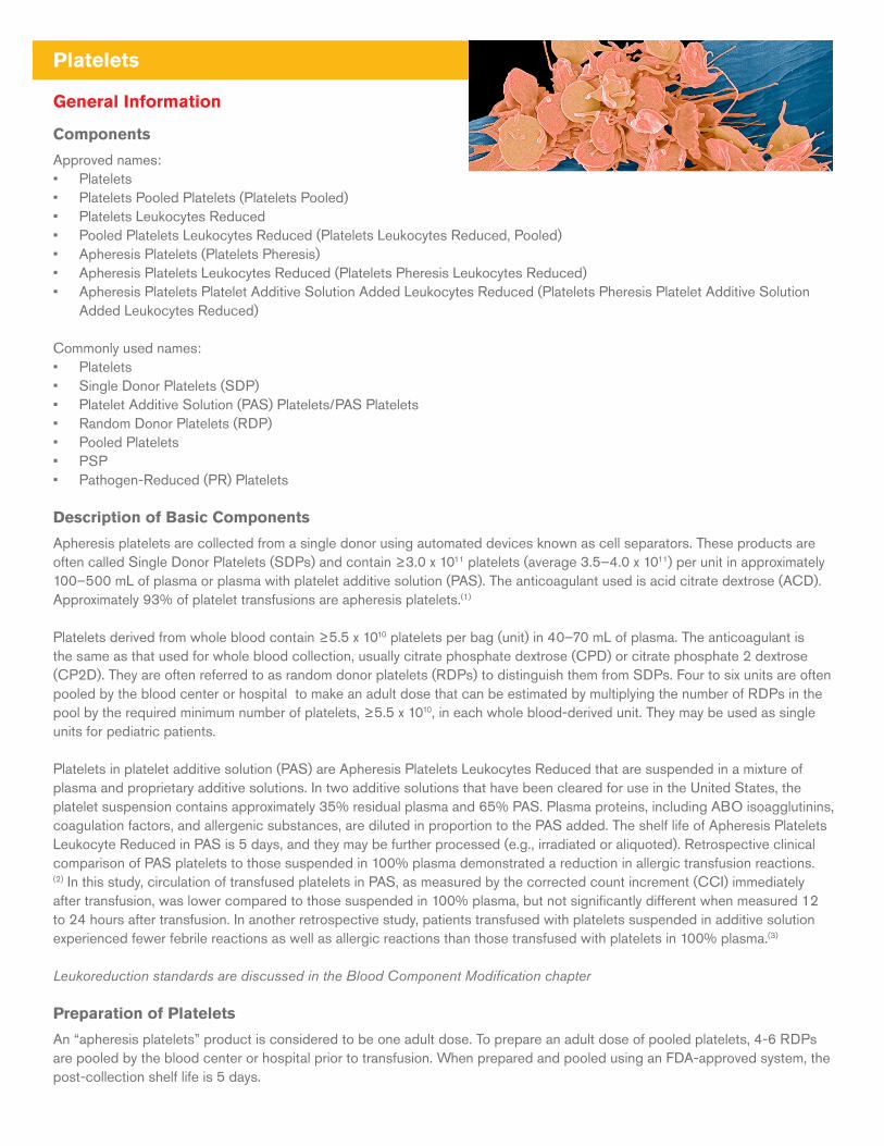
Apheresis platelets are collected from a single donor using automated devices known as cell separators. These products are often called Single Donor Platelets (SDPs) and contain ≥3.0 x 10¹¹ platelets (average 3.5–4.0 x 10¹¹) per unit in approximately 100–500 mL of plasma or plasma with platelet additive solution (PAS). The anticoagulant used is acid citrate dextrose (ACD). Approximately 93% of platelet transfusions are apheresis platelets.(1)
Platelets derived from whole blood contain ≥5.5 x 10¹⁰ platelets per bag (unit) in 40–70 mL of plasma. The anticoagulant is the same as that used for whole blood collection, usually citrate phosphate dextrose (CPD) or citrate phosphate 2 dextrose (CP2D). They are often referred to as random donor platelets (RDPs) to distinguish them from SDPs. Four to six units are often pooled by the blood center or hospital to make an adult dose that can be estimated by multiplying the number of RDPs in the pool by the required minimum number of platelets, ≥5.5 x 10¹⁰, in each whole blood-derived unit. They may be used as single units for pediatric patients.
Platelets in platelet additive solution (PAS) are Apheresis Platelets Leukocytes Reduced that are suspended in a mixture of plasma and proprietary additive solutions. In two additive solutions that have been cleared for use in the United States, the platelet suspension contains approximately 35% residual plasma and 65% PAS. Plasma proteins, including ABO isoagglutinins, coagulation factors, and allergenic substances, are diluted in proportion to the PAS added. The shelf life of Apheresis Platelets Leukocyte Reduced in PAS is 5 days, and they may be further processed (e.g., irradiated or aliquoted). Retrospective clinical comparison of PAS platelets to those suspended in 100% plasma demonstrated a reduction in allergic transfusion reactions.(2) In this study, circulation of transfused platelets in PAS, as measured by the corrected count increment (CCI) immediately after transfusion, was lower compared to those suspended in 100% plasma, but not significantly different when measured 12 to 24 hours after transfusion. In another retrospective study, patients transfused with platelets suspended in additive solution experienced fewer febrile reactions as well as allergic reactions than those transfused with platelets in 100% plasma.(3)
Leukoreduction standards are discussed in the Blood Component Modification chapter
Preparation of Platelets
An “apheresis platelets” product is considered to be one adult dose. To prepare an adult dose of pooled platelets, 4-6 RDPs are pooled by the blood center or hospital prior to transfusion. When prepared and pooled using an FDA-approved system, the post-collection shelf life is 5 days.
ABO and Rh Compatibility
Donor plasma should be ABO-compatible with the recipient’s red cells. This is particularly important when transfusing infants or giving large volumes to adults, to avoid the possibility of exposure to potentially hemolyzing isoagglutinins (anti-A and/or anti-B).
Rh-negative recipients should receive Rh-negative platelets, particularly women of childbearing age. Apheresis platelets may contain up to 0.001 mL of RBCs, and it has been suggested that Rh immune prophylaxis may not be necessary when Rh positive platelets are given to Rh negative patients.(4) In a study that included hematological, oncological and patients with other conditions, D alloimmunization by Rh(D) positive apheresis platelets given to Rh(D) negative recipients was reported to be approximately 1.4% after a median 77 day follow up.(5)
Dosing
Clinical practice guidelines for prophylactic and therapeutic platelet transfusion have been recently published and are based on systematic literature review and recommendations using the Grading of Recommendations, Assessment, Development and Evaluation (GRADE) framework.(6,22)
To treat bleeding or prepare patients for invasive procedures, transfuse as needed to maintain hemostasis or the target platelet count, whichever is applicable. Four to ten units of RDPs, one to two units of Pooled Platelets (each containing approximately 4 to 6 whole blood platelet concentrates), or one to two SDPs are generally transfused to thrombocytopenic or thrombocytopathic adults. The Prophylactic Platelet Dose on Transfusion Outcomes (PLADO) trial concluded that prophylaxis at pre-specified triggers was accomplished with equivalent effect on hemorrhage, using three RDPs or half of an SDP in adults, provided that the minimum dose exceeded 1.1 x 10¹¹ platelets per square meter of the patient’s body surface area. This strategy, however, resulted in more frequent transfusions, usually on a daily basis.(7) A higher minimum dose of 2.4 x 10¹¹ platelets per square meter for outpatients is likely to be more cost effective, minimizing the number of patient visits.(8) It is recognized that in two of the PLADO study arms, doses of platelets <3 x 10¹¹/unit would not meet current standards and practices.
Response
Recipient response to platelet transfusion can be measured by the count increment (CI), which is defined as the increase in platelet count, measured in platelets/µL usually 10-60 minutes, after transfusion (a “one hour” post transfusion platelet count). Otherwise defined, the CI is the post-transfusion count minus the pre-transfusion platelet count and is asimple method to judge response to platelet transfusion. For an adult of 70 kg, the platelet CI should be approximately 5,000–10,000/μL for each RDP or 10,000–60,000/μL for each SDP given. In neonates and infants, a dose of 5–10 mL/kg of platelets should result in a 50,000–100,000/μL increment.(9-11)
Although the CI takes into account the dilutional effect of transfusion, a more accurate calculation for response to platelet transfusion is the corrected count increment (CCI), which also includes correction for body surface area and the number of platelets transfused:
CCI = CI x (body surface area in m2)/number of platelets transfused (x1011)
Thus, a platelet count increment (CI) of 15,000/µL in a person of 1.8 m2 after transfusion of 3 x 10¹¹ platelets would be (15,000 x 1.8/3) = 9,000. Generally, CCIs measured between 10 and 60 minutes post-transfusion are expected to be >7,500, and reflect 20-30% platelet recovery due to normal platelet consumption to support endothelial function. Platelet refractoriness is defined as a CCI ≤7,500 for at least 2 sequential platelet transfusions. Refractory platelet transfusions can be due to a number of non-immune causes, including fever, infection, bleeding, DIC, extensive surgery, splenomegaly, irradiation, and concurrent amphotericin B therapy.(12)
Failure to achieve the expected response within one hour of transfusion suggests the existence of HLA alloimmunization or immunization to human platelet antigens (HPA). In the absence of a consumptive process or decreased production, post-transfusion counts may be somewhat lower than the dose administered because approximately 7,100 platelets/μL are consumed daily in endothelial support functions, the equivalent, for example, of approximately one RDP per day for a 70 kg adult with marrow failure.(13)
Indications
Use to treat bleeding due to critically decreased circulating platelet counts or functionally abnormal platelets. Use prophylactically to prevent bleeding at pre-specified low platelet counts. In general, maintain platelet count at >10,000/μL in stable, non-bleeding patients, at >20,000/μL in unstable, non-bleeding patients, and at >50,000/μL in patients who are actively bleeding or undergoing major invasive procedures or surgery.
Contraindications
Use of platelets in patients with autoimmune thrombocytopenia, thrombotic thrombocytopenic purpura/hemolytic uremic syndrome (TTP/HUS), idiopathic thrombocytopenic purpura (ITP), or heparin-induced thrombocytopenia with thrombosis (HITT) should be avoided except for life-threatening hemorrhage. Transfusion before invasive procedures or surgery in patients without thrombotic manifestations may be considered when the risk of bleeding is high.(14,15)
For side effects and hazards, please see the Appendices. For information on pathogen-reduced platelets and plasma please refer to the Blood Component Modification chapter.
Utilization Guidelines
Surgery
• Prophylactic preoperative transfusion is rarely required for counts >100,000/μL, is usually required for counts <50,000/μL, and is guided by risk factors for intermediate counts.(16)
• Intraoperative platelet counts should be obtained to guide transfusion
• Procedures with insignificant blood loss or vaginal deliveries can be performed at counts <50,000/μL without prophylactic transfusion.
• Transfusion may be required with apparently adequate counts when known or suspected platelet dysfunction results in microvascular bleeding.
• Point of care (POC) testing devices, which reflect the availability of functional platelets and/or coagulation and fibrinolytic proteins, can assess hemostatic function in bleeding surgical patients and during massive transfusion situations. These tests can guide optimal administration of blood products and reduce inappropriate component utilization.(17-19)
Cardiothoracic Surgery
Routine prophylactic transfusions do not alter bleeding or postoperative transfusion requirements, and are not recommended for non-thrombocytopenic patients. There are no published guidelines for managing patients on aspirin and P2Y12 receptor inhibitors and other anti-platelet drugs. These patients are known to be at higher risk for bleeding and reoperation, however, management of patients taking these agents may require platelet transfusions in urgent situations.(20,21) Platelet transfusion is recommended for patients who are on cardiopulmonary bypass for cardiovascular procedures and who exhibit microvascular perioperative bleeding with thrombocytopenia and/or evidence of platelet dysfunction.(22)
Specific Procedures
• When pre-procedural transfusion is deemed necessary, a post-transfusion count should be obtained to assure an appropriate increment prior to the procedure.
• • In the absence of coagulopathy or thrombocytopathy, AABB clinical practice guidelines suggest “prophylactic platelet transfusion for patients having major elective non-neuraxial surgery with a platelet count less than 50 × 109 cells/L.”(22) Procedures including paracentesis/thoracentesis, liver biopsy, sinus aspiration, and dental extraction may also require platelet transfusion.
• The AABB suggests “prophylactic platelet transfusion for patients having elective central venous catheter placement with a platelet count less than 20 × 109 cells/L.”(22) Serious bleeding complications after CVC placement are rare and are usually due to complications rather than thrombocytopenia.(6)
• The AABB suggests “prophylactic platelet transfusion for patients having elective diagnostic lumbar puncture with a platelet count less than 50 × 109 cells/L.”(22) Studies evaluated for development of these guidelines, however, included adult and pediatric patients, and showed no bleeding complications at platelet counts less than 50 × 109 cells/L or less
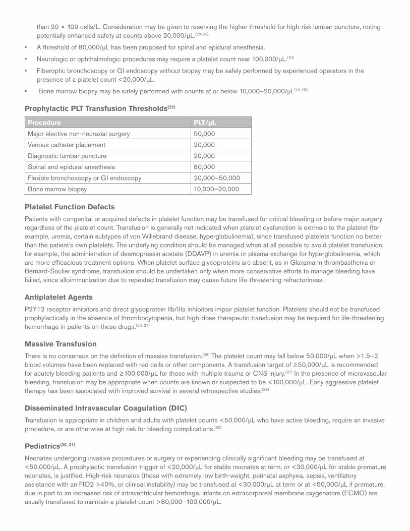
than 20 × 109 cells/L. Consideration may be given to reserving the higher threshold for high-risk lumbar puncture, noting potentially enhanced safety at counts above 20,000/μL.(23-25)
• A threshold of 80,000/μL has been proposed for spinal and epidural anesthesia.
• Neurologic or ophthalmologic procedures may require a platelet count near 100,000/μL.(10)
• Fiberoptic bronchoscopy or GI endoscopy without biopsy may be safely performed by experienced operators in the presence of a platelet count <20,000/μL.
• Bone marrow biopsy may be safely performed with counts at or below 10,000–20,000/μL(10, 25)
Prophylactic PLT Transfusion Thresholds(22)
Procedure PLT/µL
Major elective non-neuraxial surgery 50,000
Venous catheter placement 20,000
Diagnostic lumbar puncture 20,000
Spinal and epidural anesthesia 80,000
Flexible bronchoscopy or GI endoscopy 20,000 –50,000
Bone marrow biopsy 10,000–20,000
Platelet Function Defects
Patients with congenital or acquired defects in platelet function may be transfused for critical bleeding or before major surgery regardless of the platelet count. Transfusion is generally not indicated when platelet dysfunction is extrinsic to the platelet (for example, uremia, certain subtypes of von Willebrand disease, hyperglobulinemia), since transfused platelets function no better than the patient’s own platelets. The underlying condition should be managed when at all possible to avoid platelet transfusion, for example, the administration of desmopressin acetate (DDAVP) in uremia or plasma exchange for hyperglobulinemia, which are more efficacious treatment options. When platelet surface glycoproteins are absent, as in Glanzmann thrombasthenia or Bernard-Soulier syndrome, transfusion should be undertaken only when more conservative efforts to manage bleeding have failed, since alloimmunization due to repeated transfusion may cause future life-threatening refractoriness.
Antiplatelet Agents
P2Y12 receptor inhibitors and direct glycoprotein IIb/IIIa inhibitors impair platelet function. Platelets should not be transfused prophylactically in the absence of thrombocytopenia, but high-dose therapeutic transfusion may be required for life-threatening hemorrhage in patients on these drugs.(20, 21)
Massive Transfusion
There is no consensus on the definition of massive transfusion.(26) The platelet count may fall below 50,000/μL when >1.5–2 blood volumes have been replaced with red cells or other components. A transfusion target of ≥50,000/μL is recommended for acutely bleeding patients and ≥100,000/μL for those with multiple trauma or CNS injury.(27) In the presence of microvascular bleeding, transfusion may be appropriate when counts are known or suspected to be <100,000/μL. Early aggressive platelet therapy has been associated with improved survival in several retrospective studies.(28)
Disseminated Intravascular Coagulation (DIC)
Transfusion is appropriate in children and adults with platelet counts <50,000/μL who have active bleeding, require an invasive procedure, or are otherwise at high risk for bleeding complications.(29)
Pediatrics(30, 31)
Neonates undergoing invasive procedures or surgery or experiencing clinically significant bleeding may be transfused at <50,000/μL. A prophylactic transfusion trigger of <20,000/μL for stable neonates at term, or <30,000/μL for stable premature neonates, is justified. High-risk neonates (those with extremely low birth-weight, perinatal asphyxia, sepsis, ventilatory assistance with an FIO2 >40%, or clinical instability) may be transfused at <30,000/μL at term or at <50,000/μL if premature, due in part to an increased risk of intraventricular hemorrhage. Infants on extracorporeal membrane oxygenators (ECMO) are usually transfused to maintain a platelet count >80,000–100,000/μL.
Oncology
Platelets should be transfused prophylactically to patients with a platelet count of 10 × 109 cells/L or less to reduce the risk of spontaneous bleeding in hospitalized adult patients with therapy-induced hypoproliferative thrombocytopenia. AABB guidelines emphasize transfusing a single apheresis unit or equivalent. Additional transfusions are not more effective.(22) Prophylactic platelets may be given despite a higher platelet count if clinical factors such as drug-induced platelet dysfunction, fever and/or sepsis, hyperleukocytosis, tumors with greater risk of hemorrhage, use of antithymocyte globulin, acute graft-versus-host disease, or hepatic veno-occlusive disease are present. Results from a 14 center, randomized, open-label, non-inferiority trial conducted in the United Kingdom and Australia support the continued use of prophylactic platelet transfusion to reduce bleeding as compared to no prophylaxis.(32, 33)
Intracranial Hemorrhage in Patients on Antiplatelet Agents (traumatic or spontaneous)There are no clear guidelines for transfusion in patients on antiplatelet agents with intracranial hemorrhage. Clinical factors such as the extent of hemorrhage, type of procedure planned, and the patient’s level of consciousness may inform the decision to transfuse.(21)
Platelet Refractoriness(34-37)
Platelet refractoriness may be due to immune or non-immune causes and should be suspected after two or more poor responses to transfusion. The most common cause of immune refractoriness is the presence of HLA antibodies and, less frequently, antibodies to human platelet antigens (HPA), which can be confirmed by the demonstration of HLA or HPA antibodies, respectively. Post-transfusion platelet counts obtained 10–60 minutes after infusion should be obtained whenever transfusion refractoriness is suspected. HLA or HPA matched SDPs are the product of choice for alloimmunized patients. Alternatives include antibody compatible or crossmatched platelets. When possible, ABO identical units should be used(38) Successful transfusion is defined as a CCI ≥7,500. This specific calculation may not always be warranted, as refractory patients typically have a one hour post count increment of less than 5000/uL; such a finding suggests HLA alloimmunization as the likely cause of refractoriness. Post-infusion counts at 24 hours assess platelet survival, which is sensitive to non-immune and immune conditions.
HLA matched platelets must undergo gamma or x-ray irradiation to prevent transfusion-associated graft versus host disease (TA-GVHD).
Many algorithms exist for obtaining appropriate platelets for these patients. Early consultation with the transfusion service medical director is essential to initiating the process of obtaining these special products.(38, 39)
Idiopathic Thrombocytopenic Purpura (ITP)(40)
Patients who experience major, life-threatening bleeding or intraoperative hemorrhage should receive high-dose platelet transfusions as well as steroids, intravenous immunoglobulin (IVIG), and any other appropriate second-line therapies. Prophylactic transfusions are usually inappropriate since transfused platelets do not survive any longer than the patient’s own platelets. Administration of IVIG may be considered before minor surgery with platelet counts ≤50,000/μL or major surgery with counts ≤80,000/μL.
Thrombotic Thrombocytopenic Purpura/Hemolytic Uremic Syndrome (TTP/HUS) and Heparin-Induced Thrombocytopenia with Thrombosis (HITT)
Due to the significant risk of fatal thrombosis, platelets should be transfused only for life-threatening hemorrhage or, possibly, before invasive procedures in patients without thrombotic manifestations.(14,15)
Post-transfusion Purpura (PTP)
IVIG is the treatment of choice for PTP. Platelets may be administered for severe bleeding, but transfusion of platelets is usually ineffective unless the patient lacks the specific platelet antigen. Though efficacy is not well documented, HPA-1a-negative platelets, if available, are frequently given empirically pending specific alloantibody testing results, as 70% of cases of PTP are due to HPA-1a antibodies.
Neonatal Alloimmune Thrombocytopenia (NAIT)(41)
While awaiting a response to IVIG, platelet transfusions are indicated for severe thrombocytopenia and/or bleeding. Ideally, platelets should lack the HPA recognized by circulating maternal antibodies. Until appropriate platelets are found, available platelets may be transfused. If maternal platelets are used, they should be washed or volume-reduced and irradiated. HPA-1a-negative platelets are often used empirically, as more than 75% of infants with NAIT are assumed to have exposure to HPA-1a antibodies.
Aplastic Anemia
Transfuse stable patients prophylactically at counts ≤5,000/μL and patients with fever or minor hemorrhage at counts 6,000–10,000/μL(42)
References1. Whitaker BI, Rajbhandary S, et al. The 2013 AABB National Blood Collection, Utilization, and Patient Blood Management Survey Report. Bethesda, MD: AABB; 2015. http://
www.aabb.org/research/hemovigilance/bloodsurvey/Pages/default.aspx
2. Tobian AA, Fuller AK, et al. The impact of platelet additive solution apheresis platelet units on allergic transfusion reactions and corrected count increment. Transfusion 2011;54:1523-9
3. Cohn CS, Stubbs J, et al. A comparison of adverse reaction rates for PAS C versus plasma platelet units. Transfusion 2014;54:1927-34.
4. O’Brien KL, Haspel RL, et al. Anti-D alloimmunization after D-incompatible platelet transfusions: a 14 year single institution retrospective review. Transfusion 2014;54:650-4.
5. Cid J, Lozano M, et al. Low frequency of anti-D alloimmunization following D+ platelet transfusion: the Anti-D Alloimmunization after D-incompatible Platelet Transfusions (ADAPT) study. Br J Haematol 2015;168:598-603.
6. Zeidler K, Arn K, et al. Optimal pre-procedural platelet transfusion threshold for central venous catheter insertions in patients with thrombocytopenia. Transfusion 2011;51:2269-76.
7. Slichter SJ, Kaufman RM, et al. Dose of prophylactic platelet transfusions and prevention of hemorrhage. New Engl J Med 2010;362:600-13.
8. Nahirniak S, Slichter SJ, et al. Guidance for platelet transfusion for patients with hypoproliferative thrombocytopenia. Transfus Med Rev 2015;29:3-13.
9. Cooper ES, Bracey AW, et al., for the Fresh-Frozen Plasma, Cryoprecipitate, and Platelets Administration Practice Guidelines Development Task Force of the College of American Pathologists. Practice parameter for the use of fresh-frozen plasma, cryoprecipitate, and platelets. JAMA 1994;271:777–81.
10. Heddle NM, Arnold DM, et al. Comparing the efficacy and safety of apheresis and whole blood-derived platelet transfusions: a systematic review. Transfusion 2008;48:1447-58.
11. Poterjoy BS, Josephson CD. Platelets, frozen plasma, and cryoprecipitate: what is the clinical evidence for their use in the neonatal intensive care unit? Semin Perinatol 2009;33:66–74.
12. Slichter SJ, Davis K, et al. Factors affecting post-transfusion platelet increments, platelet refractoriness, and platelet transfusion intervals in thrombocytopenic patients. Blood 2005;105:4106-14.
13. Estcourt LJ, Stanworth SJ, et al. Platelet transfusions for patients with haematological malignancies: who needs them? Br J Haematol 2011;154:425-40.
14. Hopkins CK, Goldfinger D. Platelet transfusions in heparin-induced thrombocytopenia: a report of four cases and a review of the literature. Transfusion 2008;48:2128–32.
15. Swisher KK, Terrell DR, et al. Clinical outcomes after platelet transfusions in patients with thrombotic thrombocytopenic purpura. Transfusion 2009;49:873-87.
16. Practice guidelines for perioperative blood transfusion and adjuvant therapies: an updated report by the American Society of Anesthesiologists Task Force on Perioperative Blood Transfusion and Adjuvant Therapies. Anesthesiology 2015;122:1-35.
17. Enriquez LJ, Shore-Lesserson L. Point-of-care coagulation testing and transfusion algorithms. Br J Anaesth 2009;103(suppl 1):i14-22.
18. Gibbs NM. Point-of-care assessment of antiplatelet agents in the perioperative period: a review. Anaesth Intensive Care 2009;37:354-69.
19. Dickinson KJ, Troxler M, et al. The surgical application of point-of-care haemostasis and platelet function testing. Br J Surg 2008;95:1317-30.
20. Vilahur G, Choi BG, et al. Normalization of platelet reactivity in clopidogrel-treated subjects. J Thromb Haemost 2007;5:82-90.
21. Sarode, R. How do I transfuse platelets (PLT) to reverse anti-PLT drug effect? Transfusion 2012;52:695-701.
22. Kaufman RM, Djulbegovic B, et al. Platelet transfusion: A clinical practice guideline from the AABB. Ann Int Med 2015;162:205-13.
23. Howard SC, Gajjar A, et al. Safety of lumbar puncture for children with acute lymphoblastic leukemia and thrombocytopenia. JAMA 2000;284:2222-4.
24. German Medical Association. Cross-sectional guidelines for therapy with blood components and plasma derivatives. 4th ed. Revised January 2011. Available at: bundesaerztekammer.de/fileadmin/user_upload/downloads/Querschnittsleitlinie_Gesamtdokument-englisch_07032011.pdf. Last accessed February 19, 2016.
25. The C17 Guidelines Committee. Guideline for platelet transfusion thresholds for pediatric hematology/oncology patients. Updated March 2011. Available at: c17.ca/index.php/download_file/view/41. Last accessed April 4, 2016.
26. Zatta AJ, McQuilten ZK, et al. Elucidating the clinical characteristics of patients captured using different definitions of massive transfusion. Vox Sang 2014;107:60-70.
27. Nishijima DK, Zehtabshi S, et al. Utility of platelet transfusion in adult patients with traumatic intracranial hemorrhage and preinjury antiplatelet use: a systematic review. J Trauma Acute Care Surg 2012;72:1658-63.
28. Hallet J, Lauzier F, et al. The use of higher platelet:RBC transfusion ratio in the acute phase of trauma resuscitation: a systematic review Crit Care Med 2013;41:2800-11.
29. Levi M, Toh CH, et al., for the British Committee for Standards in Haematology. Guidelines for the diagnosis and management of disseminated intravascular coagulation. Br J Haematol 2009;145:24-33.
30. Del Vecchio A, Motta M. Evidence-based platelet transfusion recommendations in neonates. J Matern Fetal Neonatal Med 2011;24(Suppl 1):38-40.
31. Roseff SD, Luban NLC, et al. Guidelines for assessing appropriateness of pediatric transfusion. Transfusion 2002;42:1398-1413.
32. Stanworth SJ, Estcourt LJ, et al. A no-prophylaxis platelet-transfusion strategy for hematologic cancers. N Engl J Med 2013;368:1771-80.
33. Slichter SJ. Eliminate prophylactic platelet transfusions? N Engl J Med 2013;368:1837-8.
34. Pavenski K, Rebulla P, et al. Efficacy of HLA-matched platelet transfusions for patients with hypoproliferative thrombocytopenia: a systematic review. Transfusion 2013;53:2230-42.
35. Vassallo RR, Fung M, et al. Utility of crossmatched platelet transfusions in patients with hypoproliferative thrombocytopenia: a systematic review. Transfusion 2014;54:1180-91.
36. Hod E, Schwartz J. Platelet transfusion refractoriness. Br J Haematol 2008;142:348-60.
37. Vassallo RR. New paradigms in the management of alloimmune refractoriness to platelet transfusions. Curr Opin Hematol 2007;14:655-63.
38. Kopko PM, Warner P, et al. Methods for the selection of platelet products for alloimmune refractory patients. Transfusion 2015;55:235-44.
39. Fung MK, Grossman BJ, et al, eds. Technical Manual. 18th ed. Bethesda, MD: AABB; 2014.
40. Provan D, Stasi R, et al. International consensus report on the investigation and management of primary immune thrombocytopenia. Blood 2010;115:168-86.
41. Peterson JA, McFarland JG, et al. Neonatal alloimmune thrombocytopenia: pathogenesis, diagnosis and management. Br J Haematol 2013;161:3-14.
42. Sagmeister M, Oec L, et al. A restrictive platelet transfusion policy allowing long-term support of outpatients with severe aplastic anemia. Blood 1999;93:3124-6.
Whole Blood
General Information
Approved Names:• Whole Blood• Heparin Whole Blood• Whole Blood, antihemophilic factor removed
Commonly Used names: • Whole blood• Leukocytes reduced whole blood• Leukocytes reduced low titer whole blood• Leukocytes reduced low titer TRALI mitigated whole blood
Description
Whole blood provided by the Red Cross is leukoreduced using an approved filter and is collected from TRALI-safe aspirin-free donors into approved blood bags with anticoagulants CPD or CPD-A. The product is Group O positive or negative with low isoagglutinin titers (<1:200). The bags are made of polyvinyl chloride (PVC) that are plasticized with diethylhexyl phthalate (DEHP). Each unit contains a standard amount of anticoagulant which is specified on the label. Whole blood must be cooled towards 1-10°C as it is transported from the collection site, and must be stored at 1-6°C (21 CFR 640.2 c 3). Expiration is based on the anti-coagulant into which it is drawn, in general, 21 days. The product is available within five days of collection.
Selection and Preparation
Whole blood must be tested for the presence of all required infectious disease markers, and the ABO type must be confirmed by forward and back typing and undergo standard testing for the Rh type and RBC antibody screening. In practice, transfusing physicians may limit WB use to a time period shorter than the approved expiration (see History and Current Utilization, below). The 31st edition of the AABB Standards for Blood Bank and Transfusion Services no longer requires whole blood to be ABO identical (but ABO compatible)(1). The use of a leukoreduction filter may also spare the majority of platelets, although no labeling language is approved to denote the presence of active platelets in the unit of whole blood. If platelet-sparing leukofiltration is performed, whole blood must undergo leukocyte reduction in accordance with the manufacturer’s instructions, typically within eight hours of collection. The relative abundance of isohemagglutinins, determined either from a concurrently drawn blood sample or a segment taken from the collected whole blood unit, can be determined. This information can be used as a release criterion, with only units found to have a titer less than a pre-determined cut-off suitable to be labeled for final distribution as whole blood. This information is not reflected on the label, but may be included on a tie tag. AABB standards identify whole blood as a high plasma product and require it to be appropriately TRALI risk mitigated(1).
History and Current Utilization
From the early days of such transfusion pioneers as James Blundell, MD(²) and Lieutenant Oswald Hope Robertson, MD(3) whole blood was administered to provide lifesaving resuscitation to rapidly exsanguinating patients. The development of blood component manufacturing technologies, particularly the introduction of plasticized polymer blood bags during the Korean war(4), led to the use of specific blood components to provide replacement of blood constituents across the clinical spectrum. Research into optimal resuscitation of patients with massive bleeding from trauma was once again advanced by the recent experience of physicians treating wounded soldiers. Initial insights supported ratios of transfusion of blood components(5) but advocacy for return to the use of whole blood grew within some fields of combat.(6)
Massive Hemorrhage Associated with Trauma
In military(7) and some civilian(8) settings, whole blood from group O donors has been the preferred initial intervention to address trauma associated massive hemorrhage. Low titer group O whole blood (LTOWB) is used by some facilities to replace the initial massive transfusion pack, as this product provides red cells, plasma and a low dose of platelets (approximately one-sixth of the dose in an apheresis platelet). Some hospitals track ABO incompatible plasma transfusion, and perform a direct antiglobulin test (DAT) after a patient has received more than a pre-determined volume of incompatible plasma. However, as of the writing of this discussion, published studies have not demonstrated that LTOWB is associated with hemolysis in civilian trauma patients.(9)
Pediatric Open-Heart Surgery
The use of whole blood in pediatric cardiothoracic surgery has been debated for many years. Recent evidence(10) continues to suggest that transfusion needs—and the concomitant risk of adverse reactions from multiple donor exposures —can be reduced in these cases by the use of fresh whole blood. The value of functional platelets in whole blood processed with platelet sparing leukocytes reduction filters has not yet been fully determined or characterized
Non-trauma associated hemorrhage
The appropriateness of using whole blood in civilian settings, particularly in non-trauma or routine surgical settings has not been determined, and randomized controlled trials to compare outcomes with component therapy vs. whole blood are warranted as evidence-based transfusion therapy continues to advance.(11, 12). Limited data(13) suggest a clinical benefit in obstetrical bleeding, while no significant data are available for other common causes of non-traumatic massive bleeding, such as gastrointestinal, urogenital, or cardiovascularprocedures.
References1. Standards for Blood Banks and Transfusion Services, 31st Edition. AABB, Bethesda 2017.
2. Bause GS Blundell Gravitator. Anesthes. 2009; 110: 1416
3. Robertson OH A method of citrated blood transfusion Brit. Med. J. 1918; 1: 477-79
4. Hess JR & Thomas MJG Blood use in war and disaster: lessons from the past century. Transfusion 2003; 43: 1622-33.
5. Borgman MA, Spinella PC, Perkins JG, Grathwohl KW, Repine T, Beekley AC, Sebesta J, Jenkins D, Wade CE, Holcomb JB.The ratio of blood products transfused affects mortality in patients receiving massive transfusions at a combat support hospital. J Trauma. 2007 Oct (63); #4:805-13.
6. Cap AP, Pidcoke HF, Spinella P, Strandenes G, Borgman MA, Schreiber M, Holcomb J, Tien HC, Beckett AN, Doughty H, Woolley T, Rappold J, Ward K, Reade M, Prat N, Ausset S, Kheirabadi B, Benov A, Griffin EP, Corley JB, Simon CD, Fahie R, Jenkins D, Eastridge BJ, Stockinger Z. Damage Control Resuscitation. Mil Med. 2018 Sep 1; 183(suppl_2):36-43
7. Cap AP, Beckett A, Benov A, Borgman M, Chen J, Corley JB, Doughty H, Fisher A, Glassberg E, Gonzales R, Kane SF, Malloy WW, Nessen S, Perkins JG, Prat N, Quesada J, Reade M, Sailliol A, Spinella PC, Stockinger Z, Strandenes G, Taylor A, Yazer M, Bryant B, Gurney Whole Blood Transfusion. J. Mil Med. 2018 Sep 1; 183(suppl_2):44-51.
8. Seheult JN, Anto VP, Farhat N, Stram MN, Spinella PC, Alarcon L, Sperry J, Triulzi DJ, Yazer MH. Application of a recursive partitioning decision tree algorithm for the prediction of massive transfusion in civilian trauma: the MTPitt prediction tool. Transfusion. 2018 Dec 12. doi: 10.1111/trf.15078
9. Seheult JN, Stram MN, Sperry J, Spinella PC, Triulzi DJ, Yazer MH.In silico model of the dilutional effects of conventional component therapy versus whole blood in the management of massively bleeding adult trauma patients. Transfusion. 2018 Nov 9. doi: 10.1111/trf.14983. [Epub ahead of print]
10. Jobes DR, Sesok-Pizzini D, Friedman D Reduced transfusion requirement with use of fresh whole blood in pediatric cardiac surgical procedures.. Ann Thorac Surg. 2015 May;99(5):1706-11.
11. Repine TB, Perkins JG, Kauvar DS, Blackborne L. The use of fresh whole blood in massive transfusion. J Trauma. 2006 Jun;60(6 Suppl): S59-69. Review.
12. Sommer N, Schnüriger B, Candinas D, Haltmeier T.Massive Transfusion Protocols in Non-Trauma Patients: A Systematic Review and Meta-Analysis. J Trauma Acute Care Surg. 2018 Oct 29. doi: 10.1097/TA.0000000000002101. [Epub ahead of print]
13. Alexander JM, Sarode R, McIntire DD, Burner JD, Leveno KJ. Whole blood in the management of hypovolemia due to obstetric hemorrhage. Obstet Gynecol. 2009 Jun;113(6):1320-6
Plasma
General Information(1, 2)
Components
Approved names and abbreviations: • Fresh Frozen Plasma (FFP) • Plasma Frozen within 24 hours after Phlebotomy (PF24)• Plasma Cryoprecipitate Reduced (“Cryo poor plasma”)• Plasma Frozen within 24 Hours after Phlebotomy Held at Room Temperature up to 24 Hours after Phlebotomy (PF24RT24)• Octaplas®, Pooled Plasma (Human), Solvent/Detergent Treated Solution for Intravenous Infusion • Thawed Plasma• Thawed Plasma Cryoprecipitate Reduced• Liquid Plasma
Commonly used names:• Plasma• Cryo poor plasma• Liquid Plasma• Thawed Plasma
Description of Components
Plasma consists of the noncellular portion of blood that is separated and frozen after collection. It contains coagulation factors, fibrinolytic proteins, immunoglobulins, albumin, and other proteins. Plasma may be prepared from whole blood or collected by apheresis. The anticoagulant solution used and the component volume are indicated on the label. Units prepared from whole blood are approximately200–250 mL. Apheresis-derived units contain 400–600 mL.
FFP is frozen within 6–8 hours of collection at – 18°C or colder. It contains physiological quantities of all coagulation factors. PF24 is frozen at – 18°C or colder within 24 hours of collection. FFP, PF24, or PF24RT24 can be relabeled as Thawed Plasma when stored at 1 to 6°C, for a total of five days, including the initial 24-hour post-thaw period. PF24, PF24RT24, and Thawed Plasma contain variably reduced levels of the labile factors V and VIII.(3,4) Despite these differences, FFP, PF24, PF24RT24, and Thawed Plasma are generally used for the same indications, and are referred to as Plasma in this publication.
Liquid Plasma is separated no later than five days after the expiration date of the corresponding whole blood unit, and is stored at 1–6°C. Coagulation factor levels in Liquid Plasma are variable and change over time.(5,6) Liquid Plasma can be used for initial treatment of patients requiring massive transfusion because of life-threatening trauma or hemorrhage and who have clinically significant coagulation deficiencies.
Octaplas® was approved by the FDA for use in the United States in January 2013. It is produced in pools of plasma from 630–1,520 donors, undergoes 1 μM filtration, solvent-detergent reagent treatment, and affinity column filtration to bind prion proteins. Units are supplied in ABO-specific 200 mL volumes.(7)
Plasma Cryoprecipitate Reduced is produced after thawing, centrifugation, and removal of cryoprecipitate from FFP. It has decreased levels of fibrinogen, Factor VIII and von Willebrand factor, fibronectin, and Factor XIII.(8) Proteins such as albumin and other coagulation factors remain at approximately the same levels as in FFP. FFP, PF24, PF24RT24 and Plasma Cryoprecipitate Reduced have equivalent levels of ADAMTS13, the protein that is deficient or has reduced activity in thrombotic thrombocytopenic purpura (TTP). ADAMTS13 activity should remain stable for the duration of the shelf life of these thawed products.(9)
Coagulation factor half-life should be considered when plasma is given prior to invasive procedures. For example, for a patient with Factor VII deficiency, the 4–6 hour in vivo half-life of Factor VII requires transfusion of plasma as close as possible to the time of the procedure to achieve hemostatic factor levels.(10)
Selection and Preparation
Plasma for transfusion must be ABO-compatible with the recipient’s red cells. For example, group A Plasma is suitable for transfusion to group A and group O patients. Group AB Plasma is suitable for transfusion to patients of all blood types, but should be reserved for AB patients.
Frozen plasma must be thawed in an FDA-approved water bath or other FDA-approved device at 30 to 37°C, and transfused immediately. It can also be stored at 1–6°C for no longer than 24 hours when stored at this temperature. Alternatively, once thawed, FFP, PF24 and PF24RT24 may be relabeled as Thawed Plasma and used as a source of stable coagulation factors for up to five days. Collection occurs in a closed system, so Plasma Cryoprecipitate Reduced can be used for up to five days post-thaw and relabeled as Thawed Plasma Cryoprecipitate Reduced.(11) Octaplas® has a 24 hour shelf life post-thaw if stored at 1°C to 6°C or 8 hours if stored at 20°C to 25°C.(7)
Dosing
Plasma dose is determined by patient weight and clinical condition. Plasma should be administered in doses calculated to achieve plasma factor concentrations of at least 30%, which is the minimum hemostatic level for most coagulation factors.(12-14) This is usually achieved with the administration of 10–20 mL/kg patient weight, though more may be required depending on the clinical situation.
When used to correct isolated coagulation factor deficiencies for which no concentrated preparation is available (for example, Factors V and XI), dosing will depend on the pre-transfusion level of the factor, the desired post-transfusion level, the needed duration of higher levels, and the factor’s half-life and volume of distribution.(15)
When used to correct multiple coagulation factor deficiencies, plasma transfusion should be guided by coagulation testing. A prothrombin time (PT) greater than 1.5 times the mid-range of normal, an activated partial thromboplastin time (aPTT) greater than 1.5 times the upper level of the normal range(15) or an INR of greater than 1.7(16) would warrant plasma transfusion. When such testing is not readily available, clinical evidence of bleeding may be used to direct transfusion decisions.
Thrombocytic thrombocytopenic purpura (TTP) initially requires the exchange of 1–1.5 plasma volumes daily. In clinical practice, plasma exchange is often tapered as disease activity declines, although this has not been studied prospectively.(17) (Please see the Cryoprecipitate chapter for plasma and blood volume calculations).
The efficacy of plasma is questionable in many clinical settings, but in general, plasma transfusion is more effective at higher INR values.(12)
Response
Plasma used to correct coagulation abnormalities should bring the aPTT, PT, and INR within the hemostatic range, but transfusion will not always correct these values, or the correction may be transient.(12)
Plasma used to treat TTP should result in an increasing platelet count associated with a decrease in serum lactate dehydrogenase.(17)
Indications and Contraindications (1,7, 12, 14-24)
Plasma is indicated for use in patients with the following conditions:
• Active bleeding or risk of bleeding due to deficiency of multiple coagulation factors
• Severe bleeding due to warfarin therapy or urgent reversal of warfarin effect when 4 factor prothrombin complex concentrate is not available
• Massive transfusion with coagulopathic bleeding
• Bleeding or prophylaxis of bleeding for a known single coagulation factor deficiency for which no concentrate is available
• Thrombotic thrombocytopenic purpura (Plasma or Plasma Cryoprecipitate Reduced)
• Rare specific plasma protein deficiencies for which no concentrate is available, e.g., fibronectin
Octaplas® is indicated for:
• Replacement of multiple coagulation factors in patients with acquired deficiencies due to liver disease and in patients undergoing cardiac surgery or liver transplant.
• Plasma exchange in patients with thrombotic thrombocytopenic purpura.
Plasma should not be used for:
• Increasing blood volume or albumin levels.
• A coagulopathy that can be corrected by adjusting warfarin dose and/or administration of vitamin K.
• Normalizing abnormal coagulation screen results in the absence of bleeding.
For side effects and hazards, please see the Appendices.
Utilization Guidelines(12, 18-44)
Liver Disease
Plasma may be used to replace multiple coagulation factor deficiencies due to liver disease in patients who are actively bleeding or prior to an invasive procedure that would create a risk of bleeding. However, the response may be unpredictable, and complete normalization of the hemostatic defect may not occur. Post-transfusion coagulation testing may be necessary to evaluate efficacy. Patients with liver disease may safely undergo operative or invasive procedures when the PT is ≤1.5 times the mid-range of normal.
Warfarin
Patients on warfarin who experience serious bleeding are treated with INR-based doses of vitamin K and plasma or prothrombin complex concentrates as clinically warranted. Recent guidelines suggest that 4-factor prothrombin complex concentrates are preferable to plasma transfusion for situations requiring urgent reversal of warfarin.(25-27) Three-factor prothrombin complex concentrates have been proposed as an alternative without supporting randomized controlled trial data. These suggestions are based on limited evidence. When prothrombin complex concentrates are not immediately available, plasma transfusion may be necessary. As in liver disease, patients on warfarin may safely undergo operative or invasive procedures when the PT is ≤1.5 times the mid-range of normal.
Massive Transfusion and Cardiopulmonary Bypass
Plasma may be used to treat excessive microvascular bleeding, as determined on joint visual assessment of the operative field by the anesthesiologist and surgeon when the coagulation screening test results are abnormal or are not available in a timely fashion. However, microvascular bleeding may be a result of hypofibrinogenemia or residual heparin effect.
For massive transfusion, recent trends based on retrospective studies advocate using a high plasma-to-RBC ratio to improve survival. A recent randomized controlled trial comparing two high plasma-to-RBC ratios to each other but not to a laboratory driven model showed no difference in the mortality outcomes studied.(32) However, other studies have shown this strategy may increase the risk of multiple organ failure, adult respiratory distress syndrome and other forms of respiratory morbidity.(33) Further studies, including randomized controlled trials, are necessary to determine the risks or benefits of a high ratio strategy compared to a laboratory driven approach.
Thrombotic Thrombocytopenic Purpura
If plasma exchange is not immediately available, simple transfusion of plasma might be an alternative until exchange can be initiated. With equivalent levels of ADAMTS13, plasma and Plasma Cryoprecipitate Reduced are equally efficacious in the treatment of TTP and newly diagnosed TTP.(37) If ADAMTS13 levels are used to diagnose and/or monitor the response, a level should be obtained prior to initiation of treatment.
Specific Plasma Protein/Factor Deficiencies
Deficiencies of other isolated plasma proteins and factors in a setting for which concentrates are not readily available are also treated with plasma and include:
• Prophylactic correction of a known factor deficiency for which specific concentrates are unavailable (would be guided by recommended perioperative hemostatic levels for each type of procedure).
• Treatment or prophylaxis of thromboembolism in antithrombin, protein C, and protein S deficiencies.
• Therapy of acute angioedema or preoperative prophylaxis in hereditary C1-inhibitor deficiency.
• Factor V deficiency (no plasma concentrate available).
• Factor XI deficiency (factor concentrate not available in the US).
Pediatrics(42-44)
The indications for transfusion of plasma in children are essentially the same as for adults. In infants less than 6 months of age, the levels of vitamin K-dependent coagulants, anticoagulants, and fibrinolytic proteins are decreased, resulting in prolongation of coagulation assays compared to older children and adults. Despite these differences, hemostatic balance is maintained in the healthy newborn, and spontaneous bleeding or thrombosis are rarely observed. The reserve capacity to respond to pathologic insults in a sick premature infant during the first week of life, however, may be limited.
References1. AABB, American Red Cross, America’s Blood Centers, Armed Services Blood Program. Circular of Information for the Use of Human Blood and Blood Components. Revised
November 2013. Available at: aabb.org.
2. Fung MK, Grossman BJ, et al., eds. Technical Manual. 18th ed. Bethesda, MD: AABB; 2014.
3. Smak Gregoor PJH, Harvey MS, et al. Coagulation parameters of CPD fresh-frozen plasma and CPD cryoprecipitate-poor plasma after storage at 4°C for 28 days. Transfusion 1993;33:735-8.
4. Scott E, Puca K, et al. Evaluation and comparison of coagulation factor activity in fresh-frozen plasma and 24-hour plasma at thaw and after 120 hours of 1 to 6°C storage. Transfusion 2009;49:1584-91.
5. Gosselin RC, Marshall C, et al. Coagulation profile of liquid-state plasma. Transfusion 2013;53:579-90.
6. Matijevic N, Wang YW, et al. Better hemostatic profiles of never-frozen liquid plasma compared with thawed fresh frozen plasma. J Trauma Acute Care Surg 2013;74:84-90.
7. Package insert, Octaplas, Pooled Plasma (Human), Solvent/Detergent Treated Solution for Intravenous Infusion. Octapharma; revised March 2015.
8. Gerhard GS, Hoffman SM, et al. Coagulation parameters of ABO group-specific cryosupernatant. Am J Clin Pathol 1998:109:379-86.
9. Scott EA, Puca KE, et al. Comparison and stability of ADAMTS13 activity in therapeutic plasma products. Transfusion 2007;47:120-5.
10. Bick RL. Disorders of Thrombosis and Hemostasis: Clinical and Laboratory Practice, 3rd ed. Philadelphia: Lippincott, Williams, and Wilkins; 2002. p. 121.
11. Ooley P, Chair. Standards for Blood Banks and Transfusion Services. 30th ed. Bethesda, MD: AABB; 2015.
12. Tinmouth A. Evidence for a rational use of frozen plasma for the treatment and prevention of bleeding. Transfus Apher Sci 2012;46:293-8.
13. Burns ER, Goldberg SN, et al. Paradoxic effect of multiple mild coagulation factor deficiencies on the prothrombin time and activated partial thromboplastin time. Am J Clin Pathol 1993;100:94-8.
14. Dzik W, Rao A. Why do physicians request fresh frozen plasma? Transfusion 2004;44:1393-4.
15. Cooper ES, Bracey AW, et al., for the Fresh-Frozen Plasma, Cryoprecipitate, and Platelets Administration Practice Guidelines Development Task Force of the College of American Pathologists. Practice parameter for the use of fresh-frozen plasma, cryoprecipitate, and platelets. JAMA 1994;271:777–81.
16. Holland LL, Brooks JP. Toward rational fresh frozen plasma transfusion: The effect of plasma transfusion on coagulation test results. Am J Clin Pathol 2006;126:133-9.
17. Schwartz J, Padmanabhan,Aet al. Guidelines on the use of therapeutic apheresis in clinical practice – evidence-based approach from the Writing Committee of the American Society for Apheresis: the seventh special issue. J Clin Apher 2016; 31:149-338.
18. Stanworth SJ, Grant-Casey J, et al. The use of fresh-frozen plasma in England: high levels of inappropriate use in adults and children. Transfusion 2011;51:62-70.
19. O’Shaughnessy DF, Atterbury C, et al. for the British Committee for Standards in Haematology, Blood Transfusion Task Force. Guidelines for the use of fresh-frozen plasma, cryoprecipitate and cryo¬supernatant. Br J Haematol 2004;126:11-28.
20. German Medical Association. Cross-sectional guidelines for therapy with blood components and plasma derivatives. 4th ed. Revised January 2011. Available at: http://www.bundesaerztekammer.de/downloads/Querschnittsleitlinie_Gesamtdokument-englisch_07032011.pdf. Last accessed March 28, 2016.
21. Dutch Institute for Healthcare Improvement CBO Working Group. Blood transfusion guideline. 2011. Available at: http://www.sanquin.nl/repository/documenten/en/prod-en-dienst/287294/blood-transfusion-guideline.pdf, Last accessed March 28, 2016.
22. Roback JD, Caldwell S, et al. Evidence-based practice guidelines for plasma transfusion. Transfusion 2010;50:1227-39.
23. Society of Thoracic Surgeons Blood Conservation Guideline Task Force. 2011 update to the Society of Thoracic Surgeons and the Society of Cardiovascular Anesthesiologists blood conservation clinical practice guidelines. Ann Thorac Surg 2011;91:944-82.
24. Dzik W. Reversal of drug-induced anticoagulation: old solutions and new problems. Transfusion 2012;52(Suppl 1):45S-55S.
25. Holbrook A, Schulman S, et al. for the American College of Chest Physicians. Evidence-based management of anticoagulant therapy: Antithrombotic therapy and prevention of thrombosis, 9th ed: American College of Chest Physicians Evidence-Based Clinical Practice Guidelines. Chest 2012;141(Suppl 2):e152S-184S.
26. Keeling D, Baglin T, et al. for the British Committee for Standards in Haematology. Guidelines on oral anticoagulation with warfarin − fourth edition. Br J Haematol 2011;154:311-24.
27. Tran HA, Chunilal SD, et al. for the Australasian Society of Thrombosis and Haemostasis. An update of consensus guidelines for warfarin reversal. Med J Aust 2013;198:198-9.
28. Malone DL, Hess JR, et al. Massive transfusion practices around the globe and a suggestion for a common massive transfusion protocol. J Trauma 2006;60(Suppl 6):S91-6.
29. Johansson PI, Stensballe J. Hemostatic resuscitation for massive bleeding: the paradigm of plasma and platelets – a review of the current literature. Transfusion 2010;50:701-10.
30. Shaz BH, Dente CJ, et al. Transfusion management of trauma patients. Anesth Analg 2009;108: 1760-8.
31. Sihler KC, Napolitano LM. Massive transfusion: new insights. Chest 2009;136:1654-67.
32. Holcomb JB, Tilley BC, et al. Transfusion of plasma, platelets, and red blood cells in a 1:1:1 vs a 1:1:2 ratio and mortality in patients with severe trauma: the PROPPR randomized clinical trial. JAMA 2015;313:471-82.
33. Callum JL. Plasma transfusion for patients with severe hemorrhage: what is the evidence? Transfusion 2012;52(Suppl 1):30S-37S.
34. Dzik WH, Blajchman MA, et al. Clinical review: Canadian National Advisory Committee on Blood and Blood Products – Massive transfusion consensus conference 2011: report of the panel. Crit Care 2011;15:242.
35. Wada H, Thatchil J, et al. Guidance for diagnosis and treatment of disseminated intravascular coagulation from harmonization of the recommendations from three guidelines. J Thromb Haemost 2013;11:761-7.
36. Levi M, Toh CH, et al. for the British Committee for Standards in Haematology. Guidelines for the diagnosis and management of disseminated intravascular coagulation. Br J Haematol 2009;145:24-33.
37. Michael M, Elliott EJ, et al. Interventions for hemolytic uremic syndrome and thrombotic thrombocytopenic purpura: a systematic review of randomized controlled trials. Am J Kidney Dis 2009;53:259-72.
38. Zeigler ZR, Shadduck RK, et al. for the North American TTP Group. Cryoprecipitate poor plasma does not improve early response in primary adult thrombotic thrombocytopenic purpura (TTP). J Clin Apher 2001;16:19-22.
39. Blackall DP, Uhl L, et al. for the Transfusion Practices Com¬mittee. Cryoprecipitate-reduced plasma: rationale for use and efficacy in the treatment of thrombotic thrombocytopenic purpura. Transfusion 2001;41:840-4.
40. Keeling D, Tait C, et al. Guideline on the selection and use of therapeutic products to treat haemophilia and other hereditary bleeding disorders. A United Kingdom Haemophilia Center Doctors’ Organisation (UKHCDO) guideline approved by the British Committee for Standards in Haematology. Haemophilia 2008;14:671-84.
41. Mannucci PM, Duga S, et al. Recessively inherited coagulation disorders. Blood 2004;104:1243-52.
42. Gibson BE, Todd A, et al. for the British Committee for Standards in Haematology Transfusion Task Force. Transfusion guidelines for neonates and older children. Br J Haematol 2004;124:433-53.
43. Poterjoy BS, Josephson CD. Platelets, frozen plasma, and cryo¬precipitate: what is the clinical evidence for their use in the neonatal intensive care unit? Semin Perinatol 2009;33:66-74.
44. Roseff SD, Luban NL, et al. Guidelines for assessing appro¬priateness of pediatric transfusion. Transfusion 2002;42:1398-413.
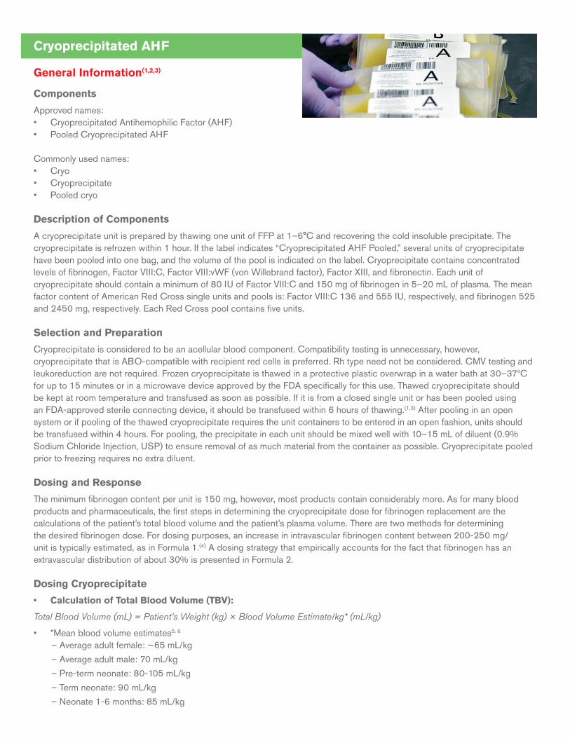
Cryoprecipitated AHF
General Information(1,2,3)
Components
Approved names:• Cryoprecipitated Antihemophilic Factor (AHF) • Pooled Cryoprecipitated AHF
Commonly used names: • Cryo• Cryoprecipitate• Pooled cryo
Description of Components
A cryoprecipitate unit is prepared by thawing one unit of FFP at 1–6°C and recovering the cold insoluble precipitate. The cryoprecipitate is refrozen within 1 hour. If the label indicates “Cryoprecipitated AHF Pooled,” several units of cryoprecipitate have been pooled into one bag, and the volume of the pool is indicated on the label. Cryoprecipitate contains concentrated levels of fibrinogen, Factor VIII:C, Factor VIII:vWF (von Willebrand factor), Factor XIII, and fibronectin. Each unit of cryoprecipitate should contain a minimum of 80 IU of Factor VIII:C and 150 mg of fibrinogen in 5–20 mL of plasma. The mean factor content of American Red Cross single units and pools is: Factor VIII:C 136 and 555 IU, respectively, and fibrinogen 525 and 2450 mg, respectively. Each Red Cross pool contains five units.
Selection and Preparation
Cryoprecipitate is considered to be an acellular blood component. Compatibility testing is unnecessary, however, cryoprecipitate that is ABO-compatible with recipient red cells is preferred. Rh type need not be considered. CMV testing and leukoreduction are not required. Frozen cryoprecipitate is thawed in a protective plastic overwrap in a water bath at 30–37ºC for up to 15 minutes or in a microwave device approved by the FDA specifically for this use. Thawed cryoprecipitate should be kept at room temperature and transfused as soon as possible. If it is from a closed single unit or has been pooled using an FDA-approved sterile connecting device, it should be transfused within 6 hours of thawing.(1,3) After pooling in an open system or if pooling of the thawed cryoprecipitate requires the unit containers to be entered in an open fashion, units should be transfused within 4 hours. For pooling, the precipitate in each unit should be mixed well with 10–15 mL of diluent (0.9% Sodium Chloride Injection, USP) to ensure removal of as much material from the container as possible. Cryoprecipitate pooled prior to freezing requires no extra diluent.
Dosing and Response
The minimum fibrinogen content per unit is 150 mg, however, most products contain considerably more. As for many blood products and pharmaceuticals, the first steps in determining the cryoprecipitate dose for fibrinogen replacement are the calculations of the patient’s total blood volume and the patient’s plasma volume. There are two methods for determining the desired fibrinogen dose. For dosing purposes, an increase in intravascular fibrinogen content between 200-250 mg/unit is typically estimated, as in Formula 1.(4) A dosing strategy that empirically accounts for the fact that fibrinogen has an extravascular distribution of about 30% is presented in Formula 2.
Dosing Cryoprecipitate
• Calculation of Total Blood Volume (TBV):
Total Blood Volume (mL) = Patient’s Weight (kg) × Blood Volume Estimate/kg* (mL/kg)
• *Mean blood volume estimates5, 6
– Average adult female: ~65 mL/kg
– Average adult male: 70 mL/kg
– Pre-term neonate: 80-105 mL/kg
– Term neonate: 90 mL/kg
– Neonate 1-6 months: 85 mL/kg
– Child, 6 months—12 years: 75 mL/kg
– Pregnancy: can increase 45%; estimate per kg depends on gestational age, multiple gestation, and pre-gravid weight7-9.
• Calculation of Plasma Volume (PV):
Plasma Volume (mL) = Total Blood Volume (mL) × (1.0 – Hematocrit (%)) Hematocrit needs to be expressed as a decimal.
• Calculation of Fibrinogen Required (mg) and number of cryoprecipitate units: 2 methods:
Formula 1 estimates intravascular distribution:
Fibrinogen desired (mg) =
PV (mL) x (desired fibrinogen – current fibrinogen (mg/dL)) x 0.01 (dL/mL)
Estimate 250 mg fibrinogen average intravascular content per unit
# units = Fibrinogen desired (mg) ÷ 250 mg/unit
Formula 2 accounts empirically for extravascular fibrinogen:
Fibrinogen desired (mg) =
PV (mL) x (desired fibrinogen – current fibrinogen (mg/dL)) x 0.01 (dL/mL) ÷ 0.7210
Use actual ARC average fibrinogen content of 525 mg per unit
# units = Fibrinogen desired (mg) ÷ 525 mg/unit
For example, to increase fibrinogen from 50 to 100 mg/dL in an adult female (65 kg, 40% hematocrit):
1. 65 kg × 65 mL/kg = 4,225 mL blood volume
2. 4,225 mL × (1.0–0.4) = 2,535 mL plasma volume
3. Fibrinogen required =
Using Formula 1:
(100 mg/dL–50 mg/dL) × 2,535 mL × 0.01 dL/mL = 1267.5 mg intravascular fibrinogen. Thus, 1267.5 mg ÷ 250 mg average estimated intravascular fibrinogen rise per unit ≈ 5 units
Using Formula 2:
(100 mg/dL–50 mg/dL) × 2,535 mL × 0.01 dL/mL ÷ 0.72 = 1760.4 mg total body fibrinogen. Thus, 1760.4 mg ÷ 525 mg average unit content ≈ 3.4 units
Pre-transfusion and post-transfusion fibrinogen levels should be determined to assess the adequacy of the cryoprecipitate dose. The frequency of dosing depends on the rate of consumption, degree of fibrinogen recovery, and half-life, so serial sampling would be warranted. The half-life is approximately 4 days in the absence of increased consumption such as disseminated intravascular coagulation or major bleeding.
General Information
Indications and Contraindications
Cryoprecipitate is indicated for bleeding associated with fibrinogen deficiencies. Routine use of cryoprecipitate as an alternative treatment for congenital fibrinogen deficiency, dysfibrinogenemia, Factor XIII deficiency, hemophilia A, or von Willebrand disease is not recommended and should be considered only when the specific factor concentrate is not available.(11) Use of this component may be considered for uremic bleeding after other modalities have failed.(12)
For side effects and hazards, please see the Appendices.
Acquired Fibrinogen Deficiency and Bleeding
Cardiac surgery is the most common surgical circumstance for cryoprecipitate transfusion. Excessive bleeding associated with worsened morbidity and mortality may result from coagulopathy due to exposure of the patient’s blood to artificial surfaces,
hemodilution, hypothermia, and/or acidosis.(13) Established general guidelines have recommended maintaining fibrinogen levels above a critical low level of 100 mg/dL in bleeding patients,(14) although this threshold was not based on clinical trials. More recent studies in obstetric, trauma, and cardiac surgery patients indicate that higher levels (150–200 mg/dL)(15-17) improve clot strength(18) and in vitro parameters, and may improve clinical outcomes.(19) Substituting alternative fibrinogen sources for cryoprecipitate in bleeding is an area of active investigation with promise in many clinical settings. However, further data on timing and impact on clinical outcomes are required.(20)
Fibrin Sealant
Although allogeneic cryoprecipitate had been used in the past as part of a hemostatic surgical adhesive, safer options are now available.(21) Several commercially produced, virus-inactivated, allogeneic sealants and autologous fibrin sealant systems are FDA-approved and are preferable to cryoprecipitate with respect to safety and efficacy for topical use.(22-24)
Massive Transfusion
Transfusion for bleeding is often required after one or more blood volumes have been replaced, with rapid consumption of fibrinogen.(25, 26) Algorithms employing early fibrinogen infusion have not been validated for efficacy or safety with cryoprecipitate, but higher levels (150–200 mg/dL) may be beneficial in treating trauma, obstetric, and cardiac surgery patients(15-17, 25-29) and are recommended by some international interdisciplinary groups.(17)
Uremic Bleeding
Other modalities such as 1-deamino-8-D-arginine vasopressin (DDAVP or desmopressin acetate) are preferred. Cryoprecipitate is used in the failure or absence of other treatments, though effectiveness has not been uniformly observed.(12,28)
Disseminated Intravascular Coagulation (DIC)
Although transfusion in DIC may be empirical and not necessarily based on lab values, severe hypofibrinogenemia (<100–150 mg/dL) that persists despite FFP replacement may be treated with cryoprecipitate.(30)
Congenital Factor Deficiencies
Congenital Fibrinogen Deficiency
In 2009, a human-derived, virus-inactivated fibrinogen concentrate was approved by the FDA and is now considered first-line treatment for congenital fibrinogen deficiency.(11) For spontaneous bleeding prior to surgery or to prevent fetal loss throughout pregnancy, recommendations are to keep fibrinogen levels above 100 mg/dL. After surgical or spontaneous bleeding is stopped, levels above 50 mg/dL should be maintained until wound healing is complete.(30)
Hemophilia A and von Willebrand Disease (vWD)
Cryoprecipitate is not recommended unless recombinant or virus-inactivated Factor VIII:C or Factor VIII:vWF concentrates are not available. Recombinant vWF has been recently approved to control bleeding in adults with vWD.(31,32) DDAVP is the treatment of choice for type 1 vWD.(31-35)
Factor XIII Deficiency
Deficiency of Factor XIII presents risk for severe bleeding, spontaneous abortion, and is associated with a 25–40% risk of spontaneous intracranial hemorrhage. Cryoprecipitate is not recommended and only used if virus-inactivated Factor XIII concentrates are not available.(11) Due to the high incidence of intracranial hemorrhage, newborns and some adults receive prophylactic dosing.
References1. AABB, American Red Cross, America’s Blood Centers, Armed Services Blood Program. Circular of Information for the Use of Human Blood and Blood Components, Revised
November 2013. Available at: aabb.org.
2. Fung MK, Grossman BJ, et al, eds. Technical Manual. 18th ed. Bethesda, MD: AABB; 2014.
3. Ooley P, Chair. Standards for Blood Banks and Transfusion Services. 30th ed. Bethesda, MD: AABB; 2015.
4. Hillyer CD, Silberstein LE, et al. Blood Banking and Transfusion Medicine: Basic Principles & Practice. 2nd ed. Philadelphia: Elsevier Health Sciences; 2007.
5. Barcelona SL, Thompson AA, et al. Intraoperative pediatric blood transfusion therapy: a review of common issues. Part II: transfusion therapy, special considerations, and reduction of allogenic blood transfusions. Paediatr Anaesth 2005;15:814-30.
6. Riley AA, Arakawa Y, et al. Circulating blood volumes: a review of measurement techniques and a meta-analysis in children. ASAIO J 2010;56:260-64.
7. Hytten F. Blood volume changes in normal pregnancy. Obstet Gynecol Surv 1986;41:426-28.
8. Ouzounian JG, Elkayam U. Physiologic changes during normal pregnancy and delivery. Cardiol Clin 2012;30:317-29.
9. Tan EK, Eng LT. Alterations in physiology and anatomy during pregnancy. Best Pract Res Cl Ob 2013;27:791-802.
10. Collen D, Tytgat GN, et al. Metabolism and distribution of fibrinogen. I. Fibrinogen turnover in physiological conditions in humans. Br J Haematol 1972;22:681-700.
11. National Hemophilia Foundation. MASAC Recommendations Concerning Products Licensed for the Treatment of Hemophilia and Other Bleeding Disorders. MASAC document #240 (Revised February 2016). Available at: hemophilia.org. Last accessed March 29, 2016.
12. New York State Council on Human Blood and Transfusion Services. Guidelines for transfusion options and alternatives. 1st Ed. New York State Department of Health, 2010. Available at: wadsworth.org/labcert/blood_tissue/pdf/txoptsaltsfixed122811.pdf. Last accessed: March 29, 2016.
13. Karlsson M, Ternström L, et al. Prophylactic fibrinogen infusion reduces bleeding after coronary artery bypass surgery. A prospective randomised pilot study. Thromb Haemost 2009;102:137-44.
14. Callum JL, Karkouti K, et al. Cryoprecipitate: the current state of knowledge. Transfus Med Rev 2009;23:177-88.
15. Ahmed S, Harrity C, et al. The efficacy of fibrinogen concentrate compared with cryoprecipitate in major obstetric haemorrhage—an observational study. Transfus Med 2012;22:344-49.
16. Charbit B, Mandelbrot L, et al. The decrease of fibrinogen is an early predictor of the severity of postpartum hemorrhage. J Thromb Haemost 2007;5:266-73.
17. Spahn DR, Bouillon B, et al. Management of bleeding and coagulopathy following major trauma: an updated European guideline. Crit Care 2013;17:R76.
18. Lee SH, Lee SM, et al. Fibrinogen recovery and changes in fibrin‐based clot firmness after cryoprecipitate administration in patients undergoing aortic surgery involving deep hypothermic circulatory arrest. Transfusion 2014;54:1379-87.
19. Levy JH, Welsby I, et al. Fibrinogen as a therapeutic target for bleeding: a review of critical levels and replacement therapy. Transfusion 2014;54:1389-1405.
20. Sachais BS, Shaz BH. Early cryoprecipitate for trauma patients is feasible, but will it improve outcome? Br J Anaesth 2105;115:3-5.
21. Spotnitz, WD. Fibrin sealant: past, present, and future: a brief review. World J Surg 2010;34:632-4.
22. Foster LJR. Bioadhesives as surgical sealants: A review. In: Bianco-Peled H, Davidovich-Pinhas M, eds. Bioadhesion and biomimetics: From nature to applications. Boca Raton, FL: CRC Press; 2015:203.
23. Alston SM, Solen KA, et al. In vivo efficacy of a new autologous fibrin sealant. J Surg Res 2008;146:143-8.
24. Buchta C, Hedrich HC, et al. Biochemical characterization of autologous fibrin sealants produced by CryoSeal® and Vivostat® in comparison to the homologous fibrin sealant product Tissucol/Tisseel®. Biomaterials 2005;26:6233-41.
25. Stainsby D, MacLennan S, et al, for the British Committee for Standards in Haematology. Guidelines on the management of massive blood loss. Br J Haematol 2006;135:634-41.
26. Dzik WH, Blajchman MA, et al. Clinical review: Canadian National Advisory Committee on Blood and Blood Products—Massive transfusion consensus conference 2011: report of the panel. Crit Care 2011;15:242.
27. Karlsson M, Ternström L, et al. Prophylactic fibrinogen infusion reduces bleeding after coronary artery bypass surgery. A prospective randomised pilot study. Thromb Haemost 2009;102:137-44.
28. Galbusera M, Remuzzi G, et at. Treatment of bleeding in dialysis patients. Semin Dial 2009;22:279-86.
29. Tziomalos K, Vakalopoulou S, et al. Treatment of congenital fibrinogen deficiency: overview and recent findings. Vasc Health Risk Manag 2009;5:843-8.
30. Wada H, Thatchil J, et al. Guidance for diagnosis and treatment of disseminated intravascular coagulation from harmonization of the recommendations from three guidelines. J Thromb Haemost 2013;11:761-7.
31. Mannucci PM, Kempton C, et al. Pharmacokinetics and safety of a novel recombinant human von Willebrand factor manufactured with a plasma-free method: a prospective clinical trial. Blood 2013;122:648-57.
32. Gill JC, Castaman G, et al. Hemostatic efficacy, safety, and pharmacokinetics of a recombinant von Willebrand factor in severe von Willebrand disease. Blood 2015;126:2038-46.
33. Archer NM, Samnaliev M, et al. The utility of the DDAVP challenge test in children with low von Willebrand factor. Br J Haematol 2015;170:884-6.
34. Federici AB. Prophylaxis in patients with von Willebrand disease: who, when, how? J Thromb Haemost 2015;13:1581-4.
35. Windyga J, Dolan G, et al. Practical aspects of DDAVP use in patients with von Willebrand disease undergoing invasive procedures: a European survey. Haemophilia 2016;22:110-20.
Blood Component Modification (1, 2, 3, 4)
1. Leukocyte-Reduced Components
Description and Preparation of Components
Alternative terminology: • Leukocyte reduction• Leukoreduction• Leukoreduced• LR• Leukocyte-poor• Leuko-poor
Leukoreduction is a process by which the white blood cell content of cellular blood components is reduced. This may be accomplished in-process during apheresis collection or by filtration of the blood product either in the manufacturer’s laboratory (pre-storage), or at the patient’s bedside (post-storage).(3) Prestorage leukocyte reduction is preferable to post-storage as it facilitates appropriate quality control and removes leukocytes prior to the release of cytokines, cellular debris, and intracellular microorganisms. Leukoreduction may also reduce erythrocyte storage-induced damage and transfusion reactions, and decrease the risk of infection.(5) Blood products that are customarily leukoreduced include red blood cells (RBCs), apheresis platelets and whole blood-derived (WBD) platelets.
The average whole blood unit contains ≥1 x 10⁹ leukocytes at collection. To meet quality standards, a leukoreduced component, whether a unit of RBCs, apheresis platelets or prestorage-pooled WBD platelets, must have a final white blood cell count of <5 x 10⁶.(3) In order for pooled WBD platelets to meet this criterion, each single WBD platelet unit must have a residual leukocyte count of <8.3 x10⁵ per unit.
Indications(6-11)
• To reduce the incidence of recurrent febrile non-hemolytic transfusion reactions (FNHTR) (incidence reduced by up to 60%.)
• To reduce HLA alloimmunization and HLA-mediated platelet refractoriness (incidence reduced by 50–80%).
• To prevent transfusion transmission of intracellular pathogens such as cytomegalovirus and Human T-Lymphotropic Virus, types I and II (HTLV – I/II). Reduction of viral load has been demonstrated for Epstein-Barr virus (EBV), without published clinical effectiveness data.(12)
Additional Comments
• Apheresis Granulocytes is, by definition, a blood product transfused for its white blood cell content and thus must not be leukoreduced.
• Leukoreduction does not eliminate the presence of all white blood cells. The presence of residual donor leukocytes may result in microchimerism, which can lead to transfusion-associated graft-versus-host disease [(TA-GVHD). For patients at risk of TA-GVHD, cellular products must be irradiated.(13) (refer also to “Irradiated Components” section).
2. Cytomegalovirus (CMV) Reduced-Risk Components
Description and Preparation of Components
CMV is an intracellular virus, transmissible by cellular blood components, that may have detrimental effects in immunocompromised patients. Of potential U.S. donors over age 17, 50–90% have been exposed to CMV.(14) Blood products that are considered to have reduced risk include the following:(15,16)
• CMV seronegative cellular blood components from individuals who test negative by an FDA-approved screening test for CMV antibodies. Residual risk remains, as donors may have been tested within the “window period” of 6–8 weeks, the time before CMV antibodies develop after initial exposure. Alternately, over time, donor antibody titers may decrease to undetectable levels, resulting in false-negative serostatus.(17)
• Leukoreduced cellular blood components. Residual risk may remain due to the presence of cell-free virus and/or residual infected WBCs in the product.(15, 17, 18)
Additional Comments
• Frozen/deglycerolized RBCs that were collected and stored prior to the availability of modern leukoreduction filters may not be leukoreduced.
• Plasma products that have been frozen are not considered to represent a risk for CMV transmission.
• CMV reduced-risk components may not be considered necessary for patients receiving chemotherapy unless they are severely immunosuppressed.
• For recipients requiring CMV-reduced-risk granulocyte transfusions, the donor should be CMV-seronegative since these products cannot undergo leukoreduction.
• The CMV serostatus of chronically transfused infants should be evaluated on a regular basis if the patient is seronegative pre-transfusion.
• Final determination of product choice may depend on product availability and patient need based on a risk/benefit analysis.(19)
3. Irradiated Components
Description and Preparation of Components
A severe and almost uniformly fatal consequence of transfused allogeneic leukocytes is transfusion-associated graft-versus-host disease (TA-GVHD), a reaction that occurs in a recipient incapable of mounting an immune response against foreign donor-derived white blood cells. Viable donor WBCs are capable of recognizing foreign (i.e., recipient) HLA antigens on tissues and organs and mounting a cellular immune response that damages recipient skin, liver, the gastrointestinal tract and other tissues.(20) Inactivation of donor lymphocytes by gamma – or X-irradiation can prevent the proliferation of transfused lymphocytes and the development of TA-GVHD. AABB-recommended irradiation exposure dosages consist of the following: Central portion of the container, to a minimum of Gy (2,500 cGy), with the remainder of the irradiation container receiving ≥15 Gy.(2,3)
If a patient requires irradiated products, all cellular products (e.g., RBCs, whole blood, apheresis granulocytes, whole blood-derived platelets, apheresis platelets, and liquid, i.e., never-frozen plasma) should be irradiated. TA-GVHD has not been reported in association with use of cryoprecipitate(21,22) or frozen plasma, thus irradiation of these components would not be required.(23)
The expiration date of irradiated RBCs is shortened to 28 days post-irradiation or the original expiration date, whichever occurs sooner. Irradiation has no known deleterious effect on platelets, and the expiration date remains unchanged.(3)
For patients who are sensitive to elevated extracellular levels of potassium that can accumulate during storage of irradiated RBC-containing units, removal of residual plasma by washing is recommended.(24)
Indications(25,26)
• Intrauterine transfusion (IUT) and infants who have received IUTs
• Pediatric patients: infants and children with or suspected to have an immune deficiency
• Congenital cellular immunodeficiency, for example, severe combined immunodeficiency (SCID), DiGeorge syndrome.
• Hodgkin disease
• Granulocyte transfusions
• Blood product from a related donor (any degree relation), regardless of the patient’s immune status
• Blood product from an HLA-selected or crossmatched donor, regardless of patient’s immune status
• Allogeneic or autologous hematopoietic progenitor cell (HPC) transplant. HPC products MUST NOT be irradiated.
• Patient receiving T-cell suppression therapy. Current lists include purine nucleoside analogs and antagonists (for example, fludarabine, bendamustine, azathioprine, alemtuzumab, antithymocyte globulin)
4. Washed Cellular Components
Description and Preparation of Components
Using an automated device, a cellular blood product, (RBCs or platelets) is repeatedly washed with normal saline solution (0.9% Sodium Chloride Injection, USP). In this manner, there is a >95% reduction of plasma and its constituents(27), necessitating shortening of the expiration date to 24 hours for RBCs (open system) and four hours for platelets (whole blood-derived or apheresis). The overall product recovery yield, which is dependent on the type of automated blood cell processor and age of component, can result in an approximate cell loss of 20% for a RBC unit and 33% or more for platelet units.(3, 27)
Indications(28-30)
• Anaphylactic and recurrent significant allergic transfusion reactions that are unresponsive to pre-medication.
• For recipients with IgA deficiency, particularly those with a prior anaphylactic reaction, platelet washing is an alternative when the need is urgent and a product from an IgA-deficient donor cannot be located. IgA-deficient donor RBCs are used less commonly due to logistical challenges of obtaining them in a timely manner.
• Avoidance of hyperkalemia in patients predisposed to arrhythmia from rapid transfusion and/or large volumes (for example, neonates, patients with superior vena caval/atrial lines, or renal disease).
• Neonatal Alloimmune Thrombocytopenia (NAIT) which is severe congenital thrombocytopenia due to maternal anti-platelet antibody directed against a paternally derived fetal platelet antigen (for example, HPA-1a). Washing of maternal platelets will remove the antibody. However, because washing reduces the expiration time to 4 hours, infectious disease testing and bacterial cultures cannot be performed prior to release of maternal platelets. Frequently, platelets from recently tested platelet donors known to be HPA-1a negative are used to manage NAIT.
• Recurrent febrile non-hemolytic transfusion reactions (FNHTRs) in patients unresponsive to leukocyte-reduced products and anti-pyretic pre-treatment.
Additional Comments
• Leukoreduction decreases the occurrence of alloimmunization, CMV transmission, and FNHTRs.
• Bacterial contamination remains a risk when using an open system.
• The risk of transfusion transmission of infectious organisms such as HIV and viral hepatitis is unaffected in washed blood products.
• The risk of transfusion-associated graft-versus-host disease (TA-GVHD) is also unaffected by washing blood products.
• An alternative to washing RBC units when potassium is an issue is the use of fresher (e.g., <5-day-old) products.
• Because volume reduction may not adequately reduce the supernatant plasma proteins in platelet units, washing is preferred for the prevention of recurrent and/or severe allergic reactions and preparation of maternal units for patients with NAIT.(29)
5. Volume Reduction(1)
Volume reduction involves centrifugation of cellular products followed by aseptic removal of the supernatant which contains excess plasma and storage medium. In addition to decreasing volume, this process reduces the concentration of plasma proteins including antibodies. Some loss of platelet function through platelet activation may be anticipated. Maximum shelf-life is 24 hours at 1 to 6°C for RBCs or 4 hours at 20 to 24°C for platelets.
Indications
Cellular components with reduced plasma volume may be used when the volume status of a patient is being aggressively managed, such as a fetus undergoing in-utero transfusion, infants with compromised cardiac function, or adults who may tolerate volume poorly. This approach may also be used to prevent or limit hemolysis when platelets with ABO-incompatible plasma are transfused. Volume reduction is not a substitute for washing or for dosing with small aliquots.(1, 2)
6. Pathogen Inactivation
Technology to inactivate pathogens is now available in the United States and is widely used in European countries. These methods prevent the replication and growth of a wide variety of viruses, bacteria and parasites by damaging genetic material (i.e., nucleic acids) or disrupting cellular membranes. “Pathogen inactivation” usually refers to the methods used to treat blood components, and “pathogen reduction” describes the outcome of the process in decreasing the levels of organisms in the treated blood components.(31) None of the current pathogen inactivation methods can completely eliminate all pathogens or assure sterility in a blood component, and all have an effect on component content and quality compared to untreated components.
Pathogen-Inactivation Methods Approved in the United States
Plasma: Solvent-detergent (S/D) treatment (Octaplas, Pooled Plasma, Octapharma USA, Hoboken, NJ)Principle: S/D treatment inactivates lipid-enveloped pathogens by disrupting cellular membranes and blocking replication. Octaplas is manufactured from pooled human plasma that is treated with S/D reagents [1% tri(n-butyl) phosphate (TNBP) and 1% octoxynol].(32) Because the treatment has no effect on non-enveloped viruses, the plasma pool is tested for Parvovirus B19 DNA and Hepatitis E Virus RNA, in addition to other applicable donor screening requirements and viral marker tests.
Clinical Utility: S/D Plasma is tested for coagulation factors II, V, VII, VIII, X, XI, Protein C, Protein S, alpha-2-plasmin inhibitor, fibrinogen and ADAMTS13. Protein S and alpha-2-plasmin inhibitor are sensitive to the S/D treatment, and the process is controlled to ensure that in the final product these factors are within the normal physiologic range. However, the product still
contains a labeled warning for use with Protein S deficient patients, as historical experience with another S/D treated plasma product with lower levels of Protein S and alpha-2-plasmin inhibitor (<20%) identified thromboembolic complications.(33) S/D Plasma is approved for use in patients with acquired coagulation factor deficiencies (liver disease or liver transplant), cardiac surgery, and thrombotic thrombocytopenic purpura. The product should be ABO compatible. Octaplas must be transfused within 24 hours if stored at 1-6oC or within 8 hours if stored at 20-25oC.(34)
For Plasma and Platelet Components: Amotosalen (synthetic psoralen compound, S-59) and photoactivation by UVA light (INTERCEPT Blood System for Platelets, Cerus Corporation BV, Amersfoort, Netherlands)
Principle: The INTERCEPT Blood System is a photochemical treatment process in which amotosalen (S-59, a psoralen derivative), a chemical capable of covalent binding to nucleic acids, is added to plasma or platelets.(35) Upon exposure to UVA illumination, the amotosalen induces covalent crosslinking of bound nucleic acids, thereby blocking further cellular replication.
Clinical Utility of Plasma: All coagulation factors, including Protein S and alpha-2-plasmin inhibitor, are present in the treated plasma with only minor differences (~15%) in levels compared to conventional components. INTERCEPT plasma has provided comparable hemostatic support to patients with acquired coagulation factor deficiencies (liver disease or liver transplant) and in treating patients with thrombotic thrombocytopenic purpura.
Clinical Utility of Platelets: Platelet collections treated with the INTERCEPT Blood System for Platelets and stored for five days retained mean platelet doses that are predicted to be therapeutically effective.
Contraindications for INTERCEPT plasma and platelets include patients with a history of hypersensitivity reaction to amotosalen or other psoralens and neonatal patients treated with phototherapy devices. A precaution on the label warns that INTERCEPT plasma may cause cardiac events ranging from rate or rhythm disturbances(36) to angina pectoris(37) or cardiac arrest.(37, 38) In addition, a randomized trial for recipients of INTERCEPT platelets observed an increased incidence of Acute Respiratory Distress Syndrome (ARDS).(38)
Pathogen-Inactivation Methods Available in Other Countries or in Research Studies Several pathogen inactivation systems are not approved by FDA for use in the United States, but have met regulatory requirements in the European Union. In addition, pathogen inactivation methods for red cells and whole blood are actively under investigation in clinical research studies.
References1. AABB, American Red Cross, America’s Blood Centers, Armed Services Blood Program. Circular of Information for the Use of Human Blood and Blood Components November
2013. Available at: aabb.org/resources/bct/Pages/aabb_coi.aspx.
2. Mark K. Fung, MD, PhD et al., ed. Technical Manual. 18th ed. Bethesda, MD: AABB Press 2014.
3. Ooley P, Chair. Standards for Blood Banks and Transfusion Services. 30th ed. Bethesda, MD: AABB Press, 2015.
4. Guidance for Industry: Pre-Storage Leukocyte Reduction of Whole Blood and Blood Components Intended for Transfusion. FDA. September, 2012. Last updated 10-8-2015. http://www.fda.gov/biologicsbloodvaccines/guidancecomplianceregulatoryinformation/guidances/blood/ucm320636.htm
5. Hébert PC, Fergusson D, et al., for the Leukoreduction Study Investigators. Clinical outcomes following institution of the Canadian universal leukoreduction program for red blood cell transfusions. JAMA 2003;289:1941–1949.
6. Napier A, Chapman JF, et al., for the British Committee for Standards in Haematology, Blood Transfusion Task Force. Guidelines on the clinical use of leucocyte-depleted blood components. Transfus Med 1998;8:59–71.
7. Slichter SJ, for The Trial to Reduce Alloimmunization to Platelets Study Group. Leukocyte reduction and ultraviolet B irradiation of platelets to prevent alloimmunization and refractoriness to platelet transfusions. N Engl J Med 1997;337:1861–1869.
8. Bilgin YM, van de Watering LM, et al. Clinical effects of leucoreduction of blood transfusions. Neth J Med 2011;69:441–450.
9. Hewitt PE, Davison K, et al. Human T-lymphotropic virus look back in NHS Blood and Transplant (England) reveals the efficacy of leukoreduction. Transfusion published online Feb 5, 2013. doi: 10.1111/trf.12105.
10. Vamvakas EC. White-blood-cell containing allogeneic blood transfusion and postoperative infection or mortality: an updated meta-analysis. Vox Sang 2007;92:224-232.
11. King KE, Shirey RS, et al. Universal leukoreduction decreases the incidence of febrile nonhemolytic transfusion reactions to RBCs. Transfusion 2004;44:25–29.
12. Qu L, Xu S, Rowe D, et al. Efficacy of Epstein-Barr Virus removal by leukoreduction of red blood cells. Transfusion 2005;45:591–595.
13. Sanchez R, Lee TH, et al. Absence of transfusion-associated microchimerism in pediatric and adult recipients of leukoreduced and gamma-irradiated blood components. Transfusion 2012;52:936–945.
14. Allain JP, Stramer SL, et al. Transfusion transmitted infectious diseases. Biologicals 2009;37:71–77.
15. Wu Y, Zou S, Cable R, et al. Direct assessment of cytomegalovirus transfusion-transmitted risks after universal leukoreduction. Transfusion 2010;50:776–786.
16. Nichols WG, Price TH, et al. Transfusion-transmitted cytomegalovirus infection after receipt of leukoreduced blood products. Blood 2003;101:4195-4200.
17. Ziemann M, Juhl D, et al, The impact of donor cytomegalovirus DNA on transfusion strategies for at-risk patients. Transfusion 2013;53(10):2183-9.
18. Thiele T, Kruger W, et al. Transmission of cytomegalovirus (CMV) infection by leukoreduced blood products not tested for CMV antibodies: a single-center prospective study in high-risk patients undergoing allogeneic hematopoietic stem cell transplantation. Transfusion 2011;51:2620–2626.
19. Laupacis A, Brown J, et al. Prevention of posttransfusion CMV in the era of universal WBC reduction: a consensus statement. Transfusion 2001;41:560–569.
20. Dwyre DM, Holland PV. Transfusion-associated graft-versus-host disease. Vox Sang 2008;95:85–93.
21. Kelsey P, Murphy MF, et al., for the British Committee for Standards in Haematology, Blood Transfusion Task Force. Guidelines for the use of platelet transfusions. Br J Haematol 2003;122:10–23.
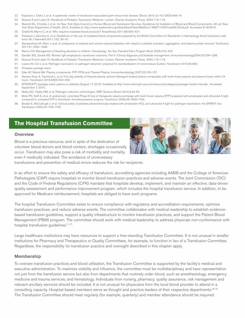
22. Kopolovic I, Ostro J, et al. A systematic review of transfusion-associated graft-versus-host disease. Blood. 2015 Jul 16;126(3):406-14.
23. Strauss R and Luban N. Handbook of Pediatric Transfusion Medicine. London: Elsevier Academic Press, 2004.113–116.
24. Skerrett DL, Chiofolo J, et al., for New York State Council on Human Blood and Transfusion Services. Guidelines for Irradiation of Blood and Blood Components. 4th ed. New York State Department of Health, 2012. Available at: http://www.wadsworth.org/sites/default/files/WebDoc/1368771646/irrad0812final.pdf. Accessed 3/18/2016.
25. Challis M, Marrin C, et al. Who requires irradiated blood products? Anaesthesia 2011;66:620–621.
26. Treleaven J, Gennery A, et al. Guidelines on the use of irradiated blood components prepared by the British Committee for Standards in Haematology blood transfusion task force. Br J Haematol 2011;152: 35–51.
27. Veeraputhiran M, Ware J, et.al. A comparison of washed and volume-reduced platelets with respect to platelet activation, aggregation, and plasma protein removal. Transfusion 2011;51:1030–1036.
28. Manno CS. Management of bleeding disorders in children. Hematology: Am Soc Hematol Educ Program Book 2005;416–422.
29. Sandler SG, Zantek ND. Review: IgA anaphylactic transfusion reactions. Part II. Clinical diagnosis and bedside management. Immunohematology2004;20:234–238.
30. Strauss R and Luban N. Handbook of Pediatric Transfusion Medicine. London: Elsevier Academic Press, 2004.113–116.
31. Lozano M, Cid J, et al. Pathogen inactivation or pathogen reduction: proposal for standardization of nomenclature (Letter). Transfusion 2015;55:690.
32. Octaplas package insert
33. Eder AF, Sebok MA. Plasma components: FFP, FP24 and Thawed Plasma. Immunohematology 2007;23:150-157.
34. Neisser-Svae A, Trawnicek L, et al. Five-day stability of thawed plasma: solvent-detergent-treated plasma comparable with fresh-frozen plasma and plasma frozen within 24 hours. Transfusion 2016.56(2):404-409.
35. INTERCEPT package insert, plasma or platelets (August 17, 2015). Available at http://www.intercept-usa.com/resources/category/package-inserts-manuals , Accessed September 7, 2015.
36. Marks DC, Faddy HM, et al. Pathogen reduction technologies. ISBT Science Series 2014;9:44-50.
37. Mintz PD, Neff A, et al. A randomized, controlled Phase III trial of therapeutic plasma exchange with fresh-frozen plasma (FFP) prepared with amotosalen and ultraviolet A light compared to untreated FFP in thrombotic thrombocytopenic purpura. Transfusion 2006;46:1693-1704.
38. Snyder E, McCullough J, et al. Clinical safety of platelets photochemically treated with amotosalen HCL and ultraviolet A light for pathogen inactivation: the SPRINT trial. Transfusion 2005;44:1732-1740.
The Hospital Transfusion Committee
Overview
Blood is a precious resource, and in spite of the dedication of volunteer blood donors and blood centers, shortages occasionally occur. Transfusion may also pose a risk of morbidity and mortality, even if medically indicated. The avoidance of unnecessary transfusions and prevention of medical errors reduces the risk for recipients.
In an effort to ensure the safety and efficacy of transfusion, accrediting agencies including AABB and the College of American Pathologists (CAP) require hospitals to monitor blood transfusion practices and adverse events. The Joint Commission (TJC) and the Code of Federal Regulations (CFR) mandate that hospitals develop, implement, and maintain an effective, data-driven quality assessment and performance improvement program, which includes the hospital transfusion service. In addition, to be approved for Medicare reimbursement, hospitals are obliged to have such programs.
The hospital Transfusion Committee exists to ensure compliance with regulatory and accreditation requirements, optimize transfusion practices, and reduce adverse events. The committee collaborates with medical leadership to establish evidence-based transfusion guidelines, support a quality infrastructure to monitor transfusion practices, and support the Patient Blood Management (PBM) program. The committee should work with medical leadership to address physician non-conformance with hospital transfusion guidelines.(1–7)
Large healthcare institutions may have resources to support a free-standing Transfusion Committee. It is not unusual in smaller institutions for Pharmacy and Therapeutics or Quality Committees, for example, to function in lieu of a Transfusion Committee. Regardless, the responsibility for transfusion practice and oversight described in this chapter apply.
Membership
To oversee transfusion practices and blood utilization, the Transfusion Committee is supported by the facility’s medical and executive administration. To maximize visibility and influence, the committee must be multidisciplinary and have representation not just from the transfusion service but also from departments that routinely order blood, such as anesthesiology, emergency medicine and trauma services, and hematology. Individuals from nursing, pharmacy, quality assurance, risk management and relevant ancillary services should be included. It is not unusual for physicians from the local blood provider to attend in a consulting capacity. Hospital-based members serve as thought and practice leaders of their respective departments.(8, 9) The Transfusion Committee should meet regularly (for example, quarterly) and member attendance should be required.
At a minimum, the Transfusion Committee should include the following identified roles:(11)
• Chair: – A physician knowledgeable about contemporary transfusion practices who has influence within the organization’s
physician community
• Committee Members: – Representatives from departments that order and/or transfuse blood products: physicians, nurses, and other health care
professionals such as physician assistants or nurse practitioners
– Transfusion Service
» Medical Director and managerial staff who:
» Serve as the primary liaisons between the transfusion and clinical care services
» Report on adverse events, transfusion errors and near misses, and issues that may have an impact on blood bank inventory or services (e.g., blood shortages, reagent shortages or software upgrades)
» Report on metrics such as blood utilization, product outdates or wastage
» Provide updates on technology (e.g., pathogen reduction or coagulation point-of-care testing instruments), products, and practices for improving transfusion safety
• Secretary appointed by the Medical Executive Committee or designated by the Transfusion Committee to record and distribute meeting minutes, track member-assigned project deadlines, and coordinate meeting schedules
• Transfusion Safety Officer/Coordinator who reports data pertaining to transfusion practices, compliance with transfusion guidelines, and hemovigilance.(10, 11)
Functions
The Transfusion Committee’s scope of responsibility extends well beyond reviewing and responding to blood utilization trends. It also advocates a culture to promote proper blood utilization and reduce errors and near-misses. The following is a list of Transfusion Committee responsibilities:(1, 5, 6, 9,12)
• Blood administration policy and procedure development
• Patient blood management– Appropriateness of blood product utilization
– Turnaround times for emergency requests
– Massive transfusion protocols
– Blood utilization trend analysis for crossmatch:transfusion ratio, usage and discard rates
– Patient outcome analysis
– Identification and implementation of clinical alternatives to blood transfusion (e.g., erythropoietin, perioperative blood recovery)
– Promotion of new services and blood products (e.g., platelet additive solution (PAS) platelets)
– Promotion of technologies such as clinical decision support services (CDSS) and computerized online entry (CPOE) to influence ordering practices in real time
– Promotion of software or web-based technologies to monitor and benchmark transfusion practices of individual physicians and specialty groups
• Compliance and regulatory oversight– Evaluate policies
– Define internal audit metrics
– Report external and internal audit survey results
– Report infectious and non-infectious adverse events
– Track medical errors, near-misses and sentinel events, including those that must be reported to accrediting or regulatory agencies, and oversee corrective and preventive actions
– Participate in follow up actions on transfusion-associated fatalities reported to the FDA and other governing and accrediting agencies
– Be remained informed about follow up actions on biological product deviations reported to the FDA and other governing and accrediting agencies
• Staff training and education– Develop communication processes to provide updates on transfusion related information
– Oversee development and implementation of training modules
– Provide resources for continuing education
– Serve as department liaison to share relevant Transfusion Committee information
Processes
The key function of the Transfusion Committee is to promote best practices in transfusion. This is accomplished by establishing institutional guidelines and policies, facilitating educational opportunities for ordering and transfusing staff, and overseeing blood utilization through internal assessments and quality audits.
• Institutional guidelines should be periodically updated and based on evidence derived from randomized controlled trials and/or professional society guidelines that are evidence-based. Technological and scientific advances should be evaluated by the transfusion and patient care committees. When required by standards of practice, regulations, or accrediting organizations, such advances should be promoted by the Transfusion Committee and through the PBM program.
• Transfusion practice information and updates should be integrated into the institution’s Continuing Medical Education (CME) and Continuing Education (CE) curricula. This can be accomplished through lectures, staff meetings, grand rounds, self-paced modular activities, and interactive laboratory and transfusion practice sessions. These activities may be coordinated by the designated Transfusion Safety Officer, medical or nursing education officers, or other educational department staff.
• The Transfusion Safety Officer or staff trained on quality assurance and compliance should perform periodic electronic or manual record review and real time observation of transfusion processes. These reviews facilitate assessment of compliance with transfusion guidelines and should identify educational opportunities or a potential need to intervene when non-conformances are noted. Increasingly, software and web-based programs direct guideline-driven blood component ordering practices and monitor and benchmark practices of individual physicians and specialty groups. Some hospitals, in an effort to automate audits, have successfully integrated clinical laboratory data into the blood bank ordering system.(13-15)
Reports
Information presented to the Transfusion Committee should be consolidated into a standard reporting format. This would include, for example, transfusion service data, transfusion practice audits, and project updates. This facilitates on-going assessment of blood administration practices. Trends in blood utilization, inventory management, and cost can be identified and analyzed on a regular basis.
Minutes and reports should be submitted by the secretary to other committees such as Medical Executive or Credentialing committees. This is to provide other peer-review committees with a record of actions to ensure appropriate transfusion-related patient care. Minutes may be protected from inappropriate legal discovery as a critical component of the institution’s quality monitoring program.
Situations in which blood administration deviates from the institution’s policies and procedures should be investigated by the committee in collaboration with the leadership of the involved department and quality assurance.(9, 13)
The Transfusion Committee and Patient Blood Management The adoption of Patient Blood Management by many hospitals strengthens the role of the Transfusion Committee in promoting safer transfusion practices. Please refer to the Patient Blood Management chapter.
References1. Fung MK, Grossman BJ, et al., eds. Technical Manual. 18th ed. Bethesda, MD: AABB; 2014.
2. Ooley P, Chair. Standards for Blood Banks and Transfusion Services. 30th ed. Bethesda, MD: AABB; 2015.
3. Comprehensive Accreditation Manual for Hospitals (CAMH). Oakbrook Terrace, IL: The Joint Commission; 2016.
4. Code of Federal Regulations. Title 42, CFR Part 493. Available at: ecfr.gov. Last accessed January 22, 2016.
5. College of American Pathologists Accreditation Checklists (Transfusion Medicine). Northfield, IL: College of American Pathologists; 2015.
6. Owens W, Gagliardi K, et al. Building better hospital transfusion committees for Ontario. Transfus Apher Sci 2012;46:323-7.
7. Liumbrono GM, Rafanelli D. Appropriateness of blood transfusion and physicians’ education: a continuous challenge for Hospital Transfusion Committees. Blood Transfus 2012;10:1-3.
8. AABB White Paper. Building a better patient blood management program: Identifying tools, solving problems, and promoting patient safety. Bethesda, MD: AABB; 2015.
9. Shulman IA, Saxena S. The transfusion services committee— responsibilities and response to adverse transfusion events. Hematology Am Soc Hematol Educ Program 2005:483–90.
10. Dunbar NM, Sczepiorkowski ZM. How do we utlize a transfusion safety officer? Transfusion 2015;55: 2064-8.
11. Whitaker BI, Rajbhandary S, et al. The 2013 AABB blood collection, utilization, and patient blood management survey report. Bethesda, MD: AABB: 2015. Available from: http://www.aabb.org/research/hemovigilance/bloodsurvey/Pages/default.aspx
12. Saxena S, ed. The Transfusion Committee: Putting Patient Safety First. 2nd ed. Bethesda, MD: AABB; 2013.
13. Saxena S, Shulman IA. Resurgence of the blood utilization committee. Transfusion 2003;43:998-1006.
14. Yazer MH, Triulzi D, et al. Effectiveness of a real-time clinical decision support system for computerized physician order entry of plasma orders Transfusion 2013;52:3120-7.
15. Adams ES, Longhurst CA, et al. Computerized physician online entry with decision support to decrease blood transfusions in children. Pediatrics 2011;127:e1113-9.
Patient Blood Management
Introduction
Transfusion of the appropriate blood component in the appropriate setting can be a life-saving medical intervention, but because of inherent risks should be administered only when clinically necessary and ideally, evidence-based. Blood is a vital but limited resource. Good stewardship is therefore essential to ensure blood availability when transfusion is truly indicated. Patient Blood Management (PBM) is a multifaceted discipline that encompasses this concept. This chapter:
• Defines PBM• Discusses the essential elements of a successful program• Presents approaches to perioperative blood management
Specific blood components are discussed in other chapters.
Rationale for Patient Blood Management
Blood transfusion is the most commonly performed medical procedure, occurring in with 11 percent of patients.(1) In 2013, the American Hospital Association identified blood utilization in inpatient services as one of five areas for hospitals to focus on to reduce non-beneficial care.(2) Per capita transfusions in the US are higher than in other Western countries, and practices vary widely.(3) In one report, RBC transfusion rates in similar patient populations ranged from 9 to 92% in orthopedic surgery, 17% to 82% in colorectal surgery, 20% to 53% in critical care and up to 28% in acute coronary syndrome.(4) Patient outcomes were not significantly different, suggesting that some transfusions may have been avoidable and possibly offered no clinical benefit, yet may have exposed the patient to risk. Evidence of hospital-specific variability in transfusion rates for adult patients undergoing coronary artery bypass graft (CABG) surgery was described in the Journal of the American Medical Association (JAMA) and showed that RBC transfusion rates varied from 7.8% to 92.8%, platelet transfusion from 0.4% to 90.4%, and plasma use was as high as 97.5%.(5)
The risks of allogeneic transfusion are well established and include but are not limited to transmission of infectious diseases and the occurrence of non-infectious complications such as alloimmunization, hemolysis, transfusion-related acute lung injury (TRALI), transfusion-associated circulatory overload (TACO), and anaphylactic reactions. A growing body of evidence suggests that RBC transfusions may be associated with unfavorable outcomes such as increased risk of patient morbidity and mortality.(6) Glance et al. performed a retrospective analysis of the association between intraoperative blood transfusion and 30-day morbidity and mortality in patients undergoing general, vascular, or orthopedic surgery. Patients receiving an intraoperative transfusion of one or more units of red blood cells had a higher risk of mortality and were more likely to have pulmonary, septic, thromboembolic, and wound complications.(7)
For side effects and hazards, please see the Appendices.
The possibility that patients are unnecessarily exposed to transfusion risks is reinforced by reports from the American Medical Association and The Joint Commission (TJC) with the Centers for Medicare and Medicaid Services that identified RBC transfusions as one of the top five overused procedures in medicine.(8) The 2013 AABB Survey of Blood Collection, Utilization, and PBM Survey noted that 78% of hospital survey respondents reported having transfusion guidelines,(9) but at least one audit of transfusion practices performed in 2011 indicated that 40%-60% of transfusions were inappropriate and given outside of guidelines.(10)
The above discussion demonstrates the need for a formal process to guide transfusion decisions and, ultimately, improve patient care. PBM is defined by the AABB as “an evidence-based, multidisciplinary approach to optimizing the care of patients who might need transfusion.”(9,11) It is wide-ranging and includes transfusion therapy based on robust evidence to maximize a patient’s oxygen-carrying capacity, minimize blood loss in surgical and trauma settings, and integrate alternative therapies, when available, into PBM practices.(12) Benefits of PBM include fewer transfusions, the avoidance of potential complications,(13) decreased length of hospitalization, fewer readmissions and a subsequent reduction in the many associated costs.(14)
AABB has provided an important framework for PBM that is supported by its PBM Standards and PBM Accreditation Program,(15) yet the 2013 AABB survey of blood collection, utilization and PBM practices reported that only 37.8% of responding hospitals had a PBM program.(9) It is important to note that TJC has leveraged its authority as a hospital accreditation organization to influence transfusion oversight and practices, and is expanding beyond previously established Performance Measures to collaborate with AABB in offering PBM certification
Elements of an effective and successful PBM program are:(15, 16)
• Commitment• Leadership• Program oversight• Transfusion practice guidelines• Integration of information technology • Non-compliance with transfusion guidelines
Commitment
Commitment ofan organization to developing and implementing a PBM program is essential and maximizes the potential for success. Without it, efforts to improve transfusion practices and safely minimize transfusion may not realize their full potential, or even flounder. The provision of financial resources for information technology, infrastructure, staffing, education, consistent messaging, and continuing focus on patient safety all characterize commitment. Leadership
High-level medical, nursing, and executive leaders who will advocate for evidence-based changes in practice and financial resources should serve on a PBM steering or program development committee. Other stakeholders would include medical and surgical departments that routinely order and transfuse blood, the hospital transfusion committee, transfusion and laboratory medicine, pharmacy, quality assurance, and risk management. If the institution has a Transfusion Safety Officer, that individual should participate as well.
Please refer to the Hospital Transfusion Committee chapter
Program Oversight
The organization should create a transfusion oversight entity to establish, monitor, promote and enforce PBM activities through audits, periodic updating of transfusion guidelines, advocacy of educational initiatives, communication with clinical and nursing staff, and continuous review and process improvement. This requirement may be met by a transfusion or stand-alone PBM committee or other committee charged with comprehensive PBM responsibilities. Optimally this multidisciplinary group should include representatives from departments that participate in the steering committee.
Transfusion practice guidelines
The organization should develop or update institutional transfusion guidelines that are based on credible evidence, current peer-reviewed literature, and/or guidelines published by professional societies. Many of these resources are provided in specific blood component sections of this Compendium and will not be covered in depth here.
Role of information technology in PBM
Information technology is increasingly recognized as an essential PBM tool that provides evidence-based transfusion recommendations in real time and enables extraction of benchmarking data for individual physicians and service lines.(16) Utilization audits can be facilitated by software designed for generating orders. Computerized physician order entry (CPOE) and clinical decision support systems (CDSS) represent established and effective IT approaches for monitoring transfusion practices.(17)
Rana et al. first described the use of CPOE with CDSS for red cell transfusions in critically ill adults. The CPOE configuration prompted ordering physician to select an indication for red cell transfusion when the pre-transfusion hemoglobin was greater than 7 g/dL in adult intensive care patients. The study showed that in the initial three months after implementation of CPOE with CDSS, a decrease in RBC transfusions of approximately 15 % and a decrease in transfusion-related complications were observed.(18) Lin et al. described the results of a hospital-wide CPOE system that was configured to prompt the ordering physician to select from a list of appropriate transfusion indications when ordering platelets.(19)
PBM-based blood order sets can be integrated into the hospital’s ordering system and evidence-based transfusion guidelines and alerts hardwired into the CPOE systemData collection and monitoring can be facilitated. In addition, CPOE provides automated, real-time clinical practice guidance when the physician is interfacing with the system to order blood.(20)
CPOE alerts are effective in reducing orders that may not meet guidelines. Rothschild et al. evaluated ordering practices prior to and post implementation of a CPOE-based CDSS. Their CPOE system had an “adaptive alert” capability that enabled the CPOE system to interact with the laboratory information system. Laboratory parameters were queried and transfusion recommendations were provided based on clinical information and laboratory test results. Results showed a decrease in inappropriate orders.(21)
Baer et al. described the implementation of an electronic transfusion ordering and monitoring system with guidelines for the neonatal intensive care unit (NICU). This resulted in an increase in compliance from 68% to 90%.(22) Yerrabothala et al. described the utilization of CPOE with a CDSS system to support the establishment of evidence-based restrictive transfusion guidelines for RBC transfusion. The study had an approved transfusion trigger of 7 g/dL with additional indications for special populations, such as actively bleeding patients or patients with a history of acute myocardial infarction. The study showed a statistically significant decrease in the pre-transfusion hemoglobin and in RBC transfusions per 1000 patient days.(23)
Other features that influence the effectiveness of CPOE with CDSS include user involvement in the development phase, the capability to provide alternate recommendations at point-of-order, and integration with the medical record or order entry system.(24)
Addressing outliers
Accredited hospitals are required by AABB, CAP, and TJC, and possibly other oversight organizations, to perform blood utilization review. Despite findings from randomized controlled trials and updated transfusion recommendations based on peer-reviewed published data, clinician knowledge gaps remain. Continuing education of physicians about evidence-based transfusion guidelines may modify transfusion practices. Strategies for continuing education could include transfusion medicine participation in clinical rounds, audits with specific feedback, and one-on-one discussions with the department chair/designee or the Transfusion Safety Officer, continuing medical education (CME), and electronic or hard-copy dissemination of hospital transfusion guidelines.(25)
Retrospective monitoring of transfusion practices using hard-copy medical records is still in use, and results are presumably reported to an institutional oversight committee for evaluation and follow-up actions. Limitations of this approach include the potential for statistically unsubstantiated selection of documents for review, untimely feedback to physicians, inconsistent review processes, occasional inaccurate identification of physicians who actually ordered the blood, and the inability to prevent unnecessary transfusion.(26) If audits can be performed within days of transfusion and are combined with strategies such as education, it may be feasible to decrease utilization.(27) Sarode et al. used a combination of institution-wide education and prospective review to achieve significant cost savings by decreasing inappropriate RBC and platelet utilization by 60% and 25% respectively.(28) Toy found that one-on-one meetings with physicians, scheduled teaching conferences, prospective audits of ordered transfusions, in addition to daily clinical rounds, were useful.(29)
While such approaches may potentially impact clinician behavior, they are labor intensive and impractical for some hospitals. Further, real time interventions in ordering may cause a delayin issuing blood products. Meaningful effective change requires continuous monitoring and the collection of ordering and transfusion practice data.(30) Physicians and physician groups who routinely order outside of established hospital guidelines can be made aware of this through data sharing and comparison of their practices with physicians of the same specialty. Subsequent interventions to address outlier ordering behavior include peer interaction or targeted education or feedback from medical committees and department chairs.
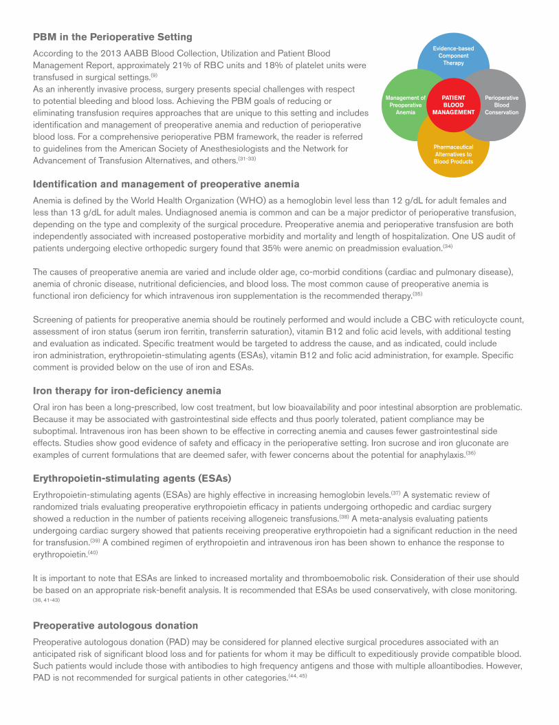
PBM in the Perioperative Setting
According to the 2013 AABB Blood Collection, Utilization and Patient Blood Management Report, approximately 21% of RBC units and 18% of platelet units were transfused in surgical settings.(9) As an inherently invasive process, surgery presents special challenges with respect to potential bleeding and blood loss. Achieving the PBM goals of reducing or eliminating transfusion requires approaches that are unique to this setting and includes identification and management of preoperative anemia and reduction of perioperative blood loss. For a comprehensive perioperative PBM framework, the reader is referred to guidelines from the American Society of Anesthesiologists and the Network for Advancement of Transfusion Alternatives, and others.(31-33) Identification and management of preoperative anemia
Anemia is defined by the World Health Organization (WHO) as a hemoglobin level less than 12 g/dL for adult females and less than 13 g/dL for adult males. Undiagnosed anemia is common and can be a major predictor of perioperative transfusion, depending on the type and complexity of the surgical procedure. Preoperative anemia and perioperative transfusion are both independently associated with increased postoperative morbidity and mortality and length of hospitalization. One US audit of patients undergoing elective orthopedic surgery found that 35% were anemic on preadmission evaluation.(34)
The causes of preoperative anemia are varied and include older age, co-morbid conditions (cardiac and pulmonary disease), anemia of chronic disease, nutritional deficiencies, and blood loss. The most common cause of preoperative anemia is functional iron deficiency for which intravenous iron supplementation is the recommended therapy.(35)
Screening of patients for preoperative anemia should be routinely performed and would include a CBC with reticuloycte count, assessment of iron status (serum iron ferritin, transferrin saturation), vitamin B12 and folic acid levels, with additional testing and evaluation as indicated. Specific treatment would be targeted to address the cause, and as indicated, could include iron administration, erythropoietin-stimulating agents (ESAs), vitamin B12 and folic acid administration, for example. Specific comment is provided below on the use of iron and ESAs.
Iron therapy for iron-deficiency anemia
Oral iron has been a long-prescribed, low cost treatment, but low bioavailability and poor intestinal absorption are problematic. Because it may be associated with gastrointestinal side effects and thus poorly tolerated, patient compliance may be suboptimal. Intravenous iron has been shown to be effective in correcting anemia and causes fewer gastrointestinal side effects. Studies show good evidence of safety and efficacy in the perioperative setting. Iron sucrose and iron gluconate are examples of current formulations that are deemed safer, with fewer concerns about the potential for anaphylaxis.(36)
Erythropoietin-stimulating agents (ESAs)
Erythropoietin-stimulating agents (ESAs) are highly effective in increasing hemoglobin levels.(37) A systematic review of randomized trials evaluating preoperative erythropoietin efficacy in patients undergoing orthopedic and cardiac surgery showed a reduction in the number of patients receiving allogeneic transfusions.(38) A meta-analysis evaluating patients undergoing cardiac surgery showed that patients receiving preoperative erythropoietin had a significant reduction in the need for transfusion.(39) A combined regimen of erythropoietin and intravenous iron has been shown to enhance the response to erythropoietin.(40)
It is important to note that ESAs are linked to increased mortality and thromboemobolic risk. Consideration of their use should be based on an appropriate risk-benefit analysis. It is recommended that ESAs be used conservatively, with close monitoring. (36, 41-43)
Preoperative autologous donation
Preoperative autologous donation (PAD) may be considered for planned elective surgical procedures associated with an anticipated risk of significant blood loss and for patients for whom it may be difficult to expeditiously provide compatible blood. Such patients would include those with antibodies to high frequency antigens and those with multiple alloantibodies. However, PAD is not recommended for surgical patients in other categories.(44, 45)
Management ofPreoperative
Anemia
Evidence-basedComponent
Therapy
PharmaceuticalAlternatives to Blood Products
Perioperative Blood
Conservation
PATIENTBLOOD
MANAGEMENT
Although PAD has a role, albeit a limited one, for minimizing exposure to allogeneic blood, associated risks must be considered. A 2001 Cochrane review of 14 randomized controlled trials (RCTs) showed a mean decrement in the preoperative hemoglobin of approximately 1.1 g/dL in the PAD group compared to the non-PAD group, with an associated 24% increased risk of transfusion among patients in the PAD arm.(46) If PAD is ordered, prescription of iron supplements should be considered to enhance erythropoiesis in the perioperative and postoperative periods. Although AABB Standards permit PAD in patients with a hemoglobin as low as 11.5g/dL, it should be noted that each donation may either cause preoperative anemia or worsen pre-existing anemia. In addition, although perceived as being a safer alternative to allogeneic RBCs, units may be lost due to collection or processing problems, and complications such as TACO can occur. Further, the frequency of outdating for PAD units approached 45% in one publication.(47)
Anticoagulant and anti-platelet therapy
The perioperative management of patients who are receiving antithrombotic anticoagulant and/or anti-platelet therapy is a frequent and often challenging situation for clinicians. New oral anticoagulants (NOACs) such as dabigatran, a direct thrombin inhibitor, and rivaroxaban, a direct factor Xa inhibitor, introduce additional complexity to perioperative blood management. (In October 2015, the U.S. Food and Drug Administration (FDA) approved idarucizumab, a monoclonal antibody fragment that directly binds dabigatran for the urgent reversal of the anticoagulant effects of dabigatran in emergency situations).(48) Currently, there are few clinical trials that provide definitive guidance regarding best practices. The experience of Sarode may provide assistance in some circumstances.(49)
Decisions regarding perioperative management of patients receiving antithrombotic therapy can be guided by an assessment of thromboembolic risk versus risk of perioperative bleeding.(50) Guidelines from the American College of Chest Physicians stratify patients into the following thromboembolic risk categories:
• High risk (>10% annual risk for thromboembolism)
• Moderate risk (5-10% annual risk for thromboembolism)
• Low risk (<5% annual risk for thromboembolism)
Consideration regarding use of bridging therapy is often required for patients at greater risk for thromboembolism. Bridging therapy is commonly defined as the use of a short acting anticoagulant such as unfractionated heparin or low molecular weight heparin (LMWH) during the period of time that warfarin is being withheld. Observational studies with a systematic approach to perioperative anticoagulant management showed lower rates of thromboembolic and bleeding episodes.(51)
The American College of Chest Physicians recommends the following processes for inclusion into a standardized perioperative management protocol:
• Assessing patients at least 7 days preoperatively to plan perioperative anticoagulant management
• Developing a schedule with the timing of warfarin and anti-platelet drug discontinuation and resumption, dose and timing of LMWH bridging, and the INR measurement schedule
• Ensuring that the perioperative management strategy for vitamin K antagonists and anti-platelet drug interruption and initiation of LMWH bridging accounts for pharmacokinetics and thromboembolic and bleeding risks
• Training of the patient or caregiver to administer LMWH
• When indicated and feasible, performing INR testing on the day before surgery to identify patients with INRs that are elevated enough to presumably allow timely administration of vitamin K--Assessing hemostasis, preferably on the day of surgery and on the first postoperative day, to facilitate safe resumption of anticoagulant drugs.(50)
Intraoperative blood management
Intraoperative blood management strategies generally focus on minimizing blood loss, collecting and reinfusing blood from the operative field, and improving tolerance of anemia. These approaches are generally considered standard practice:
Acute normovolemic hemodilution (ANH)
ANH is a blood conservation technique that may be considered when significant blood loss is anticipated during the surgical procedure. The procedure is performed in the operating room immediately prior to surgery and involves the simultaneous removal of a specific volume of whole blood and replacement with crystalloid or colloid solution to maintain adequate volume. A dilutional anemia is created, reducing surgical blood loss. Blood collected by ANH is stored at room temperature and returned
to the patient within 8 hours, possibly preserving the function of platelets and coagulation factors. The benefits of ANH have not been firmly established and disparate results have been reported.(52, 53)
Autologous red blood cell recovery
Autologous blood recovery (previously referred to as “salvage”) is another blood conservation technique that may be considered for surgical procedures in which substantial blood loss (greater than 1000 mL) is expected.(75) The procedure involves the collection of red blood cells from the surgical field. In intraoperative salvage, blood that is collected from the surgical field is centrifuged, washed, filtered and suspended in normal saline for reinfusion. The hematocrit is 45% to 55%. The minimum volume for reinfusion is 200 mL which is roughly equivalent to one RBC unit. This technique is often used in vascular, cardiothoracic, orthopedic, gynecologic and urologic surgery. Cell recovery has been shown to decrease the need for allogeneic transfusion.(54)
Pharmacologic Agents
Antifibrinolytic drugs
Antifibrinolytic drugs may be useful for reducing perioperative blood loss. Antifibrinolytic drugs act by inhibiting the fibrinolytic system which has a major role in controlling clot formation and dissolution. Tranexamic acid (TXA) and epsilon-aminocaproic acid (EACA) are lysine analogues that reversibly inhibit fibrinolysis by interfering with plasminogen activation. A Cochrane review determined that antifibrinolytics provided significant reduction in perioperative blood loss and the need for allogeneic transfusions without serious adverse effects.(55) The results of a literature review by Ortmann et al. showed that the use of TXA prophylactically reduced perioperative blood loss in cardiac and non-cardiac major surgery.(56) In the Clinical Randomization of an Antifibrinolytic in Significant Haemorrhage-2 (CRASH-2) trial, 20,000 trauma patients were randomized to receive either TXA or placebo. Results of the trial showed that early administration of TXA (within 3 hours) was associated with reduced mortality.(57)
Aprotinin is a serine protease inhibitor that inhibits fibrinolysis by directly inhibiting plasmin. A multicenter trial showed an increase in mortality with its use, and it was withdrawn from the market. However, subsequent reevaluation of data from the Blood Conservation Using Antifibrinolytics in a Randomized Trial (BART) study and other data resulted in its reintroduction and use for cardiac surgery in Canada and Europe.(58)
Topical Hemostatic Agents
Several commercially available, virally inactivated, allogeneic sealants and autologous fibrin sealant systems are FDA-approved and are preferable to cryoprecipitate with respect to safety and efficacy for topical use.(59-61) A Cochrane review showed a 37% reduction in allogeneic RBC transfusion and reduced blood loss when fibrin sealant was used.(62) Evaluation of 124 patients undergoing total knee arthroplasty in a randomized trial showed that topical application of TXA resulted in a 20%-25% reduction in postoperative blood loss without report of adverse effects.(63)
Point of Care Testing
Steurer and Ganter describe three primary testing methods for point of care assessment of coagulation:
• Simple anticoagulation monitoring devices for activated clotting time (ACT), whole blood PT/INR and whole blood aPTT.
• Point of care coagulation tests to assess primary hemostasis and platelet function, for example, PFA-100/200 and modified platelet aggregometry.
• Viscoelastic coagulation monitoring devices that assess global clotting processes from initial thrombin generation to maximum clot formation and ultimately clot lysis. Thromboelastography (TEG; Haemonetics Corp., Braintree, MA) and rotational thromboelastrometry (ROTEM, International GmbH, Munich, Germany) are commonly used platforms.(64)
Viscoelastic Coagulation Technology
According to the 2013 AABB survey on blood utilization, RBC transfusion in cardiac surgery patients accounted for 7% of all RBC transfusions.(9) The hemostatic management of patients undergoing cardiovascular surgery may be challenging, and requires balancing anticoagulation during cardiopulmonary bypass with management of hemostatic function after the procedure has been completed. Conventionally, transfusion algorithms use standard coagulation tests such as platelet count, PT, aPTT and the Clauss fibrinogen assay to screen for coagulation abnormalities and guide treatment.(65) Turnaround times may not be optimal, and poor predictive value for bleeding limits their clinical utility, leading clinicians to make empirical decisions.
(66-68) Increased interest in the use of viscoelastic point of care testing (POCT) has been driven in part by the limitations of conventional assays for hemostasis. Thromboelastography differs from conventional coagulation tests in that it uses whole blood to produce a two dimensional, computer-generated tracing in real time that provides information about the development, stabilization and dissolution of clots.(69) This tracing also provides a comprehensive composite view of the interaction of coagulation factors, platelets, fibrinogen, and rapid assessment of thrombosis and fibrinolysis.(70) TEG measurement of clot strength is important in determining whether intraoperative bleeding is due to coagulopathy or other causes and, as such, has a major role in TEG-based transfusion algorithms.(71) Randomized clinical trials indicate that use of TEG-based decision algorithms during cardiac surgery resulted in fewer transfusions.(72)
Viscoelastic coagulation monitoring instruments have become important tools in the POCT hemostatic assessment of patients, and have been implemented in many institutions.(65) TEG based transfusion algorithms have also been shown to improve blood utilization in liver transplant patients(73) and in major trauma patients in conjunction with massive transfusion protocols.(74)
When considering implementation of POCT, operational and quality considerations should include the logistical challenges associated with the need for rapid assay initiation, the need for daily calibration, and delineation of roles in interpreting results.
Summary
Patient blood management is a patient-centered, evidence-based approach to transfusion practice, with the overall goal of improving patient outcomes. It is now the standard of practice and care. Successful implementation of an effective patient blood management program has been shown to improve outcomes, reduce transfusion rates, and decrease associated costs.
References1. Pfuntner A, Wier LM, et al. Most frequent procedures performed in US Hospitals, 2010. HCUP Statistical Brief # 149. Agency for Healthcare Research and Quality, Rockville,
MD. Feb 2013:1-9.
2. 2. Combes JR, Arespacochaga E. Appropriate use of medical resources. American Hospital Association’s Physician Leadership Forum, Chicago, IL. November 2013.
3. 3. MacPherson J, Mahoney C, et al. Contribution of blood to hospital revenue in the United States. Transfusion 2007;47:114S-116S.
4. Farmer S, Trentino K, et al. A programmatic approach to patient blood management – reducing transfusion and improving patient outcomes. The Open Anesthesiology Journal 2015;9:6-16. DOI: 10.2174/1874321801509010006
5. Bennett-Guerrero E, Zhao Y, et al. Variation on use of blood transfusion in coronary artery bypass graft surgery. JAMA 2010;304:1568-75.
6. Hopewell S, Omar O, et al. A systematic review of the effect of red blood cell transfusion on mortality: Evidence from large-scale observational studies published between 2006 and 2010. BMJ Open 2013;3e002154. doi:10.1136/bmjopen-2012-002154.
7. Glance LG, Dick AW, et al. Association between intraoperative blood transfusion and mortality and morbidity in patients undergoing noncardiac surgery. Anesthesiology 2011;114:283-92.
8. 8. Proceedings from the National Summit on Overuse. Organized by the Joint Commission and the American Medical Association − Convened Physician Consortium for Performance Improvement®(PCPI®) 2012;1-37.
9. Whitaker BI, Rajbhandary S, et al. The 2013 AABB blood collection, utilization, and patient blood management survey report. Bethesda, MD: AABB, 2015.
10. Shander A, Fink A, et al. Appropriateness of allogeneic red blood cell transfusion: the international consensus conference on transfusion outcomes. Transfus Med Rev 2011;25:232-246.
11. Aubuchon JP, Puca K, et al. Getting Started in Patient Blood Management. Bethesda, MD: AABB, 2011.
12. AABB White Paper. Building a better patient blood management program. Bethesda MD: AABB, 2015.
13. Shander A, Hofmann A, et al. Activity-based costs of blood transfusion in surgical patients at four hospitals. Transfusion 2010;50:753-65.
14. Kotzé A, Carter LA, et al. Effect of a patient blood management programme on preoperative anaemia, transfusion rate, and outcome after primary hip or knee arthroplasty: a quality improvement cycle. Br J Anaesth 2012;108:943-52.
15. Holcomb JB, Chair. Standards for Patient Blood Management, 1st ed. Bethesda, MD: AABB, 2014.
16. Dunbar NM, Szczepiorkowski ZM. Hardwiring patient blood management: harnessing information technology to optimize transfusion practice. Curr Opin Hematol 2014;21:515-520.
17. Collins RA, Triulzi DJ, et al. Evaluation of real-time clinical decision support systems for platelet and cryoprecipitate orders. Am J Clin Pathol 2014;141:78-84.
18. Rana R, Afessa B, et al. Evidence-based red cell transfusion in the critically ill: quality improvement using computerized physician order entry. Crit Care Med 2006;34:1892-7.
19. Lin YC, Chang CS, et al. The appropriateness and physician compliance of platelet usage by a computerized transfusion decision support system in a medical center. Transfusion 2010;50:2565-70.
20. Goodnough LT, Shieh L, et al. Improved blood utilization using real-time clinical decision support. Transfusion 2014;54:1358-65.
21. Rothschild JM, McGurk S, et al. Assessment of education and computerized decision support interventions for improving transfusion practice. Transfusion 2007;47:228-39.
22. Baer VL, Henry E, et al. Implementing a program to improve compliance with neonatal intensive care unit transfusion guidelines was accompanied by a reduction in transfusion rate: a pre-post analysis within a multihospital health care system. Transfusion 2011;51:264-9.
23. Yerrabothala S, Desrosiers KP, et al. Significant reduction in red blood cell transfusions in a general hospital after successful implementation of a restrictive transfusion policy supported by prospective computerized order auditing. Transfusion 2014;54:2640-5.
24. Lobach D, Sanders GD, et al. Enabling healthcare decision-making through clinical decision support and knowledge management. Evid Rep Technol Assess (Full Rep) 2012;203:1-784.
25. Karafin MS, Bryant BJ. Transfusion medicine education: an integral foundation of effective blood management. Transfusion 2014;54:1208-11.
26. Lam HT, Schweitzer SO, et al. Are retrospective peer-review transfusion monitoring systems effective in reducing red blood cell utilization? Arch Pathol Lab Med 1996;120:810-6.
27. Marques MB, Polhill SR, et al. How we closed the gap between red blood cell utilization and whole blood collections in our institution. Transfusion 2012;52:1857-67.
28. Sarode R, Refaai MA, et al. Prospective monitoring of plasma and platelets in a large teaching hospital results in significant cost reduction. Transfusion 2010;50:487-92.
29. Toy P. Guiding the Decision to Transfuse. Interventions that do and do not work. Arch Pathol Lab Med 1999;123:592-4.
30. Mehra T, Seifert B, et al. Implementation of a patient blood management monitoring and feedback program significantly reduces transfusions and costs. Transfusion 2015;55:2807-15.
31. Practice guidelines for perioperative blood management: An updated report by the American Society of Anesthesiologists Task Force on Perioperative Blood Management Anesthesiology 2015;122:241-75.
32. Goodnough LT, Maniatis A, et al. Detection, evaluation and management of preoperative anaemia in the elective orthopaedic surgical patient: NATA guidelines. Br J Anaesth 2011;106:13-22.
33. Clevenger B, Mallett SV, et al. Patient blood management to reduce surgical risk. Br J Surg 2015;102:1325-37.
34. Bierbaum BE, Callaghan JJ, et al. An analysis of blood management in patients having a total hip or knee arthroplasty. J Bone Joint Surg Am 1999;81:2-10.
35. Auerbach M, Coyne D, et al. Intravenous iron: from anathema to standard of care. Am J Hematol 2008;83:580-8.
36. Lin DM, Lin ES, et al. Efficacy and safety of erythropoietin and intravenous iron in perioperative blood management: a systematic review. Transfus Med Rev 2013;27:221-34.
37. Goodnough LT, Shander A: Update on erythropoiesis-stimulating agents. Best Pract Res Clin Anaesthesiol 2013;27:121-9.38. Laupacis A, Fergusson D. Erythropoietin to minimize perioperative blood transfusion: a systematic review of randomized trials. The International Study of Peri-operative
Transfusion (ISPOT) Investigators. Transfus Med 1998;8:309-317.39. Alghamdi AA, Albanna MJ, et al. Does the use of erythropoietin reduce the risk of exposure to allogeneic blood transfusion in cardiac surgery? A systematic review and meta-
analysis. J Card Surg 2006;21:320-6.
40. Rineau E, Chaudet A, et al. Ferric carboxymaltose increases epoetin-alpha response and prevents iron deficiency before elective orthopaedic surgery. Br J Anaesth 2014;113:296-8.
41. Rodgers GM, Becker PS, et al. Cancer – and chemotherapy-induced anemia. J Natl Compr Canc Netw 2012;10:628-53.
42. National Comprehensive Cancer Network (NCCN). NCCN clinical practice guidelines in oncology. Cancer and chemotherapy-induced anemia. http://www.nccn.org. Version 2.2016. Last accessed: January 30, 2016.
43. Stowell CP, Jones SC, et al. An open-label, randomized, parallel-group study of perioperative epoetin alpha versus standard of care for blood conservation in major elective spinal surgery: safety analysis. Spine 2009;34:2479-85.
Vassallo R, Goldman M, et al. Preoperative autologous blood donation: waning indications in an era of improved blood safety. Transfus Med Rev 2015;29:268-75.
45. British Committee for Standards in Haematology, Transfusion Task Force, Boulton FE, James V. Guidelines for policies on alternatives to allogeneic blood transfusion. 1. Predeposit autologous blood donation and transfusion. Transfus Med 2007;17:354-65.
46. Henry DA, Carless PA, et al. Pre-operative autologous donation for minimising perioperative allogeneic blood transfusion (Review). Cochrane Database Syst Rev 2001:CD003602.
47. Department of Health and Human Services. The 2011 National Blood Collection and Utilization Survey Report. Washington, DC:DHHS;2013.
48. Pollack CV, Reilly PA, et al. Idarucizumab for Dabigatran reversal. N Engl J Med 2015;373:511-20.
49. Sarode R. How do I transfuse platelets (PLTs) to reverse anti-PLT drug effect? Transfusion 2012;52:695-701.
50. Douketis J, Spyropoulos A C, et al. Perioperative management of antithrombotic therapy: antithrombotic therapy and prevention of thrombosis, 9th ed: American College of Chest Physicians Evidence-Based Clinical Practice Guidelines. Chest 2012;141 2Suppl:e326S-50S.
51. Wysokinski WE, McBane RD. Periprocedural bridging management of anticoagulation. Circulation 2012;126:489-90.
52. Bennett J, Haynes S, et al. Acute normovolemic hemodilution in moderate blood loss surgery: A randomized control trial. Transfusion 2006;46:1097-103.
53. Frankel TL, Fischer M, et al. Selecting patients for acute normovolemic hemodilution during hepatic resection: a prospective randomized evaluation of nomogram-based allocation. J Am Coll Surg. 2013:217:10-20.
54. Ashworth A, Klein AA. Cell salvage as part of a blood conservation strategy in anaesthesia. Br J Anaesth 2010;105:401-16.
55. Henry DA, Carless PA, et al. Anti-fibrinolytic use for minimising perioperative allogeneic blood transfusion. Cochrane Database Syst Rev 2011;CD001886.
56. Ortmann E, Besser MW, et al. Antifibrinolytic agents in current anaesthetic practice. Br J Anaesth 2013;111:549-63.
57. Shakur H, Roberts I, et al. Effects of tranexamic acid on death, vascular occlusive events, and blood transfusion in trauma patients with significant hemorrhage (CRASH-2): a randomized, placebo – controlled trial. Lancet 2010;376:23-32.
58. McMullan V, Alston RP. III. Aprotinin and cardiac surgery: A sorry tale of evidence misused. Br J Anaesth 2013:110:675-8.
59. Foster LJR. Bioadhesives as surgical sealants: a review. In: Bianco-Peled H, Davidovich-Pinhas M, eds. Bioadhesion and biomimetics: from nature to applications. Boca Raton, FL: CRC Press, 2015.
60. Alston SM, Solen KA, et al. In vivo efficacy of a new autologous fibrin sealant. J Surg Res 2008;146:143-8.
61. Buchta C, Hedrich HC, et al. Biochemical characterization of autologous fibrin sealants produced by CryoSeal® and Vivostat® in comparison to the homologous fibrin sealant product Tissucol/Tisseel®. Biomaterials 2015;26:6233-41.
62. Carless PA, Henry DA, et al. Fibrin sealant use for minimising perioperative allogeneic blood transfusion. Cochrane Database Syst Rev 2013;CD004171.
63. Wong J, Abrishami A, et al. Topical application of tranexamic acid reduces postoperative blood loss in total knee arthroplasty: a randomized, controlled trial. J Bone Joint Surg Am 2010;92:2503-13.
64. Steurer MP, Ganter MT. Point of Care Coagulation Testing. American Society of Anesthesiologists Newsletter 2015;79(6).
65. Bolliger D, Seeberger MD, et al. Principles and practice of thromboelastography in clinical coagulation management and transfusion practice. Transfus Med Rev 2012;26:1-13.
66. Avidan MS, Alcock EL, et al. Comparison of structured use of routine laboratory tests or near-patient assessment with clinical judgment in the management of bleeding after cardiac surgery. Br J Anaesth 2004;92:178-86.
67. Spiess B, Gillies B, et al. Changes in transfusion therapy and reexploration rate after institution of a blood management program in cardiac surgical patients. J Cardiothorac Vasc Anesth 1995;9:168-73.
68. Shore-Lessorson L, Manspeizer HE, et al. Thromboelastography-guided transfusion algorithm reduces transfusions in complex cardiac surgery. Anesth Analg 1999;88:312-9.
69. Goodnough LT, Hill CC. Use of point-of-care testing for plasma therapy. Transfusion 2012;52 Suppl 1:56S-64S.
70. Chitlur M, Sorensen B, et al. Standardization of thromboelastography: a report from the TEG-ROTEM working group. Haemophilia 2011;17:532-7.
71. Ganter MT, Hofer CK. Coagulation monitoring: current techniques and clinical use of viscoelastic point-of-care coagulation devices. Anesth Analg 2008;106:1366-75.
72. Royston D, van Kier S. Reduced haemostatic factor transfusion using heparinase-modified thromboelastography during cardiopulmonary bypass. Br J Anaesth 2001;86:575-8.
73. Harding SA, Mallett SV, et al. Use of heparinase modified thromboelastography in liver transplantation. Br J Anaesth 1997;78:175-9.
74. Young PP, Cotton BA, et al. Massive transfusion protocols for patients with substantial hemorrhage. Transfus Med Rev 2011;25:293-303.
75. Avidan M, Silvergleid AJ, et al. Surgical blood conservation: Blood salvage. UpToDate 2015.
44.
Appendices
Appendix I: Side Effects and Hazards for Whole Blood and All Blood Components
The following section is reproduced from the November 2013 Circular of Information (COI) for blood and blood components.
Immunologic complications
1. Hemolytic transfusion reaction, the destruction of red cells, is discussed in detail in the COI.
2. Immune-mediated platelet destruction, one of the causes of refractoriness to platelet transfusion, is the result of alloantibodies in the recipient to HLA or platelet-specific antigens on transfused platelets; this is discussed in detail in the COI.
3. Febrile non-hemolytic reaction is typically manifested by a temperature elevation of ≥1° C or 2° F occurring during or shortly after a transfusion and in the absence of any other pyrexic stimulus. This may reflect the action of antibodies against white cells or the action of cytokines either present in the transfused component or generated by the recipient in response to transfused elements. Febrile reactions may occur in less than 1% of transfusions of leukocyte-reduced red cell components and about 5% of leukocyte-reduced apheresis platelet components. Febrile reactions occur more frequently in patients receiving non-leukocyte-reduced components and those previously alloimmunized by transfusion or pregnancy. No routinely available pre – or post-transfusion tests are helpful in predicting or preventing these reactions. Antipyretics usually provide effective symptomatic relief. Patients who experience repeated, severe febrile reactions may benefit from receiving leukocyte-reduced components. If these reactions are caused by cytokines in the component, prestorage leukocyte reduction may be beneficial.
4. Allergic reactions frequently occur (i.e., 1-3% of plasma-containing components) as mild or self-limiting urticaria or wheezing that usually respond to antihistamines. More severe manifestations, including respiratory and cardiovascular symptoms, are more consistent with anaphylactoid/anaphylactic reactions and may require more aggressive therapy (see below). No laboratory procedures are available to predict these reactions.
5. Anaphylactoid/anaphylactic reactions, characterized by hypotension, tachycardia, nausea, vomiting and/or diarrhea, abdominal pain, severe dyspnea, pulmonary and/or laryngeal edema, and bronchospasm and/or laryngospasm, are rare but dangerous complications requiring immediate treatment with epinephrine. These reactions have been reported in IgA-deficient patients who develop antibodies to IgA antibodies. Such patients may not have been previously transfused and may develop symptoms after infusion of very small amounts of IgA-containing plasma in any blood component. Similar reactions have also been described in patients with haptoglobin deficiency. In certain circumstances, patients may benefit from the use of washed cellular components to prevent or reduce the severity of allergic reactions not minimized by treatment with medication alone.
6. Transfusion-related acute lung injury (TRALI) is characterized by the acute onset of hypoxemia and non-cardiogenic pulmonary edema within 6 hours of a blood or blood component transfusion in the absence of other causes of acute lung injury or circulatory overload. Various stimuli in blood components, most commonly white blood cell (WBC) antibodies from donors sensitized during pregnancy or prior transfusion or transplantation, or pro-inflammatory molecules that accumulate in stored blood components, may cause TRALI. These mechanisms may not be mutually exclusive and may act synergistically with underlying patient factors to lead to a final common pathway of acute lung injury. These stimuli may trigger an inflammatory response, granulocyte activation and degranulation, and injury to the alveolar capillary membrane, and the development of permeability pulmonary edema. Although most TRALI cases are associated with donor anti-leukocyte antibodies, rare cases have implicated recipient anti-leukocyte antibodies that reacted with donor leukocytes. Widespread leukoreduction of blood components has likely mitigated this latter risk. Laboratory testing of blood donors for anti-leukocyte antibodies or blood components for biologic mediators does not alter management of this reaction which is diagnosed on clinical and radiographic findings. Treatment of TRALI involves aggressive respiratory support, and often mechanical ventilation. The preferential use of plasma collected from male donors has been associated with a significant reduction in the number of reported TRALI cases and associated fatalities. Transfusion services should immediately report suspected TRALI to the blood collection facility to facilitate the retrieval of other components associated with the involved donation(s) or prior donations.
Immunologic Complications, Delayed
1. Delayed hemolytic reaction is described in detail in the COI.
2. Alloimmunization to antigens of red cells, white cells, platelets, or plasma proteins may occur unpredictably after transfusion. Blood components may contain certain immunizing substances other than those indicated on the label. For example, platelet components may also contain red cells and white cells. Primary immunization does not become apparent until days or weeks after the immunizing event, and does not usually cause symptoms or physiologic changes. If components that express the relevant antigen are subsequently transfused, there may be accelerated removal of cellular elements from the circulation and/or systemic symptoms. Clinically significant antibodies to red cell antigens will ordinarily be detected by pre-transfusion testing. Alloimmunization to antigens of white cells, platelets, or plasma proteins can be detected only by specialized testing.
3. Post-transfusion purpura (PTP) is a rare syndrome characterized by the development of dramatic, sudden, and self-limited thrombocytopenia, typically 7 to 10 days after a blood transfusion, in a patient with a history of sensitization by either pregnancy or transfusion. Although the immune specificity may be to a platelet-specific antigen the patient lacks, both autologous and allogeneic platelets are destroyed. High-dose Immune Globulin, Intravenous (IVIG) may correct the thrombocytopenia.
4. Transfusion-associated graft-vs-host disease (TA-GVHD) is a rare but extremely dangerous condition that occurs when viable T lymphocytes in the transfused component engraft in the recipient and react against recipient tissue antigens. TA-GVHD can occur if the host does not recognize and reject the foreign transfused cells, and it can follow transfusion of any component that contains even very small numbers of viable T lymphocytes. Recipients with severe cellular immunodeficiency (except for HIV infection) are at greatest risk (e.g., fetuses receiving intrauterine transfusions, recipients of hematopoietic progenitor cell transplants, and selected patients with severe immunodeficiency conditions), but TA-GVHD has also been reported in recipients receiving purine analogues (e.g., fludarabine, cladribine) for oncologic and rheumatologic diseases, and in immunologically normal recipients who are heterozygous for a tissue antigen haplotype for which the donor is homozygous. Tissue antigen haplotype sharing is most likely to occur when the transfused component is from a blood relative or has been selected for HLA compatibility. TA-GVHD remains a risk with leukocyte-reduced components because they contain sufficient residual T lymphocytes. Irradiation of the component renders T lymphocytes incapable of proliferation and is presently the only approved means to prevent TA-GVHD.
Non-immunologic Complications
1. Because Whole Blood and blood components are made from human blood, they may carry a risk of transmitting infectious agents [e.g., viruses, bacteria, parasites, the variant Creutzfeldt-Jakob disease (vCJD) agent, and, theoretically, the CJD agent]. Careful donor selection and available laboratory tests do not totally eliminate these hazards. Also, septic and toxic reactions can result from transfusion of bacterially contaminated blood and blood components. Such complications are infrequent, but may be life-threatening. Infectious disease transmission may occur despite careful selection of donors and testing of blood. Donor selection criteria are designed to screen out potential donors with increased risk of infection with HIV, HTLV, hepatitis, and syphilis, as well as other agents (see section on testing of donor blood). These procedures do not totally eliminate the risk of transmitting these agents. Transfusion services should immediately report infections that may be related to the blood donor or to the manufacture of the blood components to the collection facility.
2. Cytomegalovirus (CMV) may be present in white-cell-containing components from donors previously infected with this virus, which can persist for a lifetime despite the presence of serum antibodies. Up to 70% of donors may be CMV seropositive. Transmission of CMV by transfusion may be of concern in low-birth weight (≤1200 g) premature infants born to CMV-seronegative mothers and in intrauterine transfusions and/or certain other categories of immunocompromised individuals such as hematopoietic progenitor cell or solid organ transplant patients, if they are CMV seronegative. For at-risk recipients, the risk of CMV transmission by cellular components can be reduced by transfusing CMV-seronegative or leukocyte-reduced components. For other infectious agents (e.g., Babesia spp, Leishmania spp, and Plasmodia spp) there are no routinely available tests to predict or prevent disease transmission. All potential blood donors are subjected to screening procedures intended to reduce to a minimum the risk that they will transmit infectious agents.
3. Bacterial sepsis occurs rarely, but can cause acute, severe, sometimes life-threatening effects. Onset of high fever (≥2°C or ≥3.5°F increase in temperature), severe chills, hypotension, or circulatory collapse during or shortly after transfusion should suggest the possibility of bacterial contamination and/or endotoxin reaction in the transfused products. Although platelet components stored at room temperature have been implicated most frequently, previously frozen components thawed by immersion in a water bath and red cell components stored for several weeks at 1 to 6 C have also been implicated. Although most platelet components are routinely tested for bacterial contamination, this does not completely
eliminate the risk. Both gram-positive and gram-negative organisms have been identified as causing septic reactions. Organisms capable of multiplying at low temperatures (e.g., Yersinia enterocolitica) and those using citrate as a nutrient are most often associated with components containing red cells. A variety of pathogens as well as skin contaminants, have been found in platelet components. Endotoxemia in recipients has resulted from multiplication of gram-negative bacteria in blood components. Prompt recognition of a possible septic reaction is essential, with immediate discontinuation of the transfusion and aggressive therapy with broad-spectrum antimicrobials and vasopressor agents, if necessary. In addition to prompt sampling of the patient’s blood for cultures, investigation should include examination of material from the blood container by Gram stain, and cultures of specimens from the container and the administration set. It is important to report all febrile transfusion reactions to the transfusion service for appropriate investigation. If post-transfusion sepsis is suspected, the transfusion service should immediately report the reaction to the blood collection facility to facilitate retrieval of other potentially contaminated components associated with the collection.
4. Transfusion-associated circulatory overload (TACO) leading to cardiogenic (hydrostatic) pulmonary edema can occur after transfusion of excessive volumes or at excessively rapid rates. This is a particular risk in individuals with underlying cardiopulmonary or renal disease, the very young and the elderly, and in patients with chronic severe anemia in whom low red cell mass is associated with high plasma volume. Small transfusion volumes can precipitate symptoms in at-risk patients who already have a positive fluid balance. Pulmonary edema should be promptly and aggressively treated, and infusion of colloid preparations, including plasma components and the supernatant fluid in cellular components, reduced to a minimum.
5. Hypothermia carries a risk of cardiac arrhythmia or cardiac arrest and exacerbation of coagulopathy. Rapid infusion of large volumes of cold blood or blood components can depress body temperature, and the danger is compounded in patients experiencing shock or surgical or anesthetic manipulations that disrupt temperature regulation. A blood warming device should be considered if rapid infusion of blood or blood components is needed. Warming must be accomplished using an FDA-cleared blood warming device so as not to cause hemolysis.
6. Metabolic complications may accompany large-volume transfusions, especially in neonates and patients with liver or kidney disease.
a. Citrate “toxicity” reflects a depression of ionized calcium caused by the presence in the circulation of large quantities of citrate anticoagulant. Because citrate is promptly metabolized by the liver, this complication is rare. Patients with severe liver disease or those with circulatory collapse that prevents adequate hepatic blood flow may have physiologically significant hypocalcemia after rapid, large-volume transfusion. Citrated blood or blood components administered rapidly through central intravenous access may reach the heart so rapidly that ventricular arrhythmias occur. Standard measurement of serum calcium does not distinguish ionized from complexed calcium. Ionized calcium testing or electrocardiogram monitoring is more helpful in detecting physiologically significant alteration in calcium levels.
b. Other metabolic derangements can accompany rapid or large-volume transfusions, especially in patients with preexisting circulatory or metabolic problems. These include acidosis or alkalosis (deriving from changing concentrations of citric acid and its subsequent conversion to pyruvate and bicarbonate) and hyperkalemia or hypokalemia.
Fatal Transfusion Reactions
When a fatality occurs as a result of a complication of blood or blood component transfusion, the Director, Office of Compliance and Biologics Quality, Center for Biologics Evaluation and Research (CBER), should be notified as soon as possible (telephone: 240-402-9160; efax 301-827-0333; e-mail: [email protected]). Within 7 days after the fatality, a written report must be submitted to the FDA/CBER, Director, Office of Compliance and Biologics Quality, Attn: Fatality Program Manager, 1401 Rockville Pike, Suite 200N, Rockville, MD 20852-1448. A copy of the report should be sent to the collecting facility, if appropriate.
Refer to the COI for references for this Appendix.
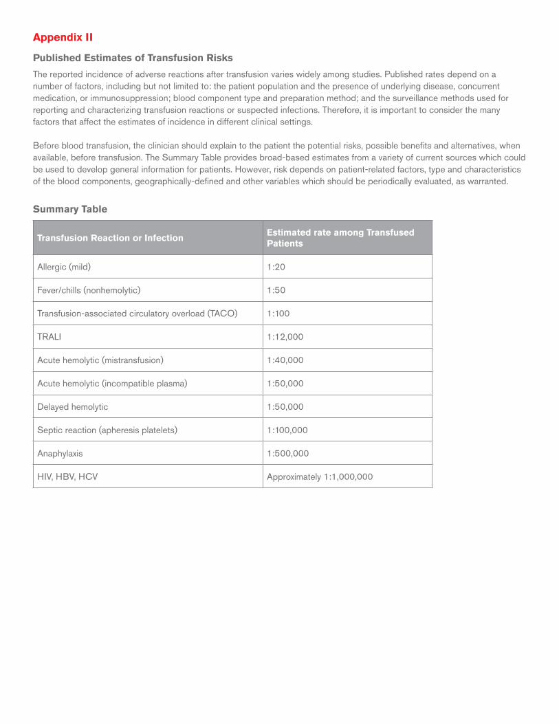
Appendix II
Published Estimates of Transfusion Risks
The reported incidence of adverse reactions after transfusion varies widely among studies. Published rates depend on a number of factors, including but not limited to: the patient population and the presence of underlying disease, concurrent medication, or immunosuppression; blood component type and preparation method; and the surveillance methods used for reporting and characterizing transfusion reactions or suspected infections. Therefore, it is important to consider the many factors that affect the estimates of incidence in different clinical settings.
Before blood transfusion, the clinician should explain to the patient the potential risks, possible benefits and alternatives, when available, before transfusion. The Summary Table provides broad-based estimates from a variety of current sources which could be used to develop general information for patients. However, risk depends on patient-related factors, type and characteristics of the blood components, geographically-defined and other variables which should be periodically evaluated, as warranted.
Summary Table
Transfusion Reaction or InfectionEstimated rate among Transfused Patients
Allergic (mild) 1:20
Fever/chills (nonhemolytic) 1:50
Transfusion-associated circulatory overload (TACO) 1:100
TRALI 1:12,000
Acute hemolytic (mistransfusion) 1:40,000
Acute hemolytic (incompatible plasma) 1:50,000
Delayed hemolytic 1:50,000
Septic reaction (apheresis platelets) 1:100,000
Anaphylaxis 1:500,000
HIV, HBV, HCV Approximately 1:1,000,000
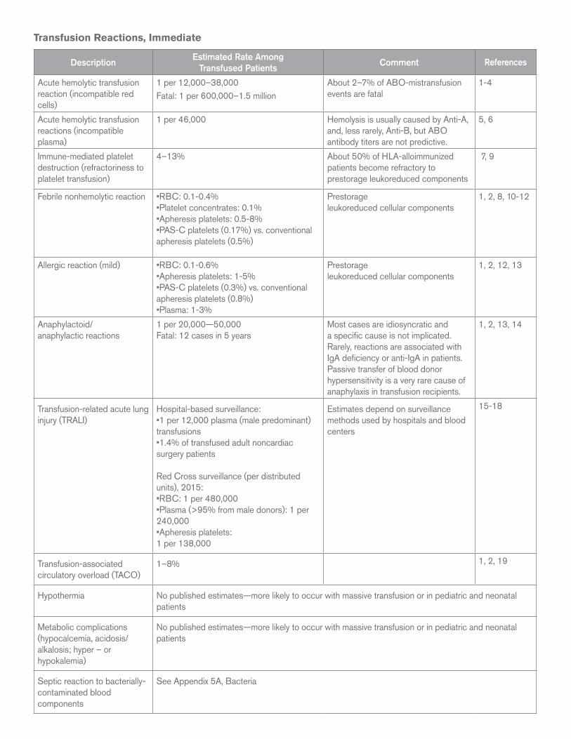
Transfusion Reactions, Immediate
DescriptionEstimated Rate Among
Transfused PatientsComment References
Acute hemolytic transfusion reaction (incompatible red cells)
1 per 12,000–38,000Fatal: 1 per 600,000–1.5 million
About 2–7% of ABO-mistransfusion events are fatal
1-4
Acute hemolytic transfusion reactions (incompatible plasma)
1 per 46,000 Hemolysis is usually caused by Anti-A, and, less rarely, Anti-B, but ABO antibody titers are not predictive.
5, 6
Immune-mediated platelet destruction (refractoriness to platelet transfusion)
4–13% About 50% of HLA-alloimmunized patients become refractory to prestorage leukoreduced components
7, 9
Febrile nonhemolytic reaction •RBC: 0.1-0.4%•Platelet concentrates: 0.1%•Apheresis platelets: 0.5-8%•PAS-C platelets (0.17%) vs. conventional apheresis platelets (0.5%)
Prestorage leukoreduced cellular components
1, 2, 8, 10-12
Allergic reaction (mild) •RBC: 0.1-0.6%•Apheresis platelets: 1-5% •PAS-C platelets (0.3%) vs. conventional apheresis platelets (0.8%)•Plasma: 1-3%
Prestorage leukoreduced cellular components
1, 2, 12, 13
Anaphylactoid/anaphylactic reactions
1 per 20,000 —50,000 Fatal: 12 cases in 5 years
Most cases are idiosyncratic and a specific cause is not implicated. Rarely, reactions are associated with IgA deficiency or anti-IgA in patients. Passive transfer of blood donor hypersensitivity is a very rare cause of anaphylaxis in transfusion recipients.
1, 2, 13, 14
Transfusion-related acute lung injury (TRALI)
Hospital-based surveillance:•1 per 12,000 plasma (male predominant) transfusions •1.4% of transfused adult noncardiac surgery patients
Red Cross surveillance (per distributed units), 2015:•RBC: 1 per 480,000•Plasma (>95% from male donors): 1 per 240,000•Apheresis platelets: 1 per 138,000
Estimates depend on surveillance methods used by hospitals and blood centers
15-18
Transfusion-associated circulatory overload (TACO)
1–8% 1, 2, 19
Hypothermia No published estimates—more likely to occur with massive transfusion or in pediatric and neonatal patients
Metabolic complications (hypocalcemia, acidosis/alkalosis; hyper – or hypokalemia)
No published estimates—more likely to occur with massive transfusion or in pediatric and neonatal patients
Septic reaction to bacterially-contaminated blood components
See Appendix 5A, Bacteria
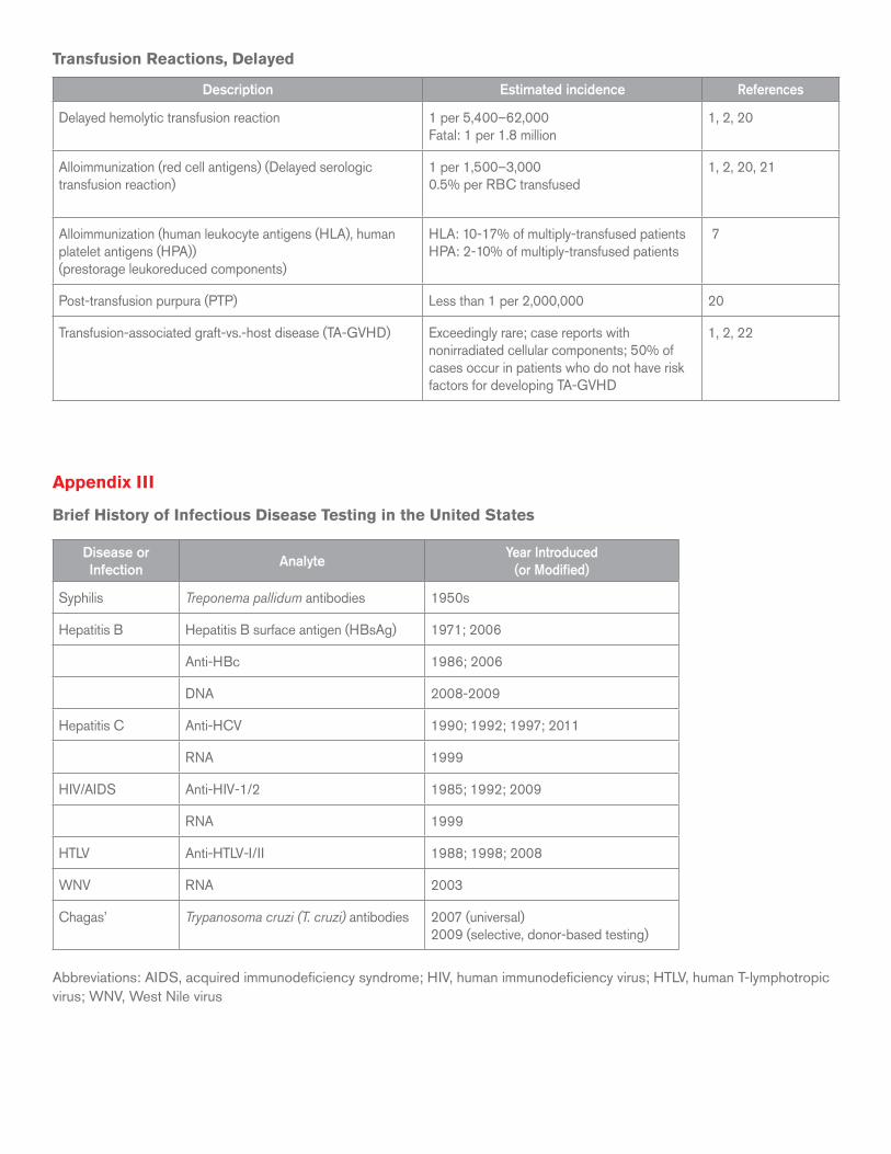
Transfusion Reactions, Delayed
Description Estimated incidence References
Delayed hemolytic transfusion reaction 1 per 5,400–62,000Fatal: 1 per 1.8 million
1, 2, 20
Alloimmunization (red cell antigens) (Delayed serologic transfusion reaction)
1 per 1,500–3,0000.5% per RBC transfused
1, 2, 20, 21
Alloimmunization (human leukocyte antigens (HLA), human platelet antigens (HPA))(prestorage leukoreduced components)
HLA: 10-17% of multiply-transfused patientsHPA: 2-10% of multiply-transfused patients
7
Post-transfusion purpura (PTP) Less than 1 per 2,000,000 20
Transfusion-associated graft-vs.-host disease (TA-GVHD) Exceedingly rare; case reports with nonirradiated cellular components; 50% of cases occur in patients who do not have risk factors for developing TA-GVHD
1, 2, 22
Appendix III
Brief History of Infectious Disease Testing in the United States
Disease or Infection
AnalyteYear Introduced
(or Modified)
Syphilis Treponema pallidum antibodies 1950s
Hepatitis B Hepatitis B surface antigen (HBsAg) 1971; 2006
Anti-HBc 1986; 2006
DNA 2008-2009
Hepatitis C Anti-HCV 1990; 1992; 1997; 2011
RNA 1999
HIV/AIDS Anti-HIV-1/2 1985; 1992; 2009
RNA 1999
HTLV Anti-HTLV-I/II 1988; 1998; 2008
WNV RNA 2003
Chagas’ Trypanosoma cruzi (T. cruzi) antibodies 2007 (universal) 2009 (selective, donor-based testing)
Abbreviations: AIDS, acquired immunodeficiency syndrome; HIV, human immunodeficiency virus; HTLV, human T-lymphotropic virus; WNV, West Nile virus
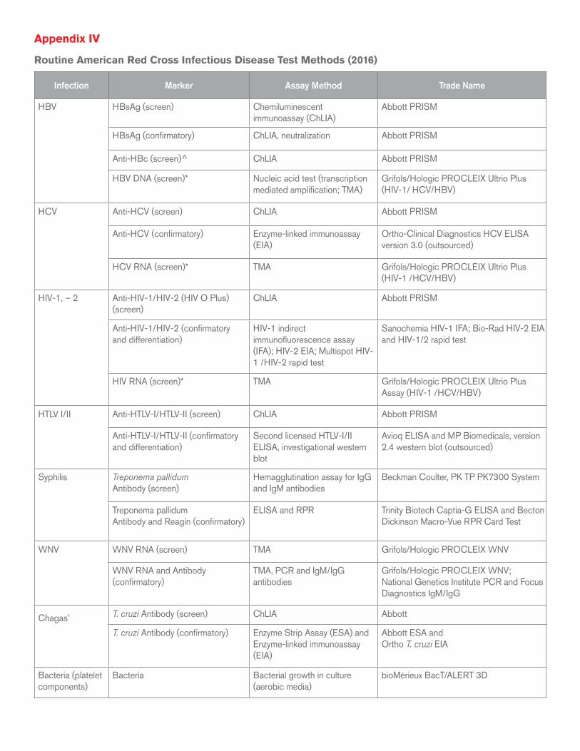
Appendix IV
Routine American Red Cross Infectious Disease Test Methods (2016)
Infection Marker Assay Method Trade Name
HBV HBsAg (screen) Chemiluminescent immunoassay (ChLIA)
Abbott PRISM
HBsAg (confirmatory) ChLIA, neutralization Abbott PRISM
Anti-HBc (screen)^ ChLIA Abbott PRISM
HBV DNA (screen)* Nucleic acid test (transcription mediated amplification; TMA)
Grifols/Hologic PROCLEIX Ultrio Plus (HIV-1/ HCV/HBV)
HCV Anti-HCV (screen) ChLIA Abbott PRISM
Anti-HCV (confirmatory) Enzyme-linked immunoassay (EIA)
Ortho-Clinical Diagnostics HCV ELISA version 3.0 (outsourced)
HCV RNA (screen)* TMA Grifols/Hologic PROCLEIX Ultrio Plus (HIV-1 /HCV/HBV)
HIV-1, – 2 Anti-HIV-1/HIV-2 (HIV O Plus) (screen)
ChLIA Abbott PRISM
Anti-HIV-1/HIV-2 (confirmatory and differentiation)
HIV-1 indirect immunofluorescence assay (IFA); HIV-2 EIA; Multispot HIV-1 /HIV-2 rapid test
Sanochemia HIV-1 IFA; Bio-Rad HIV-2 EIA and HIV-1/2 rapid test
HIV RNA (screen)* TMA Grifols/Hologic PROCLEIX Ultrio Plus Assay (HIV-1 /HCV/HBV)
HTLV I/II Anti-HTLV-I/HTLV-II (screen) ChLIA Abbott PRISM
Anti-HTLV-I/HTLV-II (confirmatory and differentiation)
Second licensed HTLV-I/II ELISA, investigational western blot
Avioq ELISA and MP Biomedicals, version 2.4 western blot (outsourced)
Syphilis
Treponema pallidumAntibody (screen)
Hemagglutination assay for IgG and IgM antibodies
Beckman Coulter, PK TP PK7300 System
Treponema pallidumAntibody and Reagin (confirmatory)
ELISA and RPR Trinity Biotech Captia-G ELISA and Becton Dickinson Macro-Vue RPR Card Test
WNV
WNV RNA (screen) TMA Grifols/Hologic PROCLEIX WNV
WNV RNA and Antibody (confirmatory)
TMA, PCR and IgM/IgG antibodies
Grifols/Hologic PROCLEIX WNV; National Genetics Institute PCR and Focus Diagnostics IgM/IgG
Chagas’
T. cruzi Antibody (screen) ChLIA Abbott
T. cruzi Antibody (confirmatory) Enzyme Strip Assay (ESA) andEnzyme-linked immunoassay (EIA)
Abbott ESA andOrtho T. cruzi EIA
Bacteria (platelet components)
Bacteria Bacterial growth in culture (aerobic media)
bioMérieux BacT/ALERT 3D
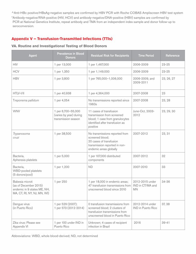
^ Anti-HBc positive/HBsAg-negative samples are confirmed by HBV PCR with Roche COBAS Ampliscreen HBV test system
* Antibody-negative/RNA-positive (HIV, HCV) and antibody-negative/DNA-positive (HBV) samples are confirmed by PCR at National Genetics Institute, repeat antibody and TMA from an independent index sample and donor follow-up to seroconversion.
Appendix V – Transfusion-Transmitted Infections (TTIs)
VA. Routine and Investigational Testing of Blood Donors
AgentPrevalence in Blood
DonorsResidual Risk for Recipients Time Period Reference
HIV 1 per 13,000 1 per 1,467,000 2006-2009 23-25
HCV 1 per 1,350 1 per 1,149,000 2006-2009 23-25
HBV 1 per 3,800 1 per 765,000–1,006,000 2006-2009, and2009-2011
23, 26, 27
HTLV-I/II 1 per 40,938 1 per 4,364,000 2007-2008 23
Treponema pallidum 1 per 4,054 No transmissions reported since 1960s
2007-2008 23, 28
WNV 1 per 6,700–55,000 (varies by year) during transmission season
11 cases of transfusion transmission from screened blood; 1 case from granulocytes identified after transfusion as positive
June-Oct, 2003-2012
23, 29, 30
Trypanosoma cruzi
1 per 38,500 No transmissions reported from screened blood; 20 cases of transfusion transmission reported in non-endemic areas globally
2007-2012 23, 31
Bacteria, Apheresis platelets
1 per 5,000 1 per 107,000 distributed components
2007-2012 32
Bacteria, WBD-pooled platelets(5 donors/pool)
1 per 1,200 ND 2007-2010 33
Babesia microti(as of December 2015) endemic in 9 states ME, NH, MA, CT, RI, NY, NJ, MN, WI)
1 per 250 1 per 18,000 in endemic areas; 47 transfusion transmissions from unscreened blood since 2010
2012-2015 under IND in CT/MA and MN
34-36
Dengue virus(in Puerto Rico)
1 per 529 (2007); 1 per 573 (2012-2014)
0 transfusion transmissions from screened blood; 2 clusters of transfusion transmissions from unscreened blood in Puerto Rico
2012-2014 under IND in Puerto Rico
37, 38
Zika virus: Please see Appendix VI
1 per 100 under IND in Puerto Rico
Unknown; 4 cases of recipient infection in Brazil
2016 39-41
Abbreviations: WBD, whole blood-derived; ND, not determined
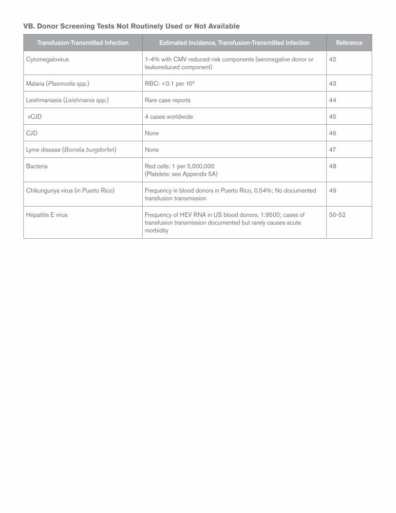
VB. Donor Screening Tests Not Routinely Used or Not Available
Transfusion-Transmitted Infection Estimated Incidence, Transfusion-Transmitted Infection Reference
Cytomegalovirus 1-4% with CMV reduced-risk components (seronegative donor or leukoreduced component)
42
Malaria (Plasmodia spp.) RBC: <0.1 per 106 43
Leishmaniasis (Leishmania spp.) Rare case reports 44
vCJD 4 cases worldwide 45
CJD None 46
Lyme disease (Borrelia burgdorferi) None 47
Bacteria Red cells: 1 per 5,000,000(Platelets: see Appendix 5A)
48
Chikungunya virus (in Puerto Rico) Frequency in blood donors in Puerto Rico, 0.54%; No documented transfusion transmission
49
Hepatitis E virus Frequency of HEV RNA in US blood donors, 1:9500; cases of transfusion transmission documented but rarely causes acute morbidity
50-52