Embed Size (px)
Citation preview
Dr. M. Elmubarak:
MBBS , Msc clinical anatomy, MD registrar of orthopedic and trauma surgery

Objective
Review the functinal component of cranial nerve
Type , neucli and applied anatomy of trigeminla
nerve
Type , neucli and applied anatomy of facial nerve
Type , neucli and applied anatomy of
vestibulocochlear nerve
CRANIAL NERVES
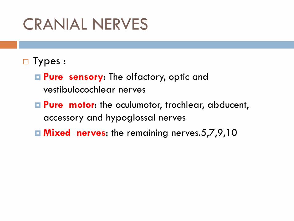
Types :
Pure sensory: The olfactory, optic and
vestibulocochlear nerves
Pure motor: the oculumotor, trochlear, abducent,
accessory and hypoglossal nerves
Mixed nerves: the remaining nerves.5,7,9,10

Functional componant of cranial nerves:
G: general, S:somatic , V:vesciral,E: efferent, A: afferent B:branchial.
GSA (exteroception)
GVA (visceral sensation)
SVA (special sense)
SSA (special senses)
GSE (sk muscle)
GVE (sm muscle, gland)
SVE (BE, pharyngeal arch)

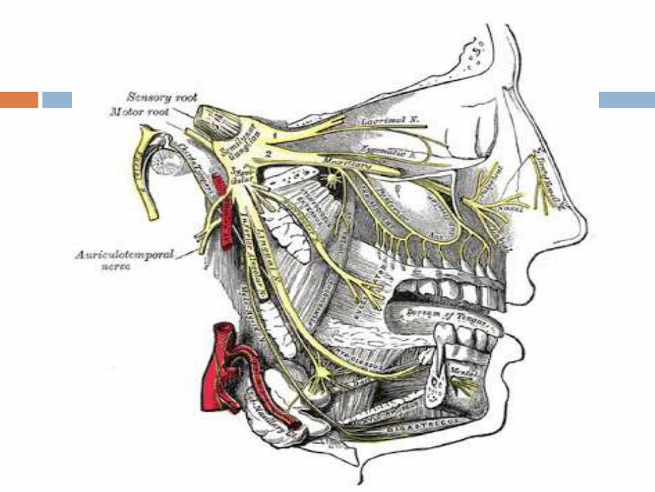
(Trigeminal (SVE, GSA
Type:
The trigeminal is mixed cranial nerve has:
Sensory supply to face, the greater part of the scalp, the
teeth, the oral & nasal cavities (axons of cells in the
trigeminal ganglion , sensory neuclus)
The motor supply to the masticatory & some other
muscles.(motor nucleus)
the largest cranial nerve
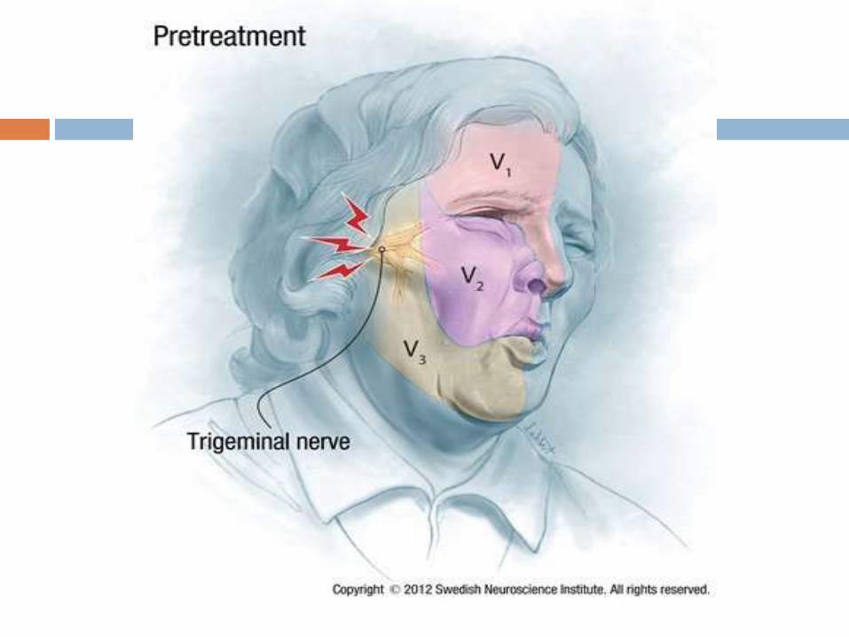
It has three divisions
1. OPHTHALMIC
2. MAXILLARY
3. MANDIBULAR.

NUCLEI:
Sensory nucleus :
Principle : in the pons , receive touch
Spinal : in medula and upper spinal cord, receive pain and
tempreture.
Mesoecephalic: in the midbrain, receive proprioception
Motor nucleus
In the pons
Send fiber to mandibular division

APPLIED ANATOMY:
LESION OF TRIGEMINAL NERVE: A lesion of the whole trigeminal nerve causes
Anaesthesia of the anterior half of the scalp, of the face (except a small area near the angle of mandible), of the cornea & conjunctiva, the mucosae of the nose,mouth and presulcal part of the tongue.
Paralysis and atrophy occur in the muscles supplied by the nerve also.
TRIGEMINAL NEURALGIA characterized by pain in the distribution of branches of the trigeminal nerve, is the most common condition affecting the sensory part of the nerve.
Facial Nerve (SVE, SVA, GVE)
Type:
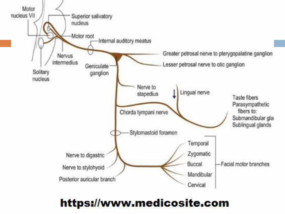
Consists of a motor & sensory (nervus intermedius) roots.
The motor root mainly supplies muscles of face, stapedius &
stylohyoid.
The sensory root conveys from the chorda tympani gustatory
fibers from the anterior 2/3 of tongue
it also carries preganglionic parasympathitic innervation of
the submandibular & sublingual salivary glands, lacrimal
glands. Nose and palat

NUCLEI:
Motor nucleus of facial : for muscle supplied by the
nerve. in the pons.
Superior Salivatory Nucleus: for pterygopalatine
Ganglion and submandibular ganglion. In the pons
Nucleus of Tractus Solitarius :for Taste fibers, anterior
2/3 of tongue. In the medulla

APPLIED ANATOMY:
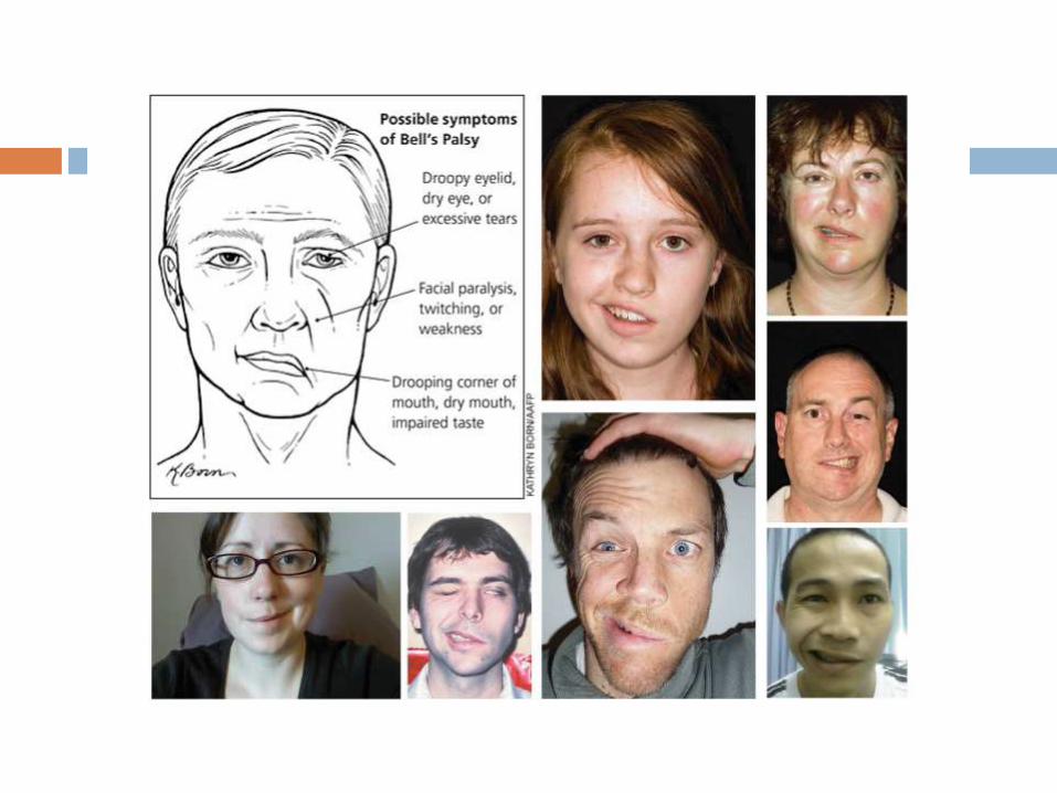
Lesion: The paralysis may be UMN or LMN:
Upper motor neuron facial paralysis,
Lower face but not the upper (forehead and orbicularisoculi) because the facial nerve nucleus innervating the upper part of face receives fibers from cerebral cortex of both sides whereas the lower part innervating the lower part of the face receives contralateral fibers.
However emotional movements of the lower face, as in smiling and laughing, are still possible (presumably there is an alternative pathway from the cerebrum).
Lower motor neuron fascial palsy lesions: upper and lower fascial muscle paralysis
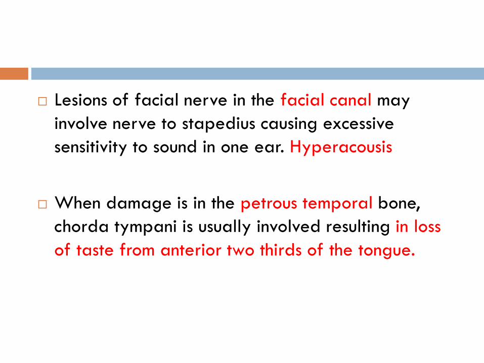
Lesions of facial nerve in the facial canal may
involve nerve to stapedius causing excessive
sensitivity to sound in one ear. Hyperacousis
When damage is in the petrous temporal bone,
chorda tympani is usually involved resulting in loss
of taste from anterior two thirds of the tongue.
VESTIBULOCOCHLEAR NERVE(8TH):
Type:
The vestibulocochlear nerve (CN-VIII), is main sensory
supply of internal ear.
It has two major sets of fibers,
Vestibular nerve, concerned with equilibration and arising
from the vestibular ganglion in the outer part of internal
acoustic meatus.
Cochlear nerve, concerned with audition arising from the
neurons in the spiral ganglion of the cochlea.
NUCLEI:
Special Somatic Afferent: Two cochlear nuclei in Pons
for HEARING.
Special Somatic Afferent: Four vestibular nuclei in Pons
& Medulla for EQUILIBRIUM.
INTRACRANIAL COURSE:
The nerve emerges in the groove between the pons and
the medulla oblongata, behind the facial nerve.

Ventral view

APPLIED ANATOMY :
Disturbances of vestibular nerve function include
giddiness (VERTIGO) and NYSTAGMUS.
Disturbances of the cochlear nerve function produce
DEAFNESS and TINNITUS.