Embed Size (px)
Citation preview

Cancers 2011, 3, 61-78; doi:10.3390/cancers3010061
cancersISSN 2072-6694
www.mdpi.com/journal/cancers
Article
Doxorubicin-Loaded PEG-PCL-PEG Micelle Using Xenograft
Model of Nude Mice: Effect of Multiple Administration of
Micelle on the Suppression of Human Breast Cancer
Nguyen-Van Cuong 1,2
, Jian-Lin Jiang 1, Yu-Lun Li
1, Jim-Ray Chen
3, Shyh-Chuan Jwo
4 and
Ming-Fa Hsieh 1,*
1 Department of Biomedical Engineering, Chung Yuan Christian University, 200, Chung Pei Rd.,
Chung Li, Taiwan; E-Mail: [email protected] (N.V.C.); [email protected] (J.L.J.);
[email protected] (Y.L.L.) 2 Department of Chemical Engineering, Ho Chi Minh City University of Industry, 12 Nguyen Van
Bao St, Ho Chi Minh, Vietnam; E-Mail: [email protected] (N.V.C.) 3 Department of Pathology, Chang Gung Memorial Hospital at Keelung, Taiwan and Chang Gung
University, College of Medicine, Taoyuan, Taiwan; E-Mail: [email protected] (J.R.C.) 4 Division of General Surgery, Chang Gung Memorial Hospital at Keelung, Taiwan and Chang Gung
University, College of Medicine, Taoyuan, Taiwan; E-Mail: [email protected] (S.C.J.)
* Author to whom correspondence should be addressed; E-Mail: [email protected];
Tel.: +886-3-265-4550; Fax: +886-3-265-4599.
Received: 11 November 2010; in revised form: 20 December 2010 / Accepted: 27 December 2010 /
Published: 28 December 2010
Abstract: The triblock copolymer is composed of two identical hydrophilic segments:
Monomethoxy poly(ethylene glycol) (mPEG) and one hydrophobic segment
poly(ε-caprolactone) (PCL); which is synthesized by coupling of mPEG-PCL-OH and
mPEG-COOH in a mild condition using dicyclohexylcarbodiimide and 4-dimethylamino
pyridine. The amphiphilic block copolymer can self-assemble into nanoscopic micelles to
accommodate doxorubixin (DOX) in the hydrophobic core. The physicochemical
properties and in vitro tests, including cytotoxicity of the micelles, have been characterized
in our previous study. In this study, DOX was encapsulated into micelles with a drug
loading content of 8.5%. Confocal microscopy indicated that DOX was internalized into
the cytoplasm via endocystosis. A dose-finding scheme of the polymeric micelle (placebo)
showed a safe dose of PEG-PCL-PEG micelles was 71.4 mg/kg in mice. Importantly, the
OPEN ACCESS

Cancers 2011, 3
62
circulation time of DOX-loaded micelles in the plasma significantly increased compared to
that of free DOX in rats. A biodistribution study displayed that plasma extravasation of
DOX in liver and spleen occurred in the first four hours. Lastly, the tumor growth of
human breast cancer cells in nude mice was suppressed by multiple injections (5 mg/kg,
three times daily on day 0, 7 and 14) of DOX-loaded micelles as compared to multiple
administrations of free DOX.
Keywords: nanoparticles; drug delivery system; monomethoxy poly(ethylene glycol);
poly(ε-caprolactone); safety evaluation; biodistribution; antitumor activity
1. Introduction
Doxorubicin (DOX), an anthracycline drug has proven very effective for the treatment of breast,
ovarian, prostate, brain, cervix and lung cancers [1-4]. Nevertheless, drawbacks such as cardiac
toxicity, short half-life and low solubility in aqueous solution have hindered its application. In addition,
some tumor cells showed multidrug resistance (MDR), which has been attributed to the P-glycoprotein
(P-gp) efflux pump on the plasma membrane [5,6]. To overcome these obstacles, strategies such as
encapsulating DOX into the core of polymeric nanoparticles by chemical conjugation or physical
entrapment have been attempted [7-11]. The employment of polymeric nanoparticles also provides an
opportunity to tailor the release profiles of encapsulated drugs [12,13].
In recent years, biodegradable polymeric nanoparticles have been extensively employed as effective
drug delivery systems to enhance the efficacy and safety of encapsulated drugs. Nanoparticle carriers
provide a better accumulation in tumor tissues through an enhanced permeability and retention (EPR)
effect [14,15], ability to overcome multidrug resistance [11], and better pharmacokinetics of drug
in vivo [16]. Nanoparticles composed of poly(ε-caprolactone) (PCL) and poly(ethylene glycol) (PEG)
have been demonstrated as great potential carriers for the delivery of anti-cancer agents. PCL is a
biodegradable, biocompatible and nontoxic thermoplastic polyester [17]. PEG is a common constituent
for the hydrophilic outer shell and is known to reduce the adhesion of plasma proteins, solubility in
water and organic solvents, stabilization of particles and lack of toxicity [4,18]. Additionally,
polymeric nanoparticles with hydrophilic PEG outer shell can potentially increase the circulation time
of drugs and can prevent recognition by macrophages of the reticuloendothelial system (RES) after
intravenous injection [18-20]. Recently, amphiphilic triblock copolymers consisting of poly (ethylene
glycol) as hydrophilic segment, and poly (ε-caprolactone) as hydrophobic block, have been used as
drug delivery systems, such as 4′-demethyl-epipodophyllotoxin [21], honokiol [22], folic acid [23],
nimodipine [24] and doxorubicin [25,26].
Previously, we reported the preparation of a triblock copolymeric (poly(ethylene glycol)/poly(ε-
caprolactone)/poly(ethylene glycol)) micelle (PEG-PCL-PEG) aiming for its application as a
nanocarrier for hydrophobic drugs [27]. A hydrophobic drug such as doxorubicin was successfully
loaded into triblock copolymeric micelle that was prepared by the self assembly method in an aqueous
solution. The release profile and cellular uptake of DOX and its cytotoxic effects against drug-sensitive
(MCF-7) breast cancer cell lines were also investigated. General prerequisites for a new drug delivery

Cancers 2011, 3
63
system should be that it is non-cytotoxic, biocompatible, and in most cases, biodegradable. Therefore,
safety evaluation of triblock copolymeric micelle including the in vitro and in vivo toxicological
studies is required before further application of triblock copolymeric micelles in biomedical fields. In
this work, we evaluated the safety of prepared PEG-PCL-PEG micelle as an intravenous drug delivery
system in a series of tests including cytotoxicity tests, in vitro nitric oxide production and hemolytic
tests and in vivo acute toxicity tests in ICR mice. Furthermore, the biodistribution of DOX-loaded
micelle was evaluated in vivo. Importantly, the therapeutic potential of single and multiple
administrations of DOX-loaded micelle were investigated using the nude mice xenograft model.
2. Results and Discussion
2.1. Physicochemical Properties of DOX-Loaded PEG-PCL-PEG Micelle
The amphiphilic triblock copolymer built with hydrophilic mPEG segments and hydrophobic PCL
segment and by a mild coupling agent-DCC/DMAP was prepared as previously reported [27]. The
triblock copolymer was characterized by proton nuclear magnetic resonance (1H NMR), gel
permeation chromatography (GPC), Fourier transform infrared spectroscopy (FT-IR), X-ray diffraction
(XRD) and differential scanning calorimetry (DSC). The molecular weight of triblock copolymer
(PEG-PCL-PEG) was 31,000 kD by 1H NMR [27]. The amphiphilic triblock copolymer can self
assemble to form a core/shell structure in an aqueous solution. The shell and the inner core of micelle
are composed of the hydrophilic PEG polymer and the hydrophobic PCL polymer, respectively. The
critical micelle concentration (CMC) value of the triblock copolymers is 5.1 × 10⁻⁴
mg/mL. This
copolymer contains the hydrophobic segment, enabling the encapsulation of the hydrophobic drug in
the core of micelle. As a well-known anti-cancer reagent, DOX is limited by its acute toxicity of free
drugs to normal tissues, low water solubility and inherent multi-drug resistance effects. In an attempt
to overcome the disadvantages of toxicity and drug-resistance and increase selectivity towards cancer
cells, the hydrophobic DOX was physically entrapped in the core of micelle. The drug loading
efficiency (DLE) and drug loading content (DLC) were calculated in a range of 55.4 to 57% and 5.4 to
8.5%, respectively (Table 1).
Table 1. Characteristics of prepared DOX-loaded PEG-PCL-PEG micelle.
Sample Mn
a CMC
(mg/mL)
Micellar size
(nm)b
Micellar size
(nm)c
DLE (%) DLC (%)
ECE 31,000 5.1 × 10⁻⁴
85.3 ± 2.1 91.6 ± 7.2 57 8.5 a Mn was calculated from 1H NMR spectra;
b Micelle before DOX-loading (average size with ten measurements);
c Micelle after DOX-loading
2.2. Stealth Property of PEG-PCL-PEG Micelle: The Response of Macrophage Cells toward the
PEG-PCL-PEG Micelle
Delivery of therapeutic agents by using polymeric nanoparticles can be effectively targeted to
specific organs or reduce the drug side effects. However, intravenously injected polymeric

Cancers 2011, 3
64
nanoparticles are rapidly eliminated from the systemic circulation because of the activation of
macrophages of the reticuloendothelial system (RES). Consequently, various inflammatory substances
including nitric oxide (NO), cytokines and reactive oxygen species (ROS) are produced. We performed
NO and ROS assays to evaluate the in vitro cytotoxicity of polymeric micelle on macrophages. The
results showed that micelles did not affect NO production up to 0.5 mg/mL (Figure 1). The NO
production by micelles was close to that of the control group. In contrast, the lipopolysaccharides
(LPS) (100 ng/mL) significantly increased the NO production by macrophage cells. Moreover, DOX-
loaded PEG-PCL-PEG micelle exhibited higher NO level than that of PEG-PCL-PEG micelle at
concentrations from 0.1 to 1.0 mg/mL (DOX concentrations were 78.5, 39.2 and 7.85 μg/mL for 1.0,
0.5 and 0.1 mg/mL polymeric micelle, respectively.). DOX-loaded PEG-PCL-PEG showed NO
production 2-fold higher than that of control. In contrast, NO secretion of PEG-PCL-PEG micelle was
increase only 1.13-fold over the control group at a micelle concentration of 0.5 mg/mL.
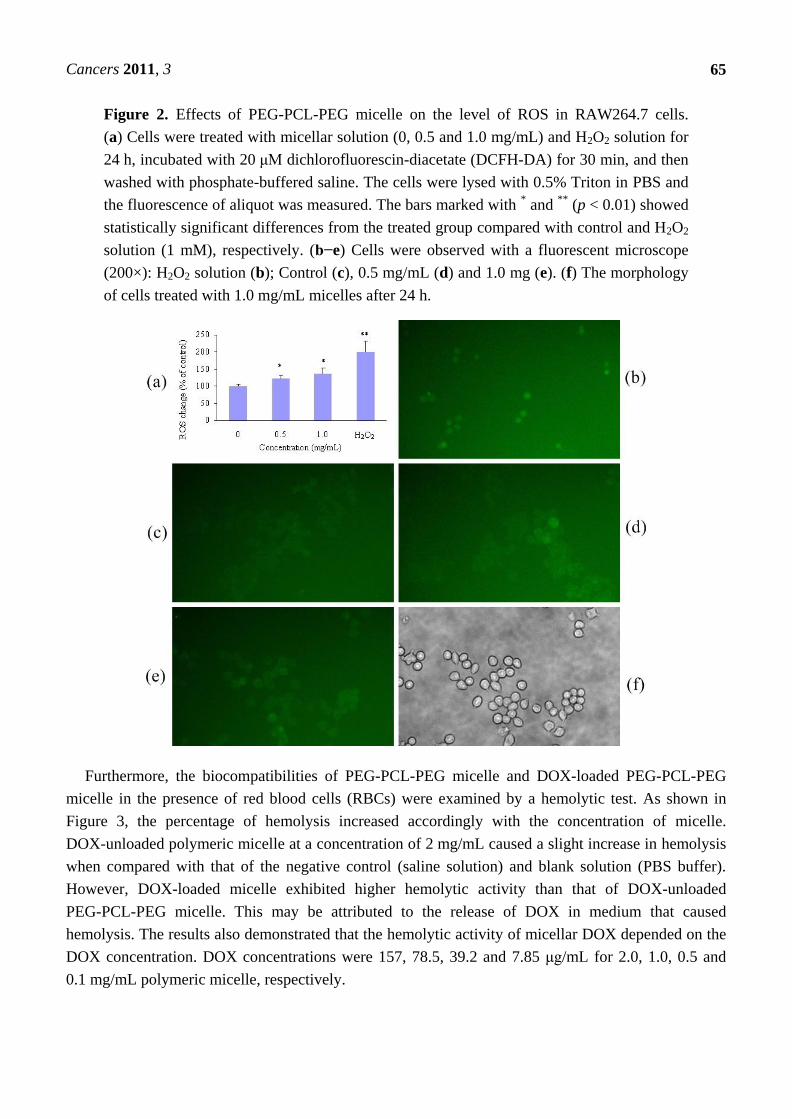
Nanoparticles were reported to induce toxicity by ROS-mediated oxidative stress [28]. Therefore,
ROS was measured to evaluate the level of oxidative stress in RAW264.7 cells treated with polymeric
micelles at 0.5 and 1.0 mg/mL. The fluorescence intensity of dichlorofluorescein (DCF), a permeative
indicator of oxidative stress, increased accordingly with increased concentration of polymeric micelle.
ROS generation at 1.0 mg/mL was elevated to approximately 130% of the control. However, cells
exposed to H2O2 solution (1 mM) showed approximately 200% ROS generation compared to the
control group. Figure 2 shows the representative figures of ROS formation visualized under a
fluorescent microscope.
Figure 1. Effects of PEG-PCL-PEG micelle and DOX-loaded PEG-PCL-PEG micelle on
the level of nitric oxide in RAW264.7 cells. Data represents the mean ± standard error of
the mean of four experiments (p < 0.01 is significantly different from the LPS).
Cancers 2011, 3
65
Figure 2. Effects of PEG-PCL-PEG micelle on the level of ROS in RAW264.7 cells.
(a) Cells were treated with micellar solution (0, 0.5 and 1.0 mg/mL) and H2O2 solution for
24 h, incubated with 20 μM dichlorofluorescin-diacetate (DCFH-DA) for 30 min, and then
washed with phosphate-buffered saline. The cells were lysed with 0.5% Triton in PBS and
the fluorescence of aliquot was measured. The bars marked with * and
** (p < 0.01) showed
statistically significant differences from the treated group compared with control and H2O2
solution (1 mM), respectively. (b−e) Cells were observed with a fluorescent microscope
(200×): H2O2 solution (b); Control (c), 0.5 mg/mL (d) and 1.0 mg (e). (f) The morphology
of cells treated with 1.0 mg/mL micelles after 24 h.
Furthermore, the biocompatibilities of PEG-PCL-PEG micelle and DOX-loaded PEG-PCL-PEG
micelle in the presence of red blood cells (RBCs) were examined by a hemolytic test. As shown in
Figure 3, the percentage of hemolysis increased accordingly with the concentration of micelle.
DOX-unloaded polymeric micelle at a concentration of 2 mg/mL caused a slight increase in hemolysis
when compared with that of the negative control (saline solution) and blank solution (PBS buffer).
However, DOX-loaded micelle exhibited higher hemolytic activity than that of DOX-unloaded
PEG-PCL-PEG micelle. This may be attributed to the release of DOX in medium that caused
hemolysis. The results also demonstrated that the hemolytic activity of micellar DOX depended on the
DOX concentration. DOX concentrations were 157, 78.5, 39.2 and 7.85 μg/mL for 2.0, 1.0, 0.5 and
0.1 mg/mL polymeric micelle, respectively.

Cancers 2011, 3
66
Figure 3. Hemolytic test on PEG-PCL-PEG micelle and DOX-loaded PEG-PCL-PEG
micelle. Data represents the mean ± standard error of the mean of three experiments
(p < 0.01 compared to saline group).
The cytotoxicity of PEG-PCL-PEG micelle to MCF-7 cells was evaluated using MTT assays.
Figure 4 demonstrated the cell viability after 24 h incubation with polymeric micelle of triblock
copolymer at various concentrations (0.001, 0.01, 0.1, 0.5 and 1.0 mg/mL). The results showed that
cell viability of MCF-7 cell decreased accordingly as the concentration of micelle increased. However,
the lowest cell viability approximately 89% was observed at a concentration of 1.0 mg/mL. The cells
viability assay indicates that the PEG-PCL-PEG micelle has generally low cytotoxicity to the MCF-7
cells with concentration up to 1.0 mg/mL. The data of macrophage response and hemolysis test suggest
that the PEG-PCL-PEG micelle prepared in this study had moderate in vitro toxicity and could be
safely used for intravenous injection in animal.
Figure 4. The cytotoxicity of PEG-PCL-PEG micelle against MCF-7 cells. Data represents
the mean ± standard error of the mean of four measurements (p > 0.4 compared to control).

Cancers 2011, 3
67
2.3. In Vitro Cytotoxicity of DOX-Loaded Micelle
The in vitro cytotoxic effect of DOX-loaded micelle was studied using a tetrazolium dye (MTT
assay) in MCF-7 cells. The cell viability was determined by incubating cells in 10 μg/mL DOX for
different periods of time. Figure 5 shows the cell viability of MCF-7 treated with DOX-loaded micelle.
The cell viability decreased significantly with increased time of treatment (2–96 h). The reason that
DOX-loaded micelle did not show cytotoxicity after a 2-h incubation could be attributed to the lag
phase of DOX. Similar results were obtained with 3-h incubation of DOX in other formulations [29,30].
However, significant cytotoxicity was observed at 24 h and 48 h. As shown in Figure 4, the cell
viability decreased from 98.7% at 2 h to 43.2% and 13.2% at 24 h and 48 h, respectively, compared to
control. Similarly, the cell viability was 7.7% and 6.6% for 72 h and 96 h, respectively. These results
indicate that the cell viability did not significantly decrease when the treatment time was prolonged,
which could be attributed to the sustained effect of DOX-loaded micelle and/or loss of cells. From the
above results, we confirmed that the optimal treatment time for cell viability assay of DOX-loaded
micelle was approximately 48 h, and prolonging the treatment time did not lead to more cell death.
Figure 5. Cytotoxicity of DOX-loaded micelle against MCF-7 cells. The cells were
incubated with DOX-loaded micelle (DOX concentration 10 µg/mL) for 2, 24, 48, 72 and
96 h at 37 °C. Each bar represents the mean of five measurements ± S.D. Bar marked with
* (p = 0.89) showed no significant difference between 2 h incubation and control. Bars
marked with ** (p < 0.001) showed significant difference between 2 h and 24 h and 48 h
incubation. *** p > 0.27 as compared to 48 h incubation.
2.4. Confocal Image of DOX-Loaded Micelle in MCF-7 Cells
Confocal microscopy was employed to visualize the cellular uptake and the internalization of
DOX-loaded micelle in MCF-7 cells. As shown in Figure 6, the distribution of DOX in cells was
different at 24 and 48 h. Red fluorescence of DOX was found to localize in the nuclei of MCF-7 cells,
showing a co-localization of nucleus (blue fluorescence of Hoechst) after 48 h. Additionally, the cell

Cancers 2011, 3
68
number was relatively low compared with that of 24-hour exposure to DOX-loaded micelle. This is
attributed to the cytotoxicity effect of DOX. Conversely, only weak fluorescence was observed in
nuclei of cells after 24 h exposure and most of DOX was accumulated outside nuclei of cells. The
signal observed in the nuclei was attributed to the release of DOX molecules from the micelles.
Additionally, red fluorescence was observed only outside the nuclei of cells after 2 h exposure [27].
Furthermore, when the MCF-7 cells were incubated with free DOX, fluorescence signals were
observed only in the nuclei of cells not in the cytoplasm at all time-points [27]. The observation of
fluorescence in cytoplasm indicated that the DOX-loaded micelle was internalized by the cells through
endocytosis and DOX was distributed in the cytoplasm after escaping from the endosome and/or the
lysosome [31,32].
Figure 6. Confocal laser microscopy (CLSM) of MCF-7 cells incubated with DOX-loaded
micelle for 24 h and 48 h with DOX concentration of 10 μg/mL (scale bar 50 μm).
2.5. Acute Toxicity of Micelle In Vivo
The PEG-PCL-PEG micelle was intravenously injected to ICR mice at a dose of 71.4 mg/kg for
multiple and single modes for evaluation of possible toxicities of PEG-PCL-PEG micelle. The body
weight of mice was not significantly different from control group (injected with PBS) in both single
and multiple doses after 14 days. Additionally, no mice died during the whole observation period.
Histological examination was performed to evaluate toxicity in kidneys and livers. As the results show
in Figure 7, no particular toxicity, no degeneration, necrosis, neutrophils or activation of

Cancers 2011, 3
69
immunoresponse were observed in liver and kidney when the mice were administered with single or
multiple doses. This demonstrated that prepared PEG-PCL-PEG micelle was safe in vivo for micellar
concentrations of less than 71.4 mg/kg.
Figure 7. In vivo acute toxicity of PEG-PCL-PEG micelle treated mice. Each organ was
evaluated by H&E staining after 14 days post-injection. (a), (d) Control; (b), (e) Single
dose; (c), (f) multiple doses (scale bar: 100 μm).
2.6. Biodistribution of DOX-Loaded Micelle
Biodistribution profile of free DOX and DOX-loaded micelles were examined in Wistar rat. The
animal was intravenously administered a dose of 5 mg/kg DOX equivalent. DOX content in plasma,
heart, liver, lung, kidney and spleen were measured at three intervals (1, 4 and 8 h). The results

Cancers 2011, 3
70
indicated that DOX-loaded micelle could prolong the DOX in plasma and exhibited higher DOX
concentration in plasma than free DOX (Figure 8). At the 1 and 4 h time-points, the DOX level of
DOX-loaded micelle in plasma was 2.3 and 3.6-fold higher than that of free DOX, respectively.
Interestingly, at 8 h time-point, the DOX level of free DOX was not detected in plasma, in contrast, it
still remained at high concentration in DOX-loaded micelle formulation. Biodistribution patterns to the
hearts, lungs, and kidneys did not show substantial accumulation in DOX-loaded micelle and free
DOX groups and were not significantly different. The uptake by liver was observed to be higher for
free DOX as compared to DOX-loaded micelle at 1 h post-injection.
Figure 8. Biodistribution of DOX-loaded micelle (a) and free DOX (b) after
administration at the equivalent 5 mg/kg DOX. Each bar represents the mean of three
measurements ± S.D. Bars marked with * (p < 0.05) are significantly different between 1 h
time-point and 4 and 8 h. Bars marked with ** and *** correspond to p values 0.073 and
0.065, respectively.

Cancers 2011, 3
71
2.7. Antitumor Activity of DOX-Loaded Micelle In Vivo
The antitumor efficacy of free DOX and DOX-loaded micelles was examined with MCF-7 human
breast tumor bearing nude mice. The tumor growth rates of mice treated with free DOX, DOX-loaded
micelle and PBS are presented in Figure 9. The free DOX and DOX-loaded micelle exhibited similar
effectiveness in preventing tumor growth when mice were administered with single dose (data not
shown). However, when the mice were treated with three injections, the DOX-loaded micelle
demonstrated the greater growth inhibition of tumor volume in comparison to free DOX. Tumor
volumes were decreased up to 60.0% by DOX-loaded micelle and 45.4% by free DOX, compared to
that of control group, respectively (Figure 9). Based on the above results, DOX-loaded micelle showed
higher tumor targeting efficiency and more therapeutic effects than free DOX. Hence, the hydrophobic
drug encapsulated in PEG-PCL-PEG micelle has advantages of prolonged blood circulation, RES
uptake prevention, and passive targeting of polymeric micelles to tumor tissue through EPR
effect [14,33].
Figure 9. Antitumor effect of free DOX and DOX-loaded micelle in MCF-7 tumor bearing
mice. Mice were administered PBS (●) and free DOX (■) and DOX-loaded micelle (▲) i.v
at the equivalent 5 mg/kg DOX.
3. Experimental Section
3.1. Materials
Monomethoxy poly(ethylene glycol) (mPEG, Mn = 5000), -caprolactone, doxorubicin hydrochloride
(DOX·HCl) and dimethylsulfoxide (DMSO) were purchased from Sigma-Aldrich (St.Louis, MO,
U.S.). Stannous 2-ethyl hexanoate (stannous octoate, Sn(Oct)2) was obtained from MP Biomedicals,
Inc. Methanol, tetrahydrofuran (THF) and acetonitrile (ACN) were HPCL grade and obtained from
ECHO chemical (Taiwan).
Human breast cancer cell lines (MCF-7) were kindly provided by Dr. Y.H. Chen of the School of
Pharmacy, College of Medicine, National Taiwan University. 3-(4,5-dimethylthiazol-2-yl)-2,5-diphenyl

Cancers 2011, 3
72
tetrazolium bromide (MTT) was purchased from Sigma-Aldrich (St. Louis, MO, U.S.). Dulbecco's
modified eagle's medium (DMEM) and antibiotic/antimycotic were purchased from GIBCO (NY,
U.S.). The fetal bovine serum (FBS) was obtained from HyClone (Ultah, U.S.).
3.2. Preparation and Characterizations of DOX-loaded PEG-PCL-PEG Micelle
The preparation of PEG-PCL-PEG micelle has been reported elsewhere [27]. The loading of DOX
in the micelle was done by first neutralizing 3.0 mg of DOX.HCl with 10 μL TEA in 2.0 mL THF and
stirred for 3 h. The resulting solution was added to 20 mg of PEG-PCL-PEG copolymer under stirring.
This solution was added to 2.0 mL of double distilled water under stirring for 3 h to form DOX-loaded
micelle. To remove un-trapped DOX and TEA, the mixture was next transferred for dialysis against
double distilled water for 24 h to produce DOX-loaded micelle (MWCO: 8,000 Da, Spectrum
Laboratories, U.S.).
The drug loading efficiency (DLE) was defined as the weight percentage of DOX in micelle relative
to the initial feeding amount of DOX. The drug loading content (DLC) was calculated from the mass
of incorporated DOX divided by the weight of polymer. The amount of DOX loaded in micelle was
determined by the absorption at 485 nm using UV-Vis spectrometry (UV-530, Jasco, Tokyo, Japan).
The DOX solutions of various concentrations were prepared, and the absorptions of the solutions were
measured to obtain a calibration curve [34,35]. The particle size was determined by dynamic light
scattering (DLS) at 25 °C using a Zetasizer 3000HSA (Malvern Instruments Ltd, U.K.) with an
excitation of 633 nm illuminated at a fixed angle of 90°.
3.3. Cytotoxicity of PEG-PCL-PEG Micelle
The in vitro cytotoxicity of micelle was tested against human breast cancer cell lines: MCF-7 by a cell
viability assay (MTT assay). MCF-7 cells were seeded in 96-well plate at a density of 5 × 10³ cells/well
and were incubated at 37 °C under a humidified atmosphere containing 5% CO2 for 24 h before assay.
After that, the cells were further incubated in media containing various concentrations of micelle. After
24 h, the medium was removed and washed with PBS. MTT solution was added to each well followed
by 4 h of incubation at 37 °C. Subsequently, the medium was removed and violet crystals were
solubilized with DMSO (200 μL). After shaking slowly twice for 5 s, the absorbance of each well was
determined using a Multiskan Spectrum spectrophotometer (Thermo Electron Corporation, Waltham,
MA, U.S.) at 570 nm and 630 nm. The cell viability (%) was calculated as the ratio of the number of
surviving cells in micelle-treated samples to that of control.
3.4. Measurement of Nitric Oxide of PEG-PCL-PEG Micelle
RAW 264.7 macrophage cells were seeded in a 96-well plate (1 × 10⁴ cells/well) and incubated in
37 °C, 5% CO2 for 1 day. Micellar solutions (PEG-PCL-PEG and DOX-loaded PEG-PCL-PEG
micelles) at various concentrations were added to the cells in a final volume of 0.2 mL. Accordingly,
polymer concentration range is from 0.001 to 1.0 mg/mL, DOX concentrations from the micelle
solutions were 78.5, 39.2, 7.85 and 0.000785 μg/mL, respectively. The supernatants were collected
after 24 h and NO production was determined by Greiss reagent (1% sulfanilamide, 2.5% H3PO4, 0.1%

Cancers 2011, 3
73
naphthylethylenediamine dihydrochloride). Briefly, 100 μL of culture medium was added to 100 μL of
Greiss reagent solution and incubated for 15 min. The absorbance was then measured at 540 nm. In the
control experiments, macrophages were incubated in a lipopolysaccharides (LPS) solution
(100 ng/mL) and a micelle-free medium. Moreover, total protein extract was determined by Micro
BCA Protein Assay.
3.5. Measurement of ROS in Macrophage Cells of PEG-PCL-PEG Micelle
A fluorometric assay using intracellular oxidation of 2,7-dichlorohydrofluorescein diacetate (DCFH-
DA) was performed [36]. Raw264.7 cells were seeded in 3.5 mm plates at a density of 3 × 10⁵ cells/plate
and were incubated at 37 °C under a humidified atmosphere containing 5% CO2 for 24 h before assay.
After that, the cells were further incubated in media containing various concentrations of micelle
(1.0 and 0.5 mg/mL). Medium and H2O2 solution (1 mM) were used as controls. After 24 h, the
medium was removed and washed with PBS. Then DCFH-DA solution (20 μM) was added to each
plate followed by 30 min of incubation at 37 °C. Subsequently, the medium was removed and
observed under fluorescent microscope (green filter with an excitation of 485 nm and an emission of
530 nm). After that cells were lysed with Triton 0.5% in PBS for 30 min, and aliquots were transferred
to the black 96-well plate. Then the fluorescence of dichlorofluorescein (DCF), which is the oxidized
product of DCFH-DA, was measured using the microplate spectrofluorometer. Data were expressed as
the percentage of the ROS level in the control group.
3.6. In Vitro Hemolytic Test of PEG-PCL-PEG Micelle
The experimental procedure described here is an adjustment of standard F-756-00 [37], which is
based on colorimetric detection of Drabkin’s solution. 0.7 mL of micellar solutions (PEG-PCL-PEG
and DOX-loaded PEG-PCL-PEG micelles) at various concentrations were incubated in 0.1 mL of
rabbit red blood cells at 37 °C and for 3 h. To make sure fresh rabbit blood was used in the test, the
hemoglobin in as-harvested plasma of rabbit blood was found to be less than 220 µg/mL, which is
regarded as basal level in the hemolysis test. Following incubation, the solution was centrifuged at
3800 rpm for 15 min. To determine the supernatant hemoglobin, 0.75 mL of Drabkin’s solution was
added to 0.25 mL of supernatant and the sample was allowed to stand for 15 min. The amount of
cyanmethemoglobin in the supernatant was measured by spectrophotometer (JASCO UV-530, Tokyo,
Japan) at a wavelength of 540 nm and then compared to a standard curve (hemoglobin concentrations
ranging from 0.003 to 1.2 mg/mL). The percent hemolysis refers to the hemoglobin concentration in
the supernatant of a blood sample not treated with micelles to the obtained percentage of micelle-
induced hemolysis. Additionally, the absorption of the micellar DOX was determined at
540 nm in order to eliminate the effect of absorption of DOX. Finally, saline solution and double
distilled water were used as negative and positive control, respectively.
3.7. In Vitro Cytotoxicity of DOX-Loaded Micelle
The in vitro cytotoxicity of DOX-loaded micelle was tested against human breast cancer cell lines:
MCF-7. The cell culture medium was composed of DMEM with 10% fetal bovine serum and

Cancers 2011, 3
74
antibiotic/antimycotic. The cell viability was determined by tetrazolium dye (MTT) assay. MCF-7
cells were seeded in 96-well plates at a density of 5 × 10³ cells/well and were incubated at 37 °C in a
humidified atmosphere containing 5% CO2 for 24 h before assay. After that, the cells were further
incubated in media containing DOX-loaded micelle (DOX concentration 10 μg/mL). The cytotoxic
effect was determined at 2, 24, 48, 72 and 96 h. For the time-points of 72 and 96 h, after 48 h
incubation, the medium containing micellar DOX was removed and culture plated was rinsed with
PBS. After that, medium-free micellar DOX was added and incubated. A solution of media with
unloaded micelle (placebo) was used as a control to be compared with results obtained from micellar
DOX. After interval time, the medium was removed and washed with PBS. MTT solution was added
to each well followed by 4 h of incubation at 37 °C. Subsequently, the medium was removed and
violet crystals were solubilized with DMSO (200 μL). After mild shaking twice for 5 s, the absorbance
of each well was determined using a Multiskan Spectrum spectrophotometer (Thermo Electron
Corporation, Waltham, MA, U.S.) at 570 nm and 630 nm. The cell viability (%) was calculated as the
ratio of the number of surviving cells in drug-treated samples to that of control.
3.8. Cellular Uptake of DOX-Loaded Micelle
The cells (MCF-7) were seeded in BD Falcon culture slides (1 × 10⁵ cells/chamber) and incubated
for 24 h. The cells were incubated with DOX-loaded micelle (DOX concentration 10 μg/mL). After 2,
24 and 48 h, the medium was removed. Hoechst 33342 (2 μg/mL) was added and incubated for
30 min. The cells were washed with cold PBS twice, and then fixed with formalin solution for 30 min,
the formalin was removed and PBS was added. The fluorescence images of cells were obtained using a
confocal laser scanning microscope (FluoView FV300, Olympus).
3.9. In Vivo Acute Toxicity of PEG-PCL-PEG Micelle
Nine female ICR mice with average body weight of 25–30 g were used in this study (Laboratory
Animals, National Taiwan University, Taiwan). The mice were housed in the animal facility under
controlled environment settings (25 °C, 60% humidity). All in-vivo experiments were carried out in
accordance with the ethical guidelines for Animal Care and Use Committee of Chung Yuan Christian
University. For acute toxicity test, mice (n = 3) were anesthetized with a cocktail of Zoletil
(0.01 mL/kg) and Xylazine (0.01 mL/kg) solutions and then micellar solution was intravenously
administrated at a dose of 71.4 mg/kg. PBS was used as control. For multiple dosing tests, 71.4 mg/kg
micellar solution was injected on day 0, 3 and 6. The body weight of the mice was then monitored for
a test period of 14 days post-injection. On day 14, the animals were sacrificed and livers and kidneys
were collected. Histological examinations of the organs were carried out after dissection, then
abnormal fibrotic tissue and loosen morphology were counted as adverse reaction due to the injection
of micelle observed under a microscope (Eclipse 50i, Nikon, Japan).
3.10. Biodistribution of DOX-Loaded PEG-PCL-PEG Micelle
The in vivo biodistribution of DOX-loaded micelle was measured using female Wistar rats weighing
250–300 g. 18 rats were randomly divided into 2 groups as follows: Group A with intravenous (i.v.)

Cancers 2011, 3
75
injection of the pristine DOX solution and Group B with i.v. injection of DOX-loaded micelle. Three
rats were injected i.v. with DOX or DOX-loaded micelle at the equivalent DOX 5 mg/kg dose at 1, 4
and 8 h time point. At the end of each time point, animals were sacrificed and blood was collected.
Heart, liver, lung, kidney and spleen were harvested and homogenized in 20 mM KH2PO. After
centrifugation, the supernatant was collected and kept at −20 °C until analyzed. For HPLC analysis,
0.4 mL of supernatant was mixed with 0.4 ml CH3CN, vortexed for 30 s and centrifuged at 15000× g
for 10 min. The supernatant was collected and the solvent was removed at 40 degree under a stream of
N2. The remaining sample was mixed with 0.25 mL of HPLC mobile phase solution [methanol: 0.1 %
acetic acid and 0.1% ammonium hydroxide (25%), pH = 4.0; ratio: 60:40], filtered and transferred to
auto sampler vials containing limited-volume inserts (150 μL) [38]. A C-18 column was used and the
mobile phase was delivered at a rate of 1 mL/min. Sample (50 μL) was injected and the column
effluent was detected with a UV detector (λex = 485 nm, λemx = 580 nm). Several concentrations of
DOX in plasma were used to create the standard curve (concentration: 25 ng/mL–1,000 ng/mL).
3.11. Antitumor Activity of DOX-loaded PEG-PCL-PEG Micelle
Three 5 week-old female BALB/c mice (National Laboratory Animal Center, Taiwan) were
randomly assigned each of the following groups: Group A: control, group B: single administration of
free DOX, group C: multiple administrations of free DOX, group D: single administration of
DOX-loaded micelle, and group E: multiple administrations of DOX-loaded micelle. After quarantine
in Animal Laboratory at CYCU, bilateral oophorectomy was performed. Estradiol (50 μg/mouse) was
injected one week after oophorectomy. MCF-7 cells were trypsinized and resuspended in PBS. 5 × 10⁶
cells/0.1 mL were inoculated into the mammary fat pad of mice using a 27-gauge syringe. When the
tumor volume reached approximately 200 mm3, the mice were injected intravenously via the tail vein
at a dose of 5mg/kg (DOX equivalent) at day 0, 3 and 6 with free DOX, DOX-loaded micelle and PBS
solution, respectively. The tumor inhibition activity was assessed by the tumor volume (TV), which
was calculated as TV = (width2 × length) × ½. The dimension of the tumor measured by a caliper and
the total body weight was also measured simultaneously.
4. Conclusions
In this study, the biocompatible and non-toxic triblock copolymer micelle was prepared in a self
assembly method. In vitro NO release from macrophages and hemolytic tests confirmed that the
PEG-PCL-PEG micelle induced very minor NO and hemolysis. The in vitro cytotoxicity study
demonstrated that the micelle was safe and low cytotoxic. The cellular uptake of DOX-loaded micelle
in MCF-7 was different from that of free DOX and the optimal time for MTT assay was 48 h.
Furthermore, mice treated with prepared micelle in multiple doses did not develop toxic effects or die
during the entire period of acute toxicity test. In vivo results showed that multiple injections of
DOX-loaded micelle could prolong the circulation time and increase the therapeutic efficacy of DOX.

Cancers 2011, 3
76
Acknowledgements
The authors would like to thank National Science Council, Republic of China, for financial support
under grant numbered 98-2221-E-033-072.
References
1. Rivera, E.; Valero, V.; Arun, B.; Royce, M.; Adinin, R.; Hoelzer, K.; Walters, R.; Wade, J.L., III;
Pusztai, L.; Hortobagyi, G.N. Phase ii study of pegylated liposomal doxorubicin in combination
with gemcitabine in patients with metastatic breast cancer. J. Clin. Oncol. 2003, 21, 3249-3254.
2. Shah, J.J.; Orlowski, R.Z.; Thomas, S.K. Role of combination bortezomib and pegylated
liposomal doxorubicin in the management of relapsed and/or refractory multiple myeloma.
Therapeut. Clin. Risk Manag. 2009, 5, 151-159.
3. Swenson, C.E.; Bolcsak, L.E.; Batist, G.; Guthrie, T.H.J.; Tkaczuk, K.H.; Boxenbaum, H.;
Welles, L.; Chow, S.C.; Bhamra, R.; Chaikin, P. Pharmacokinetics of doxorubicin administered
i.v. As myocet (tlc d-99; liposome-encapsulated doxorubicin citrate) compared with conventional
doxorubicin when given in combination with cyclophosphamide in patients with metastatic breast
cancer. Anti-Cancer Drugs 2003, 14, 239-246.
4. Cuong, N.V.; Hsieh, M.F. Recent advances in pharmacokinetics of polymeric excipients used in
nanosized anti-cancer drugs. Curr. Drug Metab. 2009, 10, 842-850.
5. Wong, H.L.; Bendayan, R.; Rauth, A.M.; Xue, H.Y.; Babakhanian, K.; Wu, X.Y. A mechanistic
study of enhanced doxorubicin uptake and retention in multidrug resistant breast cancer cells
using a polymer-lipid hybrid nanoparticle system. J. Pharmacol. Exp. Ther. 2006, 317,
1372-1381.
6. Xiong, X.-B.; Ma, Z.; Lai, R.; Lavasanifar, A. The therapeutic response to multifunctional
polymeric nano-conjugates in the targeted cellular and subcellular delivery of doxorubicin.
Biomaterials 2010, 31, 757-768.
7. Marchi, N.; Hallene, K.; Kight, K.; Cucullo, L.; Moddel, G.; Bingaman, W.; Dini, G.; Vezzani,
A.; Janigro, D. Significance of mdr1 and multiple drug resistance in refractory human epileptic
brain. BMC Medicine 2004, 2, 37.
8. Zhang, Z.; Huey Lee, S.; Feng, S.S. Folate-decorated poly(lactide-co-glycolide)-vitamin e tpgs
nanoparticles for targeted drug delivery. Biomaterials 2007, 28, 1889-1899.
9. Sharma, A.K.; Zhang, L.; Li, S.; Kelly, D.L.; Alakhov, V.Y.; Batrakova, E.V.; Kabanov, A.V.
Prevention of mdr development in leukemia cells by micelle-forming polymeric surfactant.
J. Contr. Release 2008, 131, 220-227.
10. Kim, D.; Lee, E.S.; Oh, K.T.; Gao, Z.G.; Bae, Y.H. Doxorubicin-loaded polymeric micelle
overcomes multidrug resistance of cancer by double-targeting folate receptor and early endosomal
ph. Small 2008, 4, 2043-2050.
11. Jabr-Milane, L.S.; van Vlerken, L.E.; Yadav, S.; Amiji, M.M. Multi-functional nanocarriers to
overcome tumor drug resistance. Cancer Treat. Rev. 2008, 34, 592-602.
12. Blanco, E.; Kessinger, C.W.; Sumer, B.D.; Gao, J. Multifunctional micellar nanomedicine for
cancer therapy. Exp. Biol. Med. 2009, 234, 123-131.

Cancers 2011, 3
77
13. Torchilin, V.P. Multifunctional nanocarriers. Adv. Drug Deliv. Rev. 2006, 58, 1532-1555.
14. Maeda, H.; Wu, J.; Sawa, T.; Matsumura, Y.; Hori, K. Tumor vascular permeability and the epr
effect in macromolecular therapeutics: A review. J. Contr. Release 2000, 65, 271-284.
15. Arun, K.I.; Khaled, G.; Fang, J.; Maeda, H. Exploiting the enhanced permeability and retention
effect for tumor targeting. Drug Discov. Today 2006, 11, 812-818.
16. Cao, N.; Feng, S.S. Doxorubicin conjugated to d--tocopheryl polyethylene glycol 1000 succinate
(tpgs): Conjugation chemistry, characterization, in vitro and in vivo evaluation. Biomaterials
2008, 29, 3856-3865.
17. Sinha, V.R.; Bansal, K.; Kaushik, R.; Kumria, R.; Trehan, A. Poly--caprolactone microspheres
and nanospheres: An overview. Int. J. Pharm. 2004, 278, 1-23.
18. Otsuka, H.; Nagasaki, Y.; Kataoka, K. Pegylated nanoparticles for biological and pharmaceutical
applications. Adv. Drug Deliv. Rev. 2003, 55, 403-419.
19. Shuai, X.; Merdan, T.; Unger, F.; Wittmar, M.; Kissel, T. Novel biodegradable ternary
copolymers hy-pei-g-pcl-b-peg: Synthesis, characterization, and potential as efficient nonviral
gene delivery vectors. Macromolecules 2003, 36, 5751-5759.
20. Zahr, A.S.; Davis, C.A.; Pishko, M.V. Macrophage uptake of core−shell nanoparticles surface
modified with poly(ethylene glycol). Langmuir 2006, 22, 8178-8185.
21. Zhang, Y.; Zhuo, R.X. Synthesis and in vitro drug release behavior of amphiphilic triblock
copolymer nanoparticles based on poly (ethylene glycol) and polycaprolactone. Biomaterials
2005, 26, 6736-6742.
22. Gong, C.; We, X.; Wang, X.; Wang, Y.; Guo, G.; Mao, Y.; Luo, F.; Qian, Z. Biodegradable self-
assembled peg-pcl-peg micelles for hydrophobic honokiol delivery: I. Preparation and
characterization. Nanotechnology 2010, 21, 215103.
23. Xu, B.; Yuan, J.; Ding, T.; Gao, Q. Amphiphilic biodegradable poly(ε-caprolactone)-
poly(ethylene glycol)-poly(ε-caprolactone) triblock copolymers: Synthesis, characterization and
their use as drug carriers for folic acid. Polym. Bull. 2010, 64, 537-551.
24. Ge, H.; Hu, Y.; Jiang, X.; Cheng, D.; Yuan, Y.; Bi, H.; Yang, C. Preparation, characterization,
and drug release behaviors of drug nimodipine-loaded poly(caprolactone)-poly(ethylene oxide)-
poly(caprolactone) amphiphilic triblock copolymer micelles. J. Pharm. Sci. 2002, 91,
1463-1473.
25. Gou, M.; Zheng, X.; Men, K.; Zhang, J.; Zheng, L.; Wang, X.; Luo, F.; Zhao, Y.; Zhao, X.; Wei,
Y.; Qian, Z. Poly(-caprolactone)/poly(ethylene glycol)/poly(-caprolactone) nanoparticles:
Preparation, characterization, and application in doxorubicin delivery. J. Phys. Chem. B 2009,
113, 12928-12933.
26. Cuong, N.V.; Hsieh, M.F.; Chen, Y.T.; Liau, I. Doxorubicin-loaded nanosized micelles of a star-
shaped poly(-caprolactone)-polyphosphoester block co-polymer for treatment of human breast
cancer. J. Biomater. Sci. Polym. Ed. 2010, doi: 10.1163/092050610X510533.
27. Cuong, N.V.; Hsieh, M.F.; Chen, Y.T.; Liau, I. Synthesis and characterization of peg-pcl-peg
triblock copolymers as carriers of doxorubicin for the treatment of breast cancer. J. Appl. Polym.
Sci. 2010, 117, 3694-3703.
28. Eom, H.J.; Choi, J. Oxidative stress of silica nanoparticles in human bronchial epithelial cell,
beas-2b. Toxicol. Vitro 2009, 23, 1326-1332.

Cancers 2011, 3
78
29. Eliaz, R.E.; Nir, S.; Marty, C.; Szoka, F.C. Determination and modeling of kinetics of cancer cell
killing by doxorubicin and doxorubicin encapsulated in targeted liposomes. Cancer Res. 2004, 64,
711-718.
30. Upadhyay, K.K.; Bhatt, A.N.; Mishra, A.K.; Dwarakanath, B.S.; Jain, S.; Schatz, C.; Le Meins, J.-
F.; Farooque, A.; Chandraiah, G.; Jain, A.K.; Misra, A.; Lecommandoux, S. The intracellular drug
delivery and anti tumor activity of doxorubicin loaded poly(-benzyl l-glutamate)-b-hyaluronan
polymersomes. Biomaterials 2010, 31, 2882-2892.
31. Liu, S.Q.; Wiradharma, N.; Gao, S.J.; Tong, Y.W.; Yang, Y.Y. Bio-functional micelles self-
assembled from a folate-conjugated block copolymer for targeted intracellular delivery of
anticancer drugs. Biomaterials 2007, 28, 1423-1433.
32. Zhao, H.; Yung, L.Y.L. Selectivity of folate conjugated polymer micelles against different tumor
cells. Int. J. Pharm. 2008, 349, 256-268.
33. Nie, S. Understanding and overcoming major barriers in cancer nanomedicine. Nanomedicine
2010, 5, 523-528.
34. Hsieh, M.F.; Cuong, N.V.; Chen, C.H.; Chen, Y.T.; Yeh, J.M. Nano-sized micelles of block
copolymers of methoxy poly(ethylene glycol)-poly(-caprolactone)-graft-2-hydroxyethyl
cellulose for doxorubicin delivery. J. Nanosci. Nanotechnol. 2008, 8, 2362-2368.
35. Aliabadi, H.M.; Mahmud, A.; Sharifabadi, A.D.; Lavasanifar, A. Micelles of methoxy
poly(ethylene oxide)-b-poly(-caprolactone) as vehicles for the solubilization and controlled
delivery of cyclosporine a. J. Contr. Release 2005, 104, 301-311.
36. Hsieh, M.F.; Lin, T.Y.; Gau, R.J.; Chang, H.T.; Lo, Y.L.; Lai, C.H. Biodegradable polymeric
nanoparticles bearing stealth peg shell and lipophilic polyester core. J. Chin. Inst. Chem. Engrs.
2005, 36, 609-615.
37. ASTM F756-00. Standard Practice for Assessment of Hemolytic Properties of Materials; ASTM
International: West Conshohocken, PA, USA, 2000; doi: 10.1520/F0756-00.
38. Wei, G.; Xiao, S.; Si, D.; Liu, C. Improved hplc method for doxorubicin quantification in rat
plasma to study the pharmacokinetics of micelle-encapsulated and liposome-encapsulated
doxorubicin formulations. Biomed. Chromatogr. 2008, 22, 1252-1258.
© 2010 by the authors; licensee MDPI, Basel, Switzerland. This article is an open access article
distributed under the terms and conditions of the Creative Commons Attribution license
(http://creativecommons.org/licenses/by/3.0/).