Embed Size (px)
Citation preview

DOSTĘPNOŚĆ INNOWACYJNYCH METOD
LECZENIA NEUROCHIRURGICZNEGO W POLSCE
Tomasz Trojanowski
Katedra i Klinika Neurochirurgii i Neurochirurgii Dziecięcej
Uniwersytet Medyczny w Lublinie
"Innowacyjne wyroby medyczne - ocena dostępności w Polsce” - Warszawa,24.11. 2011

Innowacja innovare – tworzenie nowego
Produktowa
znaczace ulepszenia parametrów technicznych oraz funkcjonalnosci
Procesowa (technologiczna)
Organizacyjna
Marketingowa
zmiany w wygladzie produktu, opakowaniu, promocji, polityce cenowej
“zastosowanie nowej wiedzy w procesie produkcji” (D.Begg, 1997)


Neurochirurgia

Dostępność procedur
neurochirurgicznych w Polsce nie tylko neurochirurgicznych
nie tylko w Polsce
podstawowych innowacyjnych
= nowych

Dostępność procedur
podstawowych / nowych
• liczba (kontrakt)
• czas dostępu (kolejka)
• rodzaj procedury (embolizacja/klipsowanie)
• jakość (sprzęt, kadra, materiały)

Dostępność procedur
rozproszenie
Procedury Podstawowe Nowe
Zasoby osobowe
wystarczające wystarczające
Zasoby materialne
ograniczone ?

Dostępność sprzętu
Mikroskop, neuronawigacja, CUSA,
obrazowanie śródoperacyjne, monitorowanie
Endoskopia
Narzędzia chirurgiczne
Wszczepy
(zastawki, stymulatory, pompy infuzyjne, protezy elektroniczne,
materiały hemostatyczne, protezy kosci, opon, naczyń)

Dostępność
Wszczepy
zastawki
stymulatory
wszczepialne pompy infuzyjne
protezy elektroniczne
materiały hemostatyczne
protezy i substytuty kosci
oponowe
naczyń

Neurochirurgia małoinwazyjna innowacyjność
głowa
kręgosłup
dostępność

Nowotwory, guzy
Badania
obrazowe (czynnościowe, spektroskopia, traktografia, DSA)
Operacja
– neuronawigacja
– ssak ultradźwiękowy, mikroskop, histopatologia
– obrazowanie śródoperacyjne (MR, TK, fluorescencja)
– opieka pooperacyjna


ALA (kwas 5 amino-lewulinowy)

Roboty

Tętniaki - leczenie
operacyjne / embolizacja
jak najwcześniej / kto

Radiochirurgia
Nóż gamma
Ciężkie jony (protony)
Przyspieszacz liniowy
Nóz cybernetyczny (cyber knive)

1949 - Radiochirurgia
Karolinska Institute
1968 - Gamma Knife

Kręgosłup

Innowacje w medycynie
Etyka
Dążenie do sukcesu
Konkurencja
(indywidualna, instytucjonalna, komercyjna)
Naciski (przemysł, srodki przekazu, organizacje)
Dla dobra chorego

Proton beam therapy
and the medical arms race
by PAUL LEVY in PHYSICIAN
hadrony
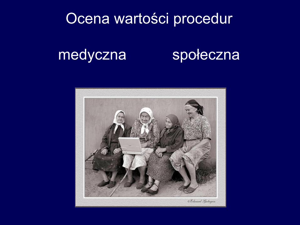
Ocena wartości procedur
medyczna społeczna

Then & Now
**Star Wars Technology in Neurosurgery** April 21, 2004
A thermal imaging camera,
one of Dr. Levy’s most important tools in the OR
What do the operating room at Children’s and Pasadena’s Jet Propulsion
Laboratory (JPL) have in common’ The thermal imaging camera, developed
for the Defense Department at JPL, is one of the most important tools
Dr. Michael Levy uses in Children’s operating rooms.
The milk carton-sized device gives images in a range of colors that
indicate the temperature of the area being viewed. The camera is used
when correcting a brain condition called arteriovenous malformation
(AVM). Areas where these abnormal vessels occur have an increased blood
supply, and are warmer. The color image helps locate the malformation.
Dr. Levy can tell when he has surgically corrected it, because the area
cools down when its blood flow is eliminated.
Thanks to Dr. Levy’s innovative use of technology during
surgery, these kinds of results are light years ahead of
what was happening at Children’s only a few years ago.
/Linda A. Canada/
Media Room
Search
Articles and Stories
Then & Now
StarWars Technology in Neurosurgery April 21, 2004
What do the operating room at Children’s
and Pasadena’s Jet Propulsion Laboratory
(JPL) have in common? The thermal
imaging camera, developed for the Defense
Department at JPL, is one of the most
important tools Dr. Michael Levy uses in
Children’s operating rooms.
The milk carton-sized device gives images
in a range of colors that indicate the
temperature of the area being viewed. The
camera is used when correcting a brain
condition called arteriovenous
malformation (AVM). Areas where these abnormal vessels occur have an
increased blood supply, and are warmer. The color image helps locate the
malformation. Dr. Levy can tell when he has surgically corrected it, because
the area cools down when its blood flow is eliminated.
Thermal imaging was particularly
helpful in the case of Jacquelyn
Riley, a 10 year old operated on
by Dr. Levy just a month ago.
Jackie was in a coma due to the
rupture of an AVM in her brain
stem. In an hour and a half
surgery, Dr. Levy was able to fix
the problem, and Jackie’s
recovery is going well. AVMs
commonly occur within families.
Jackie’s cousin, Cooper, had 13
AVMs and underwent surgery at
Children’s on March 1st.
If Jackie and Cooper had come to Children’s Hospital in the early 1970s,
diagnosing their conditions would have been much more difficult. Modern
brain imaging techniques like MRI and CT scans had not been perfected. Even
20 years ago, surgeons here might have considered their conditions
A thermal imaging camera, one of
Dr. Levy’s most important tools in
the OR
Jackie and her cousin Cooper share more than love:
They were both successfully operated on by Dr.
Levy for the same brain condition.

- Neuroscience Institute Uses RGB Spectrum's
SuperView Video Processor in
Pioneering Neurosurgery Technology
"The plasma screen/SuperView system
allows all data to be easily viewed,
even from across the room without
having to look around.”
Wallace-Kettering
Neuroscience Institute Uses RGB Spectrum's
SuperView� Video
Processor in Pioneering
Neurosurgery Technology
SuperView Used in Neurosurgery Operating Room to Display Critical Neuro-Navigation and Medical Visuals
Surgeons at the Wallace-Kettering Neuroscience Institute (WKNI) in Dayton, Ohio, have been using computer-guided surgery since 1994. Using complex computer technology to fuse anatomical scans (MRI/CT) with biochemical scans
(PET - Positron Emission Tomography), this computer guidance allows the neurosurgeons to target lesions in the brain with safer, more accurate procedures. This neuronavigation technology works much like a GPS (Global Positioning System), to enable neurosurgeons to map and navigate through each patient's brain.
Surgeons at the Wallace-Kettering Neuroscience Institute in Dayton, Ohio use computer-guided surgery to target lesions in the brain with safer, more accurate procedures. This neuronavigation
technology uses a SuperView� multi-input display
processor to display multiple signals on a single screen in the operating room. (Photos courtesy of Wallace-Kettering Neuroscience Institute.)
RGB Spectrum's SuperView� processor feeds
signals to this 42 inch NEC plasma screen in the

Przedwczesne publikacje w środkach
masowego przekazu F. Loew, Acta Neurochir 1992,116:187

Health Care's Six Money-Wasting Problems by Parija B. Kavilanz
Monday, August 10, 2009

A fool
with a tool
is still a fool
Lars Leksell

Postęp w medycynie

Innowacja
Metoda dostępna
Stosowana niezgodnie z EBM / wskazaniami
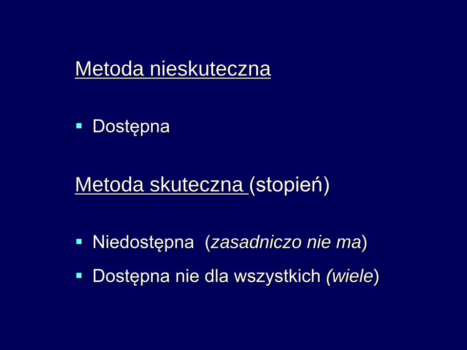
Metoda nieskuteczna
Dostępna
Metoda skuteczna (stopień)
Niedostępna (zasadniczo nie ma)
Dostępna nie dla wszystkich (wiele)