Embed Size (px)
Citation preview

Angela Karambatsakidou, Enheten för Röntgen och IJ-Strålning, VO Sjukhusfysik
Philips image gallery Siemens image gallery
Dosimetry in angiography and interventional radiology

Angiography and Interventional radiology, 2014
General information about interventional radiology
Dosimetry in interventional radiology
Special topic: Paediatric interventional cardiology
Agenda
Interventional
Radiology
CT
Radiography
Angiography and Interventional radiology, 2014
IAEA Training Material on Radiation Protection in
Diagnostic and Interventional Radiology
Dose levels

Angiography and Interventional radiology, 2014
Pioneer in interventional cardiology: Andreas Gruntzig, Zurich
2000st lyckade ballongdilatationer
Gick bort 5 mån innan introduktionen av det 1:a coronara stentet
Interventional era started in 1977
37-årig man

Angiography and Interventional radiology, 2014
Angiography and Interventional radiology

Angiography and Interventional radiology, 2014
Philips image gallery
Angiography and Interventional radiology

Angiography and Interventional radiology, 2014
DSA quantification (Siemens image gallery)
Stenosis of the right carotis communis.
3-D RA quantification (Siemens image gallery)
Basilar aneurysm of the top end of the basilar artery.
DynaCT (Siemens image gallery) AVM
embolization 3D roadmap (Philips image gallery)
Real-time image guidance for catheter navigation.
Different interventional techniques with clinical images

Angiography and Interventional radiology, 2014
Interventional cardiology
PTCA
Case: bifurcation lesion
AP, 38 CR
LAD-D1
LAO 50, 38 CR
D1
IAEA
Different interventional techniques with clinical images

Angiography and Interventional radiology, 2014
Case 1 bifurcation lesion
A
stent and balloon inflation
B
balloon angioplasty (PTCA)technique
PTCA & stentingtechnique
IAEA
Different interventional techniques with clinical images

CANCER
STOCHASTIC EFFECTS
DETERMINISTIC EFFECTS
LENS INJURIES
SKIN INJURIES
HEREDITARY DISORDERS IN THE DESCENDANTS
IAEA Training Material on Radiation Protection in
Diagnostic and Interventional Radiology
Radiation effects on humans
Angiography and Interventional radiology, 2014

Risks in angiography and interventional procedures
Erythema
(>2 Gy, 1-10 d)
Depilation
(>3 Gy, 3 wk)
Dry desquamation
(>14 Gy, 4 wk)
Moist desquamation
(>18 Gy, 4 wk) Dermal necrosis
(>12 Gy?, >1 yr)
Cataract
(>1-2 Gy, >5 år) 0.5 Gy

Angiography and Interventional radiology, 2014
Symptoms usually delayed by 1-3 weeks Patients often unaware of symptoms of injury associated
with radiation delivery Experience show a lack of association between lesion and
previous cardiologic procedure
IAEA: Radiation Protection in Cardiology, Lecture 1
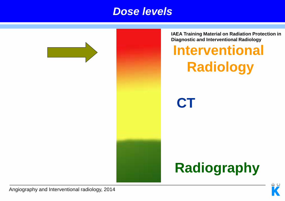
Experiences with skin injures in interventional cardiology

Angiography and Interventional radiology, 2014
Electrophysiological and ablation procedure
8 days after the third procedure
3 procedures in 4 months, each with more than 100 minutes of fluoroscopy
Vlietstra RE et al. J Interv Cardiol. 2004 Jun;17(3):131-42
Experiences with skin injures in interventional cardiology
Prolonged-, Multiple Procedures and Beam-overlapping

Angiography and Interventional radiology, 2014
Schueler et al. Radiographics 2006;26:1533-41
Large patients
Experiences with skin injures in interventional cardiology
Angiography and Interventional radiology, 2014
”Dose rate” = Incident air kerma rate
22 cm 30 cm
(>2 Gy, 1-10 d)
Experiences with skin injures in interventional cardiology
Steep beam angulations (arms and/or breast are in the radiation beam)

Angiography and Interventional radiology, 2014
0 0,5 1 1,5 2 2,5
Low
Normal
High
Cine
Rel. Dose
Do
se
le
ve
l
0 0,5 1 1,5 2
14
17
23
Rel. Dose
FO
V (
cm
)
The Essential Physics of Medical Imaging, figure 9-8
Experiences with skin injures in interventional cardiology

Angiography and Interventional radiology, 2014
0 mm Cu 0,5 mm Cu
Schueler et al. Radiographics 2006;26:1533-41
Experiences with skin injures in interventional cardiology
Tube filtering

Angiography and Interventional radiology, 2014
Experiences with skin injures in interventional cardiology
Collimation
Date of download:
2/7/2014
Copyright © The American College of Cardiology.
All rights reserved.
Cine Fluoroscopic Images in the Lateral Projection in a 6.5-Year-Old
Female Child With a PDA
(A) Angiogram in the descending aorta demonstrating a 2-mm patent
ductus arteriosus (PDA) at its narrowest diameter (arrow),
angiographic type A.
J Am Coll Cardiol Intv. 2008;1(6):603-611. doi:10.1016/j.jcin.2008.07.007
Collimation with 1 cm
REID ~25%
Skin Dose ~ Same REID

Operator experience
Angiography and Interventional radiology, 2014
Experiences with skin injures in interventional cardiology
3
4
5
6
mS
v/G
ycm
2
median: 4.1
median: 3.4
Operator A Operator B
Coronary angiography (adults)
Karambatsakidou et al. Br J Radiol. 2005 Sep;78(933):803-9

Angiography and Interventional radiology, 2014
Geijer et al Eur. Radiol. 12 (2002)
Calculated
effective
dose rate
KAP rate
Operator
dose rate
Combined Actions
Relative
dose rate
Dose Reduction
+ angulation Start + filtration +collimation
LAO caud PA 2 cm
Angiography and Interventional radiology, 2014
PTW
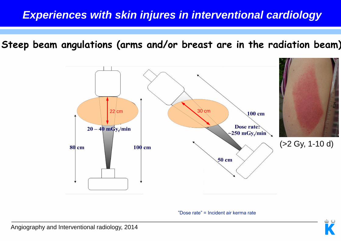
Dosimetric parameter for risk estimation
Kerma-Area Product (KAP)

Angiography and Interventional radiology, 2014
May over- or underestimate skin dose depending on:
how well IRP coincides with the skin surface
backscattered factor (BSF)
radiation geometry
Dosimetric parameter for risk estimation
Reference dose

Angiography and Interventional radiology, 2014
•Image acquisition
•Fluorography
•Exposure
•Cineradiography
•Stationary acquisition
Fluoroscopy
Dose Report

Angiography and Interventional radiology, 2014
Fluoroscopy Time
Chida Koichi, AJR 2006; 186:774–778
Graph shows correlation between maximum skin dose and fluoroscopic time in percutaneous coronary intervention (r = 0.628, p < 0.0001).
Dose Report
Measures Time, Not Dose!
Correlates Poorly With Maximum Skin Dose!

Angiography and Interventional radiology, 2014
Kerma-Area Product (KAP)
Miller, D.L, J Vasc Interv Radiol 2003;14:977-990
Figure Graph shows correlation between maximum skin dose (MESD) and kerma-area product (KAP) for 709 procedures (r = 0.848, p < 0.000001).
Dose Report
Good Correlation With Maximum Skin Dose
Tool For Cancer Risk And
Skin Dose Estimations
MESD (Gy)
KAP (Gycm2)

Angiography and Interventional radiology, 2014
Reference Dose
Figure Graph shows correlation between maximum skin dose (MESD) and reference dose for 709 procedures (r = 0.862, p < 0.000001).
Miller, D.L, J Vasc Interv Radiol 2003;14:977-990
Dose Report
Good Correlation With Maximum Skin Dose
Poor Estimate Of Stochastic Risk
MESD (Gy)
Reference Dose (Gy)
Angiography and Interventional radiology, 2014
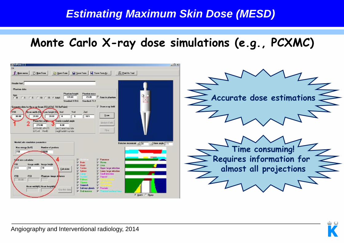
Estimating Maximum Skin Dose (MESD)
Monte Carlo X-ray dose simulations (e.g., PCXMC)
Accurate dose estimations
Time consuming! Requires information for
almost all projections

28
Estimating Maximum Skin Dose (MESD)
Based on reference dose. Included in dose reports
Time consuming! Requires information for
almost all projections Omar A. et al.,SPIE MI 14: 9033-63 (2014)

Angiography and Interventional radiology, 2014
Khodadadegan et al, J Digit Imaging. 2011 Aug;24(4):626-39
Estimating Maximum Skin Dose (MESD)
DICOM dose structured report (Dose SR)
Includes most parameters required for MESD
Ideally to use for automated monitoring of skin dose by an external program

Angiography and Interventional radiology, 2014
Journal of the ICRU Vol 5 No 2 (2005) Report 74
Figure Three dimensional representation of the patient skin dose distribution (mGy) as measured with a grid of TLDs for a coronary angiography in a biplane setting.
Estimating Maximum Skin Dose (MESD)
Thermoluminescent dosimeter (TLD)
Accurate
A rectangular array of TLDs is required for high spatial resolution
Nearly tissue equivalent

Angiography and Interventional radiology, 2014
Estimating Maximum Skin Dose (MESD)
Gafchromic film
Philips image gallery
Do not require film processing
Can be handled in normal light conditions
Do not obscure diagnostic information
Gives an accurate skin dose distribution at the level of the skin
Allows for accurate skin dose estimations

Angiography and Interventional radiology, 2014
Paediatric interventional cardiology
Kosair Children´s Hospital Konstantinos & Leandros
Angiography and Interventional radiology, 2014
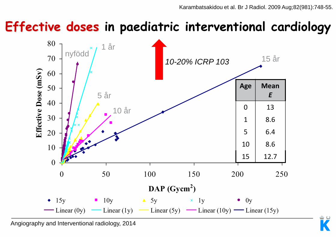
Effective doses in paediatric interventional cardiology
Karambatsakidou et al. Br J Radiol. 2009 Aug;82(981):748-55.
0
10
20
30
40
50
60
70
80
0 50 100 150 200 250
DAP (Gycm2)
Eff
ecti
ve
Do
se (
mS
v)
15y 10y 5y 1y 0y
Linear (0y) Linear (1y) Linear (5y) Linear (10y) Linear (15y)
nyfödd 1 år
5 år
10 år
15 år
Age Mean E
0 13
1 8.6
5 6.4
10 8.6
15 12.7
10-20% ICRP 103

674 children who underwent cardiac catheterization due to congenital anomalies, between the years 1950-1970
Expected number of malignancies for all sites was 4.75, while the observed number was 11.0
Angiography and Interventional radiology, 2014
Int J Epidemiol. 2000 Jun;29(3):424-8
Paediatric interventional cardiology

Angiography and Interventional radiology, 2014
Paediatric interventional cardiology
Include children who undergone at least one angio/interv in the age of <10 years
Evaluate the risk of leukaemia and solid cancers
Radiation dose will be estimated retrospectively
At present time: 4500 children (2000-2011) are already been included in the cohort

Angiography and Interventional radiology, 2014
”Linear No Threshold” - LNT-modellen
French Academy of Sciences (2004)
The American Nuclear Society (2001)
Ait-Ali (2010)
Beels (2009)
?
?

Angiography and Interventional radiology, 2014
0,0
0,2
0,4
0,6
0,8
1,0
1,2
RE
IDo
rgan
/ RE
IDto
t
leukemi breast colon liver lung ovary stomach bladder other
Risk of radiation exposure-induced cancer death
Submitted in BJR

Angiography and Interventional radiology, 2014
0 1 5 10 150.000
0.001
0.002
0.003
0.004
0.005
0.006
0.007
RE
IDo
rga
n/E
qu
ivale
nt
org
an
do
se (
%/m
Sv)
Age (years)
breast (F)
lung (F)
lung (M)
Relationship between risk, age and gender
0 1 5 10 150.000
0.005
0.010
0.015
0.020
0.025
0.030
0.035
RE
IDto
t/Eff
ecti
ve d
ose (
%/m
Sv)
Age (years)
Female
Male
Submitted in BJR

Angiography and Interventional radiology, 2014
Relationship between risk, age and gender
0,0
0,1
0,2
0,3
0,4
0,5
0,6
0,7
0 1 5 10 15
RE
ID (
%)
Age (years)
Female
Male
Submitted in BJR

Angiography and Interventional radiology, 2014
Factors affecting cancer risk
Patient population (age and gender)
Complexity of procedures
Machine settings and beam geometry
Choice of mortality cancer data
Choice of DDREF

Angiography and Interventional radiology, 2014
Questions
Angela Karambatsakidou, Enheten för Röntgen och IJ-Strålning, VO Sjukhusfysik
Assessment of local high doses in clinic routine

Angiography and Interventional radiology, 2014
Skin dose estimation
Action/Alarm levels
Patient follow-up
Statistics
Agenda

IAEA: Radiation Protection in Cardiology: Why talk about radiation protection in cardiology?
Deterministic effects

Angiography and Interventional radiology, 2014
Gafchromic XR-RV3 configuration
Dose Range 0.05-15 Gy
Energy Range 20 kV-200 kV
Barium

Angiography and Interventional radiology, 2014
Precautions in measuring skin dose with Gafchromic XR-RV3
Orientation of the film ~20% for the beam energies used in cardiology
and for a Kair~0,5 Gy.
Energy dependence for white facing the tube: 9%, 80-120kV, 5Gy
Energy dependence for yellow facing the tube: 7%, 80-120kV, 5Gy
Orientation of the film and beam quality
McCabe et al. Medical Physics, Vol. 38, No. 4, April 2011

Angiography and Interventional radiology, 2014
Sensitometric response of GAFCHROMIC® film, type XR-
RV3 Comparator strip
Precautions in measuring skin dose with Gafchromic XR-RV3
± 25 %
Color Reference Chart or Flatbed Scanner
Red channel values

Angiography and Interventional radiology, 2014
Precautions in measuring skin dose with Gafchromic XR-RV3
Time period between exposure and scan
Film darkness increase compared to 1h postexposure measurement
0
0,5
1
1,5
2
2,5
0 5 10 15 20 25 30 35
Dif
fere
nc
e in
fil
m r
es
po
ns
e (
%)
Postexposure time (h)
1 Gy
2 Gy
3,8 Gy
5,7 Gy
24±4 h
McCabe et al. Medical Physics, Vol. 38, No. 4, April 2011

Angiography and Interventional radiology, 2014
Precautions in measuring skin dose with Gafchromic XR-RV3
Contour plot without using a scanner
nonuniformity correction Contour plot with correction for nonuniformity in
scanner response
Nonuniformity correction
McCabe et al. Medical Physics, Vol. 38, No. 4, April 2011
Angiography and Interventional radiology, 2014
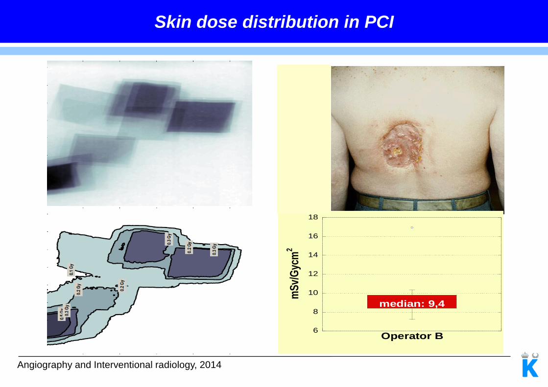
6
8
10
12
14
16
18
Operator B
mS
v/G
ycm
2
median: 9,4
Skin dose distribution in PCI
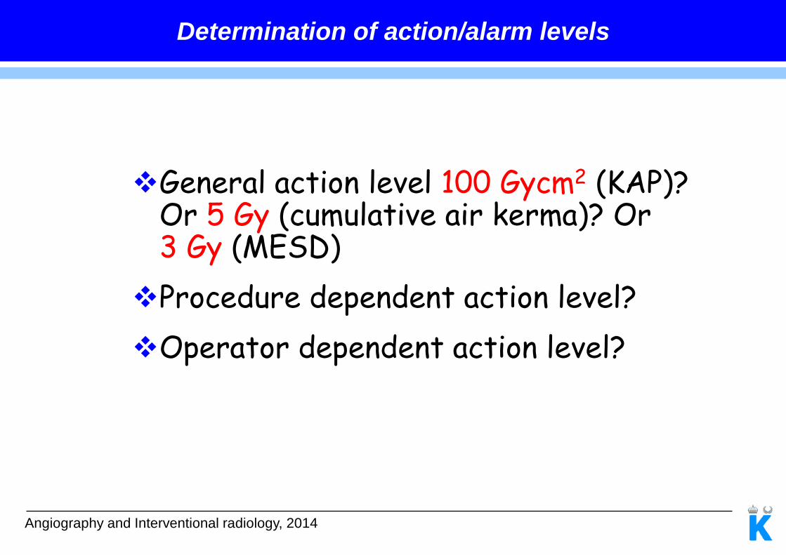
General action level 100 Gycm2 (KAP)? Or 5 Gy (cumulative air kerma)? Or 3 Gy (MESD)
Procedure dependent action level?
Operator dependent action level?
Angiography and Interventional radiology, 2014
Determination of action/alarm levels

Angiography and Interventional radiology, 2014
LARMNIVÅER
Kardiologiska angioverksamheten, Solna
Diagnostik : 350 Gycm2
Intervention: 150 Gycm2
Kombination: 200 Gycm2
Om DAP-värdet för en undersökning överskrider värdet ovan skall en kopia av
undersökningsrapporten skickas till:
Angela Karambatsakidou
Enheten för sjukhusfysik och IJ-strålning (H2:06)
VO Sjukhusfysik, Solna

Angiography and Interventional techniques, 2013
Cran
Dx Sin
Caud
Dosfördelning från en 1-sidig
panangiografisk röntgenundersökning
som inkluderar 2 st 3DRA. Den
maximala huddosen var 35 mGy och
var lokaliserad centralt på
bakhuvudet.
Large Cerebral Aneurysm
Philips image gallery
Larmnivån
DAPexp: 450 Gycm2
Skin doses in neuro angiography
Patient follow-up
Angiography and Interventional radiology, 2014
Information about previous interventional procedures
Inform the patient about potential radiation risks
Cumulative skin dose ≥3Gy (≥1Gy for procedures
likely to be repeated):Records of exposure
(position and skin dose) should be kept.
Advice to patient after the procedure about
the radiation effects (symptoms, signs)
Cumulated skin dose ≥3Gy: Follow-up patient 10-14 days after exposure
and referral to a dermatologist if serious and chronic damage are seen.
Inform personal physician about the radiation effects and doses
Threshold values not exceeded
Angiography and Interventional radiology, 2014
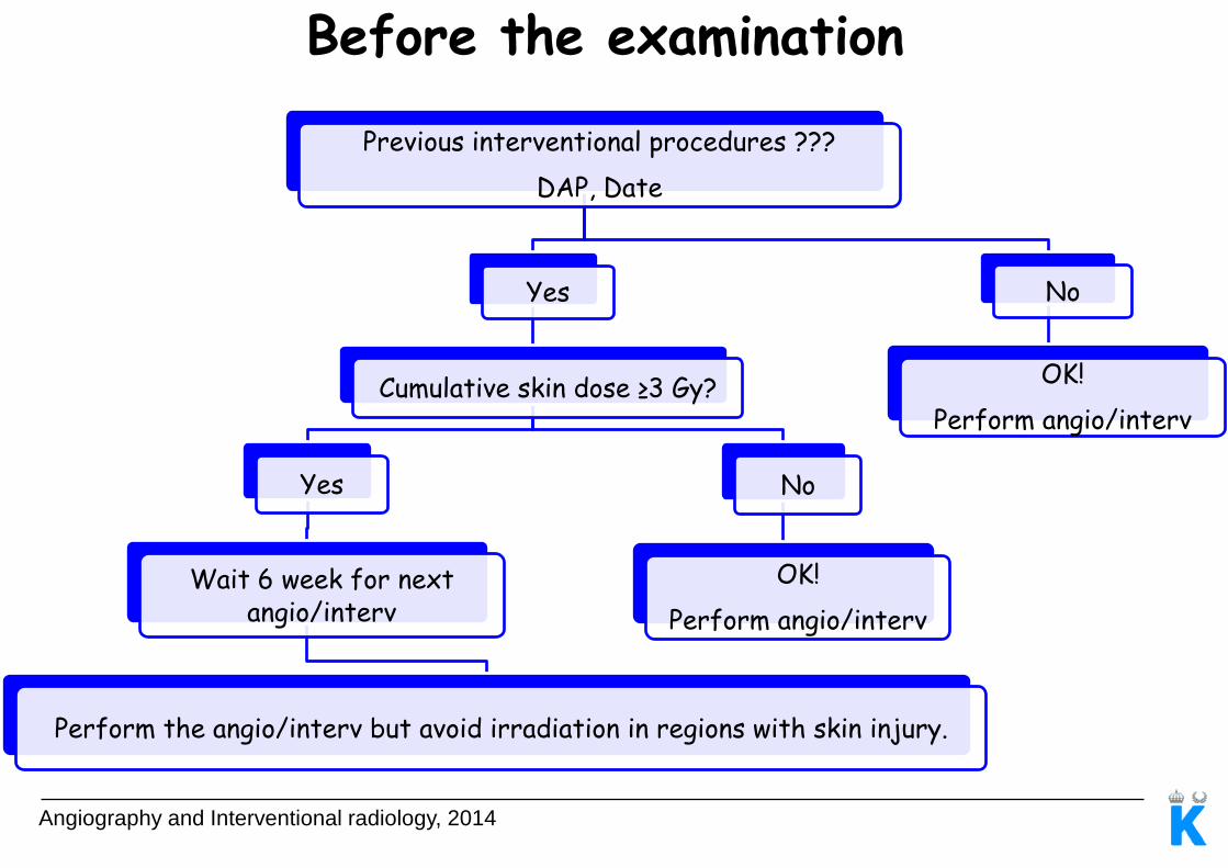
Previous interventional procedures ???
DAP, Date
Yes
Cumulative skin dose ≥3 Gy?
Yes
Wait 6 week for next angio/interv
Perform the angio/interv but avoid irradiation in regions with skin injury.
No
OK!
Perform angio/interv
No
OK!
Perform angio/interv
Before the examination
Angiography and Interventional radiology, 2014
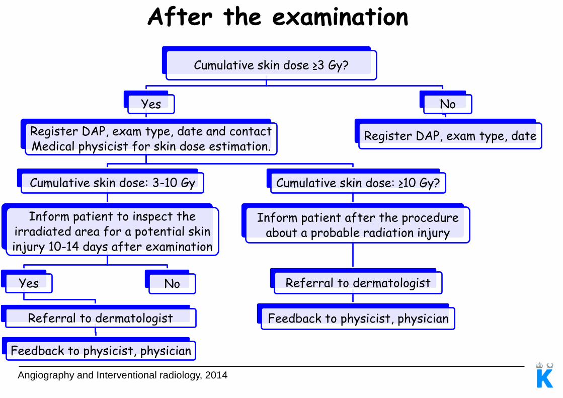
Cumulative skin dose ≥3 Gy?
Yes
Register DAP, exam type, date and contact Medical physicist for skin dose estimation.
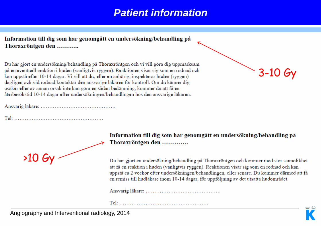
Cumulative skin dose: 3-10 Gy
Inform patient to inspect the irradiated area for a potential skin injury 10-14 days after examination
Yes
Referral to dermatologist
Feedback to physicist, physician
No
Cumulative skin dose: ≥10 Gy?
Inform patient after the procedure about a probable radiation injury
Referral to dermatologist
Feedback to physicist, physician
No
Register DAP, exam type, date
After the examination
Angiography and Interventional radiology, 2014
Patient information
>10 Gy
3-10 Gy

Angiography and Interventional radiology, 2014
Skin doses > alarm levels in the clinic at Karolinska University Hospital, Solna (2009-2013)
Skin erythema: > 2 Sv
Temporary epilation: > 3 Sv
Skin necrosis (delayed): > 12 Sv
Skin dose (Sv) Number of patients
2 - 3 44
3 - 10 32
>10 1
Skin doses

Angiography and Interventional radiology, 2014
Swedish Coronary Angiography and Angioplasty Register from 2013
Statistics from SCAAR
0
500
1000
1500
2000
2500
3000
1 2 3 4 5 6 7 8 9 10 11 12
Med
ian
DA
P (μ
Gym
2)
Operator
Coronary Angiography
10
16 172
84
13
20
33
7
98 35
50
81
0
1000
2000
3000
4000
5000
6000
7000
1 2 3 4 5 6 7 8 9 10 11 12
Med
ian
DA
P (μ
Gym
2)
Operator
Coronary Angiography + PCI
23
7
144 85
14
44
41 92
32
57
40 64

Angiography and Interventional radiology, 2014
Statistics from SCAAR
Coronarangio
Me
dia
n D
AP
(μ
Gym
2)
1
1
1
1
1
1
1 Date for procedure
2013-01-01 – 2014-01-01
Me
dia
n D
AP
(μ
Gym
2)
Date for procedure
Coronarangio + PCI

Angiography and Interventional radiology, 2014
Dose and Risk Advisor in Paediatric Interventional Radiology
y = 0,0272x
y = 0,065x
0,0
0,5
1,0
1,5
2,0
2,5
0 10 20 30 40
RE
ID (
%)
DAP (Gycm2)
Male
Female
Level of risk
(%)
Risk term
0.001-0.01 Very Low
0.01-0.1 Low
0.1-1 Moderate
>1 High
6 months – 2.5 years
UK Department of Health

Angiography and Interventional radiology, 2014
Questions

Angiography and Interventional radiology, 2014
Thank you