Embed Size (px)
Citation preview

NeuroImage xxx (2010) xxx–xxx
YNIMG-07794; No. of pages: 12; 4C: 3, 6, 7, 8, 9
Contents lists available at ScienceDirect
NeuroImage
j ourna l homepage: www.e lsev ie r.com/ locate /yn img
Dorsal and ventral stream activation and object recognition performance inschool-age children
Tessa Dekker a, Denis Mareschal a, Martin I. Sereno a,b, Mark H. Johnson a,⁎a Birkbeck College, University of London, Centre for Brain and Cognitive Development, Malet Street, London, UKb University College London, Department of Psychology, 26 Bedford Way, London, UK
⁎ Corresponding author. Fax: +44 20 7631 6587.E-mail addresses: [email protected] (T. D
[email protected] (D. Mareschal), [email protected]@bbk.ac.uk (M.H. Johnson).
1053-8119/$ – see front matter © 2010 Elsevier Inc. Aldoi:10.1016/j.neuroimage.2010.11.005
Please cite this article as: Dekker, T., et al.,NeuroImage (2010), doi:10.1016/j.neuroim
a b s t r a c t
a r t i c l e i n f oArticle history:Received 3 June 2010Revised 26 October 2010Accepted 1 November 2010Available online xxxx
We explored how developing neural artifact and animal representations in the dorsal and ventral stream playa role in children's increasingly more proficient interactions with objects. In thirty-three 6- to 10-year-oldchildren and 11 adults, we used fMRI to track the development of (1) the cortical category preference for toolscompared to animals and (2) the response to complex objects (as compared to scrambled objects) during apassive viewing task. In addition, we related a cognitive skill that improved substantially from age 6 to 10,namely the ability to recognize tools from unusual viewpoints, to the development of cortical objectprocessing. In multiple complementary analyses we showed that those children who were better atrecognizing tools from unusual viewpoints outside the scanner showed a reduced cortical response to toolsand animals when viewed inside the scanner, bilaterally in intraparietal and inferotemporal cortex. Incontrast, the cortical preference for tools in the dorsal and ventral visual stream did not predict objectrecognition performance, and was organized in an adult-like manner at six. While cortical tool preference didnot change with age, the findings suggest that animal-preferring regions in the ventral visual stream maydevelop later, concordant with previous reports of a protracted development in similar regions for faces. Wethus conclude that intraparietal and inferotemporal cortical networks that support aspects of objectprocessing irrespective of tool or animal category, continue to develop during the school-age years andcontribute to the development of object recognition skills during this period.
ekker),csd.edu (M.I. Sereno),
l rights reserved.
Dorsal and ventral stream activation and objeage.2010.11.005
© 2010 Elsevier Inc. All rights reserved.
Introduction
Objects, and children's interactions with those objects, play afundamental part in many theoretical frameworks of learning,instruction, and cognitive development. For example, Piaget (1952,1954) placed object permanence squarely at the center of his theoryof active learning in early development. In exploring the process oflearning itself in more details, Bruner (1966) proposed threemodes ofrepresentation during learning. The first (enactive representations)was action-based and was believed to bootstrap learning at all ages.The second mode (iconic representation) was image or visually basedand enabled a more concrete representation of a problem orsituations. Finally, the third mode (symbolic representation) waslanguage-based and supported abstract and hypothetical reasoning.Importantly, Bruner argued that these modes of representation wereco-existent, even in the most advanced learner, and only looselysequential in that they “translate” into each other as learning unfolds.An implication of Bruner's theory is that new material is best
presented in a way that fosters the progression from enactive toiconic to symbolic representation. Objects that support action—especially through fantasy and play—also hold a special role inVygotsky's theory of development (Vygotsky, 1934, 1978). Vygotskyargued that through play and tool use the child develops abstractmeaning separate from the objects in the world, thereby fostering theemergence of an abstract understanding of relations in the world.
While these ideas remain at the heart of the many theoreticalframeworks that underlie current educational theories, surprisinglylittle is known about the development of the neural systems thatsupport object and action processing in the developing brain. This isdespite the fact that the regions involved in object processing in theadult brain are well understood, suggesting that identifying the neuralmechanisms that underlie the development of object processing, andparticularly tool processing, during childhood is possible.
Whatwe do know from cognitive and behavioral studies is that theperception and recognition of complex objects such as faces and 3Dshapes, continues to improve dramatically during childhood and eveninto adolescence. In particular the ability to recognize objects orimages presented to the visual system in unconventional and hard-to-decode ways is known to develop late (Bova et al., 2007; Juttner et al.,2006; Mondloch et al., 2002, 2003, see Nishimura et al., 2009 for adetailed review). Further, it is well established that aspects of face
ct recognition performance in school-age children,
2 T. Dekker et al. / NeuroImage xxx (2010) xxx–xxx
processing keep on improving into adolescence (Mondloch et al.,2006, although see Crookes and McKone, 2009) and two recentstudies also indicate that tool processing keeps on developing until atleast well into the school-age years. For example, Bova et al. (2007)have shown that the ability to recognize tools from a non-canonicalviewpoint, improves rapidly between the 6th and 12th year. Inaddition, Mounoud et al. (2007) reported that seeing tool actionprimes speeds up decisions about tools in 5–9-year-olds, but not atolder ages, suggesting that the link between tool action representa-tions and tool perception is changing during childhood.
The ventral visual pathway plays an important role when adultsprocess aspects of objects for perception such as shape, color, texture,location- and size-constancy and to an extent orientation constancy(Grill-Spector, 2003, 2009). Another well-known characteristic of theadult object sensitive cortex in the ventral stream is its organizationby category; complex objects are represented in a distributed manneracross the inferotemporal cortex (ITC), but there are clustered regionswith a relatively stronger BOLD response for certain object categories(Haxby et al., 2001). These regions show a highly consistent spatialorganization across individuals that even emerges in the absence ofvisual experience with tool- and house-selective clusters are locatedmore medially in the fusiform gyrus (FFG) compared to face-, animal-and body-part selective clusters (Hasson et al., 2003; Mahon et al.,2009). Object processing in the dorsal visual pathway is lessextensively investigated. However, earlier reports from single-unitstudies in macaques (Sereno and Maunsell, 1998) suggested that inthe lateral intraparietal area (LIP), there is selectivity for objectidentity independent of location, which has been confirmed in studiesin adult humans (Konen and Kastner, 2008). In addition, dorsalcortical regions thought to be human homologues of the macaqueinferior parietal cortex (IPc) and anterior inferior parietal cortex (AIP),are thought to play a role in adult visuo-motor transformations forplanning and execution of object manipulation (Arbib, 2005;Rizzolatti and Matelli, 2003; Valyear et al., 2007). Perhaps becauseof the close link between tools and specific manual actions, apreference for tools over animate objects is distributed widely acrossa ventral and dorsal stream-spanning network. Tool preference wasmost consistently shown in the medial frontal gyrus, AIP, IPc, thepremotor cortex and an area in the medial temporal gyrus (MTG) thatresponds to non-biological tool movement and the earlier mentionedmedial FFG. This activation pattern even persists during passiveviewing tasks that do not require explicit actions or involve toolmotion, which has led to suggestions that these regions may beimportant for representing action and motion related aspects of tools(Beauchamp et al., 2003; Chao andMartin, 2000; Johnson-Frey, 2004).However, to what extent regions with a category preference performsome special computations relevant to their specific preferredcategory, and what driving principles lie behind the consistentorganization by category across the dorsal and ventral stream, iscurrently unclear (Grill-Spector, 2009). Recently, some developmen-tal fMRI studies have shed new light on this debate by investigatinghow the category selectivity in the ventral stream emerges.
Most consistently, these studies have shown that the response tofaces in the ventral stream keeps on developing until late inchildhood. According to the Interactive Specialization view (Johnson,2001, 2010), we need to discriminate between stimulus-sensitive andstimulus-selective (or preferring) tissue. For example, face-sensitivetissue is revealed by a contrast between cortical responses to faces anda baseline response to a non-object stimulus that has comparable low-level visual characteristics. Face-preferring (or selective) tissue refersto cortical regions that respond to faces considerably more thanclosely related stimuli such as other categories of complex visualobjects. As cortical regions become better tuned to particular stimuli,the Interactive Specialization view predicts increasingly focal patternsof activation of cortical tissue sensitive to stimuli such as faces withdevelopment, alongside the emergence of face-preferring tissue.
Please cite this article as: Dekker, T., et al., Dorsal and ventral stream acNeuroImage (2010), doi:10.1016/j.neuroimage.2010.11.005
These predictions have been confirmed for faces in several recentdevelopmental fMRI studies. (Aylward et al., 2005; Gathers et al.,2004; Golarai et al., 2007; Joseph et al., 2010; Passarotti et al., 2003;Scherf et al., 2007 see Johnson et al., 2009 for review). These studiesthus support the Interactive Specialization account and provideevidence against modular views of the brain that hypothesize thatregions of the cortex that perform evolutionary important functionssuch as face processing are hard-wired for that purpose from birth.
The development of tool processing and object processing in thedorsal stream in general has not been systematically investigated.Because regions that will eventually show a preference for graspableobjects during passive viewing are located across the whole brain,tracking the development of the cortical response to tools can provideimportant insights into object processing in the dorsal stream and itsdevelopmental interactions with the ventral stream. Previous devel-opmental fMRI studies on object processing have mainly focused oncategory specific regions of interest (ROIs) in the ventral stream.However, object representations do not solely depend on the regionsthat aremost specifically tuned to the category an object belongs to. Infact, substantial information about tools and other types of objects isrepresented in a distributed manner across the inferotemporal cortex(Haxby et al., 2001) and in dorsal regions of the brain (Barsalou, 2008;Konen and Kastner, 2008). The possibility that developmentalchanges take place in the object sensitive cortex outside regionswith a category preference, andmay not even be specific to a categorybut general to complex objects, has not yet been addressed.
Throughout this report, we use the word “tools” when onlyreferring to graspable utensils, the word “animals” when onlyreferring to animals, and the term “objects” when referring to bothcategories of complex objects (both animals and tools). In line withthe Interactive Specialization approach discussed earlier, we distin-guish between the development of a cortical category preference andthe development of cortical object sensitivity. More specifically, inorder to get a full picture of the developmental changes in objectprocessing across the brain during a passive viewing task in thescanner, we identify object sensitive regions (tools+animals−scrambled images) to explore the developing response distributionto objects irrespective of the local category preference and we identifytool-preferring regions (toolsNscrambledminus animalsNscrambled) andanimal-preferring regions (animalsNscrambled minus toolsNscrambledimages) toexplore thedevelopmentof a categorypreference. Additionallywe relate these two types of developmental changes to an objectrecognition skill that is still developing between age 6 and 10, theage-range of the children in the present study.
Although it is currently unknown if mere passive viewing of toolswill preferentially activate dorsal stream regions in children, we canformulate two contrasting hypotheses about the development of adorsal and ventral category preference based on previous research.Firstly, behavioral developmental studies suggest that tool represen-tations keep on developing during childhood (Bova et al., 2007;Mounoud et al., 2007) and it has been suggested that dorsal streamaction representationsmay play an important role in the developmentof tool representations (Mahon et al., 2007, 2009). We therefore mayexpect protracted tuning of regions with a tool preference, similar towhat has previously been reported for ventral regions with a facepreference (Hypothesis 1). Secondly, three recent fMRI studies havereported that an adult-like preference for abstract sculptures, bodyparts and film material in which body parts, furniture and tools wereintermixed is present in the ventral LOC from 5–8 years onwards(Golarai et al., 2007; Scherf et al., 2007; Pelphrey et al., 2009).Although these authors did not specifically compare the response totools across age and restricted their analysis to the ventral stream,their findings raise the possibility that a cortical preference for non-social objects may mature before the preference for faces. We maytherefore hypothesize that a preference for tools in the dorsal andventral stream will be adult-like in childhood, contrary to what has
tivation and object recognition performance in school-age children,

3T. Dekker et al. / NeuroImage xxx (2010) xxx–xxx
previously been reported for faces (Hypothesis 2). Finally, we canformulate a hypothesis with respect to the development of objectsensitive cortex: to the extent that object-processing skills that keepon developing until late into childhood depend on processes that arerelevant to multiple types of objects (Nishimura et al., 2009), we mayexpect that the response to both tools and animals in the objectsensitive cortex will show a protracted development with age andincreasing object experience (Hypothesis 3). Note that this thirdhypothesis is not mutually exclusive with either of the first twohypotheses.
To summarize, in the present studywe aim to getmore insight intowhich aspects of the BOLD response to objects (tool or animalpreference and/or objects in general) in dorsal and ventral corticalregions show a protracted development during childhood. In addition,we explore which developmental changes in cortical tool and animalprocessing relate to performance on an independently measuredobject recognition task that is still improving in childhood, in order togain a better understanding of the functional role that developingneural artifact- and animal representations in the dorsal and ventralstream play in children's day to day interactions with objects.
Materials and methods
Participants
Thirty-three 6- to 10-year-old children and eleven 21- to 34-year-old adults took part in a passive viewing task in the MRI scanner andin an object perception task that took place outside the scanner. Theparticipants were split into an age group of fifteen 6–7-year-olds thatconsisted of 6 boys (average age=6.9 years, SD=0.7) and 9 girls(average age=7.1 years, SD=0.5), a group of eighteen 8–10-year-old children consisting of 8 boys (average age=9.6 years, SD=0.9)and 10 girls (average age=9.4 years, SD=0.8) and 11 adultsconsisting of 5 women (average age=25.3, SD=3.0) and 6 men(average age=30, SD=3.9). All participants were right-handed withnormal or corrected vision and without any past or currentneurological or psychiatric conditions, or structural brain abnormal-ities. Children were recruited through advertisements in newspapers.Adult participants were recruited via local recruitment facilities.Before testing, adult participants and caretakers signed an informed
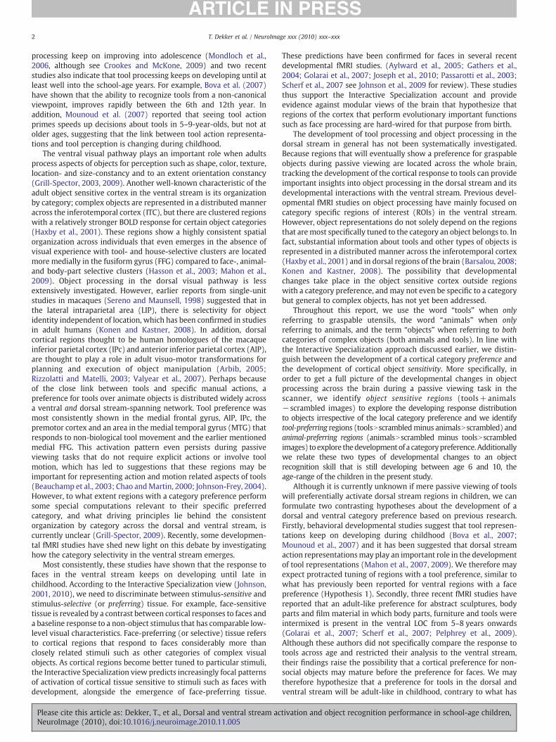
Fig. 1. A: Examples of items viewed from an unusual perspective from the object recogniparticipants were presented with in the scanner during the passive viewing task while they fi
a similar task by Bova et al., 2007) are plotted against age. Answers were considered to be corrwell as from a typical viewpoint and incorrect if only the typical viewpoint was recognized.analysis. The graph shows that performance on this task increases substantially with agepb0.000), however, performance only increases with age from 6 to 10 (R2=0.251, F (1,32)=
Please cite this article as: Dekker, T., et al., Dorsal and ventral stream acNeuroImage (2010), doi:10.1016/j.neuroimage.2010.11.005
consent form and children signed an assent form. A medical clearancequestionnaire and a metal detection test were administered to eachparticipant to ensure MR safety criteria were adhered to. The researchwas executed under the approved University protocols for the use ofhuman adult and minor participants in research.
Unusual perspective task (outside the scanner)
StimuliAll participants performed an object recognition task outside the
MRI scanner. The task was based on the “unusual perspective task”developed by Bova et al. (2007). To create the stimuli, twenty-onecommon, graspable objects were photographed from a viewpoint inwhich they are typically viewed and from which they are easy torecognize. The same objects were also photographed from an unusual,more difficult to recognize, viewpoint. Objects in the original colorphotographs were separated from their original background usingadobe Photoshop and placed on a neutral, gray background (rgb:200×200×200, 600×800 pixel size, see Fig. 1A for examples andsupplementary material for more details on the items in the task). Theobjects were presented on a 15.7 in. monitor with a viewing distanceof ca 50 cm (37°×24.2° visual angle) using E-prime stimuluspresentation software.
ProcedureThe unusual perspective task took place in a testing room separate
from the scanner suite. In this self-paced task, participants firstidentified graspable objects that appeared consecutively on the screenfrom an unusual viewpoint. Next, they identified the same objects, butnow presented from their typical viewpoint. This second part of thetask ensured that participants could recognize the objects in typicalview. All participants were instructed to look carefully at each objectpresented on the screen and to press the spacebar as soon as theywere ready to guess what the object was. After each press, a buzzersounded and the object was replaced with a blank screen with acentral fixation cross. After the participant had made a guess andrefocused attention to the cross, the experimenter initiated the nexttrial. The same procedure was followed for objects that werepresented from an unusual and from a typical viewpoint. As thiswas a recognition test and not a naming or language test, descriptions
tion task that participants performed outside the scanner. B: Examples of the stimulixated on the central cross. C: Accuracy scores on the unusual perspective task (based onect if participants correctly named or described the object from an unusual viewpoint asItems that were not correctly recognized in their typical view were excluded from thefrom childhood to adulthood (Pearson r=0.7, pb0.000, R2=0.49; F (1,43)=40.33,10.406, p=0.003) and no longer improves after the 22nd year of life, F=1.23, p=n.s.).
tivation and object recognition performance in school-age children,
4 T. Dekker et al. / NeuroImage xxx (2010) xxx–xxx
of the object or indications regarding their use (e.g., instead of saying“nailclipper”, saying: a thing to cut your nails) were accepted ascorrect answers. An item was considered as not recognized when theanswer given referred to a different object (e.g., “fork”: rake,“lightbulb”: plug). See Bova et al. (2007) for similar scoring criteria.Items were excluded from the analysis if the usual perspective wasnot recognized correctly or if the unusual perspective was recognizedincorrectly but the answer was plausible (instead of saying “mobilephone”, saying: remote control). See Supplement 1 for moreinformation about the items.
Passive viewing task (inside the scanner)
StimuliFifteen types of highly familiar animals and tools were selected for
the passive viewing task in theMRI scanner. A single color photographwas selected for each of these 30 types of objects and the animal ortool on the photo was placed on a neutral gray background usingAdobe Photoshop (background: 200×200×200 rgb, stimulus size:600×450 pixels). A scrambled version of each picture was created inMatlab, by applying a 6×9 grid to the object stimuli and shuffling thegrid cells (grid cell size 100×50 pixels). Object details were stillvisible in these scrambled stimuli but the overall object shape wasgrossly distorted. A red fixation cross with a black outline (30 pixelsheight and width) was displayed in the center, on top of all stimuliand during inter-stimulus intervals when no objects were presented(see Fig. 1B for examples of the stimuli). Images were projected onto aback projection screen (23°×14° visual angle, screen resolution800×600) attached to the bore of the scanner. Participants saw thisscreen through a mirror that was mounted on the radiofrequency coilthat surrounded their head. Stimuli were presented using Matlab 6.0(Mathworks) and Cogent 2000 extensions (www.vislab.ucl.ac.uk/cogent.php).
Procedure and taskIn the scanner, we asked all subjects to look at the screen while
fixating a cross in the center throughout the run. This task has twoimportant benefits. Firstly, task demands are very low, so participantsof a wide range of ages can do the same task with minimalconfounding influences of developmental differences in performance.Secondly, because all participants looked at the same location on thescreen, developmental differences in eye-movements, which canmask the BOLD response to objects in the retinotopic regions thatmake up about 50% of the brain, including the fusiform gyrus, regionsin the parietal and frontal cortex (Saygin and Sereno, 2008) areminimized.
Animals, tools and scrambled objects were presented in 16 blocksof 15 s (4 animal blocks, 4 tool blocks and 8 scrambled object baselineblocks). During a block, 15 items from one of the three stimulusconditions were each presented for 800 ms, followed by a 200 msfixation screen. The order of the blocks was randomized, as was theorder of the stimuli, with the constraint that no stimulus occurredmore than once during a block. The total duration of a run was16×15=240 s. Each stimulus was repeated four times during a run.Two runs were acquired for each participant. The runs were separatedby a structural scan to limit stimulus adaptation effects and to preventyoung children from getting bored with the task.
Children were trained to lie still in the scanner before theexperimental runs began. Each time anMR-compatible video camerarecorded excessive movement during the training, the scanneroperator stopped a cartoon that the child was watching andexplained that the movement he/she was making would harm theimage quality. This training continued until the child was lyingsufficiently still for a fewminutes. Children were monitored with thecamera and via an intercom throughout the session to ensure thatthey remained still, that they were fixating the central cross on the
Please cite this article as: Dekker, T., et al., Dorsal and ventral stream acNeuroImage (2010), doi:10.1016/j.neuroimage.2010.11.005
screen, and that they were comfortable during scanning. Allparticipants held an alarm button in their left hand so that theycould notify the scanner operator at any time they chose to, in casethey wanted to stop the experiment.
MR data acquisition and preprocessing
MR data were collected with a Siemens TIM Avanto 1.5 T MRIscanner using a 12-channel receive-only head coil. A high(1×1×1 mm) resolution T1-weighted 3D MPRAGE anatomicalsequence (magnetization prepared low angle spoiled gradient echo)was performed to acquire high-resolution images of the brainstructure of each participant (image matrix=224×256, 160 parti-tions, TR: 2730, TE: 3.57, effective TI 1000 ms, flip angle: 7°).Functional data were collected using an echo planar 2D imagingsequence with image-based prospective acquisition correction forhead motion (Thesen et al., 2000). Per run, we collected one hundredvolumes that covered the whole brain and consisted of thirty-twoslices, acquired in the axial plane in interleaved ascending order(bandwidth=1906 Hz/pix, TR: 2.5, TE: 39, flip angle: 90, voxel size:3.5×3.5×3.5 cm, matrix 64×64).
All functional data were converted to NIFTI format and analyzedusing FSL (http://www.fmrib.ox.ac.uk/fsl). First, the images wereflipped into the standard FSL orientation and the non-brain structuresin the co-planar high-resolution T2-weighted EPI volume and the T1-weighted MPRAGE volume were removed with FMRIB's BrainExtraction Tool (Smith, 2002). Before statistical estimation, thefollowing preprocessing steps were undertaken: the first 4 volumesof functional data from each run were discarded, brain volumes weremotion corrected to the middle volume using the Oxford Centre forFunctional Magnetic Resonance Imaging of the Brain (FMRIB) LinearImage Registration Tool (MCFLIRT) (Jenkinson et al., 2002). Spatialsmoothing was applied using a Gaussian-weighted kernel of 5 mm atfull-width half-maximum, and data were high-pass filtered to removelinear trends. Estimates of the degrees of freedom in the statisticalmodel were corrected for autocorrelation in the data by using the FSLpre-whitening technique (Woolrich et al., 2001). Only runs with lessthan 2 mm maximal absolute movements were included (53 childruns and 21 adult runs). To deal with remaining noise due to excessivesubject movement not dealt with by correction for head motion(Diedrichsen and Shadmehr, 2005), themean absolute deviation fromthe median was calculated for each volume using Afni (3DToutcount,http://afni.nimh.nih.gov/afni/). In each run, the volumes with thefifteen most extreme values were identified and modeled in thedesign matrix as regressors of no interest. Delays and undershoots inthe hemodynamic response were accounted for by convolving themodel with a double-gamma basis function. Functional images wereregistered to the high-resolution T1 weighted 3D MPRAGE using thelow resolution 3D MPRAGE acquired in the same plane as thefunctional images. The high-resolution structural T1-weighted EPIvolume was registered to the Montreal Neurological InstituteTalairach compatible MR atlas averaging 152 normal subjects usingFMRIB's Linear Image Registration Tool (FLIRT). Child brain normal-ization is an accepted method in developmental fMRI studies sincetotal cerebral volume does not change significantly with age after5 years of age to adulthood (Klingberg et al., 2002; Passarotti et al.,2007; Reiss et al., 1996) and from 6 years of age, standardnormalization procedures do not lead to artifacts (Kang et al., 2003;Muzik et al., 2000). Therefore data from 6- to 10-year-olds and adultscan be effectively transformed into the same stereotactic space.
fMRI analyses
Animal and tool blocks were modeled as regressors of interest inthe design matrix with respect to a scrambled image baseline. Eachfunctional run for a given subject was modeled separately at the first
tivation and object recognition performance in school-age children,
5T. Dekker et al. / NeuroImage xxx (2010) xxx–xxx
level. Statistics for the contrasts of interest, averaging across the runsof each subject, were estimated using fixed effects modeling. At thegroup level, random-effects components of mixed effects variancewere modeled and estimated for each contrast of interest usingFLAME1 (Beckmann et al., 2003). To identify significant clusters ofactivation, all Z-statistic images (Gaussianized T/F) were firstthresholded at an uncorrected voxel threshold of z=2.3, p=0.01.Correction for multiple comparisons was performed at the clusterlevel by applying a cluster size probability threshold of pb0.05 to theZ-statistic images. We only report clusters that survived this clustercorrected threshold.
We included adults in our study as a mature benchmark fordevelopment, but a number of changes that are not of interest whenstudying the development of cortical object processing, such as eye-movements and cognitive strategies that are unrelated to objectprocessing, motion, respiration differences, and cardiac activity, canaffect the magnitude or extension of the BOLD response. Each of thesefactors has the potential to lead to artifactual differences between agegroups.Wedealtwith these common challenges of developmental fMRIby using a robust blocked design, by minimizing eye-movements, andby applying stringent corrections for motion artifacts (see previoussection). In addition, we performed multiple, complementary types ofanalyses in which we tracked changes in activation from 6 years of ageto adulthood and investigated changes that occurred during childhoodseparately from the adult group.
We identified object sensitive regions (tools+animalsNscrambledimages), tool-preferring regions (toolsNscrambled—versus animalsNscrambled) and animal-preferring regions (animalsNscrambled−toolsNscrambled) in each age group. We explored where the objectsensitive and category-selective BOLD response varied with age inwhole brain correlation analyses as well as in a-priori, structurallydefined regions in which we expected developmental changes inobject processing to take place based on both developmental andadult literature on tool and object processing (Chao andMartin, 2000;Golarai et al., 2007; Grill-Spector, 2003; Konen and Kastner, 2008;Rizzolatti andMatelli, 2003; Sereno andMaunsell, 1998; Valyear et al.,2007). These structural ROIs encompassed the fusiform gyrus, theparietal cortex, the inferior lateral occipital cortex and the superiorlateral occipital cortex/posterior parietal cortex. Because normaliza-tion from age six onwards does not lead to artifacts (Muzik et al.,2000) we did not manually draw the borders of anatomical ROIs ineach individual anatomical scan but defined them in standard spaceafter normalization was performed, as has been done in several otherdevelopmental fMRI studies (Passarotti et al., 2003, 2007; Nelsonet al., 2003). The four structurally defined ROIs consisted of (1) thecombined occipital and temporal fusiform cortex atlas region masks,(2) the combined superior parietal cortex and supra-marginal gyrus,anterior division atlas region masks (3) the inferior LOC atlas regionmask and (4) the superior LOC atlas region mask derived from theprobabilistic Harvard–Oxford Cortical Structural Atlas (probabilisticmasks were converted to binary masks).
To further investigate the development of category-selectiveregions in the dorsal and ventral object sensitive cortex, we comparedthe animal and tool preference of 6- to 7-year-old children, 8- to 10-year-old children, and adults in independently defined functionalregions of interests. Finally, we explored where in the brain and in theearlier described a-priori defined structural ROIs, individual differ-ences in (1) tool and animal preference and (2) object sensitivity,correlatedwith age andwith individual scores on an independent toolrecognition task between age 6 to 10.
Results
The behavioral results acquired from the same subjects that weresubsequently scanned are presented first, followed by fMRI results.
Please cite this article as: Dekker, T., et al., Dorsal and ventral stream acNeuroImage (2010), doi:10.1016/j.neuroimage.2010.11.005
The unusual perspective task
The percentage of graspable objects that were correctly recognizedfrom an unusual viewpoint is plotted against age in Fig. 1C. The graphshows that accuracy increased overall with age from 6 years to34 years of age (R2=0.49, F (1,43)=40.33, pb0.001). A regressionanalyses showed that recognition of graspable objects from anunusual perspective improved markedly during childhood, betweenage 6 and 10 (R2=0.25, F (1,32)=10.406, p=0.003) but did notimprove further in the group of adults (R2=0.032, F (1,10)=0.009,pb0.92). The high adult scores on this task, which was specificallydesigned for children, may reflect a ceiling effect rather thanstabilization of the ability to recognize graspable objects from unusualviewpoints in adulthood.
The development of object sensitive cortex
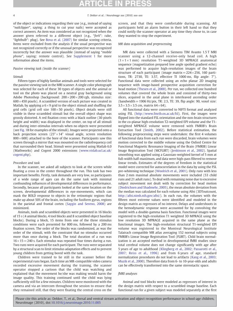
We explored how the distribution of object sensitive cortex,irrespective of its category preference, changes between age 6 andadulthood (contrasting tools+animals with scrambled images).Collapsing across all forty-four participants, we obtained a map ofobject sensitive cortex that extended from the occipital cortex into thefusiform gyrus and into the lateral occipital- and medial temporalcortex and dorsally into the left superior parietal cortex extending tothe anterior intraparietal sulcus (right panel of Fig. 2A). Objectsensitive regions are displayed separately for 6- to 7-year-olds, 8- to10-year-olds and adults in the left three panels of Fig. 2A.
We used a whole brain correlation analysis to determine if thecortical BOLD response to objects varied linearly with age in anyregion. Clusters where object sensitivity changed from the 6th year oflife to adulthood were located in the bilateral insular cortex, posteriorcingulate and visual cortex. In the insular, the response to objectsincreased with age; children showed a preference for scrambledimages vs objects and adults a preference for objects vs the scrambledimage baseline. Conversely, in the posterior cingulate the response toobjects reduced with age, with a preference for objects overscrambled images in childhood but a stronger response during thebaseline in adulthood (Fig. 2B, left). Finally, at all ages there was apreference for the baseline in the occipital pole and the lingual gyrus,but this preference reduced with age (Fig. 2B, right). The posteriorcingulate cortex and the insular gyrus are often linked to theregulation of cognitive resources and the default-network (Augustine,1996; Fransson andMarrelec, 2008; Hayden et al., 2008) we thereforesuggest that the patterns of response in these regions reflect age-related differences in cognitive strategies during the task. Forexample, the reducing response in the posterior cingulate may reflectthe allocation of fewer resources to scrambled images vs objects inadults than in children, presumably because adults were moreefficient at the passive viewing task, i.e., they engaged less with thestimuli. Likewise, the activation differences in or near primary visualcortex may have reflected slight differences in fixation strategies.Thus, we have identified several regions where the response toobjects correlated linearly with age. In later discussions of age-relateddifferences in the response to objects in childhood alone, we exploreage differences in object processing with a more complex develop-mental pattern.
The development of a cortical preference for tools and animals
We next performed multiple complementary analyses to investi-gate how cortical patterns of the preference for tools and animals inthe dorsal and ventral stream change from the 6th year of life(contrasting toolsNscrambled−animalsNscrambled to obtain tool-preferring regions and animalsNscrambled− toolsNscrambled toobtain animal-preferring regions). Collapsing across all participantswe obtained an average map of cortex with a tool preference. As can
tivation and object recognition performance in school-age children,
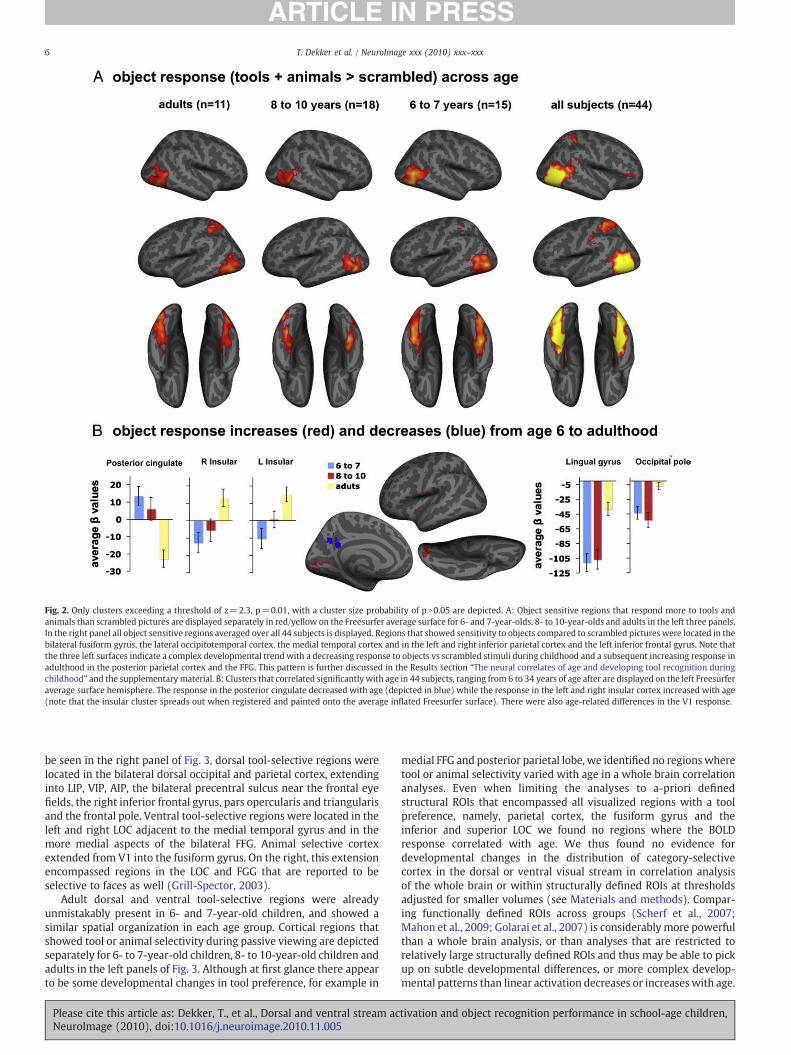
Fig. 2. Only clusters exceeding a threshold of z=2.3, p=0.01, with a cluster size probability of pN0.05 are depicted. A: Object sensitive regions that respond more to tools andanimals than scrambled pictures are displayed separately in red/yellow on the Freesurfer average surface for 6- and 7-year-olds, 8- to 10-year-olds and adults in the left three panels.In the right panel all object sensitive regions averaged over all 44 subjects is displayed. Regions that showed sensitivity to objects compared to scrambled pictures were located in thebilateral fusiform gyrus, the lateral occipitotemporal cortex, the medial temporal cortex and in the left and right inferior parietal cortex and the left inferior frontal gyrus. Note thatthe three left surfaces indicate a complex developmental trend with a decreasing response to objects vs scrambled stimuli during childhood and a subsequent increasing response inadulthood in the posterior parietal cortex and the FFG. This pattern is further discussed in the Results section “The neural correlates of age and developing tool recognition duringchildhood” and the supplementary material. B: Clusters that correlated significantly with age in 44 subjects, ranging from 6 to 34 years of age after are displayed on the left Freesurferaverage surface hemisphere. The response in the posterior cingulate decreased with age (depicted in blue) while the response in the left and right insular cortex increased with age(note that the insular cluster spreads out when registered and painted onto the average inflated Freesurfer surface). There were also age-related differences in the V1 response.
6 T. Dekker et al. / NeuroImage xxx (2010) xxx–xxx
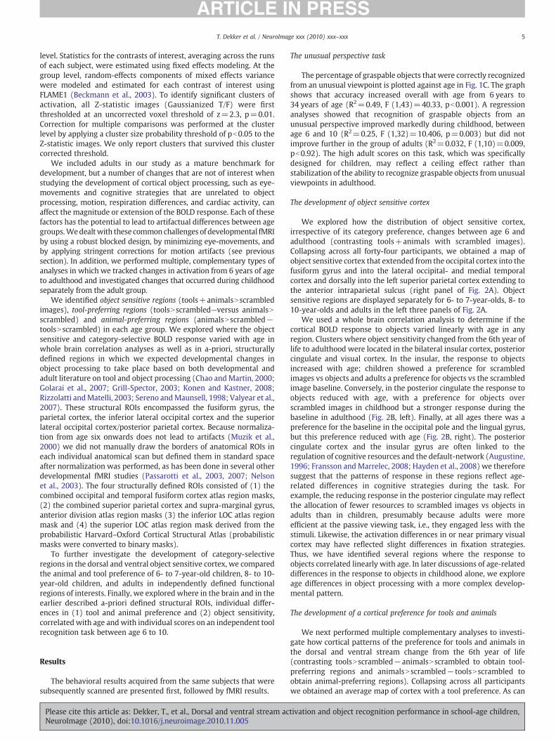
be seen in the right panel of Fig. 3, dorsal tool-selective regions werelocated in the bilateral dorsal occipital and parietal cortex, extendinginto LIP, VIP, AIP, the bilateral precentral sulcus near the frontal eyefields, the right inferior frontal gyrus, pars opercularis and triangularisand the frontal pole. Ventral tool-selective regions were located in theleft and right LOC adjacent to the medial temporal gyrus and in themore medial aspects of the bilateral FFG. Animal selective cortexextended from V1 into the fusiform gyrus. On the right, this extensionencompassed regions in the LOC and FGG that are reported to beselective to faces as well (Grill-Spector, 2003).
Adult dorsal and ventral tool-selective regions were alreadyunmistakably present in 6- and 7-year-old children, and showed asimilar spatial organization in each age group. Cortical regions thatshowed tool or animal selectivity during passive viewing are depictedseparately for 6- to 7-year-old children, 8- to 10-year-old children andadults in the left panels of Fig. 3. Although at first glance there appearto be some developmental changes in tool preference, for example in
Please cite this article as: Dekker, T., et al., Dorsal and ventral stream acNeuroImage (2010), doi:10.1016/j.neuroimage.2010.11.005
medial FFG and posterior parietal lobe, we identified no regions wheretool or animal selectivity varied with age in a whole brain correlationanalyses. Even when limiting the analyses to a-priori definedstructural ROIs that encompassed all visualized regions with a toolpreference, namely, parietal cortex, the fusiform gyrus and theinferior and superior LOC we found no regions where the BOLDresponse correlated with age. We thus found no evidence fordevelopmental changes in the distribution of category-selectivecortex in the dorsal or ventral visual stream in correlation analysisof the whole brain or within structurally defined ROIs at thresholdsadjusted for smaller volumes (see Materials and methods). Compar-ing functionally defined ROIs across groups (Scherf et al., 2007;Mahon et al., 2009; Golarai et al., 2007) is considerably more powerfulthan a whole brain analysis, or than analyses that are restricted torelatively large structurally defined ROIs and thus may be able to pickup on subtle developmental differences, or more complex develop-mental patterns than linear activation decreases or increaseswith age.
tivation and object recognition performance in school-age children,

Fig. 3. Only clusters exceeding z=2.3, p=0.01 with a cluster size probability of pN0.05 are depicted. Regions with a tool preference are depicted in red/yellow (resulting from thecontrast toolsNscrambled−animalsNscrambled) and regions with an animal preference are depicted in blue (resulting from the contrast animalsNscrambled− toolsNscrambled).Group averagemaps are displayed for 6- to 7-year-olds, 8- to 10-year-olds and adults separately in the left three panels and for all 44 participants together on the right. Regions witha tool preference in the omnibus analysis were located dorsally in the bilateral superior parietal cortex and extended into the anterior inferior parietal cortex, the bilateral precentralsulcus in the frontal eye fields and the right inferior frontal gyrus, pars opercularis and triangularis and frontal pole. More ventrally, clusters with a tool preference were located in theleft and right lateral fusiform gyrus (FFG) and lateral occipital cortex (LOC) adjacent to the medial temporal gyrus (MTG) and in the more medial aspects of the bilateral fusiformgyrus (FFG). A large cluster with a preference for animals over tools was located in V1, extending into the fusiform gyrus. Most prominently on the right, the cortical preference foranimals in V1, extended into the LOC and FGG. The blue animal-preferring region located between two tool-preferring clusters on the right inferior view and the small blue animal-preferring region on the right lateral view are located in the “fusiform face area” (FFA) and the occipital face area (OFA) (Grill-Spector, 2003). There were no regions where thepreference for tools or animals correlated with age in a whole brain analysis and correlation analyses restricted to a-priori defined structural ROIs in FFG, inferior and superior LOCand the Parietal cortex.
7T. Dekker et al. / NeuroImage xxx (2010) xxx–xxx
In the next section we therefore adopt a functionally defined ROIapproach to further explore the response patterns in object sensitivecortical regions with a category preference.
ROI analysis of the category preference in object sensitive cortex
Within the object sensitive cortex, we selected ROIs that wereorganized by category at ages 6 to 7, ages 8 to 10 and in adulthood.Wethen investigated if there were age-related changes in the responsepatterns of these functionally defined ROIs. This approach allowed usto define voxels with a category preference using a separate age group(e.g., a region with a tool preference in the adult group) from the oneused to test the experimental hypothesis (e.g. whether a group of 6- to7- and a group of 8- to 10-year-old children display a tool preferencein this adult region). Specifically, we contrasted tools with animalsand vice versa (toolsNscrambled−animalsNscrambled and animal-scrambled− toolsNscrambled) for each age group separately, withina functionally defined mask of the omnibus object sensitive cortex.The omnibus object sensitive mask is depicted in Fig. 2A, right paneland was obtained by contrasting animals+tools−scrambled images,averaging across all forty-four subjects. We identified all resultingclusters with a tool and animal preference for each age group and thenextracted from these clusters the average BOLD response to animalsand tools (both compared to a scrambled picture baseline) for eachindividual. For example, in a tool-preferring region in the adult leftparietal cortex, we extracted the response to tools and animals for allsubjects, including all children. Subsequently, we tested if the sameregion preferred tools to animals in 6- to 7- and 8- to 10-year-oldchildren using sets of ANOVA's.
Please cite this article as: Dekker, T., et al., Dorsal and ventral stream acNeuroImage (2010), doi:10.1016/j.neuroimage.2010.11.005
In Fig. 4, the responses to tools and animals in each ROI aredisplayed for all age groups. As can be seen in the graphs, allparticipants showed a tool preference in each ROI, even if the ROI fromwhich the response was extracted was defined in a different agegroup. A tool preference was already present in what will laterbecome an adult tool-specific region in both groups of children.Likewise, ROIs that had a preference for tools in 6- to 7-year-olds and8- to 10-year-olds showed a preference for tools in adults. In addition,ANOVA's showed that there were no stimulus-by-age interactions inany of the ROIs (all F's are smaller than 1.809, p=0.177). We therebyshow that no significant developmental changes such as an increasingfocalization of tool-preferring response were taking place outside thefunctionally defined adult regions.
An animal selective region in the object sensitive cortex surpassedthe statistical threshold only in adults. In accordance with previousreports (Mahon et al., 2009), this region was located in the lateraloccipital fusiform gyrus, somewhat anterior to what has been reportedas the functional occipital face area (OFA) (Grill-Spector et al., 2004). Inthe groups of children, the average BOLD response in this region (seeFig. 3A, right bottom graph) did not show a preference for animals. Tofurther explore the emergence of animal selectivity, we compared theaverage BOLD response of 6- to 7-year-olds, 8- to 10-year-olds andadults in the cluster with an animal preference that we identified in themedial FFG in the omnibus analysis of all 44 subjects (see Fig. 3, right-most panel, the medial and most anterior blue region in the inferiorview of the right hemisphere). The region showed a preference foranimals in adults (F (1,10)=12.190, p=0.006) but not in children (F(1,14)=0.131, p=0.131, F (1,17)=0.751, p=0.398). There were nosignificant age×stimulus interactions in the group average fusiform
tivation and object recognition performance in school-age children,
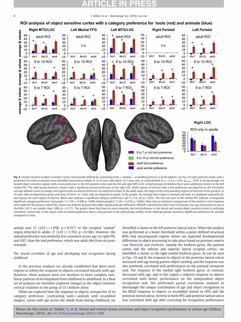
Fig. 4. Clusters located in object sensitive cortex (functionally defined by contrasting tools+animals−scrambled pictures in all 44 subjects, see Fig. 2A right panel for mask) with apreference for tools or animals were identified separately in adults, 8- to 10-year-olds and 6- to 7-year-olds, at a thresholded of z=2.3, p=0.01, pclusterb0.05. In all age groups, welocated object sensitive regions with a tool preference in the left parietal cortex and the left and right MTG/LOC. In both groups of children there were additional clusters in the leftmedial FFG. The adult group showed a cluster with a significant animal preference in the right LOC. Adult regions of interest with a tool preference are depicted on the Freesurferaverage inflated cortex in orange, and regions with an animal preference are depicted in blue. In the adult maps, the edges of the corresponding regions of interests in the group 8- to10-year-olds are depicted in green and those of the 6- to 7-year-olds are depicted in purple. In the graphs, the average beta-values to animals and tools are displayed separately foreach group, for each region of interest. Black stars indicate a significant category preference (all F'sN5.6, all p'sb0.03). The two red stars in the medial FFG indicate a marginallysignificant category preference (top graph: F (1,10)=4.308, pb0.065, bottom graph F (1,10)=4.318, pb0.064). Note that no statistical comparisons of the animal vs tool responsewere made for the group in which the region was defined, because they differ significantly per definition. ANOVA's showed that there were no stimulus-by-age interactions in any ofthe ROIs (All F's are smaller than 1.809, p=0.177). The graphs show that from six years onwards, the tool preference in the dorsal and ventral object sensitive cortex is strikinglyconsistent. Conversely, in the region with an animal preference that is only present in the adult group, neither of the child age groups showed a significant preference for animalscompared to tools.
8 T. Dekker et al. / NeuroImage xxx (2010) xxx–xxx
animal area (F (2,41)=1.058, p=0.357) or the occipital “animal”region detected in adults (F (2,41)=1.763, p=0.184). However, theanimal preference was distinctly less consistent across age (in right FFGand LOC) than the tool preference, which was adult-like from six yearsonwards.
The neural correlates of age and developing tool recognition duringchildhood
In the previous analyses we already established that there wereregions in which the response to objects correlated linearly with age.However, these analyses were not sensitive to more complex, non-linear patterns of development from childhood to adulthood. In a finalset of analyses we therefore explored changes in the object sensitivecortical response in the group of 33 children alone.
When we explored how the response to objects, irrespective of itscategory preference (contrasting tools+animals with scrambledimages), varies with age across the whole brain during childhood, we
Please cite this article as: Dekker, T., et al., Dorsal and ventral stream acNeuroImage (2010), doi:10.1016/j.neuroimage.2010.11.005
identified a cluster in the left posterior lateral sulcus.When the analysiswas performed at a lower threshold within a-priori defined structuralROIs that encompassed regions where we expected developmentaldifferences in object processing to take place based on previous reports(see Materials and methods; namely the fusiform gyrus, the parietalcortex and the inferior and superior lateral occipital cortex) weidentified a cluster in the right medial fusiform gyrus. As can be seenin Figs. 5A and B, the response to objects in the posterior lateral sulcusincreasedwith age during passive object viewing, and the responsewasalso positively correlated with performance on the unusual viewpointtask. The response in the medial right fusiform gyrus, in contrast,decreased with age, and in this region a reduced response to objectscorrelated with better performance on the independent objectrecognition task. We performed partial correlation analyses todisentangle the unique contribution of age and object recognition tothe BOLD response to objects vs scrambled stimuli in rFFG and theposterior lateral sulcus. Activity in both rFFG and posterior lateral sulcuswas correlated with age after correcting for recognition performance
tivation and object recognition performance in school-age children,

Fig. 5. A: In a whole brain correlation analysis of the BOLD response to objects (passively viewed animals+tools) and age in the group of 33 children, activation in a cluster in the leftposterior lateral sulcus correlated positively with age. We located a cluster in the right medial FFG that correlated negatively with age when restricting the analysis to the FFG. Bothregions are depicted on an average Freesurfer Surface. B: Average individual responses to objects versus scrambled objects in the clusters depicted in panel A are plotted against age(left) and performance on the unusual perspective task (right). Regression analyses show that the BOLD response in both regions explains a significant part of the variance inperformance on the unusual perspective task that participants performed outside the scanner (posterior lateral sulcus gyrus: R2=0.139, F (1,31)=4.99, p=0.033; medial FFG:R2=0.279, F (1,31)=12,01, p=0.002; note that the correlation between age and the BOLD response in these regions is significant per definition). However, in contrast to theresponse in the rFFG, the left posterior lateral sulcus cluster no longer correlated with performance after correcting for age. C: Two bilateral inferotemporal regions and two bilateralintraparietal regions in which the BOLD response to objects correlated negatively with accuracy on the unusual perspective task in 33 children are displayed on the Freesurferaverage surface. D: The average individual response to objects in the regions in panel C is plotted against age and performance. In the ventral inferotemporal regions, there was atrend towards an age-related decrease in response (right R2=0.09, F (1,31)=3.05, p=0.09; left R2=0.112, F (1,31)=3.90, pb0.057, trend indicated by red star) and there was asignificant age-related decrease in the intraparietal regions (right R2=0.160, F (1,31)=5.90, p=0.021; left R2=0.712, F (1,31)=6.46, p=0.01). Note that the correlations betweenperformance and the BOLD response in these four regions are significant by definition. Partial correlations confirmed that after correcting for age these four regions still correlatedwith performance on the usual perspective task. Age did not explain significant additional variance over and above performance.
9T. Dekker et al. / NeuroImage xxx (2010) xxx–xxx
(correlationpartial with rFFG response=−0.394, p=0.026 and withposterior lateral sulcus response=0.583, p=0.001), but only the rFFGwas correlated with recognition performance after variance due to agedifferences was partialled out (correlationpartial performance with rFFGresponse=−0.348, p=0.051 and with posterior lateral sulcusresponse=0.067, p=0.714). From this, we conclude that protracted
Please cite this article as: Dekker, T., et al., Dorsal and ventral stream acNeuroImage (2010), doi:10.1016/j.neuroimage.2010.11.005
development of complex object processing during passive viewing,with a decreasing response to objects in the right fusiform gyrus and anincreasing response to objects in posterior lateral sulcus, is correlatedwith age and the development of object recognition from unusualviewpoints that occurs between the 6th and 10th year of life. However,because object recognition performance explained no variance in the
tivation and object recognition performance in school-age children,

10 T. Dekker et al. / NeuroImage xxx (2010) xxx–xxx
activation of the posterior lateral sulcus over and above age, it ispossible that the developmental changes in this region reflect moregeneric cortical changes between 6 and 10 years, and is not necessarilydirectly related to object recognition. In line with the functional ROIanalysis in the previous section, we found no regions where a tool- oranimal preference in the BOLD response correlated with age duringchildhood in a whole brain or structurally defined ROI analysis.
Finally, we investigated where in the brain, the response to objectscorrelatedwith performance on the unusual perspective task betweenthe 6th and 10th year of life. In a whole brain analysis, we identifiedfour regions where the BOLD response decreased significantly withimproving performance. Twowere located in the left and right ventralfusiform region and two in the left and right intraparietal sulcus (seeFigs. 5C and D). There was a strong trend towards a decrease with agein the left and right fusiform response, and the response to objects inthe left and right intraparietal cortex decreased significantly between6 and 10 years of age (trends are indicated by red stars in Fig. 5D).Object recognition skills explained a substantial amount of variance inthe response in these regions over and above age (partial correlationwere between −0.369 (left intraparietal) and −0.636 (right lFFG)with p values smaller than 0.04). This suggests that the decreasingresponse in these regions may not simply reflect generic develop-mental effects but is indeed relevant to improving object-processingability. Age no longer correlated significantly with the responses inthese regions after correcting for object recognition performance (allpartial correlations were smaller than −0.220 with p values largerthan 0.226). A regression model that combined the average responseto animals+tools−scrambled in all four regions explained 50% of thevariance in performance on the unusual perspective task (R2=0.495),while only 25% of the variation in scores was accounted for byincreasing age (R2=0.251). We conclude that children whoperformed better on the unusual perspective task outside the scannerprocessed the passively viewed stimuli in the scanner differently,which expressed itself as a lower BOLD response to objects inintraparietal and inferotemporal cortex.
There were no sex differences in the average response in any ofthe clusters reported in Fig. 5. We also found no age×stimulusinteractions in the four dorsal and ventral regions that correlatedwith performance on the unusual perspective task (all four F's(2,42)b0.596, pN0.523). Moreover, in a whole brain correlationanalysis we did not identify any regions where a tool (or animal)preference in the BOLD response was correlated with performanceon the unusual perspective task during childhood. We thus showthat the BOLD response to passively viewed tools and animalspredict performance on the unusual perspective task and age of thechild and in a similar way. This suggests that the cortical processesthat underlie the development of this object-processing skillbetween age 6 and 10, are not specific to tools but are moreobject-general.
Contrary to what we found with children, adult performance onthe object recognition task was not related to the bold-response in theinferotemporal and intraparietal regions in Fig. 5C (Pearson's rb0.47,pb0.145) and the decreasing response to objects that we reportedbetween 6 and 10 years of age did not persist into adulthood either.Instead, the response to passively viewed objects compared toscrambled pictures increased again, resulting in a U-shaped develop-mental trajectory (see Supplement 1). This pattern is visualized in theleft three panels of Fig. 2A, where the response to objects vs scrambledimages is depicted separately for each age group. There is a visibledecrease in response in the intraparietal cortex and fusiform regionsfrom the younger to older groups of children, and a subsequentincrease in response in adults. It is highly unlikely that this pattern issimply due to motion differences for two reasons. Firstly we wouldexpect motion artifacts to not only appear in regions that are typicallyassociated with object processing, and secondly because in childrenthe response in these regions is highly predictive of performance on a
Please cite this article as: Dekker, T., et al., Dorsal and ventral stream acNeuroImage (2010), doi:10.1016/j.neuroimage.2010.11.005
completely independent object-processing task that participantsperformed outside the scanner.
Discussion
In the present study we explored which aspects of the BOLDresponse to objects (tool or animal-preferring and/or object-general)in dorsal and ventral cortical regions showed protracted developmentduring childhood. In addition, we explored which developmentalchanges in cortical object processing related to the developing abilityto recognize objects from unusual viewpoints.
We first replicated Bova et al.'s finding (2007) that the ability torecognize graspable objects from an unusual perspective improvesrapidly from the 6th until after the 10th year of life and we showedthat the ability has improved even more by the early twenties.Performance did not improve after the 20th year of life, suggestingthat the ability to recognize familiar graspable objects from anunusual viewpoint stabilizes sometime during the second decade.However, it remains possible that the adults performed at ceiling withthese test items that were specifically designed to capture develop-mental changes in childhood. Therefore, these results do not excludethe possibility that the ability to recognize objects from unusualperspectives can still improve with increasing object experience inadulthood.
We did not identify any age-related changes in tool-selectivetissue in any of (1) a whole brain analysis, (2) a structurally definedROI analysis, or (3) a functionally defined ROI analysis. We clearlyshow that at an age as young as six, mere passive tool viewingpreferentially activates parietal regions of the cortex, including AIP,without any explicit requirements to grasp. We did not find anyevidence for protracted tuning of any regions with a tool preference(Hypothesis 1). We therefore conclude that the dorsal and ventralstream tool network shows an adult-like spatial distribution and toolpreference relatively early (Hypothesis 2). This is in line with previousreports that a preference for other non-social objects in the ventralstream is adult-like by 5 years of age (Golarai et al., 2007; Scherf et al.,2007). Because a tool preference during passive object viewing wasalready present and organized in an adult-like way in our youngestage group, we could not explore the developmental relationshipbetween the dorsal and ventral visual stream. Future studies will needto track the development of the tool preference in the cortex at youngerages to establish whether category specialization in the two streamsdevelops (1) in tandem or (2) whether development of organization-by-category in the dorsal stream precedes and perhaps drives thedevelopment of the ventral stream (Mahon et al, 2007; Mounoud et al.,2007) or (3) whether the development of organization-by-category inthe ventral stream precedes and perhaps drives the development of thedorsal stream (Braddick et al, 2003; Klaver et al., 2008).
The consistent organization of a tool preference from 6 yearsonwards contrasts with previous reports on the developing corticalpreference for faces (Cohen-Kadosh and Johnson, 2007). The tworegions with an animal preference that we detected in the right FFG(in an omnibus analysis of all 44 subjects) and the LOC in adults are infact often reported as regions with a face preference and have beenshown to keep tuning to faces until the teenage years (Golarai et al.,2009). Compared to the developmental continuity that we found inregions with a preference for tools, there was a distinctly lessconsistent preference for animals across age in these two animal-preferring regions. In adults, but not in children the FFG and LOCshowed a significant animal preference in the BOLD response.However, direct comparison of age group differences did not reachstatistical significance. This pattern indicates that a preference foranimals in these cortical regions only gradually emerges. Furtherexploration of the possible protracted development of cortical animalprocessing and animal face processing in LOC and FFG may haveimportant implications for theories on the development of cortical
tivation and object recognition performance in school-age children,
11T. Dekker et al. / NeuroImage xxx (2010) xxx–xxx
face processing. Such research may, for example, provide insight intowhether the protracted development of a face preference in theseregions is restricted to human adult faces, or extends to the processingof animal faces as well.
In the bilateral intraparietal cortex and the FFG, we found regionswhere the sensitivity to objects (irrespective of category) wascorrelated with performance on the unusual perspective task thatimproves between age 6 and 10. Specifically, the response to objectsin the left and right intraparietal sulcus and regions in the left andright inferotemporal cortex decreased when children got better atrecognizing graspable objects from unusual viewpoints outside thescanner. The story becomes more complicated when comparingobject processing in children and adults. In adults, the response in theabove-mentioned regions was not related to performance on theunusual perspective task. In addition, the response to objects in theintraparietal cortex and fusiform regions increased after 10, resultingin a U-shaped pattern of BOLD response across age. We suggest thatthis complex developmental pattern may be the result of differentcognitive strategies employed by adults and children. Indeed, U-shaped patterns of behavior are typically associated with changes incognitive strategy (Karmiloff-Smith, 1992; Karmiloff-Smith andInhelder, 1978; Seigler, 2004). Here, for example, adults may bemore proficient at the passive viewing task and allocate fewerresources to passively viewed scrambled stimuli vs objects. Develop-mental patterns in regions that are typically associated withregulation of attention such as the posterior cingulate lend somesupport to this idea.
Possibly, the decreasing response to objects with improvingperformance in childhood reflects that children who are better atrecognizing objects process the stimuli in the scannermore efficiently,leading to a decrease in the inferotemporal and intraparietal BOLDresponse to objects in general. A developmental decrease in activationwith age and improving performance is frequently reported in thedevelopmental literature and is generally attributed to more efficientor sparse processing (Casey et al., 2005; Durston et al., 2006; Poldrack,2010). Although the neural mechanisms that result in decreasingactivation with development remain to be elucidated, Peelen et al.(2009) used an adaptation paradigm to show that developmentalchanges in the FFA were linked to increasingly selective internalrepresentations, and thus provide some evidence that this is aplausible explanation. The volume of the brain does not change muchafter age 6, but dynamic changes in long and short range connectivityand the ratio of gray to white matter density continue until late inadolescence and are most pronounced in the prefrontal cortex, theinferotemporal cortex and the intraparietal cortex (Casey et al., 2005;Fair et al., 2007; Giedd et al., 1999; Gogtay et al., 2004). In linewith theprotracted development of parietal and inferotemporal regions duringchildhood our findings suggest that the intra- and inter-regionalnetworks that support object representations in dorsal and ventralhigh-level visual regions continue to fine-tune during childhood(Johnson, 2001, 2010). A challenge for future research will be todisentangle the role that experience and brain maturation play intuning these networks for object processing.
In summary, we investigated three hypotheses with regard to thedevelopment of object processing during human development. Weobtained some evidence for the protracted developmental tuning ofcertain animal selective regions (Hypothesis 1), but we did notobserve any such developmental changes in those regions with a toolpreference, which looked adult-like from at least 6 years (Hypothesis2). The relative stability of neural representations of tools across agesis consistent with the critical role that objects and actions play inmany theories of learning and education, namely bootstrapping thedevelopment of knowledge from early ages onwards (Bruner, 1966;Mounoud et al., 2007; Piaget, 1952, 1954; Vygotsky, 1934, 1978). Inthe light of these theories one might indeed expect that by age 6,children have the neural machinery in place to learn about the world
Please cite this article as: Dekker, T., et al., Dorsal and ventral stream acNeuroImage (2010), doi:10.1016/j.neuroimage.2010.11.005
through their interactions. The early mature parietal specialization fortools that we report here is in line with this. To examine our thirdhypothesis we investigated whether the response to both tools andanimals in object sensitive cortex showed a protracted developmentwith age and increasing object-processing proficiency. The results forgeneral object sensitive cortexwere consistent with this hypothesis inthat we observed a decreasing cortical response to both types ofobjects in the dorsal and ventral higher-level object sensitive visualcortex that correlated with developing object recognition abilitybetween age 6 and 10.
Supplementarymaterials related to this article can be found onlineat doi:10.1016/j.neuroimage.2010.11.005.
Acknowledgments
We would like to thank the children and families who took part inour study for their enthusiasm and time. We would also like to thankRobert Leech for his valuable help with the data analyses. This workwas funded by a Marie Curie Centre of Excellence grant MEST-CT-2005-020725 from the European Commission. Mark Johnson isfunded by the UK Medical Research Council G0701484.
References
Arbib, M.A., 2005. From monkey-like action recognition to human language: anevolutionary framework for neurolinguistics. Behav. Brain Sci. 28, 105–124.
Augustine, J.R., 1996. Circuitry and functional aspects of the insular lobe in primatesincluding humans. Brain Res. Rev. 229–244.
Aylward, E.H., Park, J.E., Field, K.M., Parsons, A.C., Richards, T.L., Cramer, S.C., Meltzoff, A.N., 2005. Brain activation during face perception: evidence of a developmentalchange. J. Cogn. Neurosci. 17 (2), 308–319.
Barsalou, L.W., 2008. Grounded cognition. Annu. Rev. Psychol. 59, 617–645.Beauchamp, M.S., Lee, K.E., Haxby, J.V., Martin, A., 2003. fMRI responses to video and
point-light display of moving humans and manipulable objects. J. Cogn. Neurosci.15 (7), 991–1001.
Beckmann, C.F., Jenkinson, M., Smith, S.M., 2003. General multi-level linear modelingfor group analysis in FMRI. Neuroimage 20, 1052–1063.
Bova, S.M., Fazzi, E., Giovenzana, A., Montomoli, C., Signorini, S.G., Zoppello, M., Lanzi,G., 2007. The development of visual object recognition in school-age children. Dev.Neuropsychol. 31, 79–102.
Braddick, O., Atkinson, J., Wattam-Bell, J., 2003. Normal and anomalous development ofvisual motion processing: motion coherence and ‘dorsal-stream vulnerability’.Neuropsychologia 41, 1769–1784.
Bruner, J.S., 1966. On cognitive growth. In: Bruner, J.S., Olver, R.R., Greenfield, P.M.(Eds.), Studies in Cognitive Growth. Wiley.
Casey, B.J., Tottenham, N., Liston, C., Durston, S., 2005. Imaging the developing brain:what have we learned about cognitive development? Trends Cogn. Sci. 9, 104–110.
Chao, L.L., Martin, A., 2000. Representation of manipulable man-made objects in thedorsal stream. Neuroimage 12 487–484.
Cohen-Kadosh, K., Johnson, M.H., 2007. Developing a cortex specialized for faceperception. Trends Cogn. Sci. 11, 367–369.
Crookes, K., McKone, E., 2009. Early maturity of face recognition: no childhooddevelopment of holistic processing, novel face encoding, or face space. Cognition111 (2), 219–247.
Diedrichsen, J., Shadmehr, R., 2005. Detecting and adjusting for artifacts in fMRI timeseries data. Neuroimage 27 (3), 624–634.
Durston, S., Davidson, M.C., Tottenham, N.T., Galvan, A., Spicer, J., Fossella, J.A., Casey, B.J., 2006. A shift from diffuse to focal cortical activity with development. Dev. Sci. 9(1), 1–8.
Fair, D.A., Cohen, A.L., Dosenbach, N.U.F., Church, J.A., Miezin, F.M., Barch, D.M., Raichle,M.E., Petersen, S.E., Schlaggar, B.L., 2007. The maturing architecture of the brain'sdefault network. PNAS 105 (10), 4028–4032.
Fransson, P., Marrelec, G., 2008. The precuneus/posterior cingulate cortex plays apivotal role in the default network: evidence from a partial correlation networkanalysis. Neuroimage 42, 1178–1184.
Gathers, A.D., Bhatt, R., Corbly, C.R., Farley, A.B., Joseph, J.E., 2004. Developmental shiftsin cortical loci for face and object recognition. NeuroReport 15 (10), 1549–1553.
Giedd, J.N., et al., 1999. Brain development during childhood and adolescence: alongitudinal MRI study. Nat. Neurosci. 10, 861–863.
Gogtay, N., Giedd, J.N., Lusk, L., Hayashi, K.M., Greenstein, D., Vaituzis, A.C., et al., 2004.Dynamic mapping of human cortical development during childhood through earlyadulthood. Proc. Natl Acad. Sci. USA 101, 8174–8179.
Golarai, G., Ghahremani, D.G., Whitfield-Gabrieli, S., Reiss, A., Eberhardt, J.L., Gabrieli, J.D.,Grill-Spector, K., 2007. Differential development of high-level visual cortex correlateswith category-specific recognition memory. Nat. Neurosci. 10 (4), 512–522.
tivation and object recognition performance in school-age children,

12 T. Dekker et al. / NeuroImage xxx (2010) xxx–xxx
Golarai, G., Liberman, A., Yoon, J.M.D., Grill-Spector, K., 2009. Differential developmentof the ventral visual cortex extends through adolescence. Front. Hum. Neurosci. 3(80). doi:10.3389/neuro.09.080.2009.
Grill-Spector, K., 2003. The neural basis of object perception. Curr. Opin. Neurobiol. 13,159–166.
Grill-Spector, K., Knouf, N., Kanwisher, N., 2004. The fusiform face area subserves faceperception, not generic within category identification. Nat. Neurosci. 7 (5), 555–562.
Grill-Spector, K., 2009. Object perception: physiology. In: Goldstein, B. (Ed.),Encyclopedia of Perception. Sage Publications.
Hayden, B.Y., Nair, A.C., McCoy, A.N., Platt, M.L., 2008. Posterior cingulate cortexmediates outcome-contingent allocation of behavior. Neuron 60, 19–25.
Hasson, U., Harel, M., Levy, I., Malach, R., 2003. Large-scale mirror-symmetryorganization of human occipito-temporal object areas. Neuron 37, 1027–1041.
Haxby, J.V., Gobbini, M.I., Furey, M.L., Ishai, A., Schouten, J.L., Pietrini, P., 2001.Distributed and overlapping representation of faces and objects in ventral temporalcortex. Science 293, 2425–2429.
Jenkinson, M., Bannister, P.R., Brady, J.M., Smith, S.M., 2002. Improved optimisation forthe robust and accurate linear registration and motion correction of brain images.Neuroimage 17 (2), 825–841.
Johnson, M.H., 2001. Functional brain development in humans. Nat. Rev. Neurosci. 2,475–483.
Johnson, M.H., 2010. Developmental Cognitive Neuroscience, 3rd Ed. Wiley-BlackwellPublishing, Oxford.
Johnson, M.H., Grossmann, T., Cohen-Kadosh, K., 2009. Mapping functional braindevelopment: building a social brain through Interactive Specialization. Dev.Psychol. 45, 151–159.
Johnson-Frey, S.H., 2004. The neural basis of complex tool use in humans. Trends Cogn.Sci. 8 (2), 71–78.
Joseph, J.E., Gathers, A.D., Bhatt, R.S., 2010. Progressive and regressive developmentalchanges in neural substrates for face processing: testing specific predictions ofthe Interactive Specialization account. Dev. Sci, 1–15. doi:10.1111/j.1467-7687.2010.00963.x.
Juttner, M., Muller, A., Rentschler, I., 2006. A developmental dissociation of view-dependent and view-invariant object recognition in adolescence. Behav. Brain Res.175, 420–424.
Kang, H.C., Burgund, E.D., Lugar, H.M., Petersen, S.E., Schlagger, B.L., 2003. Comparisonof functional activation foci in children and adults using a common sterotacticspace. Neuroimage 19, 16–18.
Karmiloff-Smith, A., 1992. Beyond Modularity: A Developmental Perspective onCognitive Science. Mass.MIT Press/Bradford Books, Cambridge (Reprinted 1995).
Karmiloff-Smith, A., Inhelder, B., 1978. If you want to get ahead, get a theory. In:Johnson-Laid, Ph., Wason, P. (Eds.), Thinking: Readings in Cognitive Science.Cambridge University Press, Cambridge, pp. 293–306.
Klaver, P., Lichtensteiger, J., Burcher, K., Dietrich, T., Loenneker, T., Martin, E., 2008.Dorsal stream development in motion and structure-from-motion perception.Neuroimage 39, 1815–1823.
Klingberg, T., Forssberg, H., Westerberg, H., 2002. Increased brain activity in frontal andparietal cortex underlies the development of visuo-spatial working memorycapacity during childhood. J. Cogn. Neurosci. 14 (1), 1–10.
Konen, C.S., Kastner, S., 2008. Two hierarchically organized neural systems for objectinformation in human visual cortex. Nat. Neurosci. 11 (2), 224–231.
Mahon, B.Z., Milleville, S., Negri, G.A.L., Rumiati, R.I., Martin, A., Caramazza, A., 2007.Action-related properties of objects shape object representations in the ventralstream. Neuron 55 (3), 507–520.
Mahon, B.Z., Anzellotti, S., Schwarzbach, J., Zampini, M., Caramazza, A., 2009. Category-specific organization in the human brain does not require visual experience.Neuron 63 (3), 397–405.
Mondloch, C.J., Le Grand, R., Maurer, D., 2002. Configural face processing develops moreslowly than featural face processing. Perception 31, 553–566.
Please cite this article as: Dekker, T., et al., Dorsal and ventral stream acNeuroImage (2010), doi:10.1016/j.neuroimage.2010.11.005
Mondloch, C.J., Geldart, S., Maurer, D., Le Grand, R., 2003. Developmental changes inface processing skills. J. Exp. Child Psychol. 86, 67–84.
Mondloch, C.J., Ahola, S., Maurer, D., 2006. Becoming a face expert. Psychol. Sci. 17,930–934.
Mounoud, P., Duscherer, K., Moy, G., Perraudin, S., 2007. The influence of actionperception on object recognition: a developmental study. Dev. Sci. 10,836–852.
Muzik, O., Chugani, D.C., Juhasz, C., Shen, C., Chugani, H.T., 2000. Statisticalparametric mapping: assessment of application in children. Neuroimage 12 (5),538–549.
Nelson, E.E., McClure, E.B., Monk, C.S., Zarahn, E., Leibenluft, E., Pine, D.S., Ernst, M.,2003. Developmental differences in neuronal engagement during implicit encodingof emotional faces: an event-related fMRI study. J. Child Psychol. Psychiatry 44 (7),1015–1024.
Nishimura, M., Scherf, S., Behrman, M., 2009. Development of object recognition inhumans. F1000 Biol. Rep. 1, 56. doi:10.3410/B1-56.
Passarotti, A.M., Paul, B.M., Bussiere, J., Buxton, R., Wong, E., Stiles, J., 2003.Development of face and location processing: a fMRI study. Dev. Sci. 6 (1),100–117.
Passarotti, A.M., Smith, J., DeLano, M., Huang, J., 2007. Developmental differences in theneural bases of the face inversion effect show progressive tuning of face-selectiveregions to the upright orientation. Neuroimage 34 (4), 1708–1722.
Peelen, M.V., Glaser, B., Vuilleumier, P., Eliez, S., 2009. Differential development ofselectivity for faces and bodies in the fusiform gyrus. Developmental Science 12 (6),F16–F25. doi:10.1111/j.1467-7687.2009.00916.x.
Pelphrey, K.A., Lopez, J., Morris, J.P., 2009. Developmental continuity and change inresponses to social and nonsocial categories in human extrastriate visual cortex.Front. Hum. Neurosci. 3 (25). doi:10.3389/neuro.09.025.2009.
Piaget, J., 1952. The Origins of Intelligence in the Child. InternationalUniversities Press,New York.
Piaget, J., 1954. The Construction of Reality in the Child. Basic Books, New York.Poldrack, R.A., 2010. Interpreting developmental changes in neuroimaging signals.
Hum. Brain Mapp. 31, 872–878.Reiss, A.L., Abrams, M.T., Singer, H.S., Ross, J.L., Denckla, M.B., 1996. Brain
development, gender and IQ in children — a volumetric imaging study. Brain119, 1763–1774.
Rizzolatti, G., Matelli, M., 2003. Two different streams form the dorsal visual system:anatomy and function. Exp. Brain Res. 153, 146–157.
Saygin, A.P., Sereno, M.I., 2008. Retinotopy and attention in human occipital, temporal,parietal, and frontal cortex. Cereb. Cortex 18, 2158–2168.
Scherf, K.S., Behrmann, M., Humphreys, K., Luna, B., 2007. Visual category-selectivity forfaces, places and objects emerges along different developmental trajectories. Dev.Sci. 10 (4), F15–F30.
Siegler, R.S., 2004. U-shaped interest in U-shaped development–and what it means. J.Cogn. Dev. 5, 1–10.
Sereno, A.B., Maunsell, J.H., 1998. Shape selectivity in primate lateral intraparietalcortex. Nature 395, 500–503.
Smith, S.M., 2002. Fast robust automated brain extraction. Hum. Brain Mapp. 17 (3),143–155.
Thesen, S., Heid, O., Mueller, E., Schad, L.R., 2000. Prospective acquisition correction forhead motion with image-based tracking for real-time fMRI. Magn. Reson. Med. 44,457–465.
Valyear, K.F., Cavina-Pratesi, C.S., Stiglick, A.J., Culham, J.C., 2007. Does tool-related fMRIactivity within the intraparietal sulcus reflect the plan to grasp? Neuroimage 36,T94–T108.
Vygotsky Tool and symbol in child development, 1934.Vygotsky mind in society: the development of higher psychological processes, 1978.Woolrich, M.W., Ripley, B.D., Brady, J.M., Smith, S.M., 2001. Temporal autocorrelation in
univariate linear modelling of FMRI data. Neuroimage 14 (6), 1370–1386.
tivation and object recognition performance in school-age children,