Embed Size (px)
Citation preview

Anatomical Position Sec. 1.9
• Anterior/Posterior (ventral/dorsal)
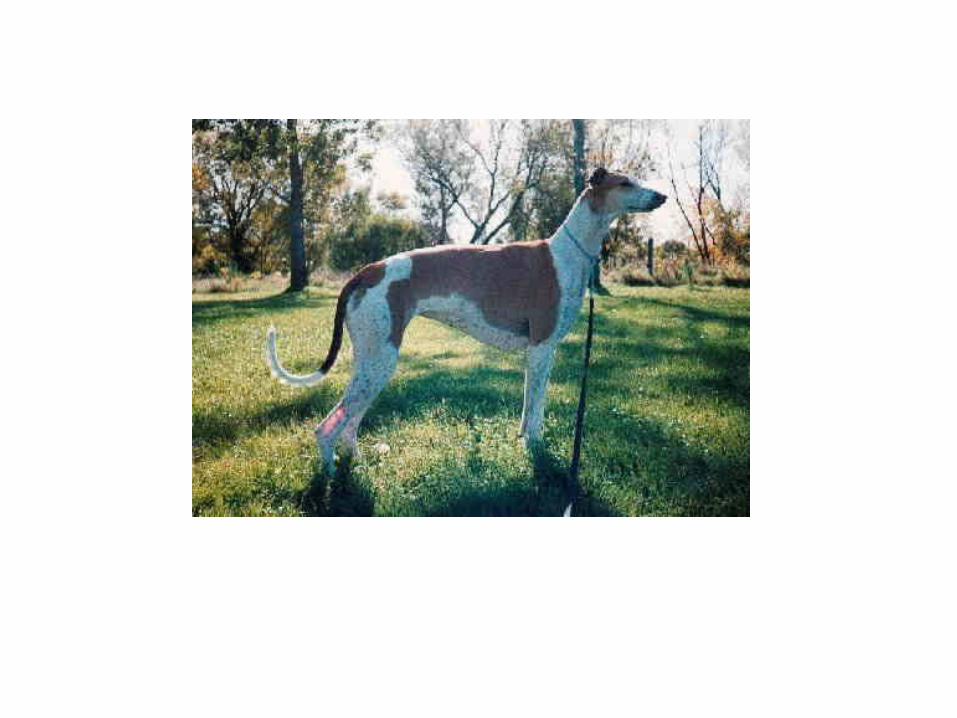

Anatomical Position
• Lateral/Medial
Anatomical Position
• Superior/Inferior


Proximal/Distal

Sections Through the Body
• 1. Transverse• 2. Frontal• 3. Saggital

Saggital Plane


Transverse

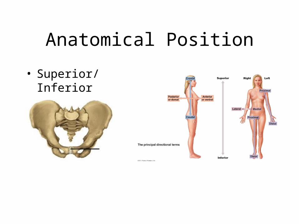
Bones of the leg pp. 248 - 249
• Tibia and Fibula

Bones of the Leg - Patella
Bones of the leg
• Femur
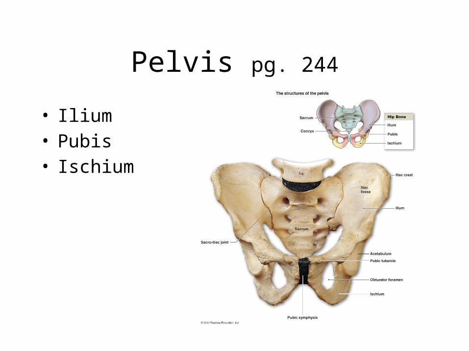
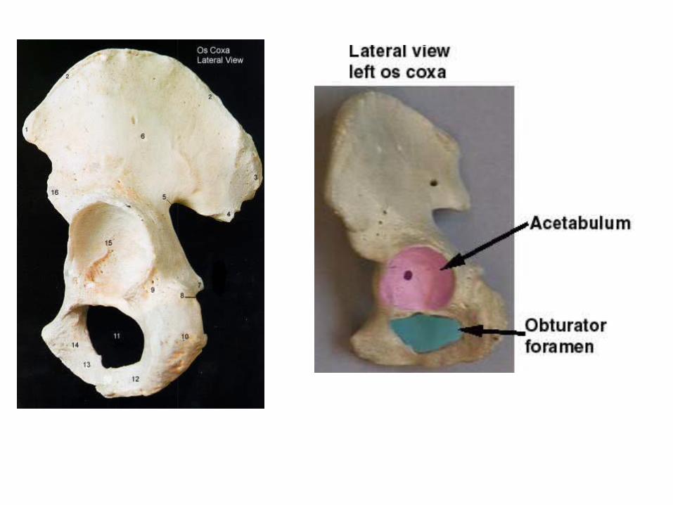
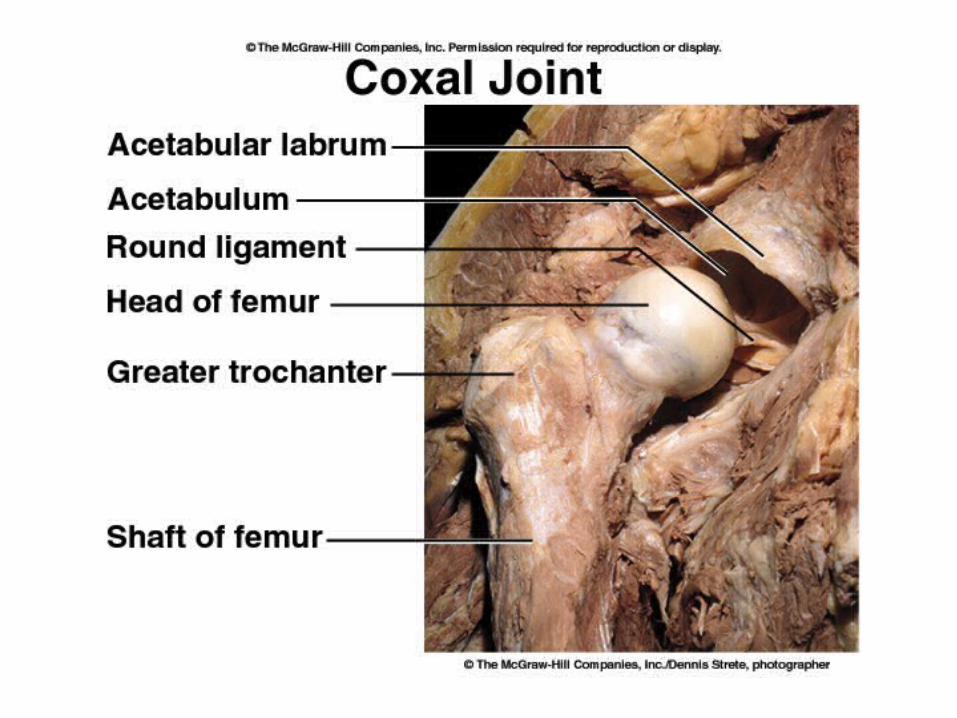
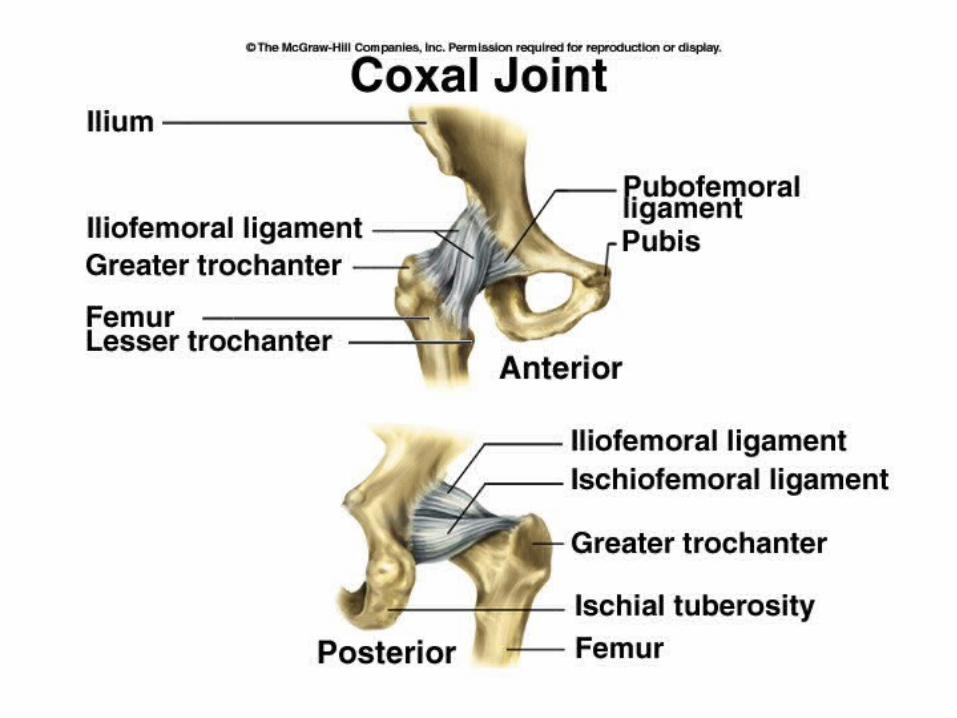
Pelvis pg. 244
• Ilium• Pubis• Ischium

Pg. 245
Welcome to BIOS 2310Human Anatomy & Physiology IInstructor: Mr. Todd Templeton
• Today’s Agenda:– Discussion of the syllabus.– Presentation of today’s topics:
• Anatomical Position• Introduction to the Skeletal System• Class will end at 4:40

Anatomical Position Sec. 1.9
• Anterior/Posterior (ventral/dorsal)

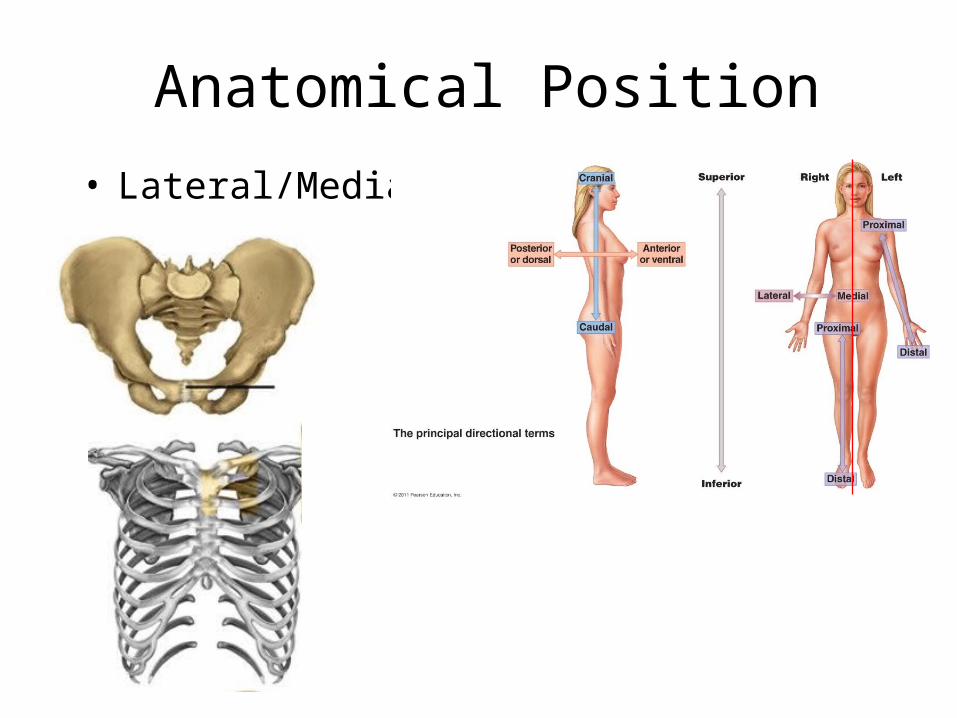
Anatomical Position
• Lateral/Medial

Anatomical Position
• Superior/Inferior
Anatomical Position
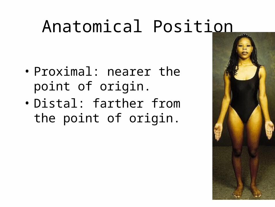
• Proximal: nearer the point of origin.
• Distal: farther from the point of origin.

Proximal/Distal

Sections Through the Body
• 1. Transverse• 2. Frontal• 3. Saggital

Saggital Plane

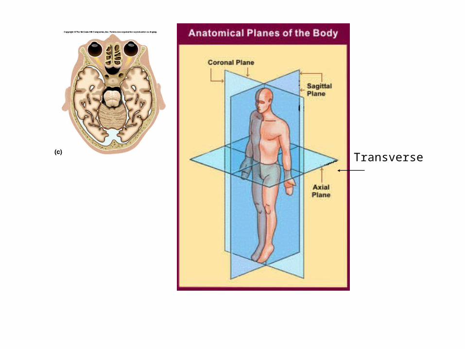
Transverse
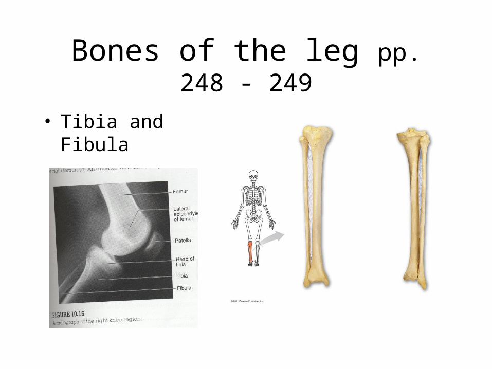
Bones of the leg pp. 248 - 249
• Tibia and Fibula
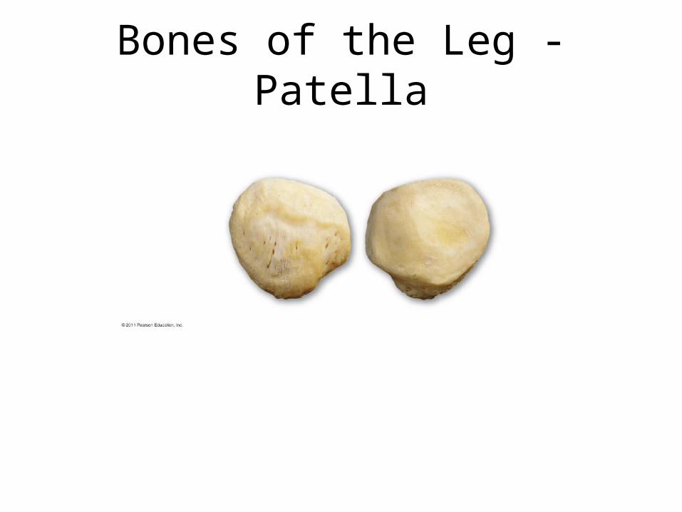
Bones of the Leg - Patella

Bones of the leg
• Femur
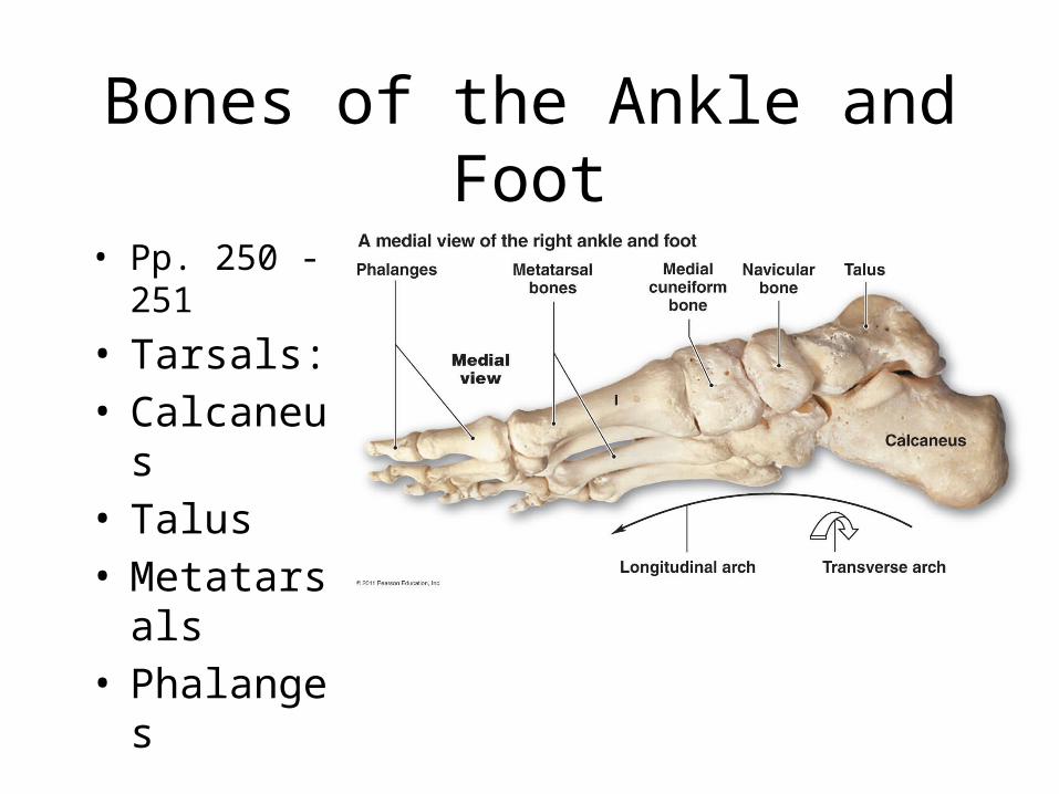
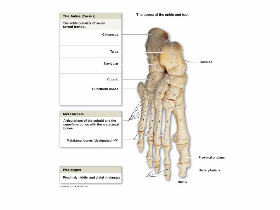
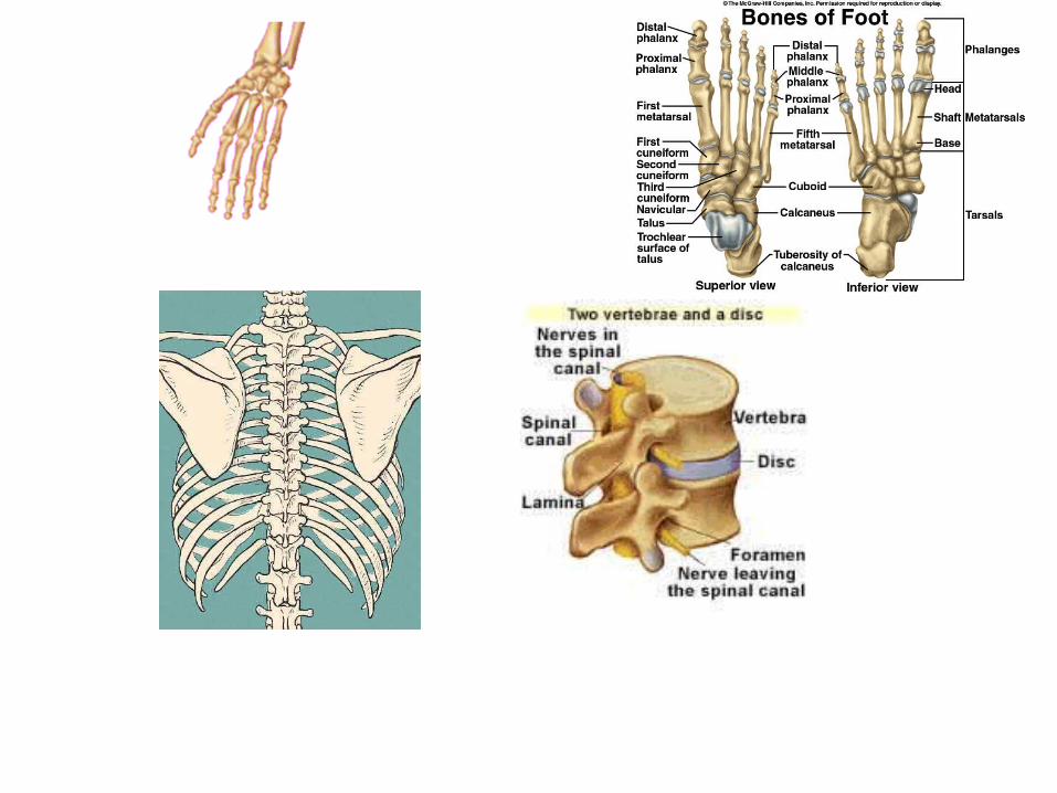
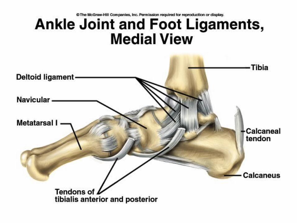
Bones of the Ankle and Foot
• Pp. 250 - 251
• Tarsals:• Calcaneus• Talus• Metatarsals• Phalanges
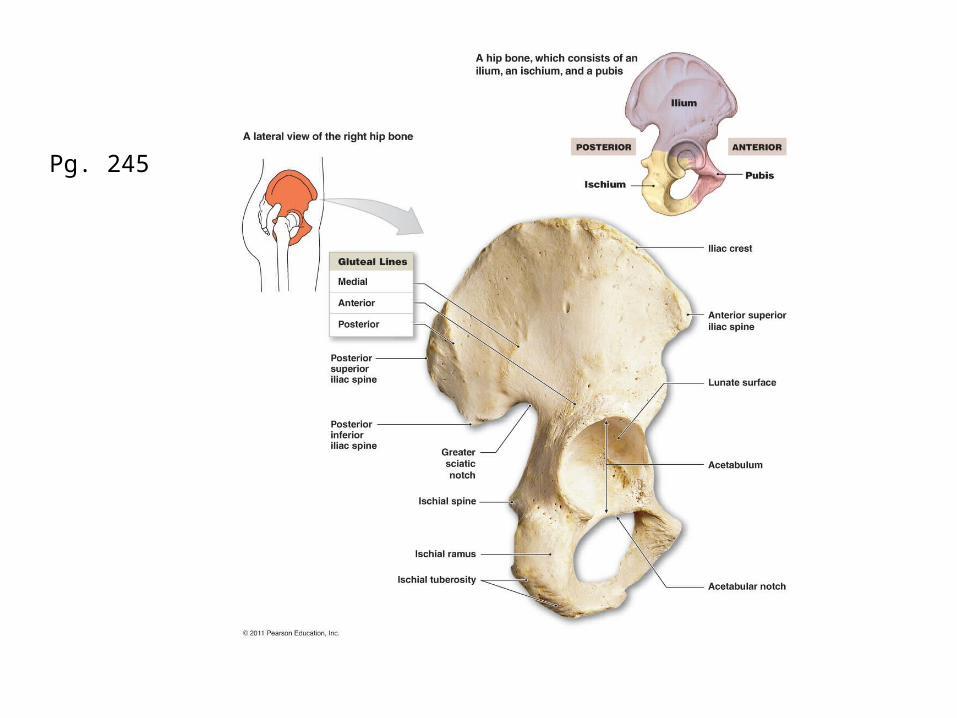
Pg. 245


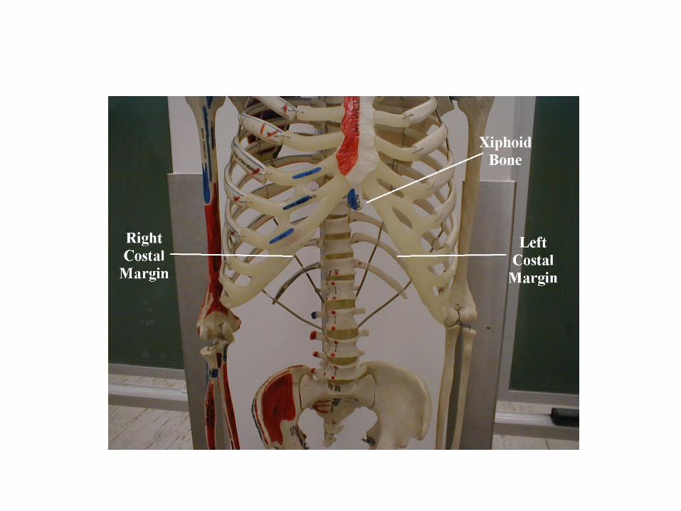
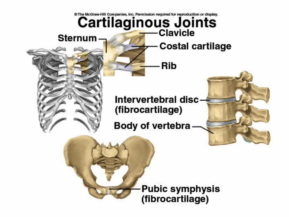
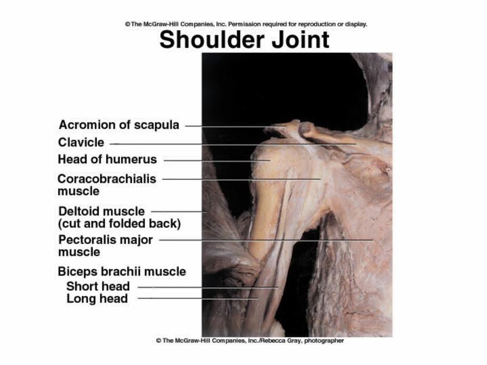
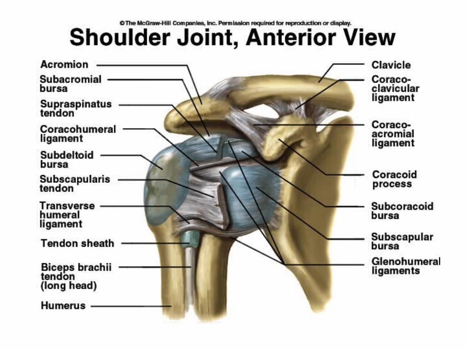
Shoulder and Thoraxpp. 238 – 239, pp. 234 - 235
• Clavicle• Scapula• Sternum• True Ribs 1 – 7
(vertebrosternal)• False Ribs 8 – 12
vertebrochondral (8 – 10). 11 & 12 are floating or vertebral ribs.
• Costal Cartilage


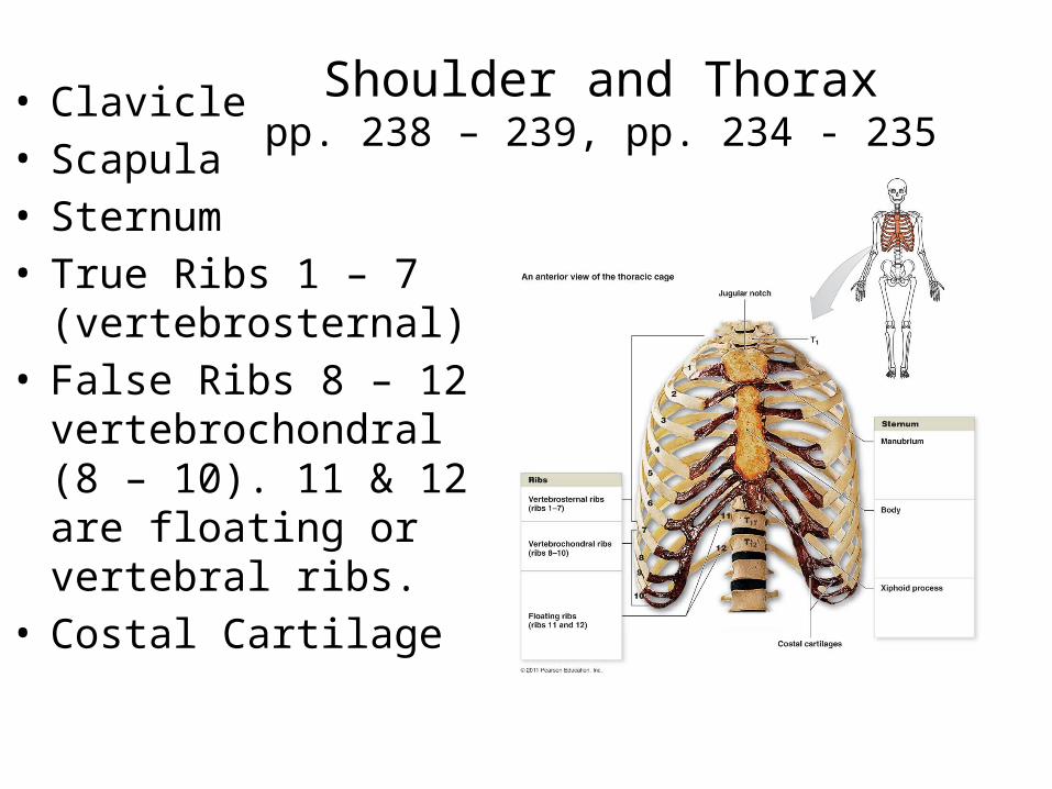
Shoulder and Thoraxpp. 238 – 239, pp. 234 - 235
• Clavicle• Scapula• Sternum• True Ribs 1 – 7
(vertebrosternal)• False Ribs 8 – 12
vertebrochondral (8 – 10). 11 & 12 are floating or vertebral ribs.
• Costal Cartilage

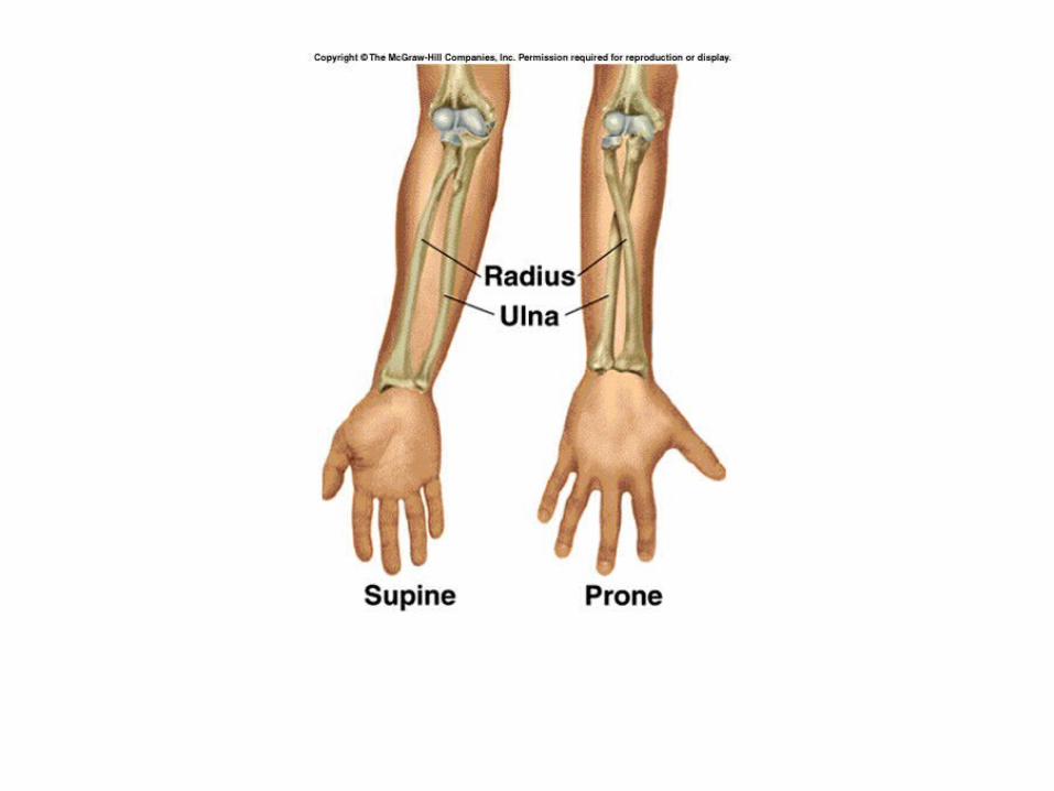
Arm• Pp. 240 - 241• Humerus• Radius• Ulna

Arm: Radius and Ulna
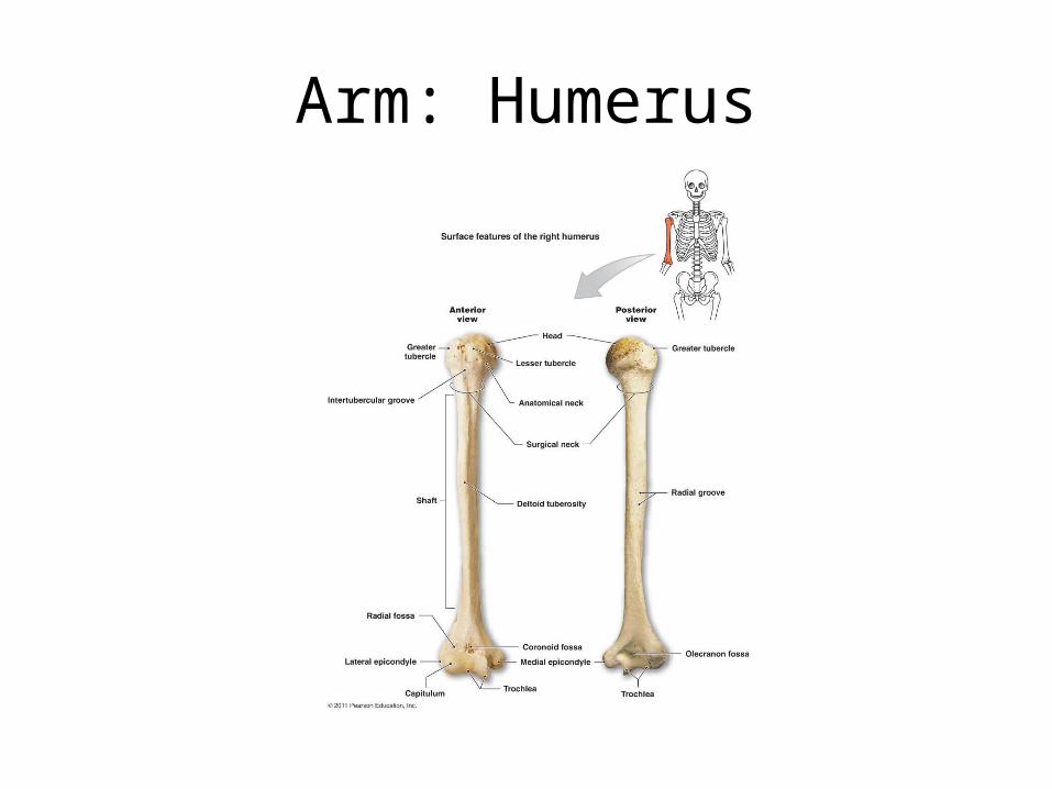
Arm: Humerus
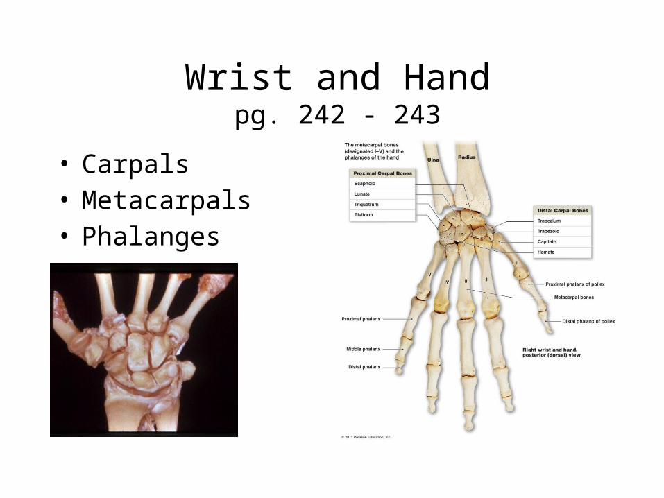
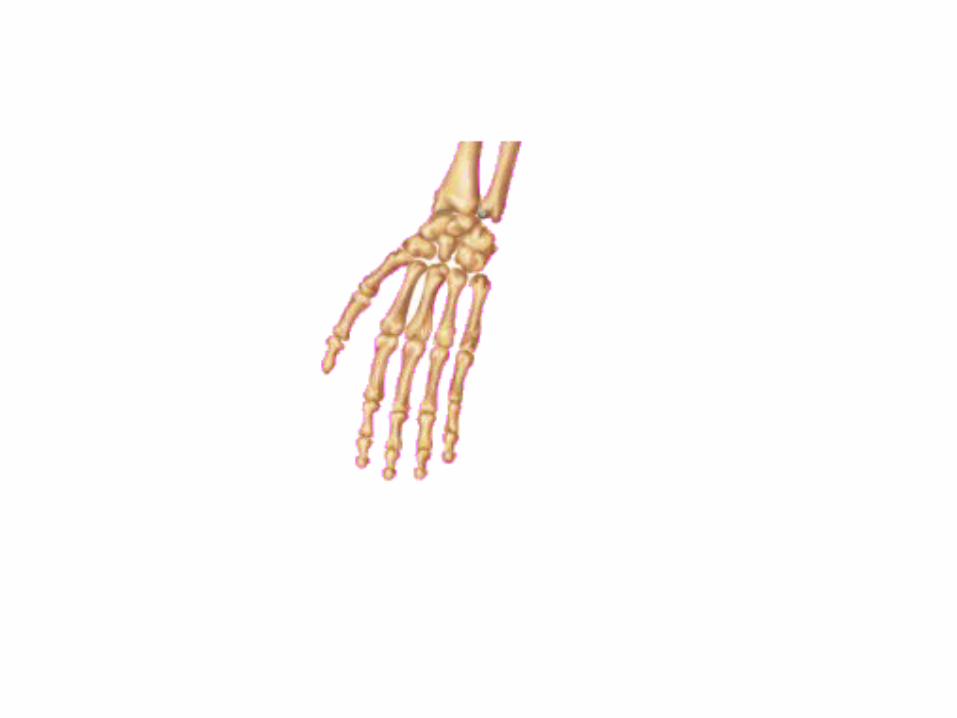
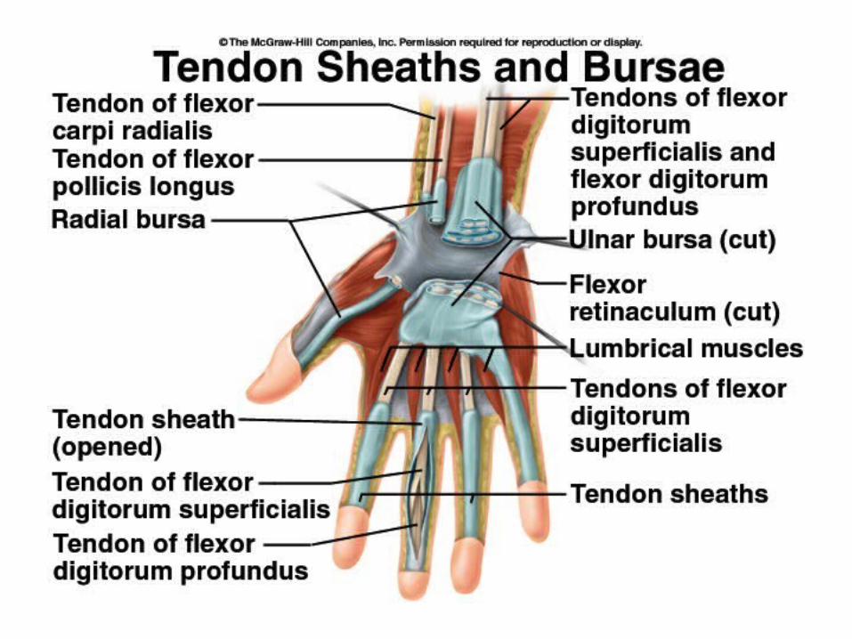
Wrist and Handpg. 242 - 243
• Carpals• Metacarpals• Phalanges


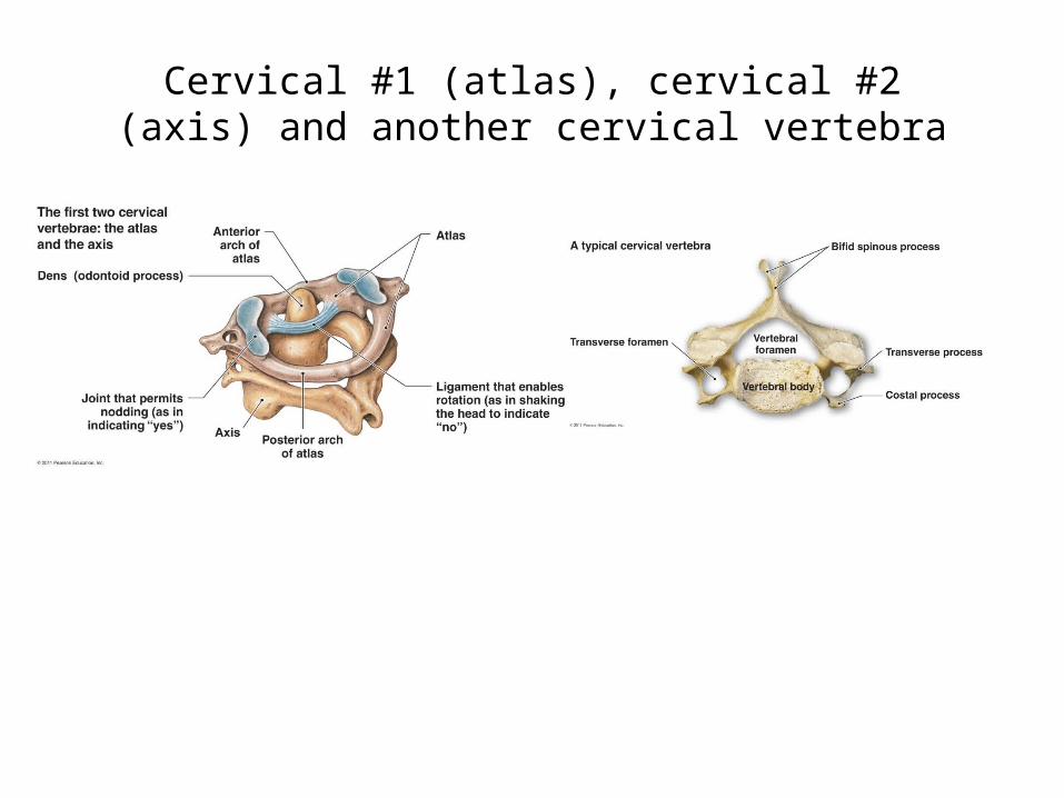
Cervical #1 (atlas), cervical #2 (axis) and another cervical vertebra

Thoracic Vertebrae12 bones pg. 231

Lumbar Vertebrae5 Bones pg. 232

Vertebrae
• Sacrum – 5 bones, fused. Pg. 233
• Coccyx – 4 bones, fused.

Day 2 Begins Here
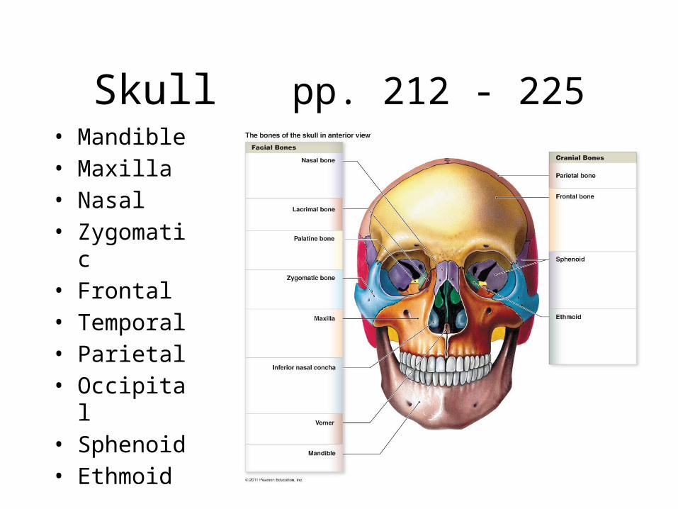
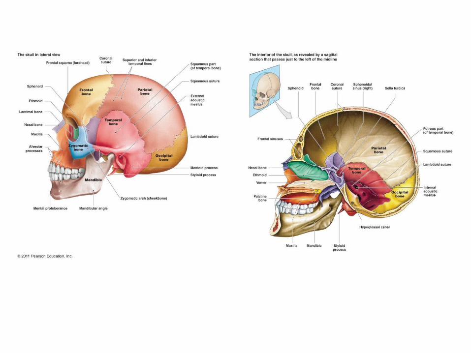
Skull pp. 212 - 225• Mandible• Maxilla• Nasal• Zygomatic• Frontal• Temporal• Parietal• Occipital• Sphenoid• Ethmoid

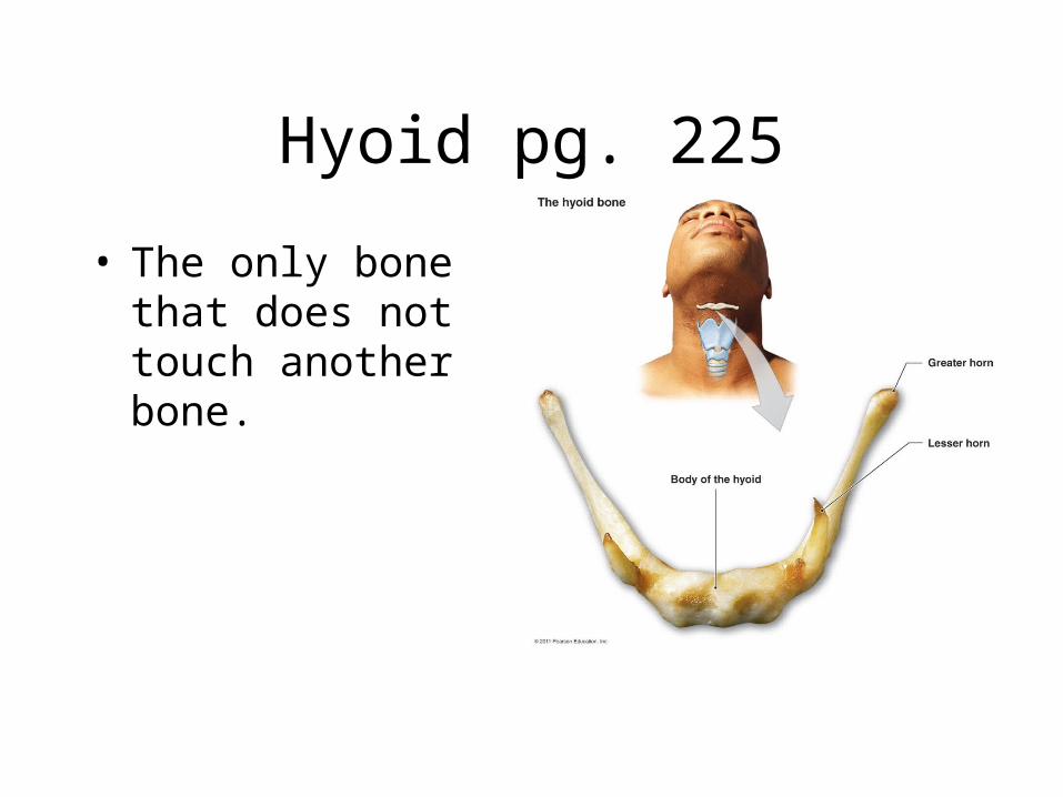
Hyoid pg. 225
• The only bone that does not touch another bone.

Axial and Appendicular Skeleton pg. 211 & pg. 237

Gender Differences

Greater Sciatic Notch

Gender Differences


Body Systems are Interrelated
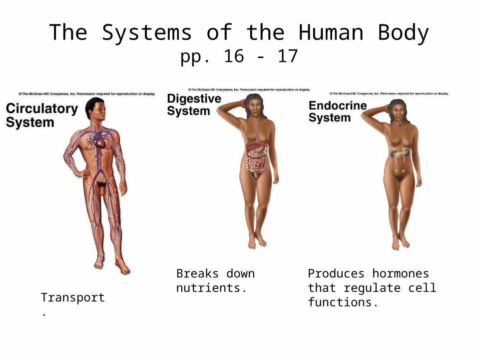
The Systems of the Human Bodypp. 16 - 17
Transport.
Breaks down nutrients. Produces hormones that regulate cell functions.
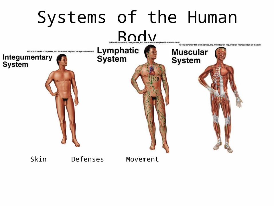
Systems of the Human Body
Skin Defenses Movement
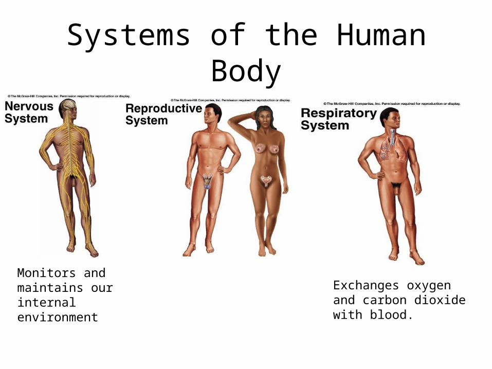
Systems of the Human Body
Monitors and maintains our internal environment Exchanges oxygen and
carbon dioxide with blood.
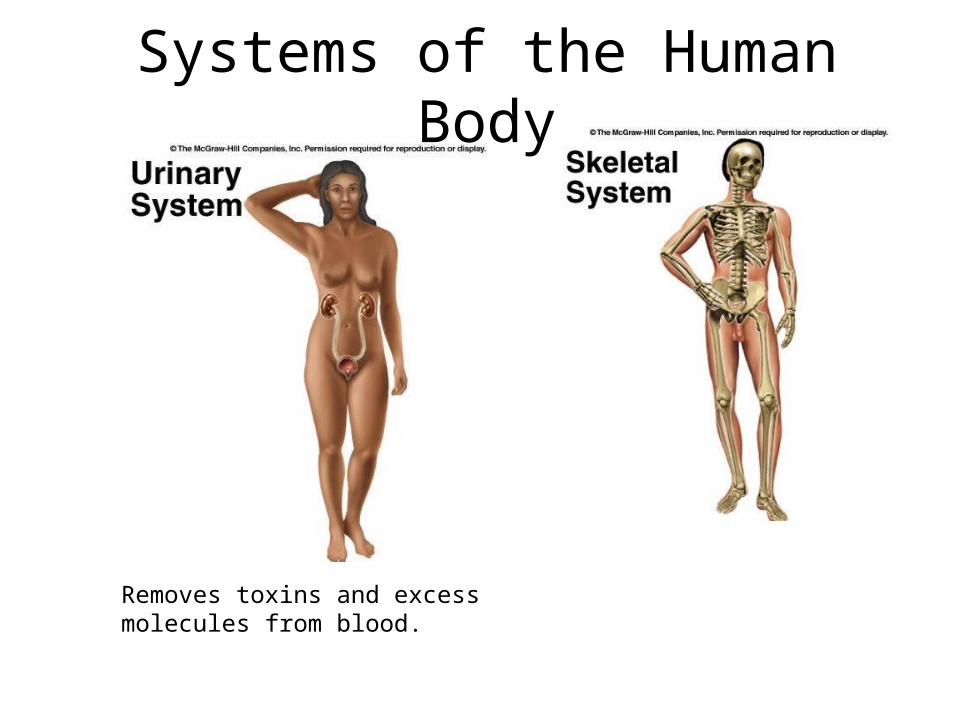
Systems of the Human Body
Removes toxins and excess molecules from blood.

Day 3 Begins Here
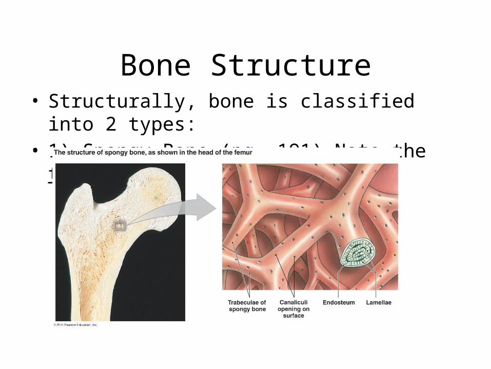
Bone Structure• Structurally, bone is classified into 2 types:• 1) Spongy Bone (pg. 191) Note the trabeculae.

Bone Structure• 2) Compact bone • pp. 191.• Note: we will discuss
the microscopic structure of bone in the next unit.
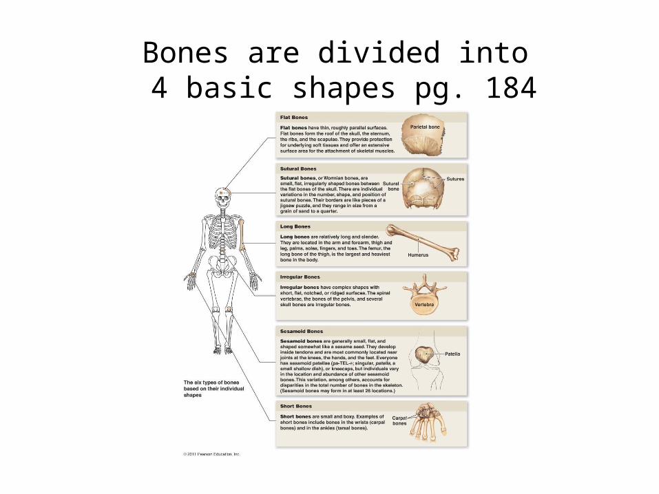
Bones are divided into 4 basic shapes pg. 184

1: Long Bones (pg. 186)• Longer than wide• Cylinder shaped.• Located in the appendicular
skeleton. • Diaphysis (compact)• Epiphysis (spongy)
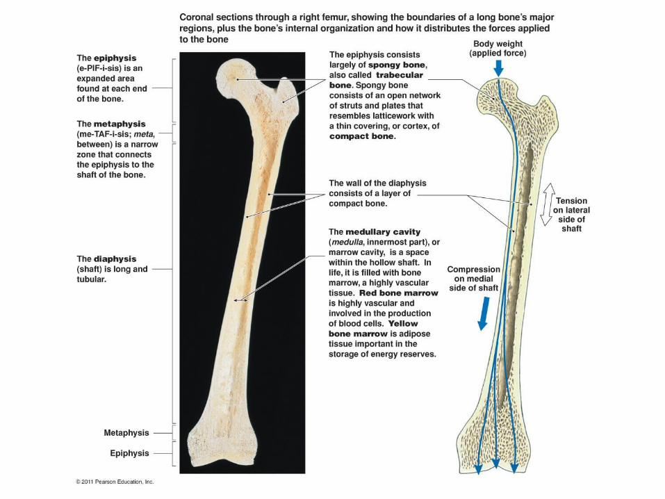
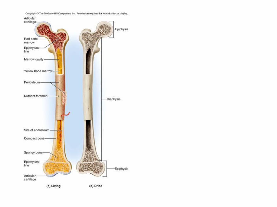

Long Bone
• Medullary Canal• Yellow marrow• Periosteum

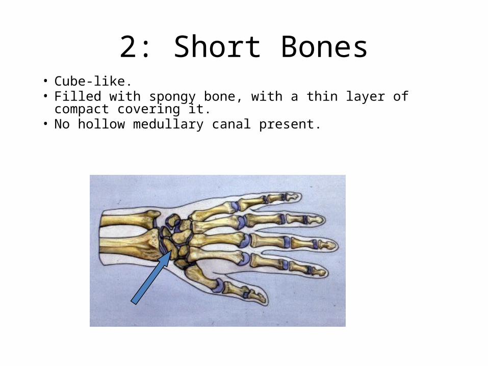
2: Short Bones• Cube-like.• Filled with spongy bone, with a thin layer of compact covering it.• No hollow medullary canal present.
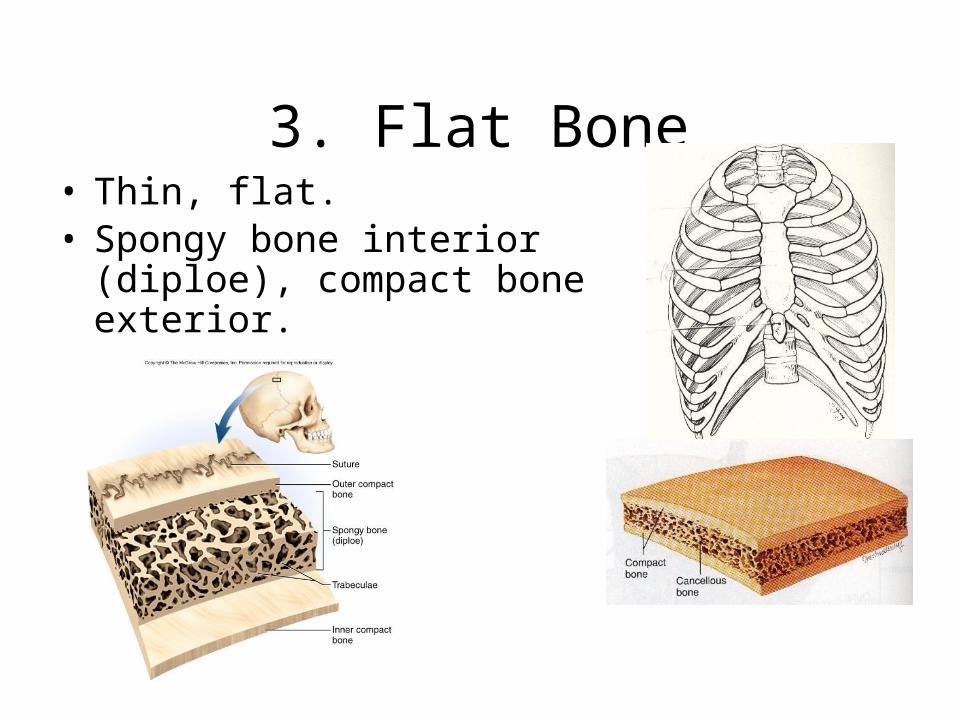
3. Flat Bone• Thin, flat.• Spongy bone interior (diploe),
compact bone exterior.

4. Irregular Bone
• Usually described as a cross between a flat bone and a short bone.
• Spongy interior. • Vertebrae and some skull bones would be
examples.
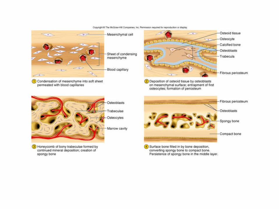
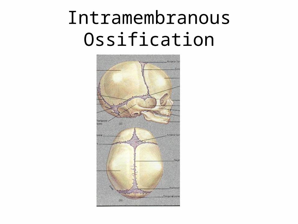
Intramembranous Ossification
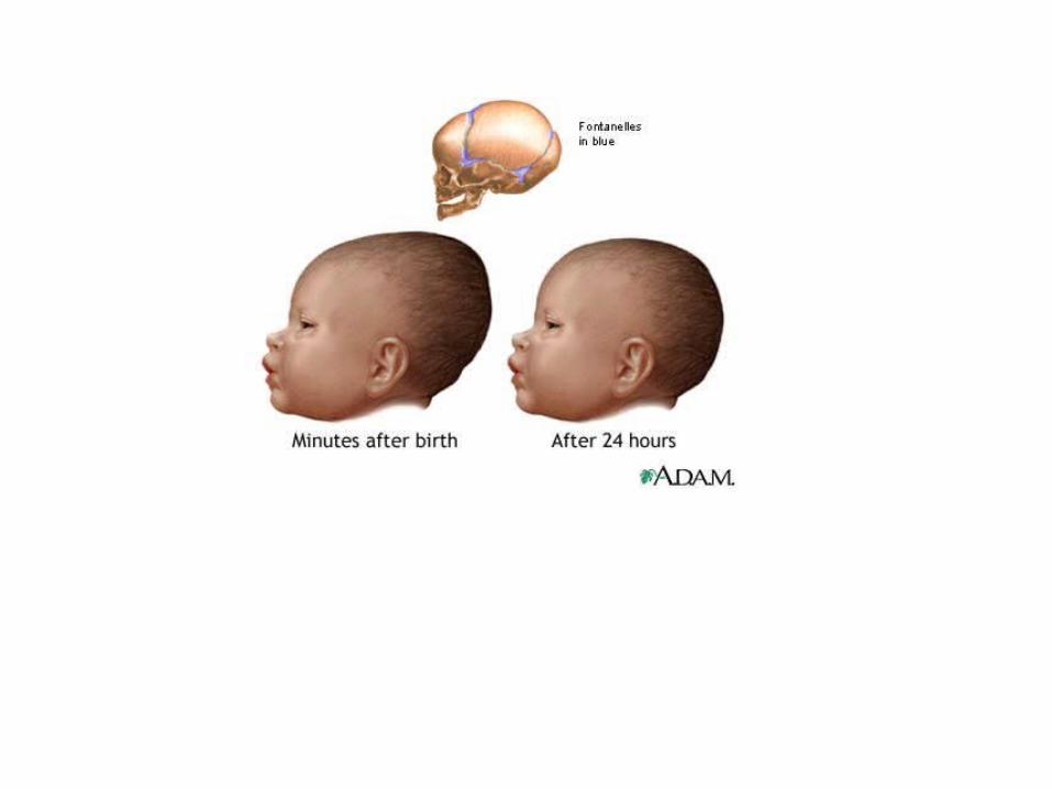

Hydroencephalus

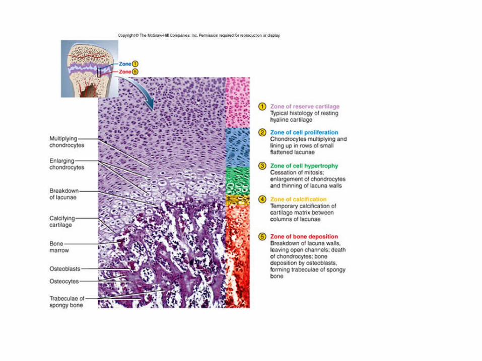
Endochondral Ossification
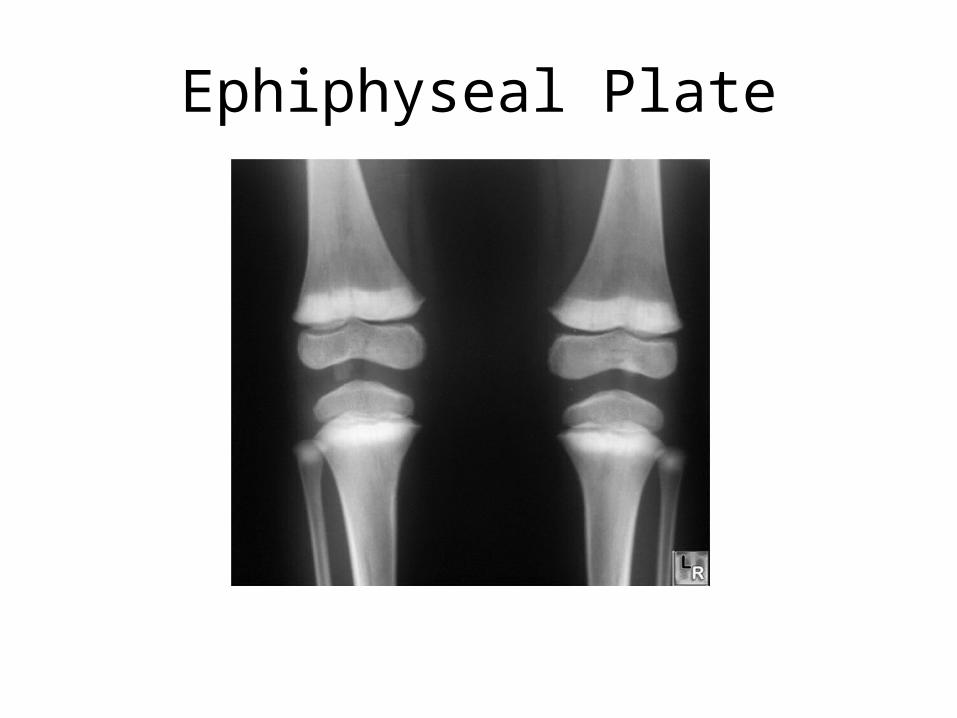
Ephiphyseal Plate

Epiphyseal (Growth) Plate
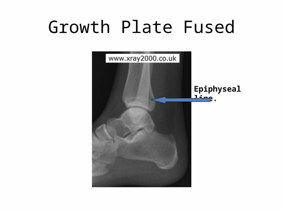
Growth Plate Fused
Epiphyseal line.

Fetal Bone Growth pg. 197

Greenstick Fracture

Day 4 Begins Here

Today’s Agenda
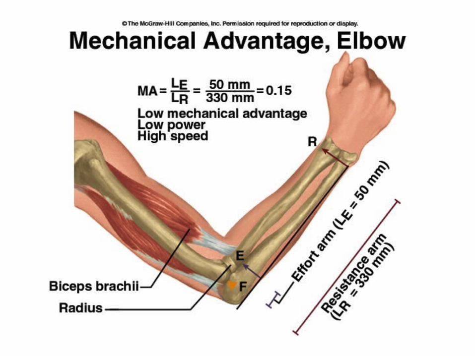
• Today we will look at joint structure and function, along with lever systems. Ch. 8
• After lab we will begin preparations for exam #1, which is the next class meeting.
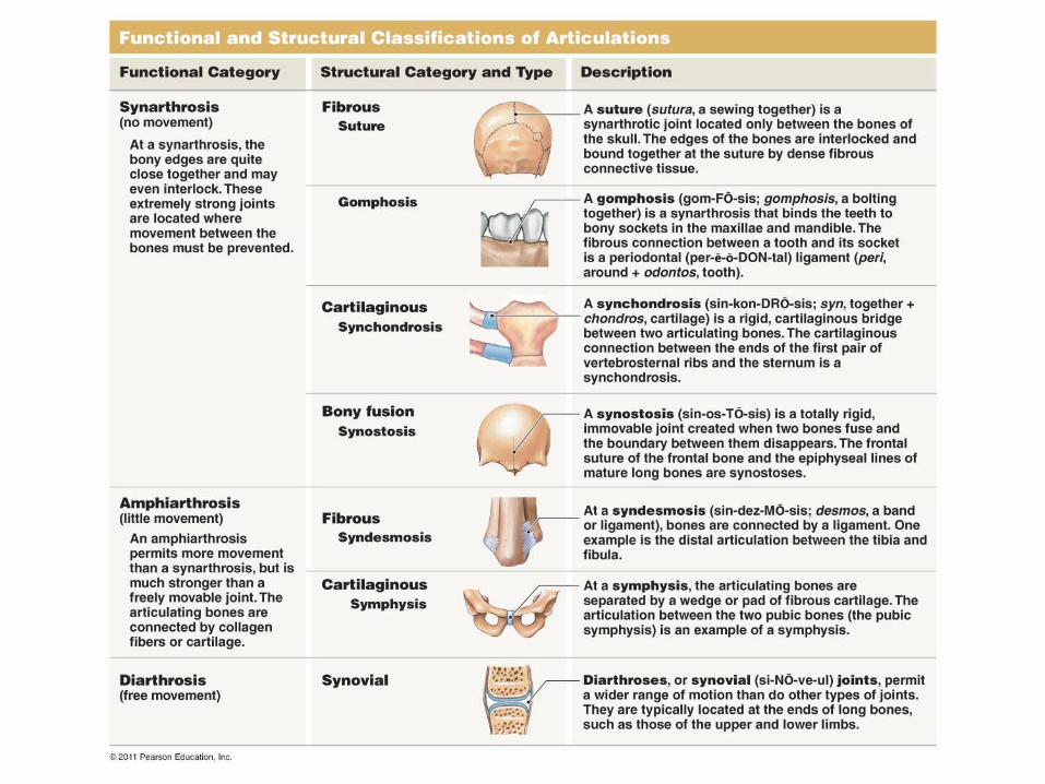
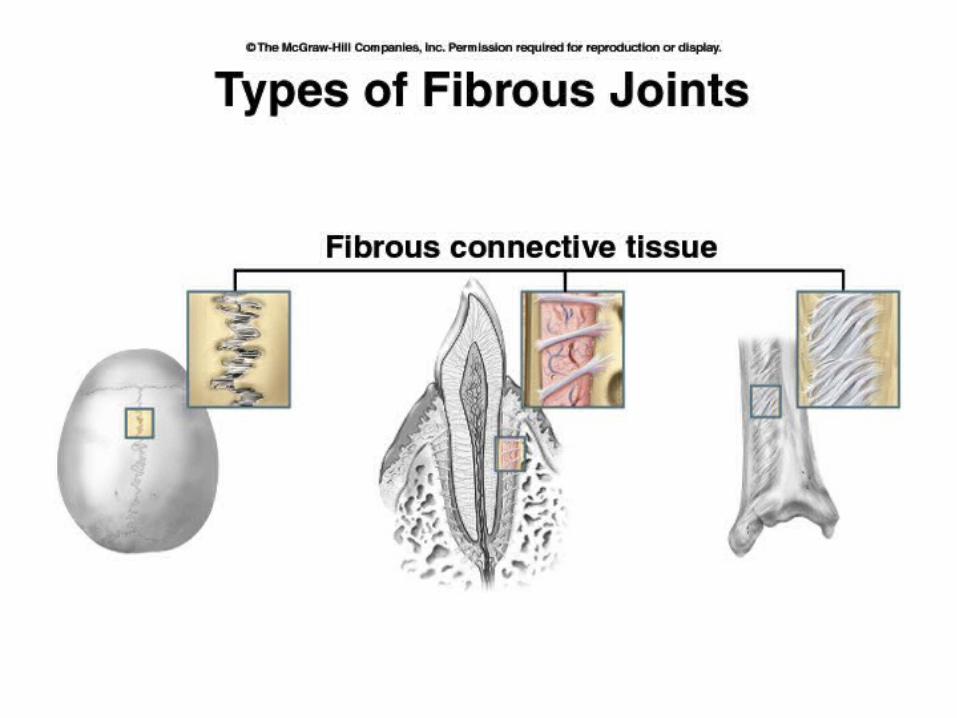

Suture Joint

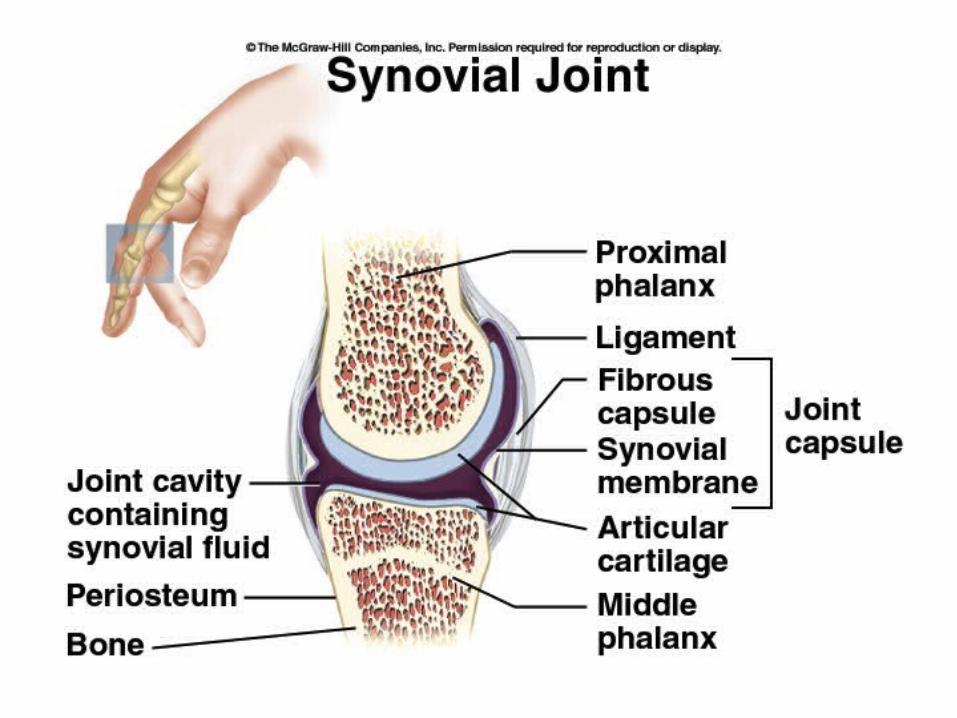
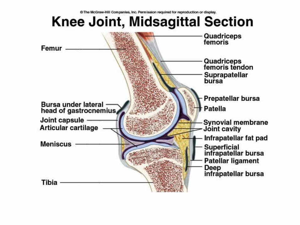

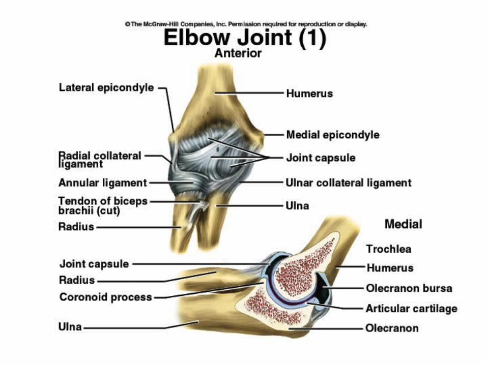
Synovial Joint


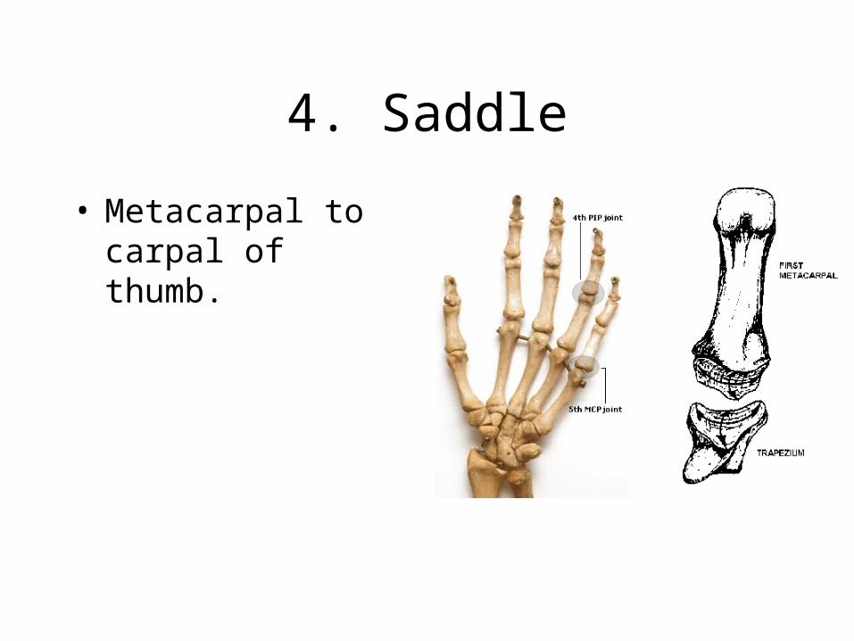
4. Saddle
• Metacarpal to carpal of thumb.

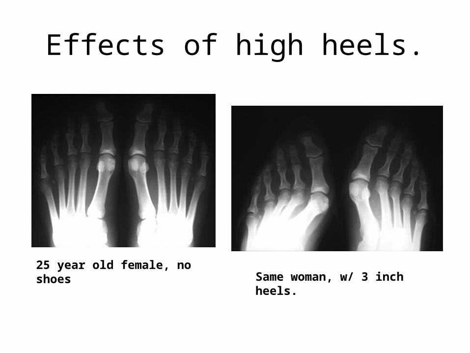
25 year old female, no shoesSame woman, w/ 3 inch heels.
Effects of high heels.
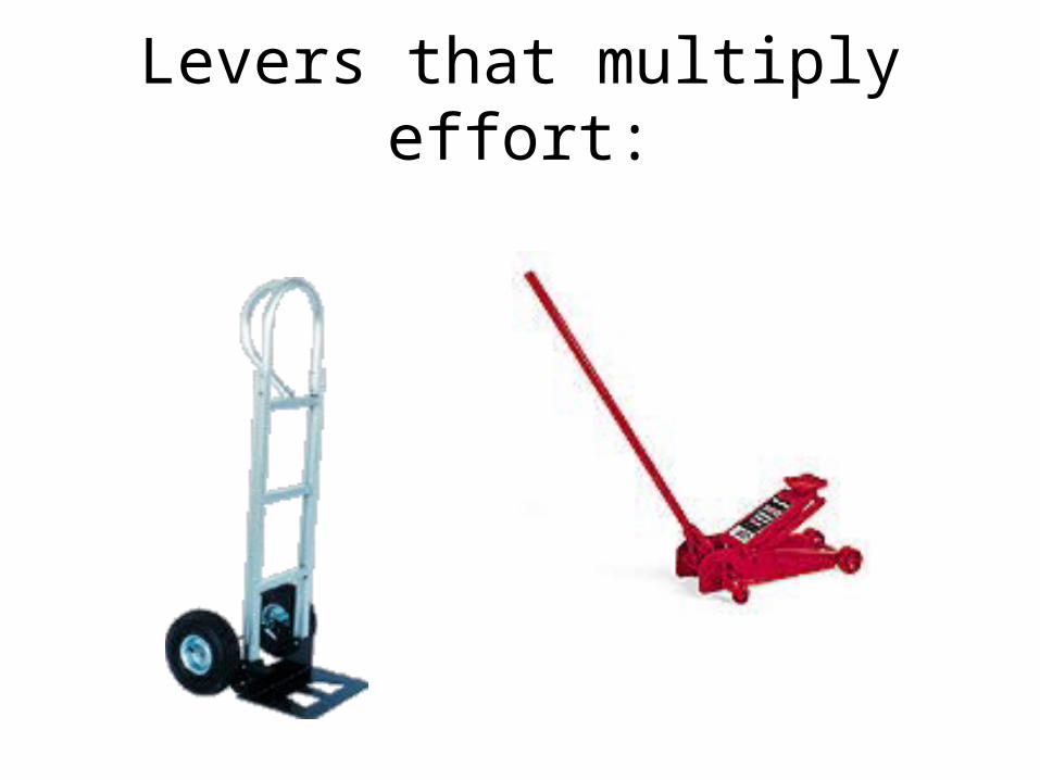
Levers that multiply effort:
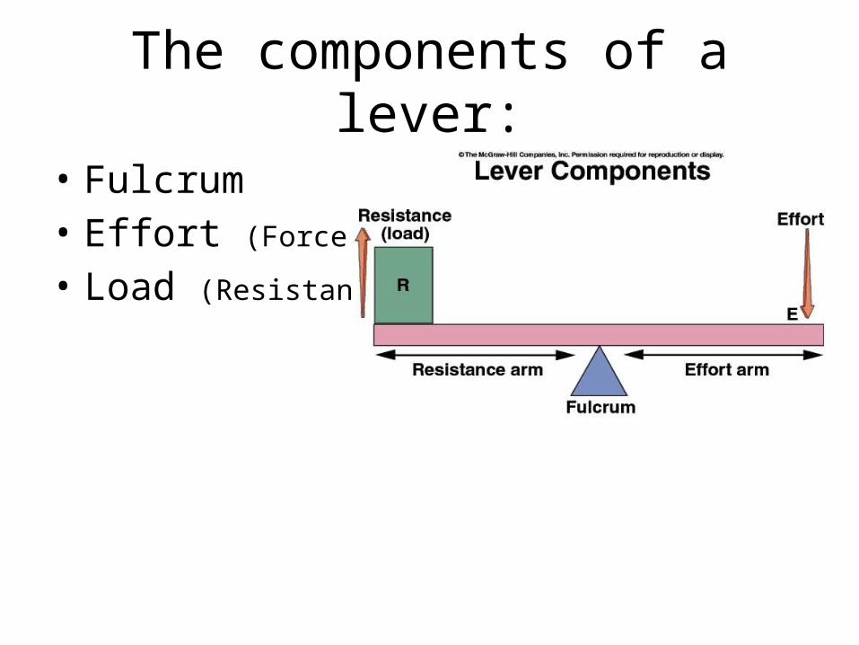
The components of a lever:
• Fulcrum• Effort (Force)
• Load (Resistance)


Speed Lever