Embed Size (px)
Citation preview

DNA Replica,on -‐ Prokaryotes

Figure 02B: Conserva,ve replica,on

Figure 02A: Semiconserva,ve replica,on

Photograph
Reproduced from M. Meselson and F. W. Stahl, Proc. Natl. Acad. Sci. USA 44 (1958): 671-‐682. Photo courtesy of MaQhew S. Meselson, Harvard University.

Figure 04: Demonstra,on of a θ structure intermediate in E. coli chromosome replica,on
Reproduced from J. Cairns, Cold Spring Harb. Symp. Quant. Biol. 28 (1964): p. 44. Copyright 1963, Cold Spring Harbor Laboratory Press. Used with permission of John Cairns, University of Oxford.

Figure 05B: Bidirec,onal replica,on

Figure 07: Evidence to support bidirec,onal θ-‐replica,on
Reprinted from J. Mol. Biol., vol. 73, E. B. Gyurasits and R. G. Wake, Bidirec,onal chromosome replica,on..., pp. 55-‐58, copyright 1973, with permission from Elsevier [hQp://www.sciencedirect.com/science/journal/00222836]. Photo courtesy of R. G. Wake, Professor Emeritus, The University of Sydney, Australia.

Figure 10: Okazaki's pulse labeling experiment
Adapted from K. Sugimoto, T. Okazaki, and R. Okazaki, Proc. Natl. Acad. Sci. USA 60 (1968): 1356-‐1362.
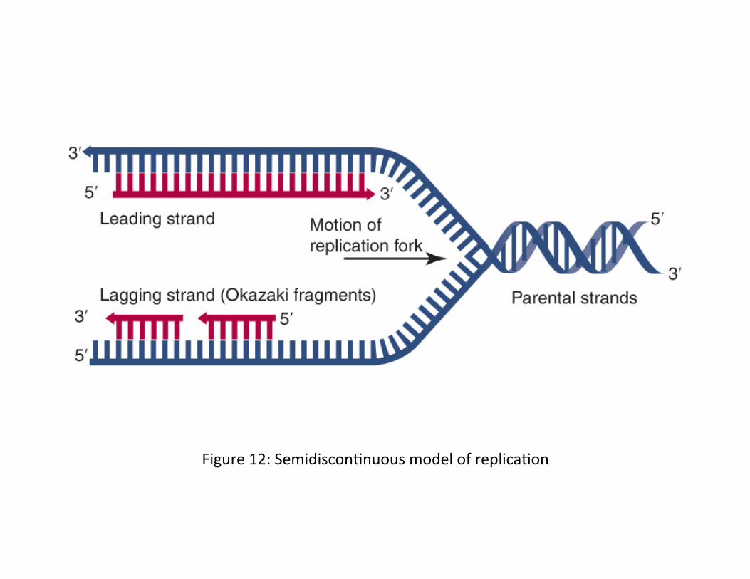
Figure 12: Semidiscon,nuous model of replica,on

Figure 16: Pathway for processing Okazaki fragments

Figure 24: DnaB orienta,on at the replica,on fork.
Adapted from K. Marians, Nat. Struct. Mol. Biol. 15 (2008): 125-‐127.

Figure 35B: Two core polymerases are involved in lagging strand synthesis Reprinted from Mol. Cell, vol. 27, P. McInerney, et al., Characteriza,on of a Triple DNA Polymerase Replisome, pp. 527-‐538, copyright 2007, with permission from Elsevier [hQp://www.sciencedirect.com/science/journal/10972765].

Figure 38: Loca,ons of the E. coli replica,on terminator (Ter ) sites
Adapted from S. Mulugu, et al., Proc. Natl. Acad. Sci. USA 98 (2001): 9569-‐9574.

Figure 39: Crystal structure of the Tus•Ter complex of E. coli showing the DNA-‐binding region of β-‐strands and the helicase blocking end
Structure from Protein Data Bank 1ECR. K. Kamada, et al., Nature 383 (1996): 598-‐603. Prepared by B. E. Tropp.

Figure 10: Conjugal transfer of DNA mediated by bacterial sex plasmids.


