Embed Size (px)
Citation preview
DNA replication and repair - Lecture 3
Jim Borowiec
September 28, 2006
QuickTime™ and aTIFF (Uncompressed) decompressor
are needed to see this picture.

Overview of DNA replication
TelomereTelomere Centromere
DNA chromosome
Specialized elements termed'origins of DNA replication’
occur many times on achromosome
Origin of DNA replication
Initiation of DNA replicationfrom origin of replicationgenerates structures termed'DNA replication bubbles'

End replication problem
DNA replication(from internal regions of the chromosome)
3’
5’
3’
5’
by leading strand synthesis
+3’
5’
by lagging strand synthesis
iDNA
RNA primer
Processing
3’
5’
DNA
replication
3’
5’
loss of DNA
After multiple rounds of DNA replication, genetic information will be lost
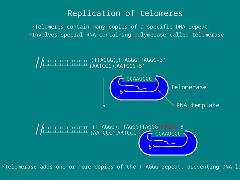
Replication of telomeres
•Telomeres contain many copies of a specific DNA repeat
(TTAGGG)nTTAGGGTTAGGG-3’(AATCCC)nAATCCC-5’
•Involves special RNA-containing polymerase called telomerase
TelomeraseCCAAUCCC
RNA template
5’
•Telomerase adds one or more copies of the TTAGGG repeat, preventing DNA loss
(TTAGGG)nTTAGGGTTAGGGTTAGGG-3’(AATCCC)nAATCCC CCAAUCCC
5’

Senescence
‘Hayflick limit’
Somatic cells
‘Hayflick limit’
Widespread cell death
Crisis
Germ line cells
Senescence
p53 mutation
Somatic cells
‘Hayflick limit’
Widespread cell death
Crisis
Germ line cells
Senescence
p53 mutation
Telomerase activation
Telomere stabilization
Somatic cells
Telomere length
Cell Divisions
Germ line cells
Telomerase needed for cell immortalization
Most somatic cells do not have telomerase activity
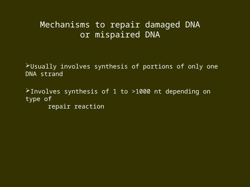
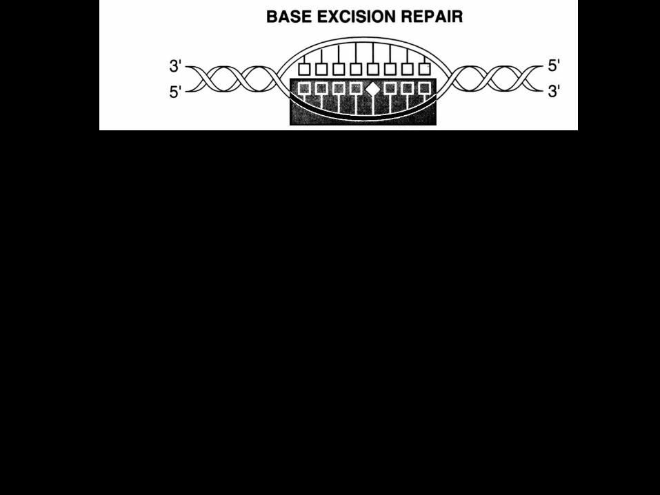
Mechanisms to repair damaged DNAor mispaired DNA
Usually involves synthesis of portions of only one DNA strand
Involves synthesis of 1 to >1000 nt depending on type of repair reaction
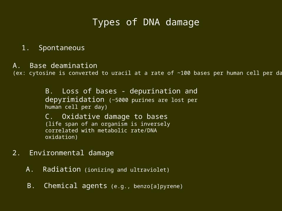
Types of DNA damage
1. Spontaneous
C. Oxidative damage to bases (life span of an organism is inversely correlated with metabolic rate/DNA oxidation)
B. Loss of bases - depurination and depyrimidation (~5000 purines are lost per human cell per day)
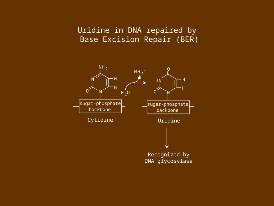
A. Base deamination (ex: cytosine is converted to uracil at a rate of ~100 bases per human cell per day)
2. Environmental damage
B. Chemical agents (e.g., benzo[a]pyrene)
A. Radiation (ionizing and ultraviolet)
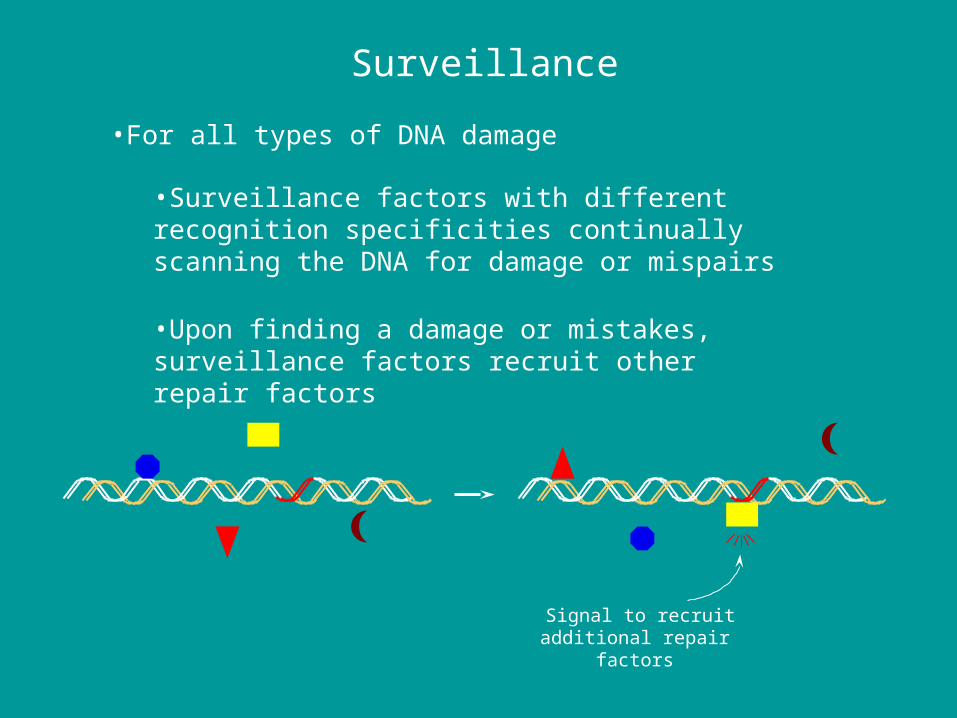
Surveillance
•For all types of DNA damage
•Surveillance factors with different recognition specificities continually scanning the DNA for damage or mispairs
•Upon finding a damage or mistakes, surveillance factors recruit other repair factors
Signal to recruitadditional repair
factors

sugar-phosphatebackbone
Cytidine
NH 2
H
O NH
N
Deamination of cytidine to uridine (spontaneous)
O
H
O NH
HN
sugar-phosphatebackbone
Uridine
NH 4+
H 2O
Cytidine
NH 2
H
O NH
N
sugar-phosphatebackbone
O
H
O NH
HN
sugar-phosphatebackbone
Uridine
NH 4+
H 2O
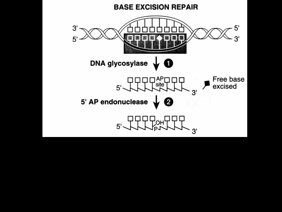
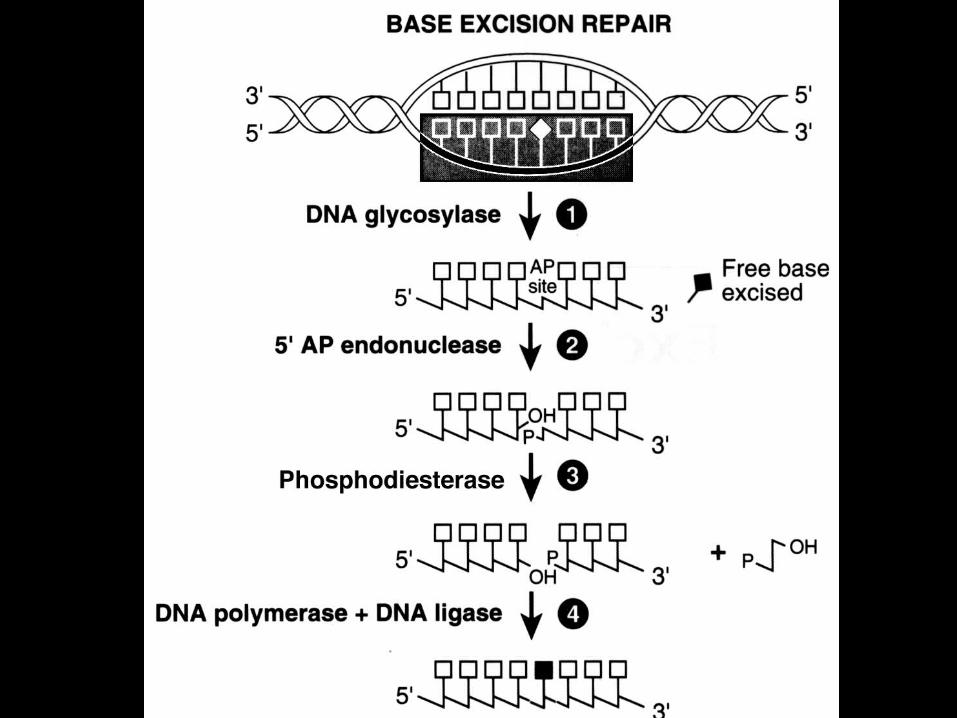
Recognized byDNA glycosylase
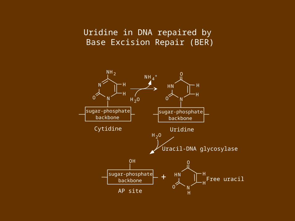
Uridine in DNA repaired by Base Excision Repair (BER)

MANY DNA GLYCOSYLASES EXIST
DIFFER IN SUBSTRATE SPECIFICITY
GENERALLY RECOGNIZE MONO-ADDUCT DAMAGE
Cytidine
NH 2
H
O NH
N
sugar-phosphatebackbone
O
H
O NH
HN
sugar-phosphatebackbone
Uridine
NH 4+
H 2O
Uracil-DNA glycosylase
sugar-phosphatebackbone
AP site
OH
H 2O
+
O
H
O NH
HN
H
Free uracil
Uridine in DNA repaired by Base Excision Repair (BER)


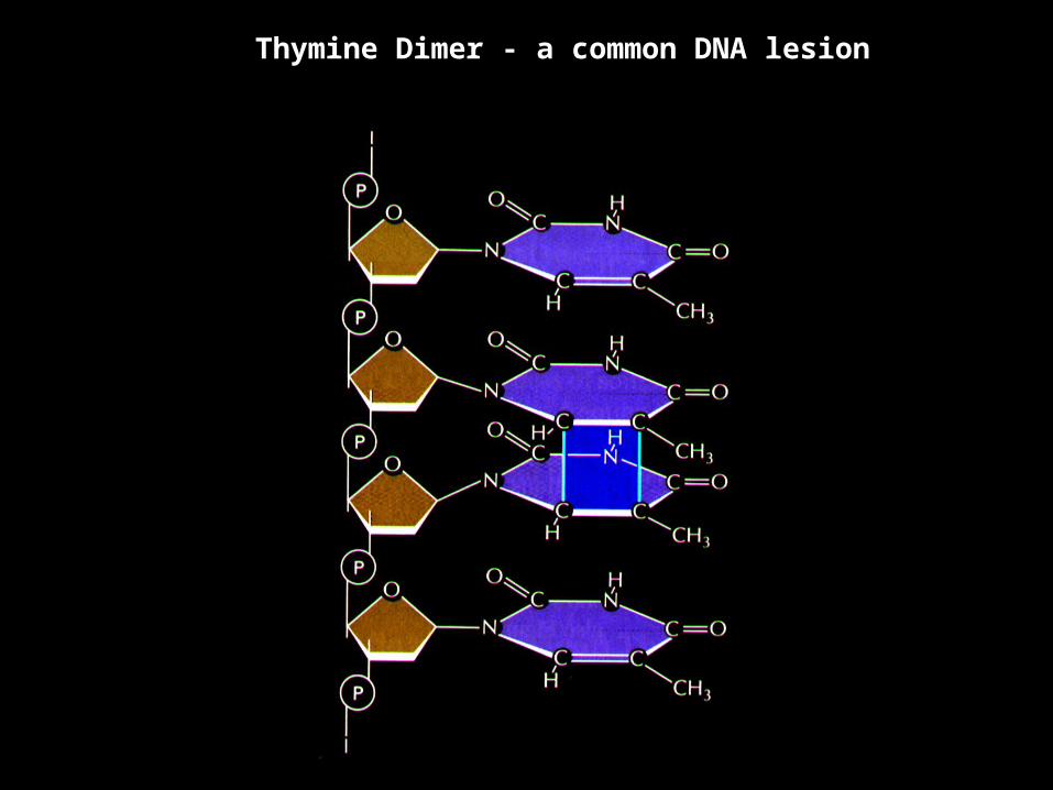
Thymine Dimer - a common DNA lesion
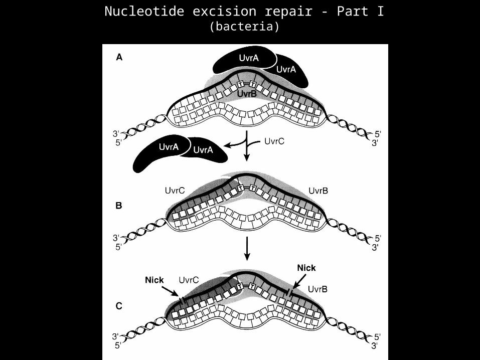
Nucleotide excision repair - Part I(bacteria)

Nucleotide excision repair - Part II(bacteria)
(uvrD)
Human nucleotide excision repair (NER)
•Xeroderma pigmentosum (XP) - an inherited disease in which patients show an extreme sensitivity to sunlight
•XP is a result of mutation of various genes involved in NER

Xeroderma Pigmentosum Society, Inc.Camp Sundown for XP children
QuickTime™ and aTIFF (Uncompressed) decompressorare needed to see this picture.
The program schedule is 9:00 p.m. to 5:00 a.m. to maximize night time hours for play and minimize need for protective arrangements.
QuickTime™ and aTIFF (Uncompressed) decompressorare needed to see this picture.
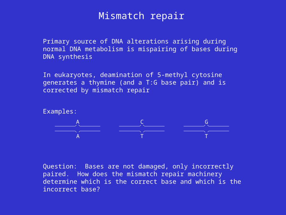
Mismatch repair
Primary source of DNA alterations arising during normal DNA metabolism is mispairing of bases during DNA synthesis
In eukaryotes, deamination of 5-methyl cytosine generates a thymine (and a T:G base pair) and is corrected by mismatch repair
A
A
Examples:
C
T
G
T
Question: Bases are not damaged, only incorrectly paired. How does the mismatch repair machinery determine which is the correct base and which is the incorrect base?
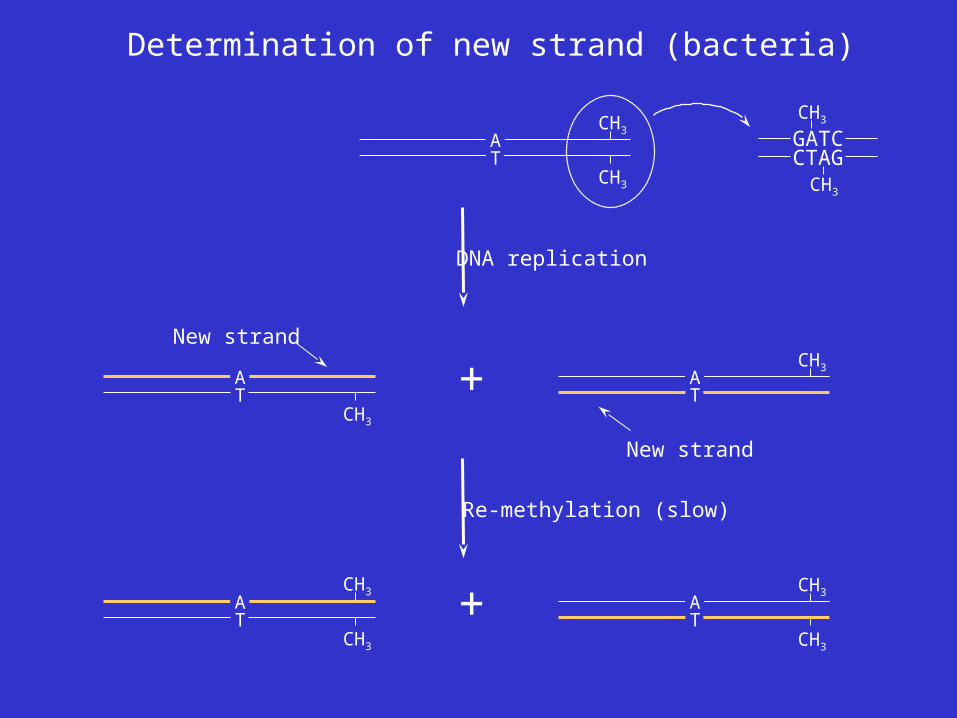
Determination of new strand (bacteria)
AT
CH3
CH3
DNA replication
New strand
AT
CH3
AT
CH3
New strand
+
CH3
CH3
Re-methylation (slow)
AT
CH3
AT
CH3+
CTAG
CH3
GATC
CH3
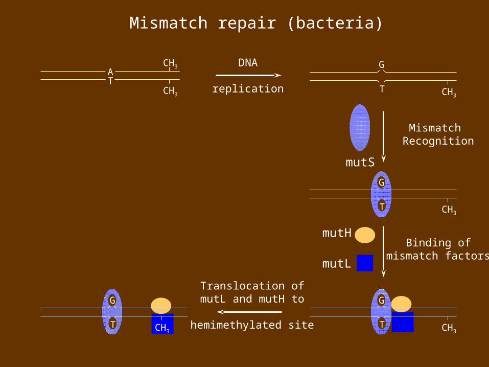
Mismatch repair (bacteria)
AT
CH3
CH3
G
T
DNA
replication CH3
Mismatch Recognition
mutS
G
T CH3
Binding ofmismatch factors
mutH
mutL
G
T CH3
G
T
Translocation ofmutL and mutH to
hemimethylated siteCH3
G
T CH3
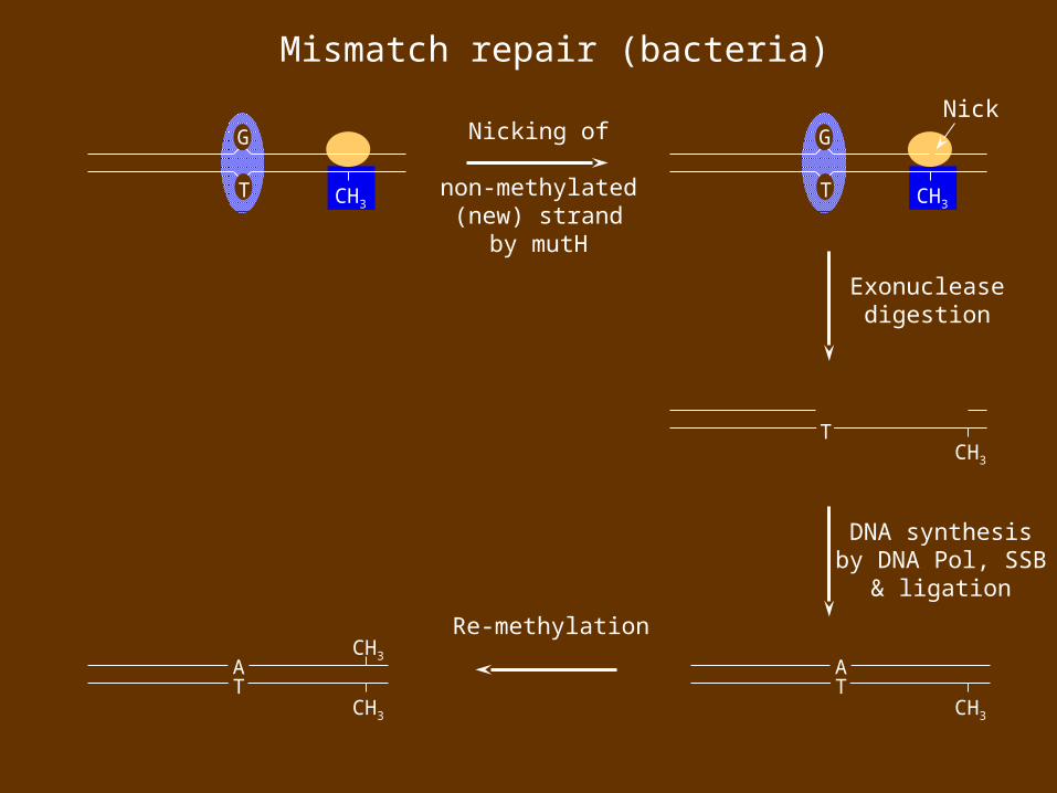
Mismatch repair (bacteria)
AT
CH3
DNA synthesisby DNA Pol, SSB
& ligation
T
Exonucleasedigestion
CH3
Nicking of
non-methylated(new) strand
by mutH
G
T CH3
Nick
AT
CH3
CH3
Re-methylation

Hereditary nonpolyposis colon cancer (HNPCC)
•HNPCC is a hereditary cancer syndrome with individuals having increased incidence of colon cancer, ovarian cancer, and endometrial tumors
•Caused by defects in human mismatch repair genes that are homologous to bacterial mismatch repair genes
- Defects in hMSH2 (human mutS homolog) account for ~60% of HNPCC cases
- Defects in hMLH1 (human mutL homolog) account for ~30% of HNPCC cases
•Cells from HNPCC patients are 100-fold more mutable than normal patients
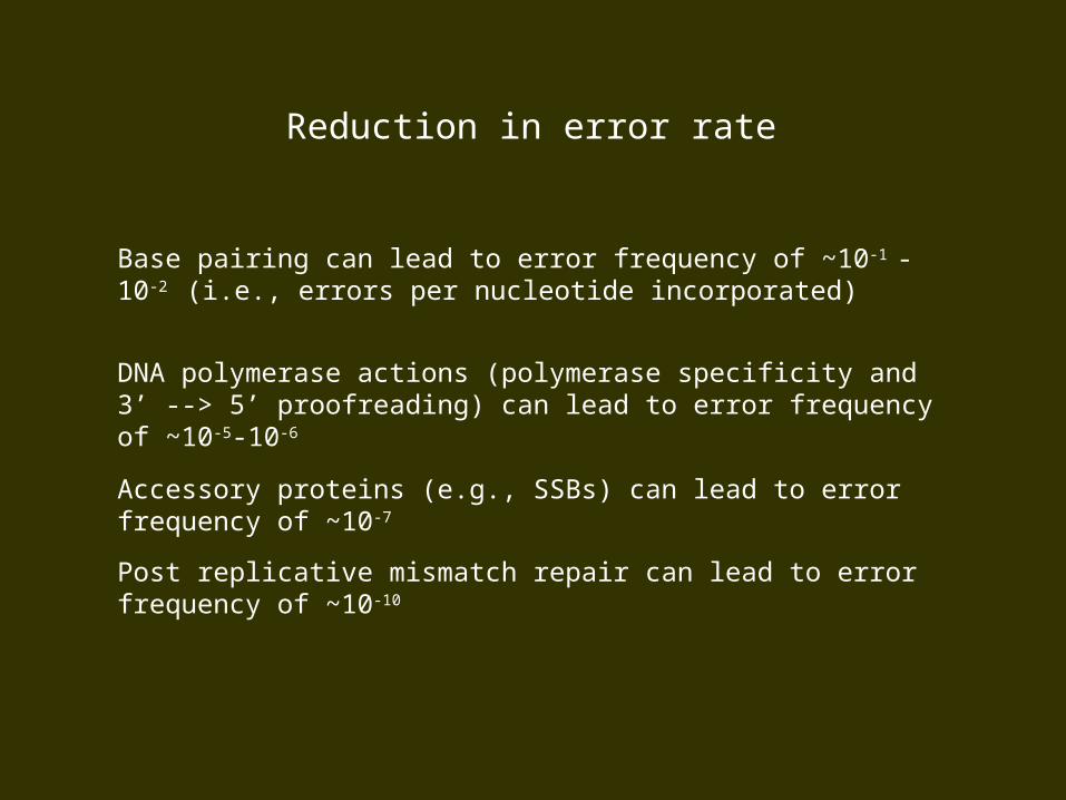
Reduction in error rate
Base pairing can lead to error frequency of ~10-1 -10-2 (i.e., errors per nucleotide incorporated)
DNA polymerase actions (polymerase specificity and 3’ --> 5’ proofreading) can lead to error frequency of ~10-5-10-6
Accessory proteins (e.g., SSBs) can lead to error frequency of ~10-7
Post replicative mismatch repair can lead to error frequency of ~10-10
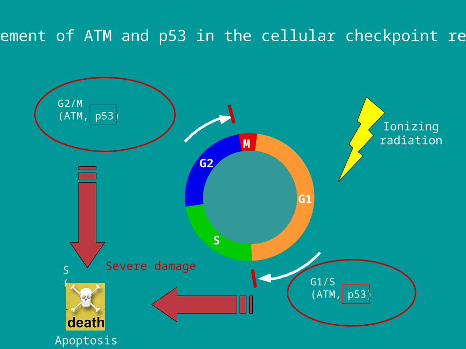
Involvement of ATM and p53 in the cellular checkpoint response
M
G1
G2
G2/M(ATM, p53)
S phase(ATM, ATR, ...) G1/S
(ATM, p53)
S
Ionizingradiation
Severe damage
Apoptosis
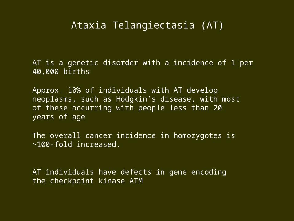
AT is a genetic disorder with a incidence of 1 per 40,000 births
Ataxia Telangiectasia (AT)
Approx. 10% of individuals with AT develop neoplasms, such as Hodgkin’s disease, with most of these occurring with people less than 20 years of age
The overall cancer incidence in homozygotes is ~100-fold increased.
AT individuals have defects in gene encoding the checkpoint kinase ATM
The tumor suppressor p53The tumor suppressor p53
•The 'guardian of the genome’
•Functions as a sequence-specific transcription factor regulating a large number of genes
•The most frequently mutated gene in cancer
•Responsive to a wide array of signals that stress the cell including:
DNA damagehypoxiahyperproliferative signals emanating from oncogenes

p53-dependent apoptosis suppresses tumor growthp53-dependent apoptosis suppresses tumor growth
Choroid plexusepithelium
Van Dyke, 1994

p53 status is a determinant of tumor response to therapyp53 status is a determinant of tumor response to therapy
p53 +/+ p53 -/-
+ adriamycin)
Lowe, Science 266:807, 1994
tum
or v
olum
e (c
m3 )
+ adriamycin

Pathway of Carcinogenesis
Non-repairedmismatchDNA replication
Non-critical gene or non-coding
sequence
Cancer
Little or no effect on cell
viability
Essential region of
essential gene
Cell death through apoptosis
Potential for unregulated cell
growth
Additional mutations(genomic
instability)
Gene involved in growth
stimulation or tumor
suppression

Pathway of Carcinogenesis(colorectal cells)
Normalcell
Mutation of: PTGS2
(proto-oncogene)
p53
(tumorsuppressor)
(tumorsuppressor)
18q LOH
Carcinoma
Ras
(proto-oncogene)
LateAdenoma
APC
(tumorsuppressor)
EarlyAdenoma