Embed Size (px)
Citation preview

7th Grade Science
DNA And Cell Division Reading Packet
BCMS


5.1 DNA, RNA, and Protein Synthesis Learning Objectives
Explain the chemical composition of DNA.
Explain how DNA synthesis works.
Explain how proteins are coded for and synthesized.
Describe the three types of RNA and the functions of each.
Introduction DNA, is the material that makes up our chromosomes and stores our genetic information. When you build a house, you need a blueprint, a set of instructions that tells you how to build. The DNA is like the blueprint for living organisms. The genetic information is a set of instructions that tell your cells what to do.
Guided Practice
What is DNA?
DNA is an abbreviation for deoxyribonucleic acid. As you may recall, nucleic acids are a type of macromolecule that store information. The deoxyribo part of the name refers to the name of the sugar that is contained in DNA, deoxyribose. DNA may provide the instructions to make up all living things, but it is actually a very simple molecule. DNA is made of a long chain of nucleotides. Nucleotides are composed of three main parts:
1. Phosphate group 2. 5-carbon sugar 3. Nitrogen-containing base
The only difference between each nucleotide is the identity of the base. There are only four possible bases that make up each DNA nucleotide: adenine (A), guanine (G), thymine (T), and cytosine (C). The various sequences of these four bases make up the genetic code of your cells. It may seem strange that there are only four letters in the “alphabet” of DNA. But since your chromosomes contain millions of nucleotides, there are many, many different combinations possible with those four letters. But how do all these pieces fit together? James Watson and Francis Crick won the Nobel Prize in 1962 for piecing together the structure of DNA. Together with the work of Rosalind Franklin and Maurice Wilkins, they determined that DNA is made of two strands of nucleotides formed into a double helix, or a two-stranded spiral, with the sugar and phosphate groups on the outside, and the paired bases connecting the two strands on the inside of the helix (Figure below and Figure below).
DNA’s three-dimensional structure is a double helix. The hydrogen bonds between the bases at the center of the helix hold the helix together.
Base-Pairing

The bases in DNA do not pair randomly. When Erwin Chargaff looked closely at the bases in DNA, he noticed that the percentage of adenine (A) in the DNA always equaled the percentage of thymine (T), and the percentage of guanine (G) always equaled the percentage of cytosine (C). Watson and Crick’s model explained this result by suggesting that A always pairs with T and G always pairs with C in the DNA helix. Therefore A and T, and G and C, are "complementary bases," or bases that always pair together. For example, if one DNA strand reads ATGCCAGT, the other strand will be made up of the complementary bases: TACGGTCA.
The chemical structure of DNA includes a chain of nucleotides consisting of a 5-carbon sugar, a phosphate group, and a nitrogen base. Notice how the sugar and phosphate form the backbone of DNA (one strand in blue), with the hydrogen bonds between the
bases joining the two strands.
DNA Replication
The base pairing rules are crucial for the process of replication. DNA replication occurs when DNA is copied to form an identical molecule of DNA. DNA replication happens before cell division. Below are the steps involved in DNA replication:
1. The DNA helix unwinds like a zipper, as the bonds between the base pairs are broken. 2. The two single strands of DNA then each serve as a template for a new stand to be created. Using DNA as a
template means that the bases are placed in the right order because of the base pairing rules. If ATG is on the "template strand," then TAC will be on the new DNA strand.
3. The new set of nucleotides then join together to form a new strand of DNA. The process results in two DNA molecules, each with one old strand and one new strand of DNA.
DNA replication occurs when the DNA strands “unzip”, and the original strands of DNA serve as a template for new nucleotides to join and form a new strand.
Protein Synthesis
The DNA sequence contains the instructions to make units called amino acids, which are assembled in a specific order to make proteins. In short, DNA contains the instructions to create proteins. Each strand of DNA has many separate sequences that code for a specific protein. Units of DNA that contain code for the creation of one protein are called genes.
Cells Can Turn Genes On or Off
There are about 22,000 genes in every human cell. Does every human cell have the same genes? Yes. Does every human cell use the same genes to make the same proteins? No. In a multicellular organism, such as us, cells have

specific functions because they have different proteins. They have different proteins because different genes are expressed in different cell types. Imagine that all of your genes are "turned off." Each cell type only "turns on" (or expresses) the genes that have the code for the proteins it needs to use. So different cell types "turn on" different genes, allowing different proteins to be made, giving different cell types different functions.
Three Types of RNA
DNA contains the instructions to create proteins, but it does not make proteins itself. DNA is located in the nucleus, while proteins are made on ribosomes in the cytoplasm. So DNA needs a messenger to bring its instructions to a ribosome located outside of the nucleus. DNA sends out a message, in the form of RNA (ribonucleic acid), describing how to make the protein. There are three types of RNA directly involved in protein synthesis:
Messenger RNA (mRNA) carries the instructions from the nucleus to the cytoplasm.
The other two forms of RNA, ribosomal RNA (rRNA) and transfer RNA (tRNA) are involved in the process of ordering the amino acids to make the protein.
All three RNAs are nucleic acids, made of nucleotides, similar to DNA. The RNA nucleotide is different from the DNA nucleotide in the following ways:
RNA contains a different kind of sugar, called ribose.
In RNA, the base uracil (U) replaces the thymine (T) found in DNA.
RNA is a single strand.
Transcription
Messenger RNA (mRNA) is created by using DNA as a template. The process of constructing an mRNA molecule from DNA is known as transcription (Figure below and Figure below). The double helix of DNA unwinds and the nucleotides follow basically the same base pairing rules to form the correct sequence in the mRNA. This time, however, U pairs with each A in the DNA. In this manner, the genetic code is passed on to the mRNA.
Each gene (a) contains triplets of bases (b) that are transcribed into RNA (c). Every triplet, or codon, encodes for a unique amino acid.

Base-pairing ensures the accuracy of transcription. Notice how the helix must unwind for transcription to take place.
Translation
The mRNA is directly involved in the protein-making process. mRNA tells the ribosome (Figure below) how to create a protein. The process of reading the mRNA code in the ribosome to make a protein is called translation (Figure below). Sets of three bases, called codons, are read in the ribosome; the organelle responsible for making amino acids which assemble together to make proteins. The following are the steps involved in translation:
1. mRNA travels to the ribosome from the nucleus. 2. The base code in the mRNA determines the order of the amino acids in the protein. The genetic code in mRNA is
read in “words” of three letters (triplets), called codons. There are 20 amino acids and different codons code for different ones. For example, GGU codes for the amino acid glycine, while GUC codes for valine.
3. tRNA reads the mRNA code and brings a specific amino acid to attach to the growing chain of amino acids. Each tRNA carries only one type of amino acid and only recognizes one specific codon.
4. tRNA is released from the amino acid. 5. Three codons, UGA, UAA, and UAG, indicate that the protein should stop adding amino acids. They are called
"stop codons" and do not code for an amino acid. Once tRNA comes to a stop codon, the protein is set free from the ribosome.
The chart in Figure below is used to determine which amino acids correspond to which codons. Transcribe and translate a gene with this interactive activity.
Ribosomes translate RNA into a protein with a specific amino acid sequence. The tRNA binds and brings to the ribosome the amino acid encoded by the mRNA. Ribosomes are made of rRNA and proteins.

This summary of how genes are expressed shows that DNA is transcribed into RNA, which is translated in turn to protein.
Mutations
The process of DNA replication is not always 100% accurate, and sometimes the wrong base is inserted in the new strand of DNA. A permanent change in the sequence of DNA is known as a mutation. Sometimes, a mutation can cause the protein to be made incorrectly, which can affect how well the protein works, or whether it works at all. Usually the loss of a protein function is detrimental to the organism. There are three types of mutations: major, minor, and neutral. A major mutation can cause harm to the organism, creating a disease or disorder. However, in rare circumstances, the mutation can be beneficial. For example, suppose a mutation in an animal’s DNA causes the loss of an enzyme that makes a dark pigment in the animal’s skin. If the population of animals has moved to a light colored environment, the animals with the mutant gene would have a lighter skin color and be better camouflaged. So in this case, the mutation is beneficial. A minor mutation will usually cause a change in appearance. Still other mutations may have no effect, or neutral. Many mutations are not caused by errors in replication. Mutations can happen spontaneously and they can be caused by mutagens in the environment. Some chemicals, such as those found in tobacco smoke, can be mutagens. Sometimes mutagens can also cause cancer. Tobacco smoke, for example, is often linked to lung cancer.
Summary
DNA stores the genetic information of the cell in the sequence of its four bases: adenine, thymine, guanine, and cytosine.
The information in a small segment of DNA, a gene, is sent by mRNA to the ribosome to synthesize a protein.
Within the ribosome, tRNA reads the mRNA in sets of three bases (triplets), called codons, which encode for the specific amino acids that make up the protein.
A mutation is a permanent change in the sequence of bases in DNA.
Vocabulary amino acid
The building blocks of proteins. codons
A triplet (3) of bases in the mRNA that codes for a specific amino acid. DNA
The material that makes up our chromosomes and stores our genetic information. DNA replication
Occurs when DNA is copied to form an identical molecule. double helix
A two stranded spiral of DNA. gene
An inherited unit of DNA that encodes for one protein. mRNA
A molecule that carries the instructions from the DNA to the rest of the cell (messenger RNA). major mutation
A type of mutation that can be harmful, in the form of disease or disorder, or beneficial, in the form of increased resistance to disease.
minor mutation A type of mutation that causes a minimal change in the organism, such as a change in appearance.
mutagen A chemical in the environment that causes mutations. Ex: tobacco smoke.

mutation A permanent change in the sequence of DNA.
neutral mutation A type of mutation that causes no change in how the protein works.
nucleotide Part of the DNA molecule made up of sugar, phosphate and nitrogen bases. Commonly called bases.
RNA A single strand of nucleic acid involved in making proteins.
transcription The process of constructing an mRNA molecule from DNA.
tRNA A molecule that carries a specific amino acid to the ribosome to make protein (transfer RNA).
translation Is the process of reading the mRNA code in the ribosome in order to make a protein.
5.2 Mitosis Learning Objectives
Explain why cells need to divide.
List the stages of the cell cycle and explain what happens at each stage.
List the stages of mitosis and explain what happens at each stage.
Introduction
How is your DNA organized? Your DNA is organized into chromosomes, the pink structures pictured above. Your DNA doesn't always look so pretty though. It only winds tightly into chromosomes when the cell is getting ready to divide. If your DNA wasn't organized into chromosomes, your DNA would look like a mass of strings and would be difficult to divide up!
Guided Practice
Why Cells Divide
Imagine the first stages of life. In humans, a sperm fertilizes an egg, forming the first cell. But humans are made up of trillions of cells, so where do the new cells come from? Remember that according to cell theory, all cells must come from existing cells. From that one cell, an entire baby will develop.

How does a new life go from one cell to so many? The cell divides in half, creating two cells. Then those two cells divide, for a total of four cells. The new cells continue to divide and divide. One cell becomes two, then four, then eight, and so on (Figure below).
Cells divide repeatedly to produce an embryo. Previously the one-celled zygote (the first cell of a new organism) divided to make two cells (a). Each of the two cells divides to yield four cells (b), then the four cells divide to make eight cells (c), and so on. Through cell
division, an entire embryo forms from one initial cell.
Besides the development of a baby, there are many other reasons that cell division is necessary for life: 1. To grow and develop, you must form new cells. Imagine how often your cells must divide during a growth spurt.
Growing just an inch requires countless cell divisions. 2. Cell division is also necessary to repair damaged cells. Imagine you cut your finger. After the scab forms, it will
eventually disappear and new skin cells will grow to repair the wound. Where do these cells come from? Some of your existing skin cells divide and produce new cells.
3. Your cells can also simply wear out. Over time you must replace old and worn-out cells. Cell division is essential to this process.
The Cell Cycle
The process of cell division in eukaryotic cells is carefully controlled. The cell cycle is the lifecycle of a cell, with cell division at the end of the cycle. Like a human lifecycle that is made up of different phases, like childhood, adolescence, and adulthood, there are a series of steps that lead to cell division. These steps can be divided into two main components, interphase and mitosis.
1. Interphase: The stage when the cell mostly performs its “everyday” functions. For example, it is when a kidney cell does what a kidney cell is supposed to do.
2. Mitosis: The stage when the cell prepares to become two cells.
Most of the cell cycle consists of interphase, the time between cell divisions. Three important changes occur during interphase:
1. The cell doubles in size and doubles the number of organelles. 2. The DNA is replicated during this phase. In other words, an identical copy of all the cell’s DNA is made. This
ensures that each new cell has a set of genetic material identical to that of the parental cell. DNA replication will be further discussed in a later lesson.
3. Proteins are synthesized (made) that will help the cell divide. At the end of interphase, the cell is ready to enter mitosis.
During mitosis, the nucleus divides. Mitosis is followed by cytokinesis, when the cytoplasm divides, resulting in two cells. After cytokinesis, cell division is complete. Scientists say that one parent cell, or the dividing cell, forms two genetically identical daughter cells, or the cells that divide from the parent cell. The term "genetically identical" means that each cell has an identical set of DNA, and this DNA is also identical to that of the parent cell. If the cell cycle is not carefully controlled, it can cause a disease called cancer, which causes cell division to happen too fast. A tumor can result from this kind of growth.
Mitosis and Chromosomes
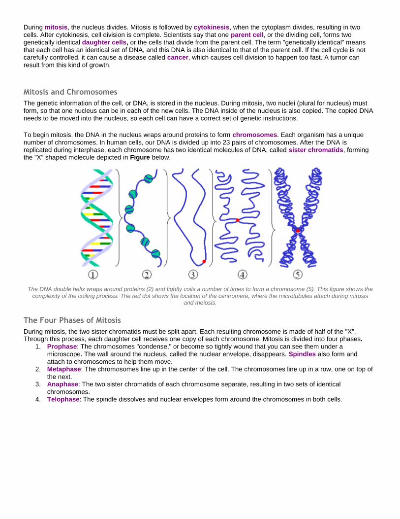
The genetic information of the cell, or DNA, is stored in the nucleus. During mitosis, two nuclei (plural for nucleus) must form, so that one nucleus can be in each of the new cells. The DNA inside of the nucleus is also copied. The copied DNA needs to be moved into the nucleus, so each cell can have a correct set of genetic instructions. To begin mitosis, the DNA in the nucleus wraps around proteins to form chromosomes. Each organism has a unique number of chromosomes. In human cells, our DNA is divided up into 23 pairs of chromosomes. After the DNA is replicated during interphase, each chromosome has two identical molecules of DNA, called sister chromatids, forming the "X" shaped molecule depicted in Figure below.
The DNA double helix wraps around proteins (2) and tightly coils a number of times to form a chromosome (5). This figure shows the complexity of the coiling process. The red dot shows the location of the centromere, where the microtubules attach during mitosis
and meiosis.
The Four Phases of Mitosis
During mitosis, the two sister chromatids must be split apart. Each resulting chromosome is made of half of the "X". Through this process, each daughter cell receives one copy of each chromosome. Mitosis is divided into four phases.
1. Prophase: The chromosomes "condense," or become so tightly wound that you can see them under a microscope. The wall around the nucleus, called the nuclear envelope, disappears. Spindles also form and attach to chromosomes to help them move.
2. Metaphase: The chromosomes line up in the center of the cell. The chromosomes line up in a row, one on top of the next.
3. Anaphase: The two sister chromatids of each chromosome separate, resulting in two sets of identical chromosomes.
4. Telophase: The spindle dissolves and nuclear envelopes form around the chromosomes in both cells.

Each new nucleus contains the exact same number and type of chromosomes as the original cell. The cell is now ready for cytokinesis, which literally means "cell movement." The cells separate, producing two genetically identical cells, each with its own nucleus. Figure below is a representation of dividing plant cells.
This is a representation of dividing plant cells. Cell division in plant cells differs slightly from animal cells as a cell wall must form. Note that most of the cells are in interphase. Can you find examples of the different stages of mitosis?
Review
Cells divide for growth, development, reproduction, and replacement of injured or worn-out cells.
The cell cycle is a series of controlled steps by which a cell divides.
During mitosis, the newly duplicated chromosomes are divided into two daughter nuclei.
This summary diagram depicts one cell dividing into two genetically identical cells. Mitosis occurs after DNA replication. A diploid cell has two sets of chromosomes, as is shown here.
Vocabulary anaphase
The third phase of mitosis in which sister chromatids separate and move to opposite sides of the cell. cancer
The uncontrollable division of cells.

cell cycle The life cycle of a cell. It is made up of two phases, interphase and mitosis.
cell division The process in which a parent cell divides to form daughter cells.
chromosome A structure within the nucleus made up of DNA and proteins. It contains the genetic material of a cell.
cytokinesis The division of the cytoplasm.
daughter cell The new cells that divide from the parent cell.
interphase The time between cell divisions when the cell performs its everyday functions. It is a time when the cell grows, makes DNA, and prepares to divide.
metaphase The second phase of mitosis in which the chromosomes meet in the middle of the cell.
mitosis Division of the nucleus.
parent cell The dividing cell.
prophase The first phase of mitosis in which the chromosomes condense, the nuclear envelope dissolves, and the spindle begins to form.
sister chromatids Two identical copies of a chromosome.
spindle A structure that helps separate the sister chromatids during mitosis.
telophase The final phase of mitosis in which a nuclear envelope forms around each of the two sets of chromosomes.
5.3 Reproduction and Meiosis Learning Objectives
Name the types of asexual reproduction.
Explain the advantage of sexual reproduction.
List the stages of meiosis and explain what happens in each stage.
Introduction
Can some animals have one parent instead of two?

Not all animals have two parents. When necessary, some animals can be produced from just one parent. Some reptiles, such as this Komodo dragon, can have only one parent. The process of creating offspring from just one individual is called asexual reproduction.
Guided Practice
What is Reproduction?
What does reproduction mean? Can an organism be considered alive if it cannot make the next generation? Since individuals cannot live forever, they must reproduce for the species to survive. Reproduction is the ability to make the next generation. Two methods of reproduction are:
1. Asexual reproduction, or the process of forming a new individual from a single parent. 2. Sexual reproduction, or the process of forming a new individual from two parents.
There are advantages and disadvantages to each method, but the result is always the same: a new life begins.
Asexual Reproduction
For humans to reproduce, DNA must be passed from the mother and father to the child. Humans cannot reproduce with just one parent, but it is possible in other organisms, like bacteria, and some insects and some fish. These organisms can reproduce asexually, meaning that the offspring (children) have a single parent and share the exact same genetic material as the parent. This is very different from humans. The advantage of asexual reproduction is that it can be very quick and does not require the meeting of a male and female organism. The disadvantage of asexual reproduction is that organisms cannot mix beneficial traits from both parents. An organism that is born through asexual reproduction only has the DNA from the one parent, and it is the exact copy of that parent. This can cause problems for the individual. For example, if the parent organism has a gene that causes cancer, the offspring will also have the gene that causes cancer. Organisms produced sexually may or may not inherit the cancerous gene because there are two parents mixing up their genes. Types of organisms that reproduce asexually include:
1. Prokaryotic organisms, like bacteria. Bacteria reproduce through binary fission, where they grow and divide in half (Figure below). First, their chromosome replicates (bacteria only have one chromosome) and the cell enlarges. After cell division, the two new cells each have one identical chromosome (mitosis is not necessary because bacteria do not have nuclei). Then, new membranes form to separate the two cells. This simple process allows bacteria to reproduce very rapidly.
2. Flatworms divide in two, then each half regenerates into a new flatworm identical to the original. 3. Different types of insects, fish, and lizards. These organisms can reproduce asexually through a process called
parthenogenesis (Figure below). Parthenogenesis happens when an unfertilized egg cell grows into a new organism. The resulting organism has half the amount of genetic material of the parent. Parthenogenesis is common in honeybees. In a hive, the sexually produced eggs become workers, while the asexually produced eggs become drones.

Bacteria reproduce by binary fission. Shown is one bacterium reproducing and becoming two bacteria.
This Komodo dragon was born by parthenogenesis.
Sexual Reproduction
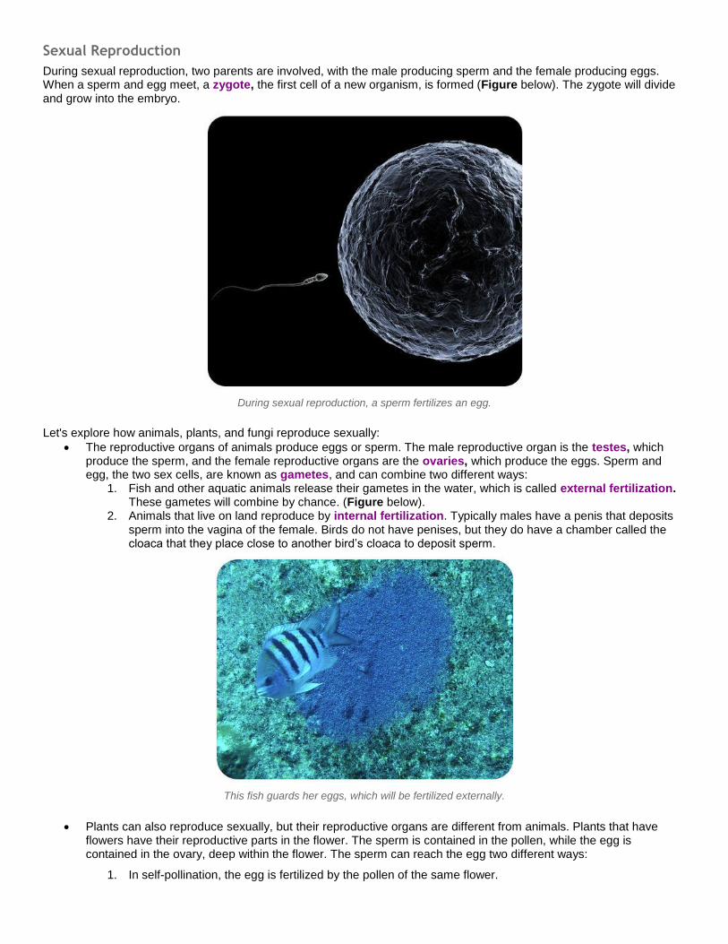
During sexual reproduction, two parents are involved, with the male producing sperm and the female producing eggs. When a sperm and egg meet, a zygote, the first cell of a new organism, is formed (Figure below). The zygote will divide and grow into the embryo.
During sexual reproduction, a sperm fertilizes an egg.
Let's explore how animals, plants, and fungi reproduce sexually: The reproductive organs of animals produce eggs or sperm. The male reproductive organ is the testes, which
produce the sperm, and the female reproductive organs are the ovaries, which produce the eggs. Sperm and egg, the two sex cells, are known as gametes, and can combine two different ways:
1. Fish and other aquatic animals release their gametes in the water, which is called external fertilization. These gametes will combine by chance. (Figure below).
2. Animals that live on land reproduce by internal fertilization. Typically males have a penis that deposits sperm into the vagina of the female. Birds do not have penises, but they do have a chamber called the cloaca that they place close to another bird’s cloaca to deposit sperm.
This fish guards her eggs, which will be fertilized externally.
Plants can also reproduce sexually, but their reproductive organs are different from animals. Plants that have flowers have their reproductive parts in the flower. The sperm is contained in the pollen, while the egg is contained in the ovary, deep within the flower. The sperm can reach the egg two different ways:
1. In self-pollination, the egg is fertilized by the pollen of the same flower.

2. In cross-pollination, sperm from the pollen of one flower fertilizes the egg of another flower. Like other types of sexual reproduction, cross-pollination allows new combinations of traits. Cross-pollination occurs when pollen is carried by the wind to another flower. It can also occur when animal pollinators, like honeybees, or butterflies (Figure below) carry the pollen from flower to flower.
Fungi can also reproduce sexually, but instead of female and male sexes, they have (+) and (-) strains. When the
filaments of a (+) and (-) fungi meet, the zygote is formed. Just like in plants and animals, each zygote receives DNA from two parent strains.
Butterflies receive nectar when they deposit pollen into flowers, resulting in cross-pollination.
Meiosis and Gametes
Meiosis is a process of cell division that produces sex cells, or gametes. Gametes are reproductive cells, such as sperm and egg. As gametes are produced, the number of chromosomes must be reduced by half. Why? The zygote must contain information from the mother and from the father, so the gametes must contain half of the chromosomes found in normal body cells. In humans, our cells have 23 pairs of chromosomes, or 46 total. For each of the 23 chromosome pairs, you received one chromosome from your father and one chromosome from your mother. The chromosomes are separated when gametes are formed. Therefore, gametes have only 23 chromosomes, not 23 pairs. Before meiosis begins, DNA replication occurs, so each chromosome contains two sister chromatids that are identical to the original chromosome. Then the cell must divide twice to resulting in 23 chromosomes in each cell. The simplified process is below.
1. The normal number of chromosomes doubles. Using humans as an example, the nucleus would go from 46 chromosomes to 92 chromosomes.
2. First Cell Division: occurs as the cell divides into two. Each cell contains 46 chromosomes; but because gametes contain 23 chromosomes, the cells must divide again.
3. Second Cell Division: occurs as the two cells divide into four. This division results in cells with 23 chromosomes each. These cells are gametes, or sex cells: the sperm for males and the eggs for females.

An overview of meiosis.
Alleles are alternate forms of genes found on chromosomes. Since the separation of chromosomes into gametes is random, it results in different combinations of chromosomes (and alleles) in each gamete. With 23 pairs of chromosomes, there is a possibility of over 8 million different combinations of chromosomes in a gamete.

Haploid vs. Diploid
A cell with two sets of chromosomes is diploid, referred to as 2n, where n is the number of sets of chromosomes. Most of the cells in a human body are diploid. A cell with one set of chromosomes, such as a gamete, is haploid, referred to as n. Sex cells are haploid. When a haploid sperm (n) and a haploid egg (n) combine, a diploid zygote will be formed (2n). In short, when a diploid zygote is formed, half of the DNA comes from each parent.
Mitosis Meiosis
Purpose: To produce new cells To produce gametes
Number of cells produced: 2 4
Rounds of Cell Division: 1 2
Haploid or Diploid: Diploid Haploid
Daughter cells identical to parent cells? Yes No
Daughter cells identical to each other? Yes No
Review
Organisms can reproduce sexually or asexually.
The gametes in sexual reproduction must have half the DNA of the parent.
Meiosis is the process of nuclear division that forms gametes.

Vocabulary alleles
Are alternate forms of genes found on chromosomes. asexual reproduction
The process of forming a new individual from a single parent. binary fission
A type of asexual reproduction that occurs when the cell divides in half. crossing-over
An exchange of DNA between homologous chromosomes that occurs during prophase I of meiosis. cross pollination
A type of sexual reproduction that occurs in flowers when the sperm from one flower fertilizes the egg of another flower.
diploid A cell with two sets of chromosomes: 2n.
external fertilization Uniting of the sperm and egg cells outside of the female body.
gamete The reproductive cells, or sex cells. Ex: sperm and egg.
haploid A cell with only one set of chromosomes. Ex: sex cells
internal fertilization Uniting of the sperm and egg cells inside the female body.
meiosis The process of cell division that makes sex cells, or gametes.
ovaries Are the female reproductive organs that make eggs.
parthenogenesis A type of asexual reproduction that occurs when an unfertilized egg cell grows into a new organism.
sexual reproduction Is the process of forming a new individual from two parents.
testes Are the male reproductive organs that make sperm.
zygote Is the first cell of a new organism.