Embed Size (px)
Citation preview

ARTICLE IN PRESS
Diversity of EntomopathogenicFungi: Which Groups Conqueredthe Insect Body?J.P.M. Ara�ujo1 and D.P. Hughes1Penn State University, University Park, PA, United States1Corresponding authors: E-mail: [email protected] and [email protected]
Contents
1. Introduction
2 2. The Major Groups of Entomopathogenic Fungi and Oomycetes 42.1 Oomycota
4 2.2 Microsporidia 6 2.3 Chytridiomycota 7 2.4 “Zygomycetes” 8 2.5 Basidiomycota 9 2.6 Ascomycota 113. Methods
12 3.1 Search Strategy 12 3.2 Dealing With Name Changes 14 3.3 Determining Host Associations 14 3.4 Monographs and Atlases 144. Results
14 4.1 Incidence of Disease on Insects Caused by Fungus and Oomycetes 154.1.1 Oomycetes
15 4.1.2 Microsporidia 18 4.1.3 Chytridiomycota 19 4.1.4 Entomophthoromycota 20 4.1.5 Basidiomycota 23 4.1.6 Ascomycota 255. Discussion
27 5.1 Factors Promoting Diversity Within Entomopathogenic Fungi and Oomycetes 275.1.1 Hemipterans as a Host Group Promoting Hyperdiversity of Entomopathogens
27 5.1.2 Broad Range of Ecologies, High Diversity of Pathogens 29 5.1.3 Susceptibility of Lepidoptera and Coleoptera Larval Stages to Fungal Infections 306. Conclusion
31Advances in Genetics, Volume 94ISSN 0065-2660http://dx.doi.org/10.1016/bs.adgen.2016.01.001
© 2016 Elsevier Inc.All rights reserved. 1 j

2 J.P.M. Ara�ujo and D.P. Hughes
ARTICLE IN PRESS
Acknowledgments
32 Supplementary Data 32 References 32Abstract
The entomopathogenic fungi are organisms that evolved to exploit insects. Theycomprise a wide range of morphologically, phylogenetically, and ecologically diversefungal species. Entomopathogenic fungi can be found distributed among five of theeight fungal phyla. Entomopathogens are also present among the ecologically similarbut phylogenetically distinct Oomycota or water molds, which belong to a differentkingdom, the Stramenopila. As a group of parasites, the entomopathogenic fungiand water molds infect a wide range of insect hosts, from aquatic larvae to adult insectsfrom high canopies in tropical forests or even deserts. Their hosts are spread among 20of the 31 orders of insects, in all developmental stages: eggs, larvae, pupae, nymphs,and adults. Such assortment of niches has resulted in these parasites evolving a consid-erable morphological diversity, resulting in enormous biodiversity, the majority ofwhich remains unknown. Here we undertake a comprehensive survey of records ofthese entomopathogens in order to compare and contrast both their morphologiesand their ecological traits. Our findings highlight a wide range of adaptations thatevolved following the evolutionary transition by the fungi and water molds to infectthe most diverse and widespread animals on Earth, the insects.
1. INTRODUCTION
The kingdom Fungi is one of the major groups of eukaryotic microbesin terrestrial and aquatic ecosystems (Mueller & Schmit, 2007). There areapproximately 100,000 described species of fungi (Kirk, Cannon, Minter,& Stalpers, 2008), which only represents a fraction of the estimated diversity,considered to be between 1.5 and 5 million species (Blackwell, 2011;Hawksworth & Rossman, 1997). Importantly, one of the hallmarks of Fungiis their propensity to form intimate interactions with other groups of life onEarth (Vega & Blackwell, 2005). According to (Hawksworth, 1988), 21% ofall described species of fungi are associated with algae as lichens and 8% formintimate relationships with plants as mycorrhiza. Few if any organisms interrestrial ecosystems exist in nature in the complete absence of fungi, andfor this reason fungi are essential players in the maintenance of ecosystemhealth (Braga-Neto, Luiz~ao, Magnusson, Zuquim, & Castilho, 2008).Another group often considered when discussing fungi are the Oomycota.These are colloquially known as water molds and belong to a very distant

Diversity of Entomopathogenic Fungi 3
ARTICLE IN PRESS
kingdomdStramenopilad(Alexopoulos, Mims, & Blackwell, 1996), moreclosely related to brown algae (Kamoun, 2003). However, it is appropriateto discuss oomycetes with fungi as they were long considered to be in thesame group and exhibit very similar ecologies, acting as parasites of bothplants and animals.
The insects, with over 900,000 described species, represent the most spe-cies-rich groups of eukaryotes (Grimaldi & Engel, 2005, p. 12). They areknown to form intimate relationships with many fungal groups that include:mutualistic endosymbionts that assist in nutrition (Suh, McHugh, Pollock, &Blackwell, 2005), fungi as food sources that are farmed as crops by leaf cutterants (Currie et al., 2003), vertically transmitted parasites (Lucarotti & Klein,1988), commensals (DeKesel, 1996), and pathogens with pronounced ef-fects on host populations (Evans & Samson, 1982, 1984). However, eventhough we know that many different fungaleinsect associations do exist,this subject remains among the most understudied fields in fungal biodiver-sity and likely harbors one of the largest reservoirs of undocumented fungalspecies (Vega & Blackwell, 2005).
A prominent characteristic of insects is their chitinous exoskeleton,which the great majority of entomopathogenic fungi and Oomycota needto penetrate (Evans, 1988). Following entry, some groups (ie, Metarhiziumand Beauveria in the order Hypocreales, phylum Ascomycota) are knownto grow inside the host as yeast-like hyphal bodies, multiplying by budding(Prasertphon & Tanada, 1968). Others, for example, some species within theEntomophthoromycota, produce protoplasts (cells without cell walls)instead (Butt, Hajek, & Humber, 1996). A third group encompassingsome species within Oomycota, Chytridiomycota and species within thegenus Entomophthora that infect aphids are known to grow directly as hyphalfilaments inside the host’s body (Lucarotti & Shoulkamy, 2000; Roberts &Humber, 1981; Samson, Ramakers, & Oswald, 1979; Zattau & McInnis,1987). The majority of entomopathogenic fungi kill their hosts before thespore production starts (as such they are termed hemibiotrophic). A fewof them, especially some in the phylum Entomophthoromycota, sporulatefrom the living body of their hosts (and as such are termed biotrophic)(Roy, Steinkraus, Eilenberg, Hajek, & Pell, 2006). All of entomopathogenicoomycetes kill the host before transmission.
All entomopathogenic fungi and water molds are transmitted via spores.There are two types to consider. The sexual spores are actively releasedinto the environment. By definition, zoospores are motile spores thatswim and, in the case of pathogenic fungi, reach their target host actively,

4 J.P.M. Ara�ujo and D.P. Hughes
ARTICLE IN PRESS
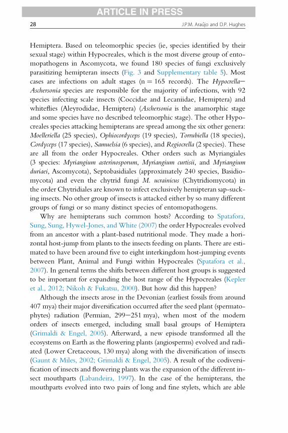
via a flagellum attached to the spore. Such motile spores occur in the Chy-tridiomycota and Oomycota. In other groups, sexual spores are named tolink them to their groups: (zygo)spores, (basidio)spores, and (asco)sporesbelonging to respectively, the “zygomycetes,” Basidiomycota and Ascomy-cota. Each of the three types exhibits unique traits (Fig. 1). The asexualmitotic spores (always called conidia regardless of taxon) are often passivelyreleased (Roberts & Humber, 1981). Spore morphology and their germina-tion behavior have been heavily relied upon in the classification and system-atics of different groups of fungi (Alexopoulos et al., 1996). We will discuss,later, the diversity of these varied spores separately for each major group ofentomopathogenic fungi.
This review has multiple aims. The first is to ask which groups of Fungiand Oomycota evolved the ability to exploit the insect body. We will thenexplore the strategies these organisms employ for both infection and subse-quent transmission. We view each group of entomopathogens within theecological framework that is its insect host, an approach that has surprisinglynot been previously considered in a broad sense. Our overarching aim is toprovide a clearer understanding of the diversity and ecology of this impor-tant group of parasites, highlight lacunae in our knowledge, and motivateother studies. Before proceeding further, however, it is necessary to intro-duce each of the fungal and oomycete groups that are known to infect in-sects. This is because many groups presented here are generally unfamiliar.
2. THE MAJOR GROUPS OF ENTOMOPATHOGENICFUNGI AND OOMYCETES
2.1 Oomycota
The species belonging to Oomycota were in the past consideredamong Fungi due to multiple ecological and morphological similarities.However, phylogenetic studies (James et al., 2006) confirmed earlier sugges-tions by some authors (Kreisel, 1969; Pringsheim, Pfeffer, & Strasburger,1858; Shaffer, 1975) that these organisms are not Fungi. They were thereforeplaced in the Stramenopila, a kingdom containing morphologically diverseorganisms such as Hyphochytriomycota and Labyrinthulomycota (Alexo-poulos et al., 1996; Beakes, Glockling, & Sekimoto, 2012). Despite havingbeen previously considered in the same group, the phylum Oomycota hasa number of biological characters that distinguish them from Fungi. The firstone is reproduction by biflagellate zoospore with a longer tinsel flagellum

Figure 1 Spore diversity within entomopathogenic fungi (in mm). (A) AscomycotadMoelleriella sloaneae (13e15 � 2.8e3 mm) (Chaverri, Liu, & Hodge, 2008); (B)OomycotadLagenidium giganteum (8e9 � 9e10 mm) (Couch, 1935); (C) Chytridio-mycotadCoelomomyces psophorae (5 � 10) (Whisler, Zebold, & Shemanchuk, 1975);(D) EntomophthoromycotadEntomophthora thripidum (10e15 � 8e12 mm) (Samsonet al., 1979); (E) MicrosporidiadNosema hyperae (3.1 � 1.7 mm) (Bulla & Cheng, 1977);(F) BasidiomycotadSeptobasidium maesae (18e19.5 � 4e5 mm) (Lu & Guo, 2009); (G)AscomycotadOphiocordyceps lloydii (4 � 1 mm) (Kobayasi & Shimizu, 1978); (H)AscomycotadHypocrella raciborskii (10e16 � 2.5e4 mm) (Liu, Chaverri, & Hodge,2006); (I) AscomycotadOphiocordyceps camponoti-rufipedis (80e95 � 2e3 mm) (Evans,Elliot, & Hughes, 2011); (J) AscomycotadOphiocordyceps blattae (40e60 � 4e6 mm)(Petch, 1924); (K) AscomycotadOphiocordyceps camponoti-melanotici (170e210 �4e5 mm) (Evans et al., 2011); (L) AscomycotadOphiocordyceps camponoti-novograna-densis (75e95 � 2.5e3.5 mm) (Evans et al., 2011).
Diversity of Entomopathogenic Fungi 5
ARTICLE IN PRESS
6 J.P.M. Ara�ujo and D.P. Hughes
ARTICLE IN PRESS
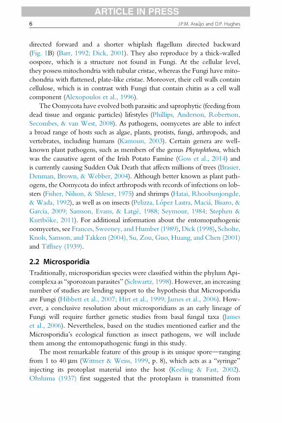
directed forward and a shorter whiplash flagellum directed backward(Fig. 1B) (Barr, 1992; Dick, 2001). They also reproduce by a thick-walledoospore, which is a structure not found in Fungi. At the cellular level,they possess mitochondria with tubular cristae, whereas the Fungi have mito-chondria with flattened, plate-like cristae. Moreover, their cell walls containcellulose, which is in contrast with Fungi that contain chitin as a cell wallcomponent (Alexopoulos et al., 1996).
TheOomycota have evolved both parasitic and saprophytic (feeding fromdead tissue and organic particles) lifestyles (Phillips, Anderson, Robertson,Secombes, & van West, 2008). As pathogens, oomycetes are able to infecta broad range of hosts such as algae, plants, protists, fungi, arthropods, andvertebrates, including humans (Kamoun, 2003). Certain genera are well-known plant pathogens, such as members of the genus Phytophthora, whichwas the causative agent of the Irish Potato Famine (Goss et al., 2014) andis currently causing Sudden Oak Death that affects millions of trees (Brasier,Denman, Brown, & Webber, 2004). Although better known as plant path-ogens, the Oomycota do infect arthropods with records of infections on lob-sters (Fisher, Nilson, & Shleser, 1975) and shrimps (Hatai, Rhoobunjongde,& Wada, 1992), as well as on insects (Pelizza, L�opez Lastra, Maci�a, Bisaro, &García, 2009; Samson, Evans, & Latgé, 1988; Seymour, 1984; Stephen &Kurtb€oke, 2011). For additional information about the entomopathogenicoomycetes, see Frances, Sweeney, and Humber (1989), Dick (1998), Scholte,Knols, Samson, and Takken (2004), Su, Zou, Guo, Huang, and Chen (2001)and Tiffney (1939).
2.2 MicrosporidiaTraditionally, microsporidian species were classified within the phylum Api-complexa as “sporozoan parasites” (Schwartz, 1998). However, an increasingnumber of studies are lending support to the hypothesis that Microsporidiaare Fungi (Hibbett et al., 2007; Hirt et al., 1999; James et al., 2006). How-ever, a conclusive resolution about microsporidians as an early lineage ofFungi will require further genetic studies from basal fungal taxa (Jameset al., 2006). Nevertheless, based on the studies mentioned earlier and theMicrosporidia’s ecological function as insect pathogens, we will includethem among the entomopathogenic fungi in this study.
The most remarkable feature of this group is its unique sporedrangingfrom 1 to 40 mm (Wittner & Weiss, 1999, p. 8), which acts as a “syringe”injecting its protoplast material into the host (Keeling & Fast, 2002).Ohshima (1937) first suggested that the protoplasm is transmitted from

Diversity of Entomopathogenic Fungi 7
ARTICLE IN PRESS
the microsporidian spore to inside the host cell. We now know that this hap-pens through the tube formed during adherence, which facilitates the sub-sequent discharge of the parasite’s intracellular content to within the host’scell (Wittner & Weiss, 1999). The discharging of the polar tube occurs bybreaking through the apex, which is the thinnest region of the spore wall.This event is compared by Keeling and Fast (2002) “to turning the fingerof a glove inside-out.”
The host range for most Microsporidia species is relatively restricted.They have been reported infecting a great number of domestic and wildanimals such as fish (Kent, Shaw, & Sanders, 2014), amphibians, reptiles,birds (Kemp & Kluge, 1975), and mammals (Snowden & Shadduck,1999), including some groups of humans, such as immunocompromisedAIDS patients (Didier & Bessinger, 1999). Detailed studies on the biologyand taxonomy of Microsporidia can be found in Bulla and Cheng (1977),Becnel and Andreadis (1999), Briano (2005), Lange (2010), Sokolova,Sokolov, and Carlton (2010), Kyei-Poku, Gauthier, Schwarz, and Franken-huyzen (2011), Hossain, Gupta, Chakrabarty, Saha, and Bindroo (2012) andVega and Kaya (2012).
2.3 ChytridiomycotaThe Chytridiomycota is the phylum suggested to be the earliest diverginglineage of the Fungi (James et al., 2006). There are reports of them datingfrom Lower Devonian (about 400 million years ago (mya)) (Taylor, Remy,& Hass, 1992) and a parasitic chytrid-like fungus dates from the AntarcticPermian (about 250e300 mya) (Massini, 2007). Chytridiomycota is theonly phylum among the kingdom Fungi that possesses motile cells at leastonce in its life cycle. These zoospores are equipped with a single posteriorlydirected whiplash flagellum, which reflects their aquatic life cycle (for de-tails, see Barr and Désaulniers (1988)). They respond to chemical gradientsallowing them to actively locate their hosts, which is especially importantfor species pathogenic on aquatic organisms (Sparrow, 1960). They canalso adaptively respond to environmental changes (eg, fluctuations in heatand humidity) in ways that reduce water loss or the collapse of the cell(Gleason & Lilje, 2009). The zoospores of chytrids are functionally equiv-alent to motile spores in the Oomycota, and so this is an example of conver-gent evolution, as both groups are aquatic.
The majority of chytrids are found as saprophytic organisms, especially infreshwaters and wet soils, but there are also some marine species (Gleasonet al., 2011). However, a significant number of species are known to be
8 J.P.M. Ara�ujo and D.P. Hughes
ARTICLE IN PRESS
parasites of plants, animals, rotifers, tardigrades, protists, and also other fungi(Dewel, Joines, & Bond, 1985; Karling, 1946; Martin, 1978; Sparrow, 1960).Diseases of insects caused by chytrids seem to be comparatively rare (Karling,1948). For further reading see Voos (1969), Whisler et al. (1975), Millay andTaylor (1978), and Padua, Whisler, Gabriel, and Zebold (1986).
2.4 “Zygomycetes”The phylum Zygomycota was traditionally organized as a single phylumand two classes, Zygomycetes and Trichomycetes (Alexopoulos, 1962;Alexopoulos et al., 1996). Both classes share common features like coeno-cytic mycelium (ie, lacking regular septation), asexual reproduction usuallyby sporangiospores and absence of flagellate cells and centrioles (Alexopouloset al., 1996). Their main general characteristic is the production of athick-walled resting spore (ie, zygospore) within a commonly ornamentedzygosporangium, formed after fusion of two specialized hyphae calledgametangia (Alexopoulos et al., 1996). The phylum is ecologically verydiverse, widely distributed, and very common, with most species occurringas saprotrophs in both soil and dung. Some of them are fast growing andthey are often found colonizing bread, fruits, and vegetables.
However, despite being placed in a single group, molecular phylogeneticstudies validated the long-suggested hypothesis concerning the polyphyly ofZygomycota species and recognized five monophyletic taxa to replace thephylum. Thus, species that form arbuscular mycorrhizal associations withplants were accommodated within the phylum Glomeromycota and allother taxa distributed among four subphyla: Entomophthoromycotina,Kickxellomycotina, Mucoromycotina, and Zoopagomycotina withoutplacement to any phylum (Hibbett et al., 2007; James et al., 2006). There-after, Humber (2012) proposed a detailed morphological and ecologicaldescription of a new phylum: Entomophthoromycota Humber, to accom-modate species previously assigned to Entomophthoromycotina. This studywas supported by a comprehensive phylogenetic study of this new phylum,which demonstrated its monophyletic nature (Gryganskyi et al., 2012). Inour study, we will use this modern classification, which is supported bymorphological, ecological, and phylogenetic data.
The Kickxellomycotina, Mucoromycotina, and Zoopagomycotina arecomposed mostly of saprobes. However, some families within Zoopagomy-cotina are known to predate nematodes (Zhang & Hyde, 2014). These arethe relatively well-known “nematode trapping fungi.” Within Zoopago-mycotina mycoparasitic species are more common (Alexopoulos et al.,

Diversity of Entomopathogenic Fungi 9
ARTICLE IN PRESS
1996). While Mucoromycotina is the largest and morphologically mostdiverse order within the zygomycetes, just one species of entomopathogenicfungi is assigned to the subphylum, ie, Sporodiniella umbellata, which occurson various insects, notably membracids (plant-feeding insects in the orderHemiptera), in cocoa farms (Evans & Samson, 1977).
The trichomycetes, currently placed within subphylum Mucoromyco-tina, order Harpellales, are fungi that exclusively inhabit the guts of variousarthropods (Horn & Lichtwardt, 1981). However, since the trichomycetesapparently do little, if any harm to their hosts under natural conditions(Horn & Lichtwardt, 1981) and the nature of their relationships is not fullyunderstood, they will not be further discussed in this study.
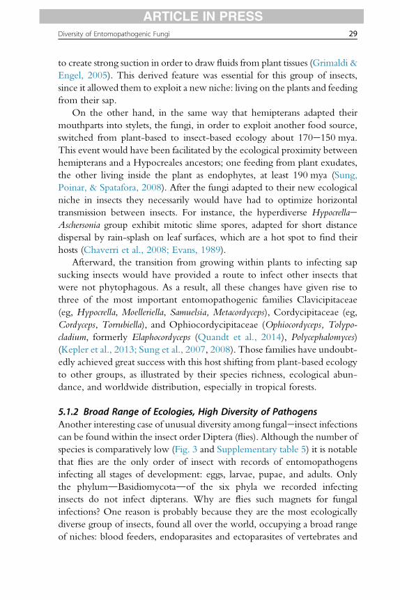
One of the most important groups of all entomopathogens is Ento-mophthoromycota, which are mainly pathogens of insects. They exhibitspecialized spore-producing cells (conidiophores) that have positive photo-trophic growth. Their spores are usually discharged forcibly and this canoccur by several different mechanisms, sometimes producing secondaryand in some species tertiary conidia (ie, Eryniopsis lampyridarum, Fig. 2)(Humber, 2012; Thaxter, 1888). They frequently occur as epizootic events,killing a large number of insects in small patches of forest or agricultural sys-tems (Roberts & Humber, 1981). For further reading see Nair and McEwen(1973), Humber (1976, 1981, 1982, 1984, 1989) and Scholte et al. (2004).
2.5 BasidiomycotaThis group, together with Ascomycota, forms the subkingdom Dikarya,which exhibits a dikaryotic phase (Hibbett et al., 2007). They containsome of the most well-known fungi such as mushrooms, puffballs, earthstars,smuts, and rust fungi. The Basidiomycota are characterized by the formationof sexual spores called basidiospores, which are formed outside specializedreproductive cells called basidia. These spores are in most cases forcibly dis-charged by specialized structures (Pringle, Patek, Fischer, Stolze, & Money,2005). Another important and unique trait for the group are clamp connec-tions. Those are structures formed during the division of the nuclei on the tipof growing hyphae, which help to ensure the dikaryotic condition (Alexo-poulos et al., 1996), and can be used to identify members of this phylum,even in fossil records (Krings, Dotzler, Galtier, & Taylor, 2011).
The basidiomycetes exhibit some important ecological traits. They colo-nize dead wood, decaying cellulose and lignin, also acting as leaf litter de-composers on the forest floor (Braga-Neto et al., 2008). Pathogenicbasidiomycetes (ie, smut and rust fungi) are familiar scourges of plants,

Figure 2 EntomophthoromycotadEryniopsis lampyridarum. (A) Cantharid beetleinfected by E. lampyridarum died with its mandibles attached to flowering plants orgrass. The elytra and wings gradually open as the fungus grows through the host’sbody; (B) Conidiophores emerges directly from the host’s body; (C) Primary conidium;(D) Primary conidium bearing mature secondary conidium at the tip; (E) Secondaryconidium; (F) Secondary conidia eventually will produce capilliconidia in absence ofa suitable host; (G) Another cantharid beetle will get infected if it touches the exposedfungal hymenium. Humber, R. A. (1984). Eryniopsis, a new genus of the Entomophthor-aceae (Entomophthorales). Mycotaxon, 21, 257e264, Roy, H. E., Steinkraus, D. C.,Eilenberg, J., Hajek, A. E., & Pell, J. K. (2006). Bizarre interactions and endgames: entomo-pathogenic fungi and their arthropod hosts. Annual Review of Entomology, 51, 331e357,and Thaxter, R. (1888). The Entomophthoreae of the United States.
10 J.P.M. Ara�ujo and D.P. Hughes
ARTICLE IN PRESS
responsible for huge losses in agriculture. In addition, forest environmentsare also attacked by species like Armillariella mellea, which attack trees andHeterobasidion annosum, attacking specifically conifers (Kendrick, 2000). Asanimal pathogens, some species in the anamorphic genus Nematoctonus(linked to the teleomorphic genus Hohenbuehelia) are known to attack nem-atodes (Barron & Dierkes, 1977). A few genera are known as pathogens ofinsects, which infect scale insects (ie, Septobasidium and Uredinella, order

Diversity of Entomopathogenic Fungi 11
ARTICLE IN PRESS
Septobasidiales) and termite eggs (ie, Fibularhizoctonia, order Atheliales,attacking eggs of the termite genus Reticulitermes).
The Septobasidiales exclusively attack scale insects (Hemiptera, Diaspidi-dae) (Evans, 1989). The order includes two genera of entomopathogens:Uredinella, attacking single insects, and Septobasidium, attacking whole col-onies of plant-feeding insects, with as many as 250 insects infected by onefungus (Couch, 1938). This character is one of the most remarkable differ-ences between both genera, but morphological differences also exist. Forexample, the presence of a binucleate uredospore in Uredinella does notoccur in Septobasidium (Couch, 1937). Due to this trait, Uredinella wasdescribed as “a new fungus intermediate between the rustsda plantpathogendand Septobasidium,” exhibiting traits of both (Couch, 1937).
Another group within Basidiomycota was described on termite eggs(Matsuura, Tanaka, & Nishida, 2000). This fungus was found living insidethe nest of termites, among their eggs, which they occasionally consume.The authors identified this fungus, based on molecular studies, within theorder Atheliales, as being a species very close related to Fibularhizoctoniasp. (asexual state of the genus Athelia), however, not describing themformally (Matsuura et al., 2000). For more details and species descriptionssee Couch (1938), Matsuura (2005, 2006), Yashiro and Matsuura (2007),Lu and Guo (2009), Matsuura, Yashiro, Shimizu, Tatsumi, and Tamura(2009) and Matsuura and Yashiro (2010).
2.6 AscomycotaThe phylum Ascomycota is the largest group in kingdom Fungi withabout 64,000 species described (Kirk et al., 2008). The majority of themare filamentous, producing regularly septate hyphae. They are character-ized by the formation of sexual spores (ie, ascospores) in sac-like structures,singularly called an ascus. As the most speciose group of fungi, it is not sur-prising that they also have diverse ecological breadth comprised by decom-posers, plant pathogens, human and animal pathogens, as well as beingknown to form mutualistic relationships (ie, lichens) (Alexopoulos et al.,1996).
The majority of entomopathogenic species within Ascomycota have awell-developed parasitic phase that infects the host’s body. Furthermore,after killing the insect, this group is able to colonize the cadaver switchingto saprophytic nutrition (hemibiotrophic), maintaining hyphal growth,even after the host’s death (Evans, 1988). According to the same author,these entomopathogens would have evolved and diversified in early, moist

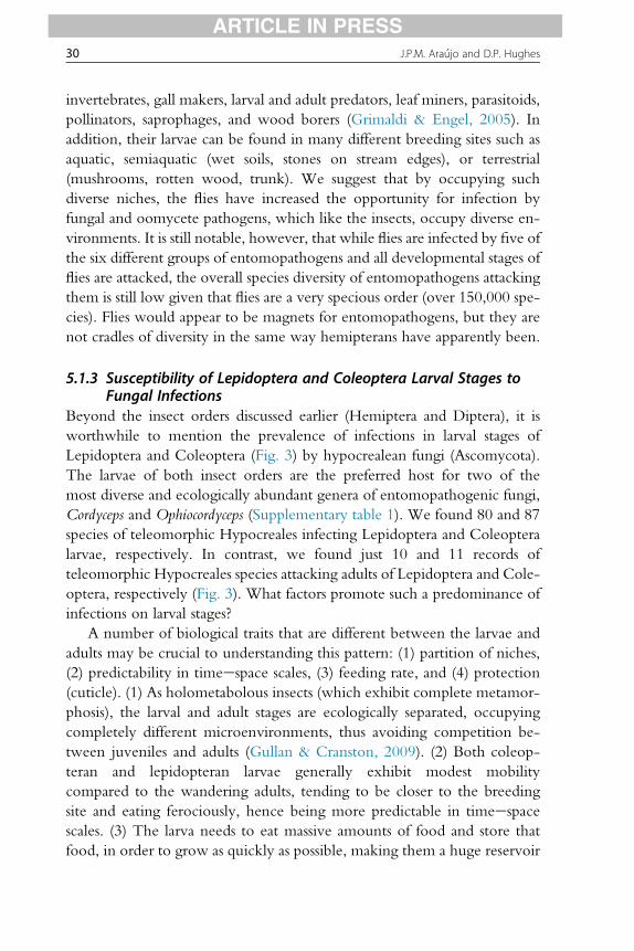
Figure 3 Hypocreales teleomorphic species. Hypocreales teleomorphic speciesnumbers (y-axis) and their distribution across the different orders of insects, furtherdivided into the stage of host development attacked. The Holometabolous ordershave complete development with a larval stage, whereas the Hemimetabolous ordershave an incomplete development with no distinct larval stage, but rather nymphs.
12 J.P.M. Ara�ujo and D.P. Hughes
ARTICLE IN PRESS
tropical forests, particularly rainforests. They are known to attack a widerange of different hosts (Fig. 3). The great majority of entomopathogenic as-comycetes form their spores inside structures called perithecia, a subgloboseor flask-like ostiolate ascoma that contains many asci (Evans et al., 2011; Kirket al., 2008; Kobayasi, 1941; Kobayasi & Shimizu, 1978). There is a widediversity of spore types and shapes (Fig. 1). The phylum ranges from insectpathogens such as Pleosporales, Myriangiales, and Ascosphaerales, whichhave relatively few species, to the biggest group of entomopathogens, thehyperdiverse Hypocreales (Samson et al., 1988).
3. METHODS
3.1 Search Strategy
We are interested in determining which species of fungi and watermolds successfully conquered the insect body. As such, our basic unit ofanalysis is the species name (binomial). The repositories for such namesare electronic databases such as Mycobank (http://www.mycobank.org)
Diversity of Entomopathogenic Fungi 13
ARTICLE IN PRESS
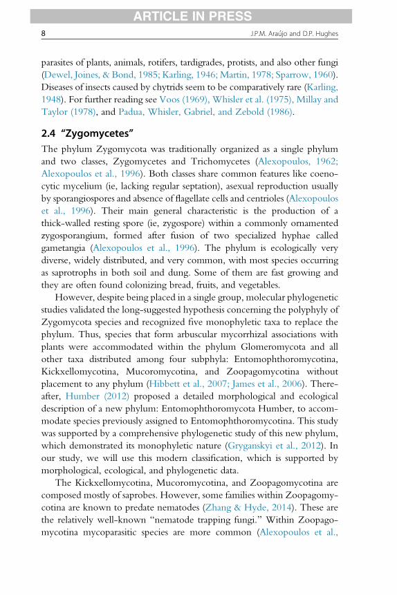
and Index Fungorum (http://www.indexfungorum.org) (Fig. 4). Myco-Bank is owned by the International Mycological Association and is an onlinedatabase aimed as a service to the mycological and scientific community bydocumenting mycological nomenclatural novelties (new names and combi-nations) and associated data, for example, descriptions and illustrations. TheIndex Fungorum, another global fungal database, coordinated and sup-ported by the Index Fungorum Partnerships, contains names of fungi(including yeasts, lichens, chromistan fungal analogues, protozoan fungal an-alogues, and fossil forms) at all ranks.
All groups (phyla) of fungi were investigated separately. Once we founda phylum containing at least one entomopathogenic species (six in total), athorough search was made within such phylum, narrowing until the ento-mopathogenic genera and finally identifying all species recorded as insectpathogens in each phylum.
Figure 4 Flowchart shows the main sources consulted (Mycobank and Index Fungo-rum), the fungal/oomycetes phyla found infecting insects, and the number of speciesand sources consulted in this study.
14 J.P.M. Ara�ujo and D.P. Hughes
ARTICLE IN PRESS
3.2 Dealing With Name ChangesSpecies names are often not static and can change as taxonomists reorganizesynonyms and as we advance with molecular phylogeny. We matched oldrecords of entomopathogenic fungi with their current valid names, avoidingany duplicated record for the same organism.
3.3 Determining Host AssociationsTo determine the host association we first consulted the original formaldescription. This information is available on both Mycobank and IndexFungorum.
The complete list of original descriptions and referencesdthat are notin the textdare listed within the tables organized by phyla, in the sup-plementary materials with species names, host association, and originalreference(s).
3.4 Monographs and AtlasesWe also consulted monographs and atlases of insect pathogenic fungi, eg,Atlas of Entomopathogenic Fungi (Samson et al., 1988), the monograph ofhypocreloid fungi (Chaverri et al., 2008), The Microsporidia and Microsporidio-sis (Wittner & Weiss, 1999), The Genus Coelomomyces (Couch & Bland,1985), the Genus Septobasidium (Couch, 1938) among others. These werealso useful for discovering both host associations and ecological aspects ofthe fungi reviewed herein.
4. RESULTS
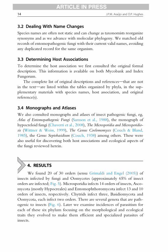
We found 20 of 30 orders (sensu Grimaldi and Engel (2005)) ofinsects infected by fungi and Oomycetes (approximately 65% of insectorders are infected; Fig. 5). Microsporidia infects 14 orders of insects, Asco-mycota (mostly Hypocreales) and Entomophthoromycota infect 13 and 10orders of insects, respectively. Chytrids infect three, Basidiomycota andOomycota, each infect two orders. There are several genera that are path-ogenic to insects (Fig. 6). Later we examine incidences of parasitism foreach of these six phylum focusing on the morphological and ecologicaltraits they evolved to make them efficient and specialized parasites ofinsects.

Figure 5 Insect orders � fungal phyla (and oomycetes): the parasitic relationship be-tween entomopathogens and their hosts. On the left, the phylogeny of insect orders(adapted from Grimaldi, D. & Engel, M. S. (2005). Evolution of the Insects. Cambridge Uni-versity Press.); on the top the phylogeny of fungal phyla and oomycetes (adapted fromJames, T.Y., Kauff, F., Schoch, C.L., Matheny, P.B., Hofstetter, V., Cox, . Miadlikowska, J.(2006). Reconstructing the early evolution of fungi using a six-gene phylogeny. Nature,443(7113), 818e822.); the table shows which fungal group infects each insect order.The uncertainty of a record is denoted with a question mark.
Diversity of Entomopathogenic Fungi 15
ARTICLE IN PRESS
4.1 Incidence of Disease on Insects Caused by Fungus andOomycetes
4.1.1 OomycetesThe entomopathogenic oomycetes are comprised of 12 species distributedamong six genera: Lagenidium (one species, Lagenidium giganteum), Leptolegnia(two species, Leptolegnia caudata and Leptolegnia chapmanii), Pythium (three
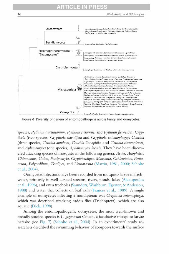
Figure 6 Diversity of genera of entomopathogens across Fungi and oomycetes.
16 J.P.M. Ara�ujo and D.P. Hughes
ARTICLE IN PRESS
species, Pythium carolinianum, Pythium sierrensis, and Pythium flevoense); Cryp-ticola (two species, Crypticola clavulifera and Crypticola entomophaga); Couchia(three species, Couchia amphora, Couchia linnophila, and Couchia circumplexa),and Aphanomyces (one species, Aphanomyces laevis). They have been discov-ered attacking species of mosquito in the following genera: Aedes, Anopheles,Chironomus, Culex, Forcipomyia, Glyptotendipes, Mansonia, Ochlerotatus, Penta-neura, Polypedilum, Tendipes, and Uranotaenia (Martin, 1981, 2000; Scholteet al., 2004).
Oomycetes infections have been recorded frommosquito larvae in fresh-water, primarily in well-aerated streams, rivers, ponds, lakes (Alexopouloset al., 1996), and even treeholes (Saunders, Washburn, Egerter, & Anderson,1988) and water that collects on leaf axils (Frances et al., 1989). A singleexample of oomycetes infecting a nondipteran was Crypticola entomophaga,which was described attacking caddis flies (Trichoptera), which are alsoaquatic (Dick, 1998).
Among the entomopathogenic oomycetes, the most well-known andbroadly studied species is L. giganteum Couch, a facultative mosquito larvaeparasite (see Fig. 7) (Scholte et al., 2004). In an experimental study re-searchers described the swimming behavior of zoospores towards the surface
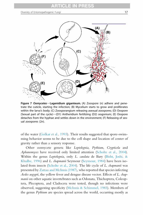
Figure 7 OomycetesdLagenidium giganteum. (A) Zoospore (n) adhere and pene-trate the cuticle, starting the infection; (B) Mycelium starts to grow and proliferateswithin the larva’s body; (C) Zoosporangium releasing asexual zoospores; (D) Oospore(Sexual part of the cycle)d(D1) Antheridium fertilizing (D2) oogonium; (E) Oosporedetaches from the hyphae and settles down in the environment; (F) Releasing of sex-ual zoospores (2n).
Diversity of Entomopathogenic Fungi 17
ARTICLE IN PRESS
of the water (Golkar et al., 1993). Their results suggested that spore-swim-ming behavior seems to be due to the cell shape and location of center ofgravity rather than a sensory response.
Other oomycete genera like Leptolegnia, Pythium, Crypticola andAphanomyces have received only limited attention (Scholte et al., 2004).Within the genus Leptolegnia, only L. caudata de Bary (Bisht, Joshi, &Khulbe, 1996) and L. chapmanii Seymour (Seymour, 1984) have been iso-lated from insects (Scholte et al., 2004). The life cycle of L. chapmanii waspresented by Zattau andMcInnis (1987), who reported that species infectingAedes aegypti, the yellow fever and dengue disease vector. Effects of L. chap-manii on other aquatic invertebrates such as Odonata, Thichoptera, Coleop-tera, Plecoptera, and Cladocera were tested, though no infections wereobserved, suggesting specificity (McInnis & Schimmel, 1985). Members ofthe genus Pythium are species spread across the world, occurring mostly as

18 J.P.M. Ara�ujo and D.P. Hughes
ARTICLE IN PRESS
soil-inhabiting organisms or plant pathogens (Alexopoulos et al., 1996).Three species of Pythium are known to infect insect larvae (Phillips et al.,2008). Aphanomyces was recorded causing seasonal epizootic in insectaries(Seymour & Briggs, 1985), but few studies were published about this genusinfecting insects.
4.1.2 MicrosporidiaMicrosporidia is a group of pathogens comprising 143 genera (Sprague &Becnel, 1999) with more than 1200 species (Wittner & Weiss, 1999).Among those, 69 genera were recorded infecting insects, attacking 12 orders(Fig. 5). According to Becnel and Andreadis (1999), the majority (42 of 69genera) infect Diptera; five genera infect Ephemeroptera and Coleoptera;four genera infect Lepidoptera, followed by Trichoptera, infected by three,Orthoptera, Odonata, and Siphonaptera each infected by two genera; andThysanura, Hymenoptera, and Isoptera with one genus of Microsporidiainfecting each of them. These accounts are data based on described generathat possess an insect as type-host. Hence, this number certainly will increasein future publications.
As will be discussed further, the dipterans are the only insect groupinfected by five different groups of Fungi/oomycetes (only the Basidiomy-cota have not been recorded as pathogens of Diptera). Among the 42 micro-sporidian genera attacking Diptera, the largest, most widespread, andcommon is Amblyospora (Andreadis, 1985), which is known to infect 79 spe-cies of Diptera in 8 genera (Becnel & Andreadis, 1999). This genus of Micro-sporidia exhibits a complex life cycle, which requires an intermediatecopepod host and two mosquito generations in order to complete its fulllife span (Sweeney & Becnel, 1991).
Another important group among the entomopathogenic Microsporidiais the genus Nosema (see Fig. 8). Some authors consider them the mostimportant and widely distributed genus (Tsai, Lo, Soichi, & Wang, 2003),being responsible for the majority of microsporidian infections in Lepidop-tera species (Tsai et al., 2003). A good example of their ecological andeconomical importance occurs with the species Nosema bombycis and Nosemaceranae that infect bees and are known to be responsible for great losses inapiculture (Higes, Martín, & Meana, 2006). These infections are restrictedto the midgut epithelial cells of bees and occur by ingestion of spores byadults (Fig. 8). Once in the midgut, the spores are chemically stimulatedto trigger the polar tube, which penetrates the host’s cells, starting the infec-tion processes (de Graaf, Raes, & Jacobs, 1994). Infectious spores are then

Figure 8 MicrosporidiadNosema sp. (A) Ingestion of spores; (B) Spore reaches the gutof the bee and is activated by its environment, triggering the polar tube to inject thesporoplasm into the host’s cell; (C) Cellular multiplication (proliferate phase); (D andE) Transition from sporoplasm to spore; (F) Spores are released into the gut again,and will be spread in the bee’s feces or will reinfect the same individual.
Diversity of Entomopathogenic Fungi 19
ARTICLE IN PRESS
released with the feces and due to the characteristic thick three-layered wallstructure, they are well adapted to resist in the environment until they areingested by another adult bee (Wittner & Weiss, 1999).
4.1.3 ChytridiomycotaAmong the chytrids, there are four genera that are entomopathogenic:Myio-phagus (one species, Myiophagus cf. ucrainicus), Coelomycidium (one species,Coelomycidium simulii), Myrmicinosporidium (one species, Myrmicinosporidiumdurum), and the most diverse genus Coelomomyces (63 species). Most of thechytrid infections in insects have been recorded for Diptera.
The genus Myiophagus was described infecting dipteran pupae (Petch,1948) and scale insects (Karling, 1948; Muma & Clancy, 1961). Doberskiand Tribe (1978) reported Catenaria auxiliaris on coleopteran larvae,although, they are not sure if the colonization occurred after the larva’s death(saprophytism) or if, in fact, parasitism occurred, leading to the death of the

20 J.P.M. Ara�ujo and D.P. Hughes
ARTICLE IN PRESS
larvae (in Fig. 5 the uncertainty of this record is denoted with a questionmark). Thus, since this relationship is not proven yet, we will not considerC. auxiliaris among the chytrids that parasitize insects.
The genus Coelomycidium is known to attack a specific group withinDiptera order, the black flies (Simuliidae) (Jitklang, Ahantarig, Kuvang-kadilok, Baimai, & Adler, 2012; McCreadie & Adler, 1999). This diseaseis identified by the observation of the larvae filled with spherical sporangiathroughout the body cavity (Kim, 2011).
One group deserves special mention because of the effect they have oninsect reproduction and behavior. The Coelomomyces species (Fig. 9) arerelatively well known because their hosts are important human disease vec-tors (Simulium and the mosquitoes Anopheles, Culex, and Aedes). Specieswithin this genus can infect eggs (Martin, 1978), larvae (the mostcommon type of infection, see details in Travland (1979)), and adults(Lucarotti & Klein, 1988). In some species (ie, Coelomomyces psophorae,Fig. 8) a copepod is required to complete the whole life cycle (Whisleret al., 1975).
In some cases, the fungus does not kill the larvae. Rather, the chytrid re-mains inside the insect as it passes through the larval and pupal stages beforematuring in the ovaries of adult females (Lucarotti, 1992). Once there andafter the first mosquito’s blood meal, the hypha matures to becomesporangia, which is the fungal structure responsible for producing zoospores(Lucarotti & Shoulkamy, 2000). Thus, instead of laying eggs, the mosquitowill ‘lay’ sporangia full of zoospores, ready to infect new larvae (Lucarotti &Klein, 1988). Fatefully, the fungus is reintroduced at the mosquito’sbreeding site by its own host.
4.1.4 EntomophthoromycotaThe phylum Entomophthoromycota is composed mostly of pathogens ofinsects, with few pathogens of other invertebrates, desmid algae, and ferngametophytes, and some that live a saprophytic life (Humber, 2012). Theentomopathogenic species are distributed among 19 genera: Entomophthora,Conidiobolus, Entomophaga, Erynia, Meristacrum, Neozygites, Strongwellsea andMassospora, Pandora, Eryniopsis (Fig. 2), Batkoa, Tarichium, Completoria, Ballo-cephala, Zygnemomyces, Ancylistes, Macrobiotophthora, Thaxterosporium, andBasidiobolus. It is difficult to say how many species of entomopathogens existsince many of these genera also infect different groups of hosts. In addition,the group is in constant taxonomic flux (Humber, 2012). However, sincethe scope of this work is not to provide a complete list of all species within

Figure 9 Chytridiomycota Coelomomyces psophorae. (A) Zoospores attach and pene-trate the copepod cuticle; (B) Development of the gametophytic phase and dispersion ofgametes into the environment; (C) Fusion of compatible gametes, inside the copepod orin the environment (plasmogamy); (D) Formation of zygote (kariogamy ¼ 2n) andattachment to the cuticle of the mosquito larva; (E) Colonization and development ofthe sporophytic phase and formation of sporangium; (F) Resting sporangium releasedinto the environment after the larva’s death; (G) Meiosis and release of asexual zoo-spores; (H) If the larvae reach the adult stage, the fungus will migrate to the ovaries.Instead of laying eggs, the mosquito will lay fungal sporangia.
Diversity of Entomopathogenic Fungi 21
ARTICLE IN PRESS
each group, but to present the diversity of morphologies and strategies toinfect their hosts, we will provide a broad overview of entomopathogenicspecies among Entomophthoromycota, presenting some representative ex-amples of their diversity.

22 J.P.M. Ara�ujo and D.P. Hughes
ARTICLE IN PRESS
The entomophthoroid fungi are well known as insect pathogens. Thisgroup attacks mainly adult insects, although two species of Entomophthora(Entomophthora aquatica and Entomophthora conglomerata) and Erynia aquaticaare known to infect aquatic larval stages of mosquitoes (Scholte et al.,2004). Transmission within entomophthoroid fungi is via forcibly dis-charged spores into the environment, with the exception of one single genus(Massospora) that releases the spores passively, with the host still alive(Humber, 1981; Thaxter, 1888) (Fig. 10). In addition to Massospora, othergroups like Strongwellsea and certain species of Entomophthora, Erynia, andEntomophaga (in addition, to the Ascomycete Lecanicillium longisporum) pro-duce spores before the host death, in or on their living bodies (Roy et al.,2006). These fungi are characteristically biotrophics, consuming the hostwhen they are still alive with no somatic growth after its death. This isone of the major differences when compared with hypocrealean fungi(discussed further in the Ascomycota section), which are all hemibiotrophic,
Figure 10 EntomophthoromycotadMassospora cicadina (A and B), Strongwellseacastrans (C and D). (A) A living cicada flying and dispersing spores while its body dis-integrates due to fungal activity. (B) Spore-producing cells (Conidiophores) in differentstages of development. (C) Fly exhibiting a hole on the abdomen caused by the fungalinfection. (D) Conidiophores exhibiting a terminal spore.

Diversity of Entomopathogenic Fungi 23
ARTICLE IN PRESS
switching from a biotrophic phase (parasitism) to a saprophytic phase,growing on or in the host’s body, even after its death (Roy et al., 2006).
The infections caused by Strongwellsea castrans in Hylemya brassicae andHylemya platura (Diptera) are classic examples of these peculiar situationswhere the sporulation occurs while the host is still alive (Nair & McEwen,1973) (Fig. 10C and D). In this case, the infected fly is characterized by thepresence of a large circular hole on the lateral side of the abdomen. How-ever, surprisingly the infected insects can be observed acting normally,despite the big hole in its body, filled with fungal tissue and conidiophores(spore-producing cells). Both, males and females were described infected byS. castrans, causing castration and premature death (Nair & McEwen, 1973).Another similar case occurs with Massospora cicadina, which attack cicadas(Fig. 10A and B). This fungus also initiates sporulation when the host is stillalive (Goldstein, 1929; Speare, 1921). Due to the pressure caused by theswelling mass of fungus, the collapse of its whole abdomen is inevitable,exposing the fungal tissue. Since the fungus maintains its growth insidethe insect, over time the abdomen falls apart until just the head and thoraxof the living insect remain (Speare, 1921). The ability to fly is retainedincreasing dispersion of spores in the environment.
Although Mucoromycotina is the largest and morphologically mostdiverse group of “zygomycetes,” the subphylum has just one single entomo-pathogenic species, S. umbellata found attacking the hemipteran genusUmbo-nia in Ecuador (Evans & Samson, 1977; Samson et al., 1988) and thelepidopteran genus Acraea in Taiwan (Chien & Hwang, 1997).
4.1.5 BasidiomycotaAlthough the phylum exhibits great diversity of speciesdover 1500 generaand 31,000 species described (Kirk et al., 2008); just three genera are knownto infect insects. Those are (1) Fibularhizoctonia spp. (an undescribed species,see Yashiro and Matsuura (2007)) infecting termite eggs, (2) Uredinella (twospecies, Uredinella coccidiophaga and Uredinella spinulosa) infecting scale insects,and (3) Septobasidium (c. 240 species attacking scale insects, Hemiptera).
The order Septobasidiales Couch (Uredinella and Septobasidium) exhibits apeculiar and complex relationship with their hosts, the Diaspididae (Hemi-ptera). Diaspididae are small, sedentary phythophagous insects, which spendtheir whole lives in one spot on a plant, a consequence of their suckingmouthpart structure (Grimaldi & Engel, 2005). To protect themselves, sincethey are not able to fly away from enemies (Heimpel & Rosenheim, 1995)and do not survive unprotected, juveniles start to secrete fine threads of

24 J.P.M. Ara�ujo and D.P. Hughes
ARTICLE IN PRESS
white wax, which within the first 24 h after their hatching will form a com-plete covering over the insect’s body (Couch, 1938).
This waxy protection is fragile but does afford some degree of defense;however, they are still exposed to external factors. An additional defensestructure can be provided when a colony of such plant-feeding insectsare infected by the fungus Septobasidium. The fungus can grow up to20 cm and creates an elaborate system of tunnels and chambers inside its“body,” which provide the Diaspididae with life-long protection (Couch,1938). However, not all insects are protected as this fungus infects somemembers of the colony often causing dwarfism and castration. The atrophyis due to penetrant haustoria that drain plant sap and nutrients from the in-sect’s body, resulting in undernourishment (Couch, 1931). Even unin-fected adult insects are surrounded and held by hyphal threads, and soare unable to escape: providing an example of a fungus farming an insect(Couch, 1931, 1938). Juveniles (crawlers) may become infected as theyattempt to move out of the parental chamber to establish a new colony(Couch, 1938).
With respect to the less speciose genus among Septobasidiales, Uredinella,there are only two described species: U. coccidiophaga and U. spinulosa (seeCouch (1937, 1941)). They can be divided based on spore shape and thesubstratum in which they infect insects; leaf and trunk for U. spinulosa andjust trunk for U. coccidiophaga. As the genus infects the body of single insect(unlike Septobasidium), the death of the insect means the death of the fungusalso. Spores are produced in the spring and reach 0.2e1.5 mm in diameter(Couch, 1937). In contrast, Septobasidium exhibits an undefined lifetime,since its body is “renewed” each season, by the infection of the newborncrawlers.
Another case of Basidiomycota parasitic on insects can be found betweenFibularhizoctonia spp. and some species of the subterranean termites Reticuli-termes. These termite workers keep their eggs inside their nest in piles, takingcare of them. Matsuura et al. (2000) found among these piles, some sclerotia,globose fungal structures, being cared for by the workers, as if they wereeggs. The same study found that these sclerotia mimic the egg diameterand texture and because these traits are similar to those of the termite eggsthemselves the worker termites mistake the sclerotium for a true egg andcare for it. In nature it is rare to observe the fungus consuming the termiteeggs, but there is the suggestion that the fungus becomes pathogenic andgrows over the true termite eggs if the termites stop caring for the fungi(Matsuura, 2006).

Diversity of Entomopathogenic Fungi 25
ARTICLE IN PRESS
4.1.6 AscomycotaAs mentioned, this diverse phylum comprises many entomopathogenicfungi: from the less speciose orders Pleosporales, Myriangiales, Ascos-phaerales to hyperdiverse groups within the relatively well-known orderHypocreales. In each case the insect dies before the fungus begins its repro-ductive phase. Here, we describe each of these groups and their maincharacteristics.
Within the order Pleosporales, the entomopathogenic species belong tothe genus Podonectria (Petch, 1921) that shares the unusual aspects of thisgroup, such as bright coloration and fleshiness. All known species havebeen found infecting scale insects, covering the whole surface of the insectsbody with a cotton-like crust on which the perithecia is produced and later,multiseptated spores that do not disarticulate into part-spores (Kobayasi &Shimizu, 1977). The related anamorphs are the genus Tetracrium (Kobayasi& Shimizu, 1977; Petch, 1921) and the genus Tetranacrium (Roberts &Humber, 1981).
The Myriangiales includes a number of species associated with plants,resins, or scale insects on plants (Alexopoulos et al., 1996). The entomopa-thogenic species exhibit perennial growth for several years or at least untilthe scale-infested branch dies, probably because of decreased nourishment.The dead host insects can be found directly under each stroma, penetratedand covered by mycelium (Miller, 1938). The stroma are sometimes formedat the side of the insect (Petch, 1924). Growth commences when the rainsbegin with the fungus increasing in diameter, producing ascomata and thenlater, ascospores. Reproduction is entirely by ascospores, and no evidence ofconidial (asexual spore) formation was found on stroma of any age or in cul-ture (Miller, 1938). For taxonomic and additional discussions, see Miller(1938) and Petch (1924).
The order Ascosphaerales contains a unique group of bee pathogenswithin the genus Ascosphaera, which has approximately 30 species. Theseparasites are specialists that exploit the provisions of bees. Most species areexclusive saprophytes on honey, cocoons, larval feces, or nest materialssuch as leaf, mud, or wax of bees (Wynns, Jensen, Eilenberg, & James,2012). However, some species are known as widespread fungal diseaseagents, attacking the brood of numerous species of solitary and social bees,causing a disease called “chalk-brood” (Klinger, James, Youssef, & Welker,2013). The infection occurs when the larva ingests fungal spores. The fungusgrows as hyphae within the body before killing the host and then developingspores on the cuticle of the dead larvae (Vojvodic, Boomsma, Eilenberg, &
26 J.P.M. Ara�ujo and D.P. Hughes
ARTICLE IN PRESS
Jensen, 2012). The morphology of Ascosphaera is very peculiar whencompared to other fungal groups. The ascoma is a small brown to blackishbrown spore cyst, which is a single enlarged cell containing ascospores(Wynns et al., 2012). Their spores also exhibit a curious similarity in appear-ance to pollen grains. For a detailed life cycle, see McManus and Youssef(1984).
The Hypocreales fungi encompass important genera of entomopatho-genic fungi such asCordyceps, Tolypocladium, Hypocrella,Ophiocordyceps,Moel-leriella, Samuelsia, and Torrubiella. In addition to these species, there are manyanamorphic species related to them, ie, Hirsutella,Metarhizium, Hymenostilbe,Akanthomyces, and many others (Roberts & Humber, 1981). In the past,these anamorphic species (which only produce asexual spores) were treatedtraditionally as a separate group within the now retired phylum Deuteromy-cota. However, with molecular techniques some of these species are nowstrongly supported or proven to be asexual stages of Ascomycota (Liuet al., 2001). Here we will address just the teleomorphic (ie, sexual stage)names since our goal is to provide the reader an overview of entomopatho-genic groups and to avoid confusion between the same species, in whichboth teleomorphic and anamorphic (ie, asexual stage) phases are known.
Within the largest group of entomopathogens, the Hypocreales, we canhighlight some genera that are notable due to their diversity and abundancein tropical forests worldwide. For example, Hypocrella (Archersonia is theanamorphic state) that was beautifully monographed by Chaverri et al.(2008). These fungi are known to infect whiteflies and scale insects in trop-ical forests, with few species recorded in the subtropics. They can cause epi-zootics in their host’s population (but epizootics are by no means confined tothis genus).
The genus Ophiocordycepsdespecially species attacking antsdare knownto cause huge infestations in small areas, called graveyards (Evans & Samson,1982, 1984; Pontoppidan, Himaman, Hywel-Jones, Boomsma, & Hughes,2009). Indeed, one of the most fascinating phenomena regarding entomopa-thogenic fungi is the zombie-ant behavior caused byOphiocordyceps unilateraliss.l. (Andersen et al., 2009; Hughes et al., 2011). This species was originallydescribed by Tulasne and Tulasne (1865) asTorrubia unilateralis. Species withinthis complex adaptively manipulate the behavior of worker ants, causing theinsect to leave the colony to find an optimum microclimate site, which isrequired by the fungus to grow and produce ascospores (Fig. 11D). Theants die biting firmly on the underside or edge of a leaf, twig, branch, etc.(the death position is related to each species or group of species of the fungus).

Figure 11 AscomycotadOphiocordyceps unilateralis s.l. (A) Ants leave the nest toforage on the forest floor; (B) Eventually they get infected with Ophiocordyceps asco-spores that were previously shot on the forest floor; (C and D) About 10 days after infec-tion (depending on the species and the geographical location) the infected ant leavesthe nest to die on an elevated position, biting the edge or the main vein of a leaf. Thefungus places the ant on a precise location, which is optimal for fungal developmentand further dispersion of the spores; (E) Two to eight weeks after the ant’s death,depending on the weather conditions, the fungus starts to produce spores and shootthem into the environment; (F) From 24 to 72 h after being shot, the spores will germi-nate and form a secondary spore, the capilliconidiospore.
Diversity of Entomopathogenic Fungi 27
ARTICLE IN PRESS
Following the ant’s death, the fungus grows a fruiting body from the back ofthe ant’s head, which will spread the ascospores on the forest floor (Fig. 11E).
5. DISCUSSION
5.1 Factors Promoting Diversity Within
Entomopathogenic Fungi and Oomycetes5.1.1 Hemipterans as a Host Group Promoting Hyperdiversity ofEntomopathogens
The host is the ecological niche for the fungus. Some ecological niches, ie,host groups, are notable because the abundance and diversity of entomopa-thogenic species infecting them are very high. For instance, the broad diver-sity found among entomopathogenic fungal species attacking sap-sucking
28 J.P.M. Ara�ujo and D.P. Hughes
ARTICLE IN PRESS
Hemiptera. Based on teleomorphic species (ie, species identified by theirsexual stage) within Hypocreales, which is the most diverse group of ento-mopathogens in Ascomycota, we found 180 species of fungi exclusivelyparasitizing hemipteran insects (Fig. 3 and Supplementary table 5). Mostcases are infections on adult stages (n ¼ 165 records). The HypocrellaeAschersonia species are responsible for the majority of infections, with 92species infecting scale insects (Coccidae and Lecaniidae, Hemiptera) andwhiteflies (Aleyrodidae, Hemiptera) (Aschersonia is the anamorphic stageand some species have no described teleomorphic stage). The other Hypo-creales species attacking hemipterans are spread among the six other genera:Moelleriella (25 species), Ophiocordyceps (19 species), Torrubiella (18 species),Cordyceps (17 species), Samuelsia (6 species), and Regiocrella (2 species). Theseare all from the order Hypocreales. Other orders such as Myriangiales(3 species: Myriangium asterinosporum, Myriangium curtisii, and Myriangiumduriaei, Ascomycota), Septobasidiales (approximately 240 species, Basidio-mycota) and even the chytrid fungi M. ucrainicus (Chytridiomycota) inthe order Chytridiales are known to infect exclusively hemipteran sap-suck-ing insects. No other group of insects is attacked either by so many differentgroups of fungi or so many distinct species of entomopathogens.
Why are hemipterans such common hosts? According to Spatafora,Sung, Sung, Hywel-Jones, andWhite (2007) the order Hypocreales evolvedfrom an ancestor with a plant-based nutritional mode. They made a hori-zontal host-jump from plants to the insects feeding on plants. There are esti-mated to have been around five to eight interkingdom host-jumping eventsbetween Plant, Animal and Fungi within Hypocreales (Spatafora et al.,2007). In general terms the shifts between different host groups is suggestedto be important for expanding the host range of the Hypocreales (Kepleret al., 2012; Nikoh & Fukatsu, 2000). But how did this happen?
Although the insects arose in the Devonian (earliest fossils from around407 mya) their major diversification occurred after the seed plant (spermato-phytes) radiation (Permian, 299e251 mya), when most of the modernorders of insects emerged, including small basal groups of Hemiptera(Grimaldi & Engel, 2005). Afterward, a new episode transformed all theecosystems on Earth as the flowering plants (angiosperms) evolved and radi-ated (Lower Cretaceous, 130 mya) along with the diversification of insects(Gaunt & Miles, 2002; Grimaldi & Engel, 2005). A result of the codiversi-fication of insects and flowering plants was the expansion of the different in-sect mouthparts (Labandeira, 1997). In the case of the hemipterans, themouthparts evolved into two pairs of long and fine stylets, which are able
Diversity of Entomopathogenic Fungi 29
ARTICLE IN PRESS
to create strong suction in order to draw fluids from plant tissues (Grimaldi &Engel, 2005). This derived feature was essential for this group of insects,since it allowed them to exploit a new niche: living on the plants and feedingfrom their sap.
On the other hand, in the same way that hemipterans adapted theirmouthparts into stylets, the fungi, in order to exploit another food source,switched from plant-based to insect-based ecology about 170e150 mya.This event would have been facilitated by the ecological proximity betweenhemipterans and a Hypocreales ancestors; one feeding from plant exudates,the other living inside the plant as endophytes, at least 190 mya (Sung,Poinar, & Spatafora, 2008). After the fungi adapted to their new ecologicalniche in insects they necessarily would have had to optimize horizontaltransmission between insects. For instance, the hyperdiverse HypocrellaeAschersonia group exhibit mitotic slime spores, adapted for short distancedispersal by rain-splash on leaf surfaces, which are a hot spot to find theirhosts (Chaverri et al., 2008; Evans, 1989).
Afterward, the transition from growing within plants to infecting sapsucking insects would have provided a route to infect other insects thatwere not phytophagous. As a result, all these changes have given rise tothree of the most important entomopathogenic families Clavicipitaceae(eg, Hypocrella, Moelleriella, Samuelsia, Metacordyceps), Cordycipitaceae (eg,Cordyceps, Torrubiella), and Ophiocordycipitaceae (Ophiocordyceps, Tolypo-cladium, formerly Elaphocordyceps (Quandt et al., 2014), Polycephalomyces)(Kepler et al., 2013; Sung et al., 2007, 2008). Those families have undoubt-edly achieved great success with this host shifting from plant-based ecologyto other groups, as illustrated by their species richness, ecological abun-dance, and worldwide distribution, especially in tropical forests.
5.1.2 Broad Range of Ecologies, High Diversity of PathogensAnother interesting case of unusual diversity among fungaleinsect infectionscan be found within the insect order Diptera (flies). Although the number ofspecies is comparatively low (Fig. 3 and Supplementary table 5) it is notablethat flies are the only order of insect with records of entomopathogensinfecting all stages of development: eggs, larvae, pupae, and adults. Onlythe phylumdBasidiomycotadof the six phyla we recorded infectinginsects do not infect dipterans. Why are flies such magnets for fungalinfections? One reason is probably because they are the most ecologicallydiverse group of insects, found all over the world, occupying a broad rangeof niches: blood feeders, endoparasites and ectoparasites of vertebrates and
30 J.P.M. Ara�ujo and D.P. Hughes
ARTICLE IN PRESS
invertebrates, gall makers, larval and adult predators, leaf miners, parasitoids,pollinators, saprophages, and wood borers (Grimaldi & Engel, 2005). Inaddition, their larvae can be found in many different breeding sites such asaquatic, semiaquatic (wet soils, stones on stream edges), or terrestrial(mushrooms, rotten wood, trunk). We suggest that by occupying suchdiverse niches, the flies have increased the opportunity for infection byfungal and oomycete pathogens, which like the insects, occupy diverse en-vironments. It is still notable, however, that while flies are infected by five ofthe six different groups of entomopathogens and all developmental stages offlies are attacked, the overall species diversity of entomopathogens attackingthem is still low given that flies are a very specious order (over 150,000 spe-cies). Flies would appear to be magnets for entomopathogens, but they arenot cradles of diversity in the same way hemipterans have apparently been.
5.1.3 Susceptibility of Lepidoptera and Coleoptera Larval Stages toFungal Infections
Beyond the insect orders discussed earlier (Hemiptera and Diptera), it isworthwhile to mention the prevalence of infections in larval stages ofLepidoptera and Coleoptera (Fig. 3) by hypocrealean fungi (Ascomycota).The larvae of both insect orders are the preferred host for two of themost diverse and ecologically abundant genera of entomopathogenic fungi,Cordyceps and Ophiocordyceps (Supplementary table 1). We found 80 and 87species of teleomorphic Hypocreales infecting Lepidoptera and Coleopteralarvae, respectively. In contrast, we found just 10 and 11 records ofteleomorphic Hypocreales species attacking adults of Lepidoptera and Cole-optera, respectively (Fig. 3). What factors promote such a predominance ofinfections on larval stages?
A number of biological traits that are different between the larvae andadults may be crucial to understanding this pattern: (1) partition of niches,(2) predictability in timeespace scales, (3) feeding rate, and (4) protection(cuticle). (1) As holometabolous insects (which exhibit complete metamor-phosis), the larval and adult stages are ecologically separated, occupyingcompletely different microenvironments, thus avoiding competition be-tween juveniles and adults (Gullan & Cranston, 2009). (2) Both coleop-teran and lepidopteran larvae generally exhibit modest mobilitycompared to the wandering adults, tending to be closer to the breedingsite and eating ferociously, hence being more predictable in timeespacescales. (3) The larva needs to eat massive amounts of food and store thatfood, in order to grow as quickly as possible, making them a huge reservoir

Diversity of Entomopathogenic Fungi 31
ARTICLE IN PRESS
of energy. (4) Furthermore, larvae need to grow at a high rate and thiswould be impossible if they had the hard exoskeleton that adult coleop-terans have. However, on the other hand, having such soft and thin skinwould make these organisms much easier to be invaded by fungal sporesequipped with their enzymatic and physical tools for infection. It is impor-tant to emphasize that the usual defenses that larvae exhibitdmimicry, apo-sematism, gregarious behavior, stinging hairsdthat are very useful againstpredators are completely useless against the effective entomopathogenicfungi. These four ecological traits that distinguish larvae and adults fromeach other may explain why entomopathogenic fungi exhibit such a greaterprevalence for infecting larvae rather than adults.
The other major holometabolous order infected by Hypocreales isHymenoptera (wasps, bees, termites, and ants). Here, however, most infec-tions are of the adult stages. There are few records (n ¼ 47) so it is harder tocontrast with Lepidoptera and Coleoptera. But it is noticeable that the hy-menopterans build nests for their larvae, and in the case of ants, some wasps,and some bees these larvae are nursed and cleaned by their siblings, which isknown to reduce fungal infections (Cremer, Armitage, & Schmid-Hempel,2007).
6. CONCLUSION
This is the first time that an extensive review encompassing all ento-mopathogenic fungal phyla and oomycetes explored entomopathogenicfungi with a fungalehost approach. Despite the importance of insectefungalassociations, they have been overlooked and their diversity is poorly studied.The lack of interaction between mycologists and entomologists might playan important role in this gap of knowledge, and efforts to address this issueare crucial to better understand the parasitic relationship between insects andthe multiple lineages of entomopathogenic fungi.
Fungi that are able to infect insects are not just comprised by a singlemonophyletic group. Different groups have arisen independently andrepeatedly in many different lineages through fungal evolution (Humber,2008). As presented here, they are spread from more basal to more complexDikaria members. The basal groups, such as aquatic chytrids, infect mostlyDiptera, while Microsporidia and Entomophthoromycota infect a widerange of hosts. Basidiomycota infects mostly Hemiptera, while Ascomycota,the most speciose group, infects a vast number of insect groups.

32 J.P.M. Ara�ujo and D.P. Hughes
ARTICLE IN PRESS
Insect pathologists, entomologists, and life scientists in general have tradi-tionally seen entomopathogenic fungi as having a single role: to kill insectpests (Vega, 2008). But the coevolution of fungi and insects across hundredsof millions of years has resulted in a wide range of complex and intricate in-teractions. The purpose of this work is to provide a wide overview of theserelationships by focusing on the impressive diversity of morphologies, ecol-ogies, and interactions between insects and fungi. Our work also highlightsthe ways that biological and ecological aspects of the hosts likely played animportant role to explain why and how some groups of insects are more sus-ceptible to fungal infection than others.
ACKNOWLEDGMENTSWe are grateful to Harry Evans, Richard Humber, Ryan Kepler, Priscila Chaverri, BhushanShrestha, and James Becnel for the help and inputs to improve this work.
SUPPLEMENTARY DATASupplementary data related to this article can be found online at http://dx.doi.org/10.1016/bs.adgen.2016.01.001.
REFERENCESAlexopoulos, C. J. (1962). Introductory mycology. Wiley.Alexopoulos, C. J., Mims, C. W., & Blackwell, M. (1996). Introductory mycology. New York:
John Wiley & Sons.Andersen, S. B., Gerritsma, S., Yusah, K. M., Mayntz, D., Hywel-Jones, N. L., Billen, J.…
Hughes, D. P. (2009). The life of a dead ant: the expression of an adaptive extendedphenotype. The American Naturalist, 174(3), 424e433.
Andreadis, T. G. (1985). Experimental transmission of a microsporidian pathogen frommosquitoes to an alternate copepod host. Proceedings of the National Academy of Sciencesof the United States of America, 82(16), 5574e5577.
Barr, D. J. S. (1992). Evolution and kingdoms of organisms from the perspective of amycologist. Mycologia, 1e11.
Barr, D. J. S., & Désaulniers, N. L. (1988). Precise configuration of the chytrid zoospore.Canadian Journal of Botany, 66(5), 869e876.
Barron, G. L., & Dierkes, Y. (1977). Nematophagous fungi: Hohenbuehelia, the perfect stateof Nematoctonus. Canadian Journal of Botany, 55(24), 3054e3062.
Beakes, G. W., Glockling, S. L., & Sekimoto, S. (2012). The evolutionary phylogeny of theoomycete “fungi”. Protoplasma, 249(1), 3e19.
Becnel, J. J., & Andreadis, T. G. (1999). Microsporidia in insects. In M. Wittner, &L. M. Weiss (Eds.), The microsporidia and microsporidiosis (pp. 447e501). Washington:Am. Soc. Microbiol. Press.
Bisht, G. S., Joshi, C., & Khulbe, R. D. (1996). Watermolds: potential biological controlagents of malaria vector Anopheles culicifacies. Current Science Bangalore, 70, 393e395.
Blackwell, M. (2011). The Fungi: 1, 2, 3 . 5.1 million species? American Journal of Botany,98(3), 426e438.
Braga-Neto, R., Luiz~ao, R. C. C., Magnusson, W. E., Zuquim, G., & Castilho, V. C.(2008). Leaf litter fungi in a Central Amazonian forest: the influence of rainfall, soil

Diversity of Entomopathogenic Fungi 33
ARTICLE IN PRESS
and topography on the distribution of fruiting bodies. Biodiversity and Conservation,17(11), 2701e2712.
Brasier, C., Denman, S., Brown, A., & Webber, J. (2004). Sudden oak death (Phytophthoraramorum) discovered on trees in Europe. Mycological Research, 108(10), 1108e1110.
Briano, J. A. (2005). Long-term studies of the red imported fire ant, Solenopsis invicta, infectedwith the microsporidia Vairimorpha invictae and Thelohania solenopsae in Argentina. Envi-ronmental Entomology, 34(1), 124e132.
Bulla, L. A., Jr., & Cheng, T. C. (1977). Comparative pathobiology. Volume 2. Systematics of themicrosporidia. Plenum Press.
Butt, T. M., Hajek, A. E., & Humber, R. A. (1996). Gypsy moth immune defenses inresponse to hyphal bodies and natural protoplasts of entomophthoralean fungi. Journalof Invertebrate Pathology, 68(3), 278e285.
Chaverri, P., Liu, M., & Hodge, K. T. (2008). A monograph of the entomopathogenicgenera Hypocrella, Moelleriella, and Samuelsia gen. nov. ( Ascomycota, Hypocreales, Clav-icipitaceae), and their aschersonia-like anamorphs in the Neotropics. Studies in Mycology,60, 1e66.
Chien, C. Y., & Hwang, B. C. (1997). First record of the occurrence of Sporodiniella umbellata(Mucorales) in Taiwan. Mycoscience, 38(3), 343e346.
Couch, J. N. (1931). Memoirs: the biological relationship between Septobasidium retiforme(B. & C.) Pat. and Aspidiotus osborni New. and Ckll. Quarterly Journal of MicroscopicalScience, 2(295), 383e438.
Couch, J. N. (1935). A new saprophytic species of Lagenidium, with notes on other forms.Mycologia, 27(4), 376e387.
Couch, J. N. (1937). A new fungus intermediate between the rusts and Septobasidium. Myco-logia, 29(6), 665e673.
Couch, J. N. (1938). The genus Septobasidium. The University of North Carolina Press.Couch, J. N. (1941). A new Uredinella from Ceylon. Mycologia, 405e410.Couch, J. N., & Bland, C. E. (1985). The genus Coelomomyces. Orlando: Academic Press.Cremer, S., Armitage, S. A. O., & Schmid-Hempel, P. (2007). Social immunity. Current
Biology, 17(16), R693eR702.Currie, C. R., Wong, B., Stuart, A. E., Schultz, T. R., Rehner, S. A., Mueller, U. G.…
Straus, N. A. (2003). Ancient tripartite coevolution in the attine ant-microbe symbiosis.Science, 299(5605), 386e388.
DeKesel, A. (1996). Host specificity and habitat preference of Laboulbenia slackensis.Mycologia,88(4), 565e573.
Dewel, R. A., Joines, J. D., & Bond, J. J. (1985). A new chytridiomycete parasitizing thetardigrade Milnesium tardigradum. Canadian Journal of Botany, 63(9), 1525e1534.
Dick, M. W. (1998). The species and systematic position of Crypticola in the Peronosporo-mycetes, and new names for Halocrusticida and species therein. Mycological Research,102(09), 1062e1066.
Dick, M.W. (2001). Straminipilous fungi: Systematics of the peronosporomycetes including accounts ofthe marine straminipilous protists, the plasmodiophorids and similar organisms.
Didier, E. S., & Bessinger, G. T. (1999). Host-parasite relationships in microsporidiosis:animal models and immunology. In M. Wittner, & L. M. Weiss (Eds.), The microsporidiaand microsporidiosis (pp. 225e257). Washington, DC: ASM Press.
Doberski, J. W., & Tribe, H. T. (1978). Catenaria auxiliaris (Chytridiomycetes: Blastocla-diales) identified in a larva of Scolytus scolytus (Coleoptera: Scolytidae). Journal of Inverte-brate Pathology, 32(3), 392e393.
Evans, H. C. (1988). Coevolution of entomogenous fungi and their insect hosts. InK. A. Pirozynski, & D. L. Hawksworth (Eds.), Coevolution of fungi with plants and animals.
Evans, H. C. (1989). Mycopathogens of insects of epigeal and aerial habitats. London: AcademicPress.

34 J.P.M. Ara�ujo and D.P. Hughes
ARTICLE IN PRESS
Evans, H. C., Elliot, S. L., & Hughes, D. P. (2011). Hidden diversity behind the zombie-antfungusOphiocordyceps unilateralis: four new species described from carpenter ants in MinasGerais, Brazil. PLoS One, 6(3), e17024.
Evans, H. C., & Samson, R. A. (1977). Sporodiniella umbellata, an entomogenous fungus of theMucorales from cocoa farms in Ecuador. Canadian Journal of Botany, 55(23), 2981e2984.
Evans, H. C., & Samson, R. A. (1982). Cordyceps species and their anamorphs pathogenic onants (Formicidae) in tropical forest ecosystems I. The Cephalotes (Myrmicinae) complex.Transactions of the British Mycological Society, 79(3), 431e453.
Evans, H. C., & Samson, R. A. (1984). Cordyceps species and their anamorphs pathogenic onants (Formicidae) in tropical forest ecosystems II. TheCamponotus (Formicinae) complex.Transactions of the British Mycological Society, 82(1), 127e150.
Fisher, W. S., Nilson, E. H., & Shleser, R. A. (1975). Effect of the fungus Haliphthorosmilfordensis on the juvenile stages of the American lobster Homarus americanus. Journal ofInvertebrate Pathology, 26(1), 41e45.
Frances, S. P., Sweeney, A. W., & Humber, R. A. (1989). Crypticola clavulifera gen. et sp.nov. and Lagenidium giganteum: oomycetes pathogenic for dipterans infesting leaf axils inan Australian rain forest. Journal of Invertebrate Pathology, 54(1), 103e111.
Gaunt, M. W., & Miles, M. A. (2002). An insect molecular clock dates the origin of theinsects and accords with palaeontological and biogeographic landmarks.Molecular Biologyand Evolution, 19(5), 748e761.
Gleason, F. H., K€upper, F. C., Amon, J. P., Picard, K., Gachon, C. M. M., Marano, A. V.…Lilje, O. (2011). Zoosporic true fungi in marine ecosystems: a review. Marine and Fresh-water Research, 62(4), 383e393.
Gleason, F. H., & Lilje, O. (2009). Structure and function of fungal zoospores: ecologicalimplications. Fungal Ecology, 2(2), 53e59.
Goldstein, B. (1929). A cytological study of the fungusMassospora cicadina, parasitic on the 17-year cicada, Magicicada septendecim. American Journal of Botany, 394e401.
Golkar, L., LeBrun, R. A., Ohayon, H., Gounon, P., Papierok, B., & Brey, P. T. (1993).Variation of larval susceptibility to Lagenidium giganteum in three mosquito species. Journalof Invertebrate Pathology, 62(1), 1e8.
Goss, E. M., Tabima, J. F., Cooke, D. E. L., Restrepo, S., Fry, W. E., Forbes, M.…Gr€unwald, N. J. (2014). The Irish potato famine pathogen Phytophthora infestans origi-nated in central Mexico rather than the Andes. Proceedings of the National Academy ofSciences of the United States of America, 201401884.
de Graaf, D. C., Raes, H., & Jacobs, F. J. (1994). Spore Dimorphism in Nosema apis (Micro-sporida, Nosematidae) developmental cycle. Journal of Invertebrate Pathology, 63(1), 92e94.
Grimaldi, D., & Engel, M. S. (2005). Evolution of the insects. Cambridge University Press.Gryganskyi, A. P., Humber, R. A., Smith, M. E., Miadlikovska, J., Wu, S., Voigt, K.…
Vilgalys, R. (2012). Molecular phylogeny of the Entomophthoromycota. MolecularPhylogenetics and Evolution, 65(2), 682e694.
Gullan, P. J., & Cranston, P. S. (2009). The insects: An outline of entomology. John Wiley &Sons.
Hatai, K., Rhoobunjongde, W., & Wada, S. (1992). Haliphthoros milfordensis isolated fromgills of juvenile kuruma prawn (Penaeus japonicus) with black gill disease. Transactions ofthe Mycological Society of Japan (Japan), 33, 185e192.
Hawksworth, D. L. (1988). The variety of fungal-algal symbioses, their evolutionary signif-icance, and the nature of lichens. Botanical Journal of the Linnean Society, 96(1), 3e20.
Hawksworth, D. L., & Rossman, A. Y. (1997). Where are all the undescribed fungi? Phyto-pathology, 87(9), 888e891.
Heimpel, G. E., & Rosenheim, J. A. (1995). Dynamic host feeding by the parasitoid Aphytismelinus: the balance between current and future reproduction. Journal of Animal Ecology,153e167.

Diversity of Entomopathogenic Fungi 35
ARTICLE IN PRESS
Hibbett, D. S., Binder, M., Bischoff, J. F., Blackwell, M., Cannon, P. F., Eriksson, O. E.…Zhang, N. (2007). A higher-level phylogenetic classification of the Fungi. MycologicalResearch, 111(5), 509e547.
Higes, M., Martín, R., & Meana, A. (2006). Nosema ceranae, a new microsporidian parasite inhoneybees in Europe. Journal of Invertebrate Pathology, 92(2), 93e95.
Hirt, R. P., Logsdon, J. M., Healy, B., Dorey, M. W., Doolittle, W. F., & Embley, T. M.(1999). Microsporidia are related to Fungi: evidence from the largest subunit of RNApolymerase II and other proteins. Proceedings of the National Academy of Sciences of the UnitedStates of America, 96(2), 580e585.
Horn, B. W., & Lichtwardt, R. W. (1981). Studies on the nutritional relationship of larvalAedes aegypti (Diptera: Culicidae) with Smittium culisetae (Trichomycetes). Mycologia,724e740.
Hossain, Z., Gupta, S. K., Chakrabarty, S., Saha, A. K., & Bindroo, B. B. (2012). Studies onthe life cycle of five microsporidian isolates and histopathology of the mid-gut of thesilkworm Bombyx mori (Lepidoptera: Bombycidae). International Journal of Tropical InsectScience, 32(04), 203e209.
Hughes, D. P., Andersen, S. B., Hywel-Jones, N. L., Himaman,W., Billen, J., & Boomsma, J. J.(2011). Behavioral mechanisms and morphological symptoms of zombie ants dying fromfungal infection. BMC Ecology, 11(1), 13.
Humber, R. A. (1976). The systematics of the genus Strongwellsea (Zygomycetes:Entomophthorales). Mycologia, 68, 1042e1060.
Humber, R. A. (1981). An alternative view of certain taxonomic criteria used in the Ento-mophthorales (Zygomycetes). Mycotaxon, 13, 191e240.
Humber, R. A. (1982). Strongwellsea vs. Erynia: the case for a phylogenetic classification of theEntomophthorales (Zygomycetes). Mycotaxon, 15, 167e184.
Humber, R. A. (1984). Eryniopsis, a new genus of the Entomophthoraceae (Entomophthorales).Mycotaxon, 21, 257e264.
Humber, R. A. (1989). Synopsis of a revised classification for the Entomophthorales(Zygomycotina). Mycotaxon, 34, 441e460.
Humber, R. A. (2008). Evolution of entomopathogenicity in fungi. Journal of InvertebratePathology, 98, 262e266.
Humber, R. A. (2012). Entomophthoromycota: a new phylum and reclassification for ento-mophthoroid fungi. Mycotaxon, 120, 477e492.
James, T. Y., Kauff, F., Schoch, C. L., Matheny, P. B., Hofstetter, V., Cox…Miadlikowska, J.(2006). Reconstructing the early evolution of fungi using a six-gene phylogeny. Nature,443(7113), 818e822.
Jitklang, S., Ahantarig, A., Kuvangkadilok, C., Baimai, V., & Adler, P. H. (2012). Parasites oflarval black flies (Diptera: Simuliidae) in Thailand. Songklanakarin Journal of Science &Technology, 34(6).
Kamoun, S. (2003). Molecular genetics of pathogenic oomycetes. Eukaryotic Cell, 2(2),191e199.
Karling, J. S. (1946). Brazilian Chytrids. VIII. Additional parasites of rotifers and nematodes.Lloydia, 9(1), 1e12.
Karling, J. S. (1948). Chytridiosis of scale insects. American Journal of Botany, 246e254.Keeling, P. J., & Fast, N. M. (2002). Microsporidia: biology and evolution of highly reduced
intracellular parasites. Annual Reviews in Microbiology, 56(1), 93e116.Kemp, R. L., & Kluge, J. P. (1975). Encephalitozoon sp. in the blue-masked lovebird, Aga-
pornis personata (Reichenow): first confirmed report of Microsporidan infection in birds.The Journal of Protozoology, 22(4), 489e491.
Kendrick, B. (2000). The fifth kingdom (3rd). Newburyport: Focus Publishing.Kent, M. L., Shaw, R. W., & Sanders, J. L. (2014).Microsporidia in fishMicrosporidia (pp. 493e
520). John Wiley & Sons, Inc.
36 J.P.M. Ara�ujo and D.P. Hughes
ARTICLE IN PRESS
Kepler, R., Ban, S., Nakagiri, A., Bischoff, J., Hywel-Jones, N., Owensby, C. A., &Spatafora, J. W. (2013). The phylogenetic placement of hypocrealean insect pathogensin the genus Polycephalomyces: an application of one fungus one name. Fungal Biology,117(9), 611e622.
Kepler, R.M., Sung, G.-H., Harada, Y., Tanaka, K., Tanaka, E., Hosoya, T.… Spatafora, J. W.(2012). Host jumping onto close relatives and across kingdoms by Tyrannicordyceps (Clavi-cipitaceae) gen. nov. and Ustilaginoidea_(Clavicipitaceae). American Journal of Botany, 99(3),552e561.
Kim, S. K. (2011). Redescription of Simulium (Simulium) japonicum (Diptera: Simuliiae) and itsentomopathogenic fungal symbionts. Entomological Research, 41(5), 208e210.
Kirk, P. M., Cannon, P. F., Minter, D. W., & Stalpers, J. A. (2008). Dictionary of the fungi(10th ed.). Wallingford, UK: CAB International.
Klinger, E. G., James, R. R., Youssef, N. N., & Welker, D. L. (2013). A multi-gene phy-logeny provides additional insight into the relationships between several Ascosphaeraspecies. Journal of Invertebrate Pathology, 112(1), 41e48.
Kobayasi, Y. (1941). The genus Cordyceps and its allies (pp. 53e260). Science Report of theTokyo Bunrika Daigaku, Section B(84).
Kobayasi, Y., & Shimizu, D. (1977). Two new species of Podonectria (Clavicipitaceae). Bulletinof National Science Museum Serie B Bot Kokuritsu Kagaku Hakubutsukan, 017574592.
Kobayasi, Y., & Shimizu, D. (1978). Cordyceps species from Japan. Bulletin of National ScienceMuseum Tokyo, 4, 43e63.
Kreisel, H. (1969). Grundz€uge eines nat€urlichen Systems der Pilze. Cramer.Krings, M., Dotzler, N., Galtier, J., & Taylor, T. N. (2011). Oldest fossil basidiomycete clamp
connections. Mycoscience, 52(1), 18e23.Kyei-Poku, G., Gauthier, D., Schwarz, R., & Frankenhuyzen, K. V. (2011). Morphology,
molecular characteristics and prevalence of a Cystosporogenes species (Microsporidia) iso-lated from Agrilus anxius (Coleoptera: Buprestidae). Journal of Invertebrate Pathology,107(1), 1e10.
Labandeira, C. C. (1997). Insect mouthparts: ascertaining the paleobiology of insect feedingstrategies. Annual Review of Ecology and Systematics, 153e193.
Lange, C. E. (2010). Paranosema locustae (Microsporidia) in grasshoppers (Orthoptera: Acri-doidea) of Argentina: field host range expanded. Biocontrol Science and Technology,20(10), 1047e1054.
Liu, M., Chaverri, P., & Hodge, K. T. (2006). A taxonomic revision of the insect biocontrolfungus Aschersonia aleyrodis, its allies with white stromata and their Hypocrella sexual states.Mycological Research, 110(5), 537e554.
Liu, Z. Y., Yao, Y. J., Liang, Z. Q., Liu, A. Y., Pegler, D. N., & Chase, M. W. (2001).Molecular evidence for the anamorpheteleomorph connection in Cordyceps sinensis.Mycological Research, 105(07), 827e832.
Lu, C., & Guo, L. (2009). Septobasidium maesae sp. nov. (Septobasidiaceae) from China.Myco-taxon, 109, 103.
Lucarotti, C. J. (1992). Invasion of Aedes aegypti ovaries by Coelomomyces stegomyiae. Journal ofInvertebrate Pathology, 60(2), 176e184.
Lucarotti, C. J., & Klein, M. B. (1988). Pathology of Coelomomyces stegomyiae in adult Aedesaegypti ovaries. Canadian Journal of Botany, 66(5), 877e884.
Lucarotti, C. J., & Shoulkamy, M. A. (2000). Coelomomyces stegomyiae infection in adultfemale Aedes aegypti following the first, second, and third host blood meals. Journal ofInvertebrate Pathology, 75(4), 292e295.
Martin, W. W. (1978). Two additional species of Catenaria (Chytridiomycetes, Blastocla-diales) parasitic in midge eggs. Mycologia, 461e467.
Martin, W. W. (1981). Couchia circumplexa, a water mold parasitic in midge eggs. Mycologia,1143e1157.
Diversity of Entomopathogenic Fungi 37
ARTICLE IN PRESS
Martin, W. W. (2000). Two new species of Couchia parasitic in midge eggs. Mycologia,1149e1154.
Massini, J. L. G. (2007). A possible endoparasitic chytridiomycete fungus from the Permian ofAntarctica. Palaeontologia Electronica, 10(3), 2493MB.
Matsuura, K. (2005). Distribution of termite egg-mimicking fungi (“termite balls”) in Retic-ulitermes spp. (Isoptera: Rhinotermitidae) nests in Japan and the United States. AppliedEntomology and Zoology, 40(1), 53e61.
Matsuura, K. (2006). Termite-egg mimicry by a sclerotium-forming fungus. Proceedings of theRoyal Society B: Biological Sciences, 273(1591), 1203e1209.
Matsuura, K., Tanaka, C., & Nishida, T. (2000). Symbiosis of a termite and a sclerotium-forming fungus: sclerotia mimic termite eggs. Ecological Research, 15(4), 405e414.
Matsuura, K., & Yashiro, T. (2010). Parallel evolution of termite-egg mimicry by sclerotium-forming fungi in distant termite groups. Biological Journal of the Linnean Society, 100(3),531e537.
Matsuura, K., Yashiro, T., Shimizu, K., Tatsumi, S., & Tamura, T. (2009). Cuckoo fungusmimics termite eggs by producing the cellulose-digesting enzyme b-glucosidase. CurrentBiology, 19(1), 30e36.
McCreadie, J. W., & Adler, P. H. (1999). Parasites of larval black flies (Diptera: Simuliidae) andenvironmental factors associated with their distributions. Invertebrate Biology, 310e318.
McInnis, T., Jr., & Schimmel, L. E. (March 22, 1985). Host range studies with the fungusLeptolegnia, a parasite of mosquito larvae (Diptera: Culicidae). Noblet Journal of MedicalEntomology, 22(2), 226e227.
McManus, W. R., & Youssef, N. N. (1984). Life cycle of the chalk brood fungus, Ascos-phaera aggregata, in the alfalfa leafcutting bee, Megachile rotundata, and its associatedsymptomatology. Mycologia, 830e842.
Millay, M. A., & Taylor, T. N. (1978). Chytrid-like fossils of Pennsylvanian age. Science,200(4346), 1147e1149.
Miller, J. H. (1938). Studies in the development of twoMyriangium species and the systematicposition of the order Myriangiales. Mycologia, 30(2), 158e181.
Mueller, G. M., & Schmit, J. P. (2007). Fungal biodiversity: what do we know? what can wepredict? Biodiversity and Conservation, 16(1), 1e5. http://dx.doi.org/10.1007/s10531-006-9117-7.
Muma, M. H., & Clancy, D. W. (1961). Parasitism of purple scale in Florida citrus groves.Florida Entomologist, 159e165.
Nair, K. S. S., &McEwen, F. L. (1973). Strongwellsea castrans (Phycomycetes: Entomophthor-aceae), a fungal parasite of the adult cabbage maggot, Hylemya brassicae (Diptera:Anthomyiidae). Journal of Invertebrate Pathology, 22(3), 442e449.
Nikoh, N., & Fukatsu, T. (2000). Interkingdom host jumping underground: phylogeneticanalysis of entomoparasitic fungi of the genus Cordyceps. Molecular Biology and Evolution,17(4), 629e638.
Ohshima, K. (1937). On the function of the polar filament of Nosema bombycis. Parasitology,29(02), 220e224.
Padua, L. E., Whisler, H. C., Gabriel, B. P., & Zebold, S. L. (1986). In vivo culture and lifecycle of Coelomomyces stegomyiae. Journal of Invertebrate Pathology, 48(3), 284e288.
Pelizza, S. A., L�opez Lastra, C. C., Maci�a, A., Bisaro, V., & García, J. J. (2009). Efecto de lacalidad del agua de criaderos de mosquitos (Diptera: Culicidae) sobre la patogenicidad einfectividad de las zoosporas del hongo Leptolegnia chapmanii (Straminipila:Peronosporomycetes). Revista de Biologia Tropical, 57(1e2), 371e380.
Petch, T. (1921). Fungi parasitic on scale insects. Transaction of the British Mycological Society,7(1e2), 18e40.
Petch, T. (1924). Studies in entomogenous fungi VMyriangium. Transaction of the British Myco-logical Society, 10, 45e80.

38 J.P.M. Ara�ujo and D.P. Hughes
ARTICLE IN PRESS
Petch, T. (1948). A revised list of British entomogenous fungi. Transactions of the British Myco-logical Society, 31(3), 286e304.
Phillips, A. J., Anderson, V. L., Robertson, E. J., Secombes, C. J., & van West, P. (2008).New insights into animal pathogenic oomycetes. Trends in Microbiology, 16(1), 13e19.
Pontoppidan, M. B., Himaman, W., Hywel-Jones, N. L., Boomsma, J. J., & Hughes, D. P.(2009). Graveyards on the move: the spatio-temporal distribution of deadOphiocordyceps-infected ants. PLoS One, 4(3), e4835.
Prasertphon, S., & Tanada, Y. (1968). The formation and circulation, in Galleria, of hyphalbodies of entomophtoraceous fungi. Journal of Invertebrate Pathology, 11(2), 260e280.
Pringle, A., Patek, S. N., Fischer, M., Stolze, J., & Money, N. P. (2005). The captured launchof a ballistospore. Mycologia, 97(4), 866e871.
Pringsheim, N., Pfeffer, W., & Strasburger, E. (1858). Jahrb€ucher f€ur wissenschaftliche Botanik(Vol. 1). Wilh. Engelmann.
Quandt, C. A., Kepler, R. M., Gams, W., Ara�ujo, J. P. M., Ban, S., Evans, H. C.…Spatafora, J. W. (2014). Phylogenetic-based nomenclatural proposals for Ophiocordyci-pitaceae (Hypocreales) with new combinations in Tolypocladium. IMA Fungus, 5(1), 121.
Roberts, D. W., & Humber, R. A. (1981). Entomogenous fungi. Biology of Conidial Fungi, 2,201e236.
Roy, H. E., Steinkraus, D. C., Eilenberg, J., Hajek, A. E., & Pell, J. K. (2006). Bizarre in-teractions and endgames: entomopathogenic fungi and their arthropod hosts. AnnualReview of Entomology, 51, 331e357.
Samson, R. A., Evans, H. C., & Latgé, J. P. (1988). Atlas of entomopathogenic fungi. Springer-Verlag GmbH & Co. KG.
Samson, R. A., Ramakers, P. M. J., & Oswald, T. (1979). Entomophthora thripidum, a newfungal pathogen of Thrips tabaci. Canadian Journal of Botany, 57(12), 1317e1323.
Saunders, G. A., Washburn, J. O., Egerter, D. E., & Anderson, J. R. (1988). Pathogenicity offungi isolated from field-collected larvae of the western treehole mosquito, Aedes sierrensis(Diptera: Culicidae). Journal of Invertebrate Pathology, 52(2), 360e363.
Scholte, E. J., Knols, B. G. J., Samson, R. A., & Takken, W. (2004). Entomopathogenicfungi for mosquito control: a review. Journal of Insect Science, 4(19), 1e24.
Schwartz, K. V. (1998). Five kingdoms: An illustrated guide to the phyla of life on earth. New York:WH Freeman.
Seymour, R. L. (1984). Leptolegnia chapmanii, an oomycete pathogen of mosquito larvae.Mycologia, 670e674.
Seymour, R. L., & Briggs, J. D. (1985). Occurrence and control of Aphanomyces (Saproleg-niales: Fungi) infections in laboratory colonies of larval Anopheles. Journal of the AmericanMosquito Control Association, 1, 100e102.
Shaffer, R. L. (1975). The major groups of Basidiomycetes. Mycologia, 1e18.Snowden, K. F., & Shadduck, J. A. (1999). Microsporidia in higher vertebrates. The microsporidia
and microsporidiosis (pp. 393e417). Washington, DC: ASM.Sokolova, Y. Y., Sokolov, I. M., & Carlton, C. E. (2010). New microsporidia parasitizing
bark lice (Insecta: Psocoptera). Journal of Invertebrate Pathology, 104(3), 186e194.Sparrow, F. K., Jr. (1960). Aquatic phycomycetes. USA: Arbor.Spatafora, J. W., Sung, G. H., Sung, J. M., Hywel-Jones, N. L., &White, J. F. (2007). Phylo-
genetic evidence for an animal pathogen origin of ergot and the grass endophytes.Molecular Ecology, 16(8), 1701e1711.
Speare, A. T. (1921). Massospora cicadina peck: a fungous parasite of the periodical cicada.Mycologia, 13(2), 72e82.
Sprague, V., & Becnel, J. J. (1999). Appendix: checklist of available generic names for micro-sporidia with type species and type hosts. In M.Wittner, & L. M.Weiss (Eds.), The Micro-sporidia and microsporidiosis (pp. 517e530). Washington, DC: ASM Press.
Diversity of Entomopathogenic Fungi 39
ARTICLE IN PRESS
Stephen, K., & Kurtb€oke, D. I. (2011). Screening of Oomycete fungi for their potential rolein reducing the biting midge (Diptera: Ceratopogonidae) larval populations in HerveyBay, Queensland, Australia. International Journal of Environmental Research and PublicHealth, 8(5), 1560e1574.
Su, X., Zou, F., Guo, Q., Huang, J., & Chen, T. X. (2001). A report on a mosquito-killingfungus, Pythium carolinianum. Fungal Diversity, 7(129), 33.
Suh, S. O., McHugh, J. V., Pollock, D. D., & Blackwell, M. (2005). The beetle gut: a hyper-diverse source of novel yeasts. Mycological Research, 109, 261e265. http://dx.doi.org/10.1017/s0953756205002388.
Sung, G. H., Hywel-Jones, N. L., Sung, J. M., Luangsa-ard, J. J., Shrestha, B., &Spatafora, J. W. (2007). Phylogenetic classification of Cordyceps and the clavicipitaceousfungi. Studies in Mycology, 57, 5e59.
Sung, G. H., Poinar, G. O., Jr., & Spatafora, J. W. (2008). The oldest fossil evidence of animalparasitism by fungi supports a Cretaceous diversification of fungalearthropod symbioses.Molecular Phylogenetics and Evolution, 49(2), 495e502.
Sweeney, A. W., & Becnel, J. J. (1991). Potential of microsporidia for the biological controlof mosquitoes. Parasitology Today, 7(8), 217e220.
Taylor, T. N., Remy, W., & Hass, H. (1992). Fungi from the lower Devonian Rhynie chert:Chytridiomycetes. American Journal of Botany, 1233e1241.
Thaxter, R. (1888). The Entomophthoreae of the United States.Tiffney, W. N. (1939). The host range of Saprolegnia parasitica. Mycologia, 31(3), 310e321.Travland, L. B. (1979). Initiation of infection of mosquito larvae (Culiseta inornata) by Coelo-
momyces psorophorae. Journal of Invertebrate Pathology, 33(1), 95e105.Tsai, S. J., Lo, C. F., Soichi, Y., & Wang, C. H. (2003). The characterization of microspori-
dian isolates (Nosematidae: Nosema) from five important lepidopteran pests in Taiwan.Journal of Invertebrate Pathology, 83(1), 51e59.
Tulasne, L. R., & Tulasne, C. (1865). Selecta Fungorum Carpologia III (p. 221). Paris Museum.Vega, F. E. (2008). Insect pathology and fungal endophytes. Journal of Invertebrate Pathology,
98(3), 277e279.Vega, F. E., & Blackwell, M. (2005). Insect-fungal associations: Ecology and evolution. Oxford:
Oxford University Press.Vega, F. E., & Kaya, H. K. (2012). Insect pathology. Academic Press.Vojvodic, S., Boomsma, J. J., Eilenberg, J., & Jensen, A. B. (2012). Virulence of mixed fungal
infections in honey bee brood. Frontiers in Zoology, 9(1), 5.Voos, J. R. (1969). Morphology and life cycle of a new chytrid with aerial sporangia.
American Journal of Botany, 898e909.Whisler, H. C., Zebold, S. L., & Shemanchuk, J. A. (1975). Life history of Coelomomyces
psorophorae. Proceedings of the National Academy of Sciences of the United States of America,72(2), 693e696.
Wittner, M., & Weiss, L. M. (1999). Microsporidia and microsporidiosis.Wynns, A. A., Jensen, A. B., Eilenberg, J., & James, R. (2012). Ascosphaera subglobosa, a new
spore cyst fungus fromNorth America associated with the solitary beeMegachile rotundata.Mycologia, 104(1), 108e114.
Yashiro, T., & Matsuura, K. (2007). Distribution and phylogenetic analysis of termiteegg-mimicking fungi “termite balls” in Reticulitermes termites. Annals of the EntomologicalSociety of America, 100(4), 532e538.
Zattau, W. C., & McInnis, T., Jr. (1987). Life cycle and mode of infection of Leptolegniachapmanii (Oomycetes) parasitizing Aedes aegypti. Journal of Invertebrate Pathology, 50(2),134e145.
Zhang, K. Q., & Hyde, K. D. (2014). Nematode-trapping fungi (Vol. 23). Springer Science &Business.