Embed Size (px)
Citation preview

Diverse set of Turing nanopatterns coat corneae acrossinsect lineagesArtem Blagodatskia,1, Anton Sergeevb, Mikhail Kryuchkova,c, Yuliya Lopatinad, and Vladimir L. Katanaevc,e,1
aInstitute of Protein Research, Russian Academy of Sciences, 142290 Pushchino, Russian Federation; bInstitute of Mathematical Problems of Biology, RussianAcademy of Sciences, 142290 Pushchino, Russian Federation; cDepartment of Pharmacology and Toxicology, Faculty of Biology and Medicine, University ofLausanne, 1005 Lausanne, Switzerland; dDepartment of Entomology, Faculty of Biology, Lomonosov Moscow State University, 119234 Moscow, RussianFederation; and eSchool of Biomedicine, Far Eastern Federal University, Vladivostok, Russian Federation
Edited by Jeremy Nathans, Johns Hopkins University, Baltimore, MD, and approved July 17, 2015 (received for review March 23, 2015)
Nipple-like nanostructures covering the corneal surfaces of moths,butterflies, and Drosophila have been studied by electron andatomic force microscopy, and their antireflective properties havebeen described. In contrast, corneal nanostructures of the majorityof other insect orders have either been unexamined or examinedby methods that did not allow precise morphological character-ization. Here we provide a comprehensive analysis of cornealsurfaces in 23 insect orders, revealing a rich diversity of insectcorneal nanocoatings. These nanocoatings are categorized intofour major morphological patterns and various transitions be-tween them, many, to our knowledge, never described before.Remarkably, this unexpectedly diverse range of the corneal nano-structures replicates the complete set of Turing patterns, thuslikely being a result of processes similar to those modeled by AlanTuring in his famous reaction−diffusion system. These findingsreveal a beautiful diversity of insect corneal nanostructures andshed light on their molecular origin and evolutionary diversi-fication. They may also be the first-ever biological example ofTuring nanopatterns.
nanocoating | cornea | insects | nanostructures | Turing
Biological patterning at the microscale and macroscale levelshas been under intensive investigation by developmental
biology, and its fundamental principles, such as the concept ofthe morphogens, have become textbook knowledge (1). In con-trast, nanoscale biological patterning is not well studied andunderstood. Among the rare known examples of biologicalnanopatterns are the 3D nanostructures covering insect cor-neal surfaces (2). They were described in moths and butter-flies and later some Dipterans as pseudoregularly spacednipple-type protrusions, up to 200 nm in height and width (3–7).These nanostructures may carry antireflective, dirt-removing/self-cleaning, and hydrophobic/antiwetting functions (2, 8–12).Later, some other insects were found to possess a very differenttype of corneal nanocoating, such as the antireflective maze-like30-nm-high evaginations covering corneae of the overwater eyesof Gyrinidae beetles (13). An attempt to analyze the variety ofcorneal nanocoatings throughout the insect class was made in theclassical study by Bernhard et al. (5). However, the scanningelectron microscopy technique of that time was mostly per-formed on platinum replicas of the insect samples and wascompromised by the partial collapse of the nanoprotrusions. Itpermitted reliable identification of 50- to 250-nm-high nipple-type protrusions in Lepidoptera, some Dipterans, Trichopterans,and, interestingly, the primitive Thysanuras, but not identifica-tion of other types of corneal nanocoatings (5).To use the corneal nanocoatings as the model to study
nanoscale biological patterning, a comprehensive investigationacross insect lineages using modern techniques must be per-formed. We recently applied atomic force microscopy (AFM),providing nanometer and subnanometer resolution of undam-aged biological material, to investigate different types of cornealnanostructures of some Dipteran and Coleopteran insects (6, 13).
Here we expand this analysis to 23 insect orders and some non-insect arthropods, describing a striking richness and beautyof the corneal nanocoatings (Fig. 1, Figs. S1–S3, Table S1, andDetailed Description of Diverse Corneal Nanostructures Orderby Order). These nanostructures can be grouped as follows.(i) Nipple-like structures (Fig. 1A and Fig. S1) include the regu-larly packed protrusions of Lepidopterans (Fig. S1A), irregularpackaging in Dipterans (Fig. S1B), and irregular packaging ofirregularly shaped nipple-like protrusions in a range of otherorders: Trichoptera (Fig. 1A), Mecoptera (Fig. S1C), Mega-loptera (Fig. S1D), Hemiptera (Fig. S1 E and F), Psocoptera(Fig. S1G), Thysanura (Fig. S1H), Raphidioptera (Fig. S1I),Neuroptera (Fig. S1J), Orthoptera (Fig. S1K), and Odonata(Fig. S1L). (ii) Maze-like nanocoatings (Fig. 1B and Fig. S2) canbe observed in Coleopterans (Fig. S2 A and B) but also in otherorders such as Trichoptera (Fig. 1B) and Hymenoptera (Fig.S2C), and in some arachnids (Fig. S2 D and E). (iii) Parallelstrands/ridges (Fig. 1C) formed by fusion of nipple-type pro-trusions can mostly be seen in Dipterans (Fig. 1 F and G) and,interestingly, in true spiders (Fig. 1C). (iv) Novel dimple-typenanocoating (Fig. 1D and Fig. S3) can be seen in different or-ders: Siphonaptera (Fig. S3A), Coleoptera (Fig. S3B), Hyme-noptera (Fig. S3C), Hemiptera (Fig. S3 D and E), Blattodea(Fig. S3F), and Dermaptera (Fig. 1D), and, interestingly, incentipedes (Fig. S3H). We also see various transitions be-tween these major forms: (v) nipples-to-maze transition (e.g., in
Significance
Corneal surfaces of some insects are coated with nipple-likenanostructures reducing the light reflection. Here we providean extensive analysis of corneae across insect groups. Usingatomic force microscopy, we discover a striking diversity ofcorneal nanocoatings, omnipresent in arthropods. These fasci-nating bionanostructures replicate the complete set of theTuring patterns—shapes resulting from the reaction−diffusionmodeling underlying many examples of patterning in bi-ological and physicochemical systems. Our work, verging onthe interface of nanotechnology and zoology, evolution andbiophysics, and ecology and genetics, sheds light on the mo-lecular origin and evolutionary diversification of a beautifuldiversity of insect corneal nanostructures. It also describes, toour knowledge, the first-ever biological example of Turingnanopatterns.
Author contributions: V.L.K. designed research; A.B., A.S., and M.K. performed research;A.S. and Y.L. contributed new reagents/analytic tools; A.B., A.S., M.K., and V.L.K. analyzeddata; and A.B. and V.L.K. wrote the paper.
The authors declare no conflict of interest.
This article is a PNAS Direct Submission.
Freely available online through the PNAS open access option.1To whom correspondence may be addressed. Email: [email protected] [email protected].
This article contains supporting information online at www.pnas.org/lookup/suppl/doi:10.1073/pnas.1505748112/-/DCSupplemental.
10750–10755 | PNAS | August 25, 2015 | vol. 112 | no. 34 www.pnas.org/cgi/doi/10.1073/pnas.1505748112

Plecoptera, Fig. 1E); (vi) maze-to-strands transition (e.g., in Dip-tera, Fig. 1F); (vii) nipples-to-strands transition (e.g., in Diptera,Fig. 1G); and (viii) dimples-to-maze transition (e.g., in Hymenop-tera, Fig. 1H).The rich diversity of these nanostructures and the easiness
with which the corneal nanopatterns merge one into another inclosely related orders and even within the orders (Fig. 2 andDetailed Description of Diverse Corneal Nanostructures Order byOrder) is striking and permits posing questions on the underlyingmolecular, developmental, and evolutionary mechanisms. De-velopmentally, the nipple-type protrusions were proposed tooriginate, during eye development, from secretion by the regu-larly spaced microvilli of the cone cells (5, 14). However, thisidea could appear plausible when the ordered Lepidopterannipple arrays were studied but, with the current diversity ofnanostructures and transitions among them, sometimes withinthe same lens (Fig. 1G), is not satisfactory. Instead, we proposethat certain mechanisms of patterning at the nanoscale are inplace, and the diverse arthropod corneal nanostructures we de-scribe here represent a model to study such nanopatterning.Further, we notice that this diversity of corneal nanostructures isremarkably similar to the complete set of the Turing patterns(Fig. 3).In his seminal paper in 1952, Alan Turing provided a system of
differential equations describing the reaction−diffusion systemof two reacting morphogens—a slowly diffusing activator anda fast diffusing inhibitor—which can model various biologic,chemical, and physical patterns (15, 16). Applicability of thismodel to biological pattern formation has been shown in severalrecent examples, such as formation of colored stripes in zebrafish(17), hair follicle spacing in mice (18), and digit specification inlimbs (19). The insect corneal nanopatterns we describe herediffer from these examples, as they reproduce not just one of themany possible forms produced by the reaction−diffusion modelbut a thorough set of possible variants including the intermediateforms (Figs. 1 and 3). This remarkable completeness of coverageof the possible set of Turing structures by the arthropod corneal
nanopatterns strongly argues in favor of the hypothesis thatthese nanopatterns are indeed a consequence of the Turingreaction−diffusion mechanisms.We hypothesize that the Turing mechanism-based reaction−diffu-
sion processes patterning the nanocoatings are mediated byorganic components of the lens, possessing different diffusionproperties and mutually influencing each other’s abundance/polymerization/aggregation, the outcome of this being the ste-reotypical formation of the nanostructures. In previous applica-tions of the Turing principles to biological processes, patterningat the microscale was modeled (17–19). Formal mathematicalanalysis shows how key parameters of the reaction−diffusionequations (primarily the diffusion coefficients of the two inter-acting morphogens) can result in the appearance of repeateddevelopmental structures with the experimentally observedmicrometer-scale wavelength (20). Our mathematical analysis(Turing modeling of corneal nanopatterns) demonstrates thatnanoscale patterns are expected to form in the reaction−diffu-sion system acting in the colloidal or liquid crystal-type envi-ronment [which is indeed the environment of the lens of the eye(21)] where diffusion properties are reduced (compared with theliquid phase).Although the molecular identity of the morphogens patterning
corneal nanocoatings remains to be revealed, simulations ofthe Turing reaction−diffusion processes provide interestinghints into the potential molecular mechanisms underlyingformation of different types of the nanocoatings and transi-tions among them (Fig. 3I and Fig. S4). Although differentsets of the reaction−diffusion coefficients (like that of TableS2 used to obtain images on Fig. 3 and Fig. S4; see also Fig.S4B for schematic description of the parameters and Fig. S5for analysis of the parameter space) can model differentnanopatterns, simulations find that three of the major types ofpatterns we observe in insect corneae occupy defined regionswithin the parameter space and transit to each other as fol-lows: dimples ↔ maze ↔ nipples (Fig. 3I and Fig. S4A). Thespace in these figures is populated by the incremental changes of
Fig. 1. The diversity of corneal nanostructural patterns among arthropod groups: (A and B) Corneal nanostructures of Trichoptera. Merged as well asundersized nipples in an irregular nipple array of the Phryganeidae family (A) and maze-like nanocoating of the Limnephilidae family (B). (C) Clearlyexpressed parallel strands in a true spider. (D) Dimpled nanopattern of an earwig (Dermaptera). (E) Nipples merging into maze on stonefly (Plecoptera)corneae. (F and G) Merging of individual Dipteran nipples into parallel strands and mazes: full merging of nipples into strands and mazes on the entirecorneal surface in Tabanidae (F); partial merging of nipples in the center of Tipulidae cornea into elongated protrusions and then complete fusion into anarray of parallel strands near the ommatidial edge (G). (H) Merging of individual burrows and dimples into a maze-like structure on bumblebee (Apidae,Hymenoptera) corneae. All image dimensions are 5 × 5 μm, except for H, which is 3 × 3 μm. Surface height in nanometers is indicated by the color scale shownnext to 2D images.
Blagodatski et al. PNAS | August 25, 2015 | vol. 112 | no. 34 | 10751
EVOLU
TION
ENGINEE
RING

two of the reaction−diffusion parameters av and bu describingthe degree of influence of the two diffusing components (acti-vator u and inhibitor v) on each other (Fig. S4B) (16, 22). In-terestingly, transition from the dimple-type nanocoating to themaze-type and then further to the nipples occurs by increasingthe absolute value of either of the two reaction−diffusion pa-rameters (Fig. 3I and Fig. S4A). In this regard, it may be spec-ulated that the initial reaction−diffusion nanopatterningsystem emerged when these parameters just exceeded theborderline, permitting the Turing patterns to appear (16, 22),and thus was likely of the dimple type. In this regard, it isinteresting to note that the dimple pattern is not only seen inmany insect groups but also in centipedes (Fig. S3H), whichare believed to retain more characteristics of the presumedarthropod ancestor than other arthropods with sequencedgenomes (23).
In the context of phylogeny, evolutionary advanced insectswere initially assumed to possess fully developed nipple-typecorneal nanocoatings, whereas simpler insects were reported tomostly carry less pronounced (and less functional) nanocoatings(5). Our findings suggest that this assumption is incorrect.Indeed, our study unequivocally shows that various types ofthe nanostructures can be seen in various insect (and wider—Arthropodan) groups without any correlation of the predominanttype of the nanocoating and the evolutionary advance of thegroup (Fig. 2). Instead, we can also apply the Turing modeling toget insights into the evolutionary transitions among differenttypes of corneal nanocoatings. Increase in the absolute value ofeither of the two av and bu parameters allowed the dimple-to-maze transition, and the further increase allowed the maze-to-nipples transition (Fig. 3I and Fig. S4A). These considerationspermit constructing a “morphogenetic tree” or “morphogramme”
Fig. 2. Distribution of basic and intermediate nanopatterns among insect orders. Each pattern type is represented by a circle of a certain color on the di-agram; double-colored circles correspond to transitional nanopatterns. Orders of which no representatives were analyzed in the present study are marked asN/A. The data on insect phylogenetic relationship are based upon ref. 24.
10752 | www.pnas.org/cgi/doi/10.1073/pnas.1505748112 Blagodatski et al.

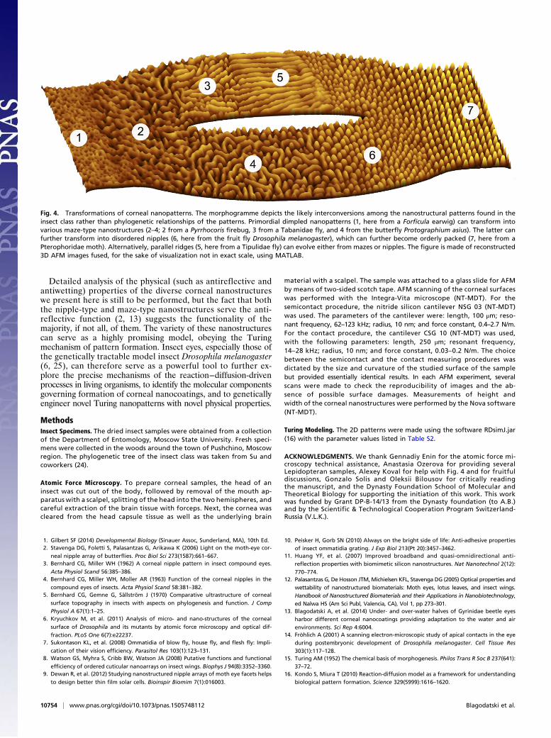
of these structures (Fig. 4). In this morphogramme, different typesof corneal nanocoatings are placed not based on the phyloge-netic hierarchy of the insect orders (24) but instead on theirmorphologies and transitions among them, as justified by theTuring modeling we performed. Originating from the dimple-type nanocoatings, this morphogramme then grows into themaze type and further into the nipples type (Fig. 4). We furtheridentify parallel ridges of some Dipterans and hexagonallypacked nipples of some Lepidopterans as developments of themaze- and nipples-type structures, respectively (Fig. 4). Bothrepresent more ordered structures and can be modeled to em-erge from their less ordered predecessors through increasingof the diffusion parameter of the activator component Du to thelevels maximally allowed within the boundaries permitting theTuring patterns to form (16, 22) (Fig. S4 C and D). In contrast,
increasing the diffusion parameter of the inhibitor componentDv leads to increase in the cross section of the nanostructures(nipples or ridges, respectively, in Fig. S4 C and D).In some Dipterans, a transition from nipples to parallel
ridges can be seen within the same lens, with nipples occu-pying the central part of the ommatidium and merging intoelongated strands away from the center (Fig. 1G). Turingmodeling predicts how such structures may be formed (Fig.S4E): Seen in flies with large ommatidia and lenses, thesenanocoatings are likely a result of a nipples-to-maze transitionwithin the same lens, happening during the lens formationafter initial nipples in the center of the cornea have beenformed. In this predefined space, parameters otherwise givingrise to mazes induce formation of parallel ridges emanatingfrom the nippled area (Fig. S4E).
Fig. 3. The insect corneal nanostructural diversityreplicates Turing patterns. Mathematically modeledTuring patterns (in black and white) and their insectcounterparts. (A and A′) Irregular nipples of varioussizes, characteristic e.g., for Hemipteran cornealnanocoatings. (B and B′) Highly ordered nipplednanoarrays (Lepidoptera). (C and C′) Strands merg-ing into a maze (Diptera, Tabanidae). (D and D′)Parallel strands (Diptera, Tipulidae). (E and E′) Nip-ples merging into a maze (Plecoptera). (F and F′)Typical maze-like structures (Coleoptera, Gyrinidae).(G and G′) Angular maze-like structures (Coleoptera,Coccinellidae). (H and H’) A typical dimpled pattern(Dermaptera). A′ is a fragment of Fig. S1F; B′ is animage from a Pterophoridae butterfly; C′ is a frag-ment of Fig. 1F; D′ is a fragment of Fig. 1G; E′ is afragment of Fig. 1E; F′ is an image from a Gyrinusbeetle [overwater eye (13)]; G′ is a fragment ofFig. S2B; and H′ is a fragment of Fig. 1D. Modelingparameters are given in Table S2. (I) Simulationsof Turing patterns formation. Step-wise changesin the av and bu parameters within the boundaryconditions produce different Turing patterns:dimples (yellow zone), mazes (blue zone), andnipples (green zone). See Fig. S4A for more de-tailed representation.
Blagodatski et al. PNAS | August 25, 2015 | vol. 112 | no. 34 | 10753
EVOLU
TION
ENGINEE
RING
Detailed analysis of the physical (such as antireflective andantiwetting) properties of the diverse corneal nanostructureswe present here is still to be performed, but the fact that boththe nipple-type and maze-type nanostructures serve the anti-reflective function (2, 13) suggests the functionality of themajority, if not all, of them. The variety of these nanostructurescan serve as a highly promising model, obeying the Turingmechanism of pattern formation. Insect eyes, especially those ofthe genetically tractable model insect Drosophila melanogaster(6, 25), can therefore serve as a powerful tool to further ex-plore the precise mechanisms of the reaction−diffusion-drivenprocesses in living organisms, to identify the molecular componentsgoverning formation of corneal nanocoatings, and to geneticallyengineer novel Turing nanopatterns with novel physical properties.
MethodsInsect Specimens. The dried insect samples were obtained from a collectionof the Department of Entomology, Moscow State University. Fresh speci-mens were collected in the woods around the town of Pushchino, Moscowregion. The phylogenetic tree of the insect class was taken from Su andcoworkers (24).
Atomic Force Microscopy. To prepare corneal samples, the head of aninsect was cut out of the body, followed by removal of the mouth ap-paratus with a scalpel, splitting of the head into the two hemispheres, andcareful extraction of the brain tissue with forceps. Next, the cornea wascleared from the head capsule tissue as well as the underlying brain
material with a scalpel. The sample was attached to a glass slide for AFMby means of two-sided scotch tape. AFM scanning of the corneal surfaceswas performed with the Integra-Vita microscope (NT-MDT). For thesemicontact procedure, the nitride silicon cantilever NSG 03 (NT-MDT)was used. The parameters of the cantilever were: length, 100 μm; reso-nant frequency, 62–123 kHz; radius, 10 nm; and force constant, 0.4–2.7 N/m.For the contact procedure, the cantilever CSG 10 (NT-MDT) was used,with the following parameters: length, 250 μm; resonant frequency,14–28 kHz; radius, 10 nm; and force constant, 0.03–0.2 N/m. The choicebetween the semicontact and the contact measuring procedures wasdictated by the size and curvature of the studied surface of the samplebut provided essentially identical results. In each AFM experiment, severalscans were made to check the reproducibility of images and the ab-sence of possible surface damages. Measurements of height andwidth of the corneal nanostructures were performed by the Nova software(NT-MDT).
Turing Modeling. The 2D patterns were made using the software RDsimJ.jar(16) with the parameter values listed in Table S2.
ACKNOWLEDGMENTS. We thank Gennadiy Enin for the atomic force mi-croscopy technical assistance, Anastasia Ozerova for providing severalLepidopteran samples, Alexey Koval for help with Fig. 4 and for fruitfuldiscussions, Gonzalo Solis and Oleksii Bilousov for critically readingthe manuscript, and the Dynasty Foundation School of Molecular andTheoretical Biology for supporting the initiation of this work. This workwas funded by Grant DP-B-14/13 from the Dynasty foundation (to A.B.)and by the Scientific & Technological Cooperation Program Switzerland-Russia (V.L.K.).
1. Gilbert SF (2014) Developmental Biology (Sinauer Assoc, Sunderland, MA), 10th Ed.2. Stavenga DG, Foletti S, Palasantzas G, Arikawa K (2006) Light on the moth-eye cor-
neal nipple array of butterflies. Proc Biol Sci 273(1587):661–667.3. Bernhard CG, Miller WH (1962) A corneal nipple pattern in insect compound eyes.
Acta Physiol Scand 56:385–386.4. Bernhard CG, Miller WH, Moller AR (1963) Function of the corneal nipples in the
compound eyes of insects. Acta Physiol Scand 58:381–382.5. Bernhard CG, Gemne G, Sällström J (1970) Comparative ultrastructure of corneal
surface topography in insects with aspects on phylogenesis and function. J Comp
Physiol A 67(1):1–25.6. Kryuchkov M, et al. (2011) Analysis of micro- and nano-structures of the corneal
surface of Drosophila and its mutants by atomic force microscopy and optical dif-
fraction. PLoS One 6(7):e22237.7. Sukontason KL, et al. (2008) Ommatidia of blow fly, house fly, and flesh fly: Impli-
cation of their vision efficiency. Parasitol Res 103(1):123–131.8. Watson GS, Myhra S, Cribb BW, Watson JA (2008) Putative functions and functional
efficiency of ordered cuticular nanoarrays on insect wings. Biophys J 94(8):3352–3360.9. Dewan R, et al. (2012) Studying nanostructured nipple arrays of moth eye facets helps
to design better thin film solar cells. Bioinspir Biomim 7(1):016003.
10. Peisker H, Gorb SN (2010) Always on the bright side of life: Anti-adhesive properties
of insect ommatidia grating. J Exp Biol 213(Pt 20):3457–3462.11. Huang YF, et al. (2007) Improved broadband and quasi-omnidirectional anti-
reflection properties with biomimetic silicon nanostructures. Nat Nanotechnol 2(12):
770–774.12. Palasantzas G, De Hosson JTM,Michielsen KFL, Stavenga DG (2005) Optical properties and
wettability of nanostructured biomaterials: Moth eyes, lotus leaves, and insect wings.
Handbook of Nanostructured Biomaterials and their Applications in Nanobiotechnology,
ed Nalwa HS (Am Sci Publ, Valencia, CA), Vol 1, pp 273–301.13. Blagodatski A, et al. (2014) Under- and over-water halves of Gyrinidae beetle eyes
harbor different corneal nanocoatings providing adaptation to the water and air
environments. Sci Rep 4:6004.14. Fröhlich A (2001) A scanning electron-microscopic study of apical contacts in the eye
during postembryonic development of Drosophila melanogaster. Cell Tissue Res
303(1):117–128.15. Turing AM (1952) The chemical basis of morphogenesis. Philos Trans R Soc B 237(641):
37–72.16. Kondo S, Miura T (2010) Reaction-diffusion model as a framework for understanding
biological pattern formation. Science 329(5999):1616–1620.
Fig. 4. Transformations of corneal nanopatterns. The morphogramme depicts the likely interconversions among the nanostructural patterns found in theinsect class rather than phylogenetic relationships of the patterns. Primordial dimpled nanopatterns (1, here from a Forficula earwig) can transform intovarious maze-type nanostructures (2–4; 2 from a Pyrrhocoris firebug, 3 from a Tabanidae fly, and 4 from the butterfly Protographium asius). The latter canfurther transform into disordered nipples (6, here from the fruit fly Drosophila melanogaster), which can further become orderly packed (7, here from aPterophoridae moth). Alternatively, parallel ridges (5, here from a Tipulidae fly) can evolve either from mazes or nipples. The figure is made of reconstructed3D AFM images fused, for the sake of visualization not in exact scale, using MATLAB.
10754 | www.pnas.org/cgi/doi/10.1073/pnas.1505748112 Blagodatski et al.
17. Nakamasu A, Takahashi G, Kanbe A, Kondo S (2009) Interactions between zebrafishpigment cells responsible for the generation of Turing patterns. Proc Natl Acad SciUSA 106(21):8429–8434.
18. Sick S, Reinker S, Timmer J, Schlake T (2006) WNT and DKK determine hair folliclespacing through a reaction-diffusion mechanism. Science 314(5804):1447–1450.
19. Raspopovic J, Marcon L, Russo L, Sharpe J (2014) Modeling digits. Digit patterning iscontrolled by a Bmp-Sox9-Wnt Turing network modulated by morphogen gradients.Science 345(6196):566–570.
20. Miura T, Maini PK (2004) Speed of pattern appearance in reaction–diffusion models:Implications in the pattern formation of limb bud mesenchyme cells. Bull Math Biol66(4):627–649.
21. Tardieu A (1988) Eye lens proteins and transparency: From light transmission theoryto solution X-ray structural analysis. Annu Rev Biophys Biophys Chem 17:47–70.
22. Miura T, Maini PK (2004) Periodic pattern formation in reaction–diffusion systems: Anintroduction for numerical simulation. Anat Sci Int 79(3):112–123.
23. Chipman AD, et al. (2014) The first myriapod genome sequence reveals conservativearthropod gene content and genome organisation in the centipede Strigamia mar-itima. PLoS Biol 12(11):e1002005.
24. Ishiwata K, Sasaki G, Ogawa J, Miyata T, Su ZH (2011) Phylogenetic relationshipsamong insect orders based on three nuclear protein-coding gene sequences. MolPhylogenet Evol 58(2):169–180.
25. Boseman A, Nowlin K, Ashraf S, Yang J, Lajeunesse D (2013) Ultrastructural analysis ofwild type and mutant Drosophila melanogaster using helium ion microscopy. Micron51:26–35.
26. Meyer-Rochow VB, Stringer IA (1993) A system of regular ridges instead of nipples ona compound eye that has to operate near the diffraction limit. Vision Res 33(18):2645–2647.
27. Clay Smith W, Butler JF (1991) Ultrastructure of the Tabanidae compound eye: Un-usual features for Diptera. J Insect Physiol 37(4):287–296.
28. Parker AR, Hegedus Z, Watts RA (1998) Solar–absorber antireflector on the eye of anEocene fly (45 Ma). Proc R Soc B 265(1398):811–815.
29. Anderson MS, Gaimari SD (2003) Raman-atomic force microscopy of the ommatidialsurfaces of Dipteran compound eyes. J Struct Biol 142(3):364–368.
30. Chu H, Norris DM, Carlson SD (1975) Ultrastructure of the compound eye of thediploid female beetle, Xyleborus ferrugineus. Cell Tissue Res 165(1):23–36.
31. Mishra M, Meyer-Rochow VB (2006) Eye ultrastructure in the pollen-feeding beetle,Xanthochroa luteipennis (Coleoptera: Cucujiformia: Oedemeridae). J Electron Microsc(Tokyo) 55(6):289–300.
32. Dey S, Dkhar B (1992) An unusual type of corneal nipple in the earwig, Forficula sp.,with a possible anti-reflection role. Micron and Microscopica Acta 23(3):337–339.
33. Dey S (1988) Scanning electron microscopic detection of corneal anti-reflectioncoating in the grasshopper, Epacromia dorsalis and its physiological significance.Vision Res 28(9):975–977.
34. Sheth R, et al. (2012) Hox genes regulate digit patterning by controlling the wave-length of a Turing-type mechanism. Science 338(6113):1476–1480.
35. Desai A, Mitchison TJ (1997) Microtubule polymerization dynamics. Annu Rev Cell DevBiol 13:83–117.
36. Alonso U, et al. (2011) Colloid diffusion coefficients in compacted and consolidatedclay barriers: Compaction density and colloid size effects. Phys Chem Earth Parts ABC36(17-18):1700–1707.
37. Pumpa M, Cichos F (2012) Slow single-molecule diffusion in liquid crystals. J PhysChem B 116(49):14487–14493.
38. Sergeev A, et al. (2015) Origin of order in bionanostructures. RSC Adv 5(78):63521–63527.
Blagodatski et al. PNAS | August 25, 2015 | vol. 112 | no. 34 | 10755
EVOLU
TION
ENGINEE
RING