Embed Size (px)
Citation preview

Divergent lineage of a novel hantavirus in thebanana pipistrelle (Neoromicia nanus) in Côted'IvoireSumibcay et al.
Sumibcay et al. Virology Journal 2012, 9:34http://www.virologyj.com/content/9/1/34 (26 January 2012)
SHORT REPORT Open Access
Divergent lineage of a novel hantavirusin the banana pipistrelle (Neoromicia nanus)in Côte d’IvoireLaarni Sumibcay1, Blaise Kadjo2, Se Hun Gu3, Hae Ji Kang4, Burton K Lim5, Joseph A Cook6, Jin-Won Song3 andRichard Yanagihara1,7*
Abstract
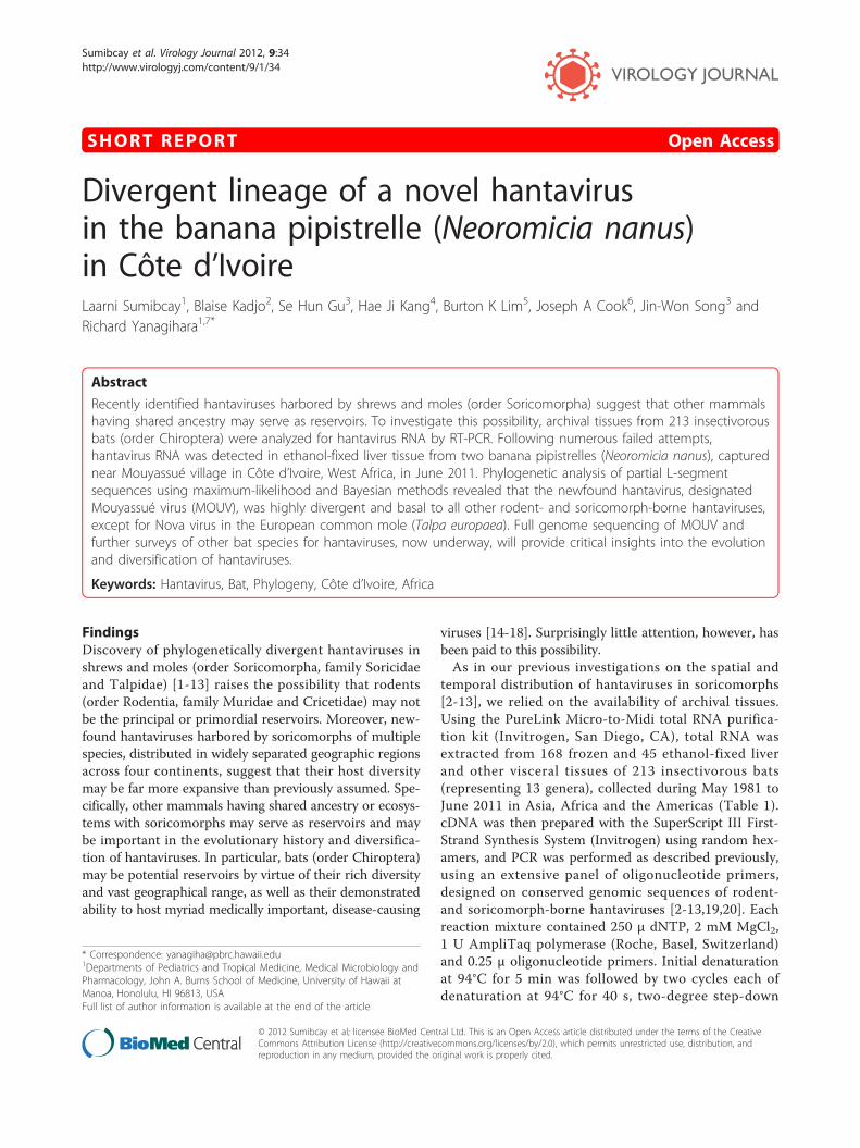
Recently identified hantaviruses harbored by shrews and moles (order Soricomorpha) suggest that other mammalshaving shared ancestry may serve as reservoirs. To investigate this possibility, archival tissues from 213 insectivorousbats (order Chiroptera) were analyzed for hantavirus RNA by RT-PCR. Following numerous failed attempts,hantavirus RNA was detected in ethanol-fixed liver tissue from two banana pipistrelles (Neoromicia nanus), capturednear Mouyassué village in Côte d’Ivoire, West Africa, in June 2011. Phylogenetic analysis of partial L-segmentsequences using maximum-likelihood and Bayesian methods revealed that the newfound hantavirus, designatedMouyassué virus (MOUV), was highly divergent and basal to all other rodent- and soricomorph-borne hantaviruses,except for Nova virus in the European common mole (Talpa europaea). Full genome sequencing of MOUV andfurther surveys of other bat species for hantaviruses, now underway, will provide critical insights into the evolutionand diversification of hantaviruses.
Keywords: Hantavirus, Bat, Phylogeny, Côte d’Ivoire, Africa
FindingsDiscovery of phylogenetically divergent hantaviruses inshrews and moles (order Soricomorpha, family Soricidaeand Talpidae) [1-13] raises the possibility that rodents(order Rodentia, family Muridae and Cricetidae) may notbe the principal or primordial reservoirs. Moreover, new-found hantaviruses harbored by soricomorphs of multiplespecies, distributed in widely separated geographic regionsacross four continents, suggest that their host diversitymay be far more expansive than previously assumed. Spe-cifically, other mammals having shared ancestry or ecosys-tems with soricomorphs may serve as reservoirs and maybe important in the evolutionary history and diversifica-tion of hantaviruses. In particular, bats (order Chiroptera)may be potential reservoirs by virtue of their rich diversityand vast geographical range, as well as their demonstratedability to host myriad medically important, disease-causing
viruses [14-18]. Surprisingly little attention, however, hasbeen paid to this possibility.As in our previous investigations on the spatial and
temporal distribution of hantaviruses in soricomorphs[2-13], we relied on the availability of archival tissues.Using the PureLink Micro-to-Midi total RNA purifica-tion kit (Invitrogen, San Diego, CA), total RNA wasextracted from 168 frozen and 45 ethanol-fixed liverand other visceral tissues of 213 insectivorous bats(representing 13 genera), collected during May 1981 toJune 2011 in Asia, Africa and the Americas (Table 1).cDNA was then prepared with the SuperScript III First-Strand Synthesis System (Invitrogen) using random hex-amers, and PCR was performed as described previously,using an extensive panel of oligonucleotide primers,designed on conserved genomic sequences of rodent-and soricomorph-borne hantaviruses [2-13,19,20]. Eachreaction mixture contained 250 μ dNTP, 2 mM MgCl2,1 U AmpliTaq polymerase (Roche, Basel, Switzerland)and 0.25 μ oligonucleotide primers. Initial denaturationat 94°C for 5 min was followed by two cycles each ofdenaturation at 94°C for 40 s, two-degree step-down
* Correspondence: [email protected] of Pediatrics and Tropical Medicine, Medical Microbiology andPharmacology, John A. Burns School of Medicine, University of Hawaii atManoa, Honolulu, HI 96813, USAFull list of author information is available at the end of the article
Sumibcay et al. Virology Journal 2012, 9:34http://www.virologyj.com/content/9/1/34
© 2012 Sumibcay et al; licensee BioMed Central Ltd. This is an Open Access article distributed under the terms of the CreativeCommons Attribution License (http://creativecommons.org/licenses/by/2.0), which permits unrestricted use, distribution, andreproduction in any medium, provided the original work is properly cited.

annealing from 48°C to 38°C for 40 s, and elongation at72°C for 1 min or 1 min 20 s, then 32 cycles of dena-turation at 94°C for 40 s, annealing at 42°C for 40 s, andelongation at 72°C for 1 min, in a GeneAmp PCR 9700thermal cycler (Perkin-Elmer, Waltham, MA). Ampli-cons were purified using the QIAQuick Gel ExtractionKit (Qiagen, Hilden, Germany), and DNA sequencingwas performed using an ABI Prism 377XL Genetic Ana-lyzer (Applied Biosystems, Foster City, CA).After innumerable failed attempts, hantavirus RNA
was detected by RT-PCR in ethanol-fixed liver tissuesfrom two of 12 banana pipistrelles (Neoromicia nanusPeters 1852), captured during June 2011 near Mouyas-sué village (5°22’07"N, 3°05’37"W) in Aboisso District,130 km from Abidjan, in the extreme southeasternregion of Côte d’Ivoire in West Africa (Figure 1). Thetaxonomic identity of the hantavirus-infected vesperbats was confirmed by phylogenetic analysis of the cyto-chrome b gene of mtDNA (GenBank JQ287717), ampli-fied by PCR as previously described [8,9]. Despitesimilarly exhaustive efforts, hantavirus RNA was notdetected in any of the other bat species tested (Table 1),including frozen liver tissue of six tiny pipistrelles (Pipis-trellus nanulus), collected in Parc National du MontPéko, 700 km northwest of Mouyassué, in February1992, and ethanol-fixed liver tissue of three tiny pipis-trelles, collected in December 2009 in Azagny, where ahantavirus was previously found in the West Africanpygmy shrew (Crocidura obscurior) [8].
A 423-nucleotide region of the RNA-dependent RNApolymerase-encoding L segment, amplified using ahemi-nested primer set (outer: 5’-GAAAGGG-CATTNMGATGGGCNTCA GG-3’, 5’-AACCADT-CWGTYCCRTCATC-3’; inner: 5’-GNAAAYTNATGT-ATGTNAGT GC-3’, 5’-AACCADTCWGTYCCRT-CATC-3’), was aligned and compared with hantavirussequences available in GenBank, using ClustalW(DNASTAR, Inc., Madison, WI) [21] and transAlign[22]. The newfound hantavirus, designated Mouyassuévirus (MOUV), exhibited low nucleotide and amino acidsequence similarity of less than 69% to all representativesoricomorph- and rodent-associated hantaviruses, exceptfor the 76.3% sequence similarity with Nova virus(NVAV), previously reported in the European commonmole (Talpa europaea) [12]. Interestingly, MOUVsequences were identical in the two banana pipistrelles(KB576 and KB577), a male-female pair captured simul-taneously and presumed to be a mating couple, suggest-ing horizontal virus transmission or common-sourceinfection.MOUV formed a uniquely divergent lineage, distant
from all other hantaviruses identified to date, except forNVAV (Figure 2), in phylogenetic trees based on L-seg-ment sequences, generated by the maximum-likelihoodand Bayesian methods, implemented in PAUP* (Phylo-genetic Analysis Using Parsimony, 4.0b10) [23], RAxMLBlackbox webserver [24] and MrBayes 3.1 [25], underthe best-fit GTR+I+Γ model of evolution established
Table 1 Detection of hantavirus RNA in tissues of insectivorous bats by RT-PCR
Genus species USA Bolivia Guyana Liberia Côte d’Ivoire Mongolia Malaysia Total
Antrozous pallidus 0/20 0/20
Corynorhinus townsendii 0/19 0/1 0/20
Eptesicus fuscus 0/21 0/21
Eptesicus gobiensis 0/20 0/20
Eptesicus sp. 0/4 0/4
Hipposideros cafer 0/14 0/5 0/19
Hipposideros cervinus 0/11 0/11
Hipposideros cyclops 0/11 0/11
Hipposideros gambianus 0/5 0/5
Lasiurus cinereus 0/20 0/20
Mops condylurus 0/2 0/2
Neoromicia nanus 2/12 2/12
Nycteris arge 0/1 0/1
Nycteris major 0/1 0/1
Nycteris thebaica 0/1 0/1
Pipistrellus nanulus 0/9 0/9
Pteronotus parnellii 0/5 0/5
Rhinolophus trifolatus 0/8 0/8
Scotophilus sp. 0/3 0/3
Tadarida brasiliensis 0/10 0/10 0/20
Total 0/90 0/11 0/5 0/23 2/45 0/20 0/19 2/213
Sumibcay et al. Virology Journal 2012, 9:34http://www.virologyj.com/content/9/1/34
Page 2 of 6

using jModeltest 0.1.1 [26]. Topologies were well sup-ported by bootstrap analysis of 100 iterations, and pos-terior node probabilities based on two runs each of 2million generations sampled every 100 generations withburn-in of 25%.
Despite the overall success of our brute-force RT-PCRapproach at identifying previously unrecognized hanta-viruses in frozen tissues [2,3,5-7,10-13] and tissues pre-served in RNAlater® RNA Stabilization Reagent [4,8],designing universal primers for the amplification of sori-comorph-borne hantaviruses has presented continuingchallenges. Thus, while it is likely that many more han-taviruses await discovery, overcoming technical barriersis essential to facilitating their detection. Viewed in thiscontext, the failure to detect hantavirus RNA in all butone bat species was not altogether unexpected and maybe attributed simply to suboptimal primer design andimperfect cycling conditions. Also, low RNA yields andpoor RNA preservation in tissues fixed in ethanol underfield conditions may have thwarted our efforts at obtain-ing more of the MOUV genome. That said, the success-ful amplification of hantavirus RNA from ethanol-fixedtissues is highly instructive and augments the pool ofarchival tissues for future exploratory studies of hanta-viruses in bats, and possibly other insectivorous smallmammals that share ancestral lineages with sorico-morphs, such as hedgehogs (order Erinaceomorpha,family Erinaceidae).Dating to the seminal discovery of Hantaan virus in
lung tissue of the striped field mouse (Apodemus agrar-ius) [27], lung has been the preferred tissue in studiesaimed at finding new hantaviruses [28-30]. However,lung is not the only tissue in which hantaviruses can bedetected [27,31]. In our search of genetically distincthantaviruses in long-stored archival tissues from shrewsand moles, lung tissue was frequently unavailable.Instead, liver tissue was more often accessible andproved to be quite suitable [4,5,12,13]. Similarly, livertissues were more often available in the present study.As in reservoir rodents and soricomorphs, hantavirusRNA is likely to be present in many tissues of persis-tently infected bats. Real-time quantitative RT-PCR ana-lysis of lung, liver and other viscera will clarify thetissue distribution of MOUV in newly captured bananapipistrelles from Mouyassué.Having their fossil origins in the Eocene epoch,
approximately 50 million years before present, batsoccur on every continent except Antarctica and areamong the most speciose orders of mammals, withmore than 1,100 extant species [32]. The banana pipis-trelle, which is distributed widely in forests and savannasacross sub-Saharan Africa (Figure 1C, inset), is one of13 species in the genus Neoromicia of the family Ves-pertilionidae and subfamily Vespertilioninae. Like othervesper bats, the banana pipistrelle is insectivorous.Unlike large fruit bats, such as the straw-colored fruitbat (Eidolon helvum) and hammer-headed bat (Hyp-signathus monstrosus), which are sold as bush meat, thebanana pipistrelle, weighing approximately 3 g, is not
Figure 1 (A) Banana pipistrelle (Neoromicia nanus) in whichhantavirus RNA was detected. (B) Capture site of bananapipistrelles near Mouyassué village in Aboisso District. (C) Map ofCôte d’Ivoire, showing Mouyassué, Azagny and Mont Péko, whereinsectivorous bats were captured. The geographic range of thebanana pipistrelle extends throughout sub-Saharan Africa (shadedarea in inset).
Sumibcay et al. Virology Journal 2012, 9:34http://www.virologyj.com/content/9/1/34
Page 3 of 6

Figure 2 Phylogenetic trees were generated by maximum-likelihood and Bayesian methods, under the GTR+I+Γ model of evolution,based on a 423-nucleotide L-genomic segment of Mouyassué virus (MOUV KB576) (GenBank JQ287716). Since tree topologies weresimilar using RAxML, PAUP* and MrBayes, the tree generated by MrBayes was displayed. The numbers at each node are posterior probabilities.The scale bar indicates nucleotide substitutions per site. The phylogenetic position of MOUV is shown in relation to representative soricomorph-borne hantaviruses, including Thottapalayam virus (TPMV VRC66412: EU001330) from the Asian house shrew (Suncus murinus), Imjin virus (MJNVCl05-11: EF641806) from the Ussuri white-toothed shrew (Crocidura lasiura), Jeju virus (JJUV SH42: HQ663935) from the Asian lesser white-toothedshrew (Crocidura shantungensis), Tanganya virus (TGNV Tan826: EF050454) from the Therese’s shrew (Crocidura theresae), Azagny virus (AZGVKBM15: JF276228) from the West African pygmy shrew (Crocidura obscurior), Cao Bang virus (CBNV CBN-3: EF543525) from the Chinese moleshrew (Anourosorex squamipes), Ash River virus (ARRV MSB73418: EF619961) from the masked shrew (Sorex cinereus), Jemez Springs virus (JMSVMSB144475: FJ593501) from the dusky shrew (Sorex monticolus), Seewis virus (SWSV mp70: EF636026) from the Eurasian common shrew (Sorexaraneus), Kenkeme virus (KKMV MSB148794: GQ306150) from the flat-skulled shrew (Sorex roboratus), Qiandao Lake virus (QDLV YN05-284:GU566021) from the stripe-backed shrew (Sorex cylindricauda), Camp Ripley virus (RPLV MSB89863: EF540771) from the northern short-tailedshrew (Blarina brevicauda), Asama virus (ASAV N10: EU929078) from the Japanese shrew mole (Urotrichus talpoides), Oxbow virus (OXBV Ng1453:FJ593497) from the American shrew mole (Neurotrichus gibbsii), Rockport virus (RKPV MSB57412: HM015221) from the eastern mole (Scalopusaquaticus), and Nova virus (NVAV MSB95703: FJ593498) from the European common mole (Talpa europaea). Also shown are rodent-bornehantaviruses, including Hantaan virus (HTNV 76-118: NC_005222), Soochong virus (SOOV SOO-1: DQ562292), Dobrava virus (DOBV Greece:NC_005235), Seoul virus (SEOV HR80-39: NC_005238), Tula virus (TULV M5302v: NC_005226), Puumala virus (PUUV Sotkamo: NC_005225), ProspectHill virus (PHV PH-1: EF646763), Andes virus (ANDV Chile-9717869: NC_003468), and Sin Nombre virus (SNV NMH10: NC_005217).
Sumibcay et al. Virology Journal 2012, 9:34http://www.virologyj.com/content/9/1/34
Page 4 of 6
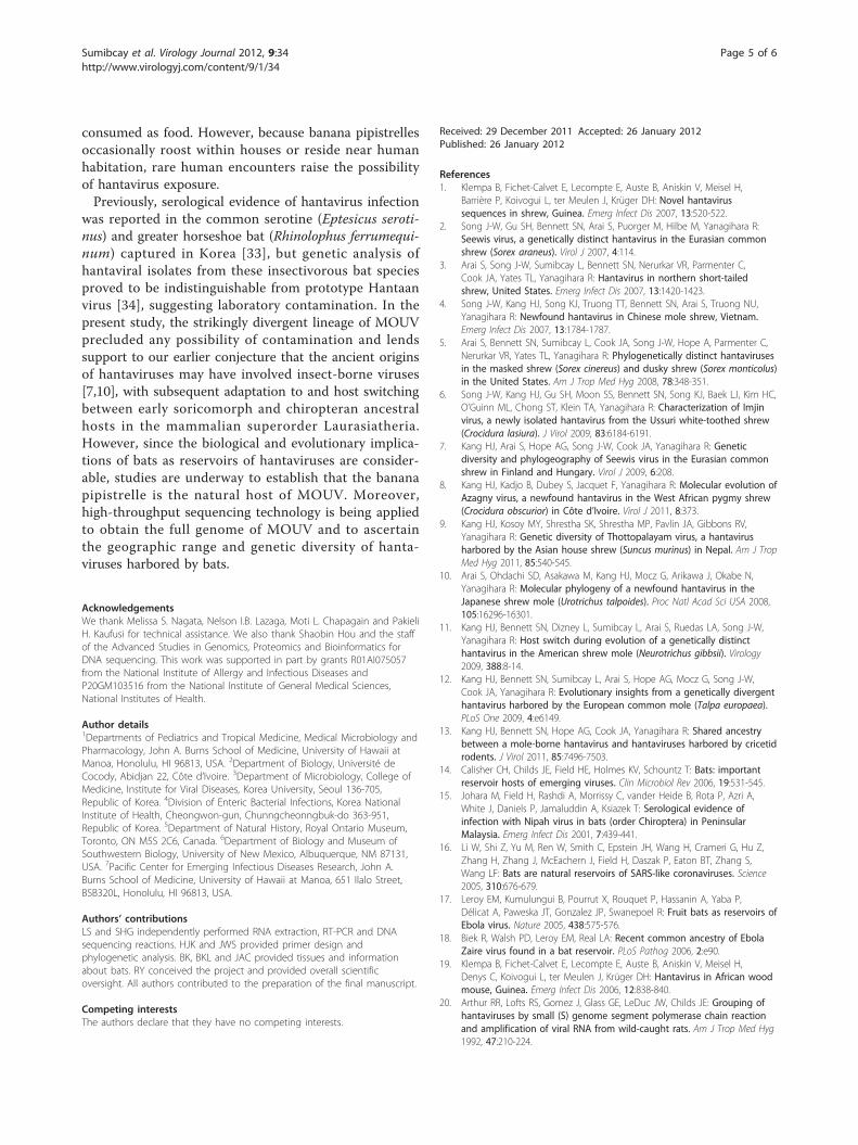
consumed as food. However, because banana pipistrellesoccasionally roost within houses or reside near humanhabitation, rare human encounters raise the possibilityof hantavirus exposure.Previously, serological evidence of hantavirus infection
was reported in the common serotine (Eptesicus seroti-nus) and greater horseshoe bat (Rhinolophus ferrumequi-num) captured in Korea [33], but genetic analysis ofhantaviral isolates from these insectivorous bat speciesproved to be indistinguishable from prototype Hantaanvirus [34], suggesting laboratory contamination. In thepresent study, the strikingly divergent lineage of MOUVprecluded any possibility of contamination and lendssupport to our earlier conjecture that the ancient originsof hantaviruses may have involved insect-borne viruses[7,10], with subsequent adaptation to and host switchingbetween early soricomorph and chiropteran ancestralhosts in the mammalian superorder Laurasiatheria.However, since the biological and evolutionary implica-tions of bats as reservoirs of hantaviruses are consider-able, studies are underway to establish that the bananapipistrelle is the natural host of MOUV. Moreover,high-throughput sequencing technology is being appliedto obtain the full genome of MOUV and to ascertainthe geographic range and genetic diversity of hanta-viruses harbored by bats.
AcknowledgementsWe thank Melissa S. Nagata, Nelson I.B. Lazaga, Moti L. Chapagain and PakieliH. Kaufusi for technical assistance. We also thank Shaobin Hou and the staffof the Advanced Studies in Genomics, Proteomics and Bioinformatics forDNA sequencing. This work was supported in part by grants R01AI075057from the National Institute of Allergy and Infectious Diseases andP20GM103516 from the National Institute of General Medical Sciences,National Institutes of Health.
Author details1Departments of Pediatrics and Tropical Medicine, Medical Microbiology andPharmacology, John A. Burns School of Medicine, University of Hawaii atManoa, Honolulu, HI 96813, USA. 2Department of Biology, Université deCocody, Abidjan 22, Côte d’Ivoire. 3Department of Microbiology, College ofMedicine, Institute for Viral Diseases, Korea University, Seoul 136-705,Republic of Korea. 4Division of Enteric Bacterial Infections, Korea NationalInstitute of Health, Cheongwon-gun, Chunngcheonngbuk-do 363-951,Republic of Korea. 5Department of Natural History, Royal Ontario Museum,Toronto, ON M5S 2C6, Canada. 6Department of Biology and Museum ofSouthwestern Biology, University of New Mexico, Albuquerque, NM 87131,USA. 7Pacific Center for Emerging Infectious Diseases Research, John A.Burns School of Medicine, University of Hawaii at Manoa, 651 Ilalo Street,BSB320L, Honolulu, HI 96813, USA.
Authors’ contributionsLS and SHG independently performed RNA extraction, RT-PCR and DNAsequencing reactions. HJK and JWS provided primer design andphylogenetic analysis. BK, BKL and JAC provided tissues and informationabout bats. RY conceived the project and provided overall scientificoversight. All authors contributed to the preparation of the final manuscript.
Competing interestsThe authors declare that they have no competing interests.
Received: 29 December 2011 Accepted: 26 January 2012Published: 26 January 2012
References1. Klempa B, Fichet-Calvet E, Lecompte E, Auste B, Aniskin V, Meisel H,
Barrière P, Koivogui L, ter Meulen J, Krüger DH: Novel hantavirussequences in shrew, Guinea. Emerg Infect Dis 2007, 13:520-522.
2. Song J-W, Gu SH, Bennett SN, Arai S, Puorger M, Hilbe M, Yanagihara R:Seewis virus, a genetically distinct hantavirus in the Eurasian commonshrew (Sorex araneus). Virol J 2007, 4:114.
3. Arai S, Song J-W, Sumibcay L, Bennett SN, Nerurkar VR, Parmenter C,Cook JA, Yates TL, Yanagihara R: Hantavirus in northern short-tailedshrew, United States. Emerg Infect Dis 2007, 13:1420-1423.
4. Song J-W, Kang HJ, Song KJ, Truong TT, Bennett SN, Arai S, Truong NU,Yanagihara R: Newfound hantavirus in Chinese mole shrew, Vietnam.Emerg Infect Dis 2007, 13:1784-1787.
5. Arai S, Bennett SN, Sumibcay L, Cook JA, Song J-W, Hope A, Parmenter C,Nerurkar VR, Yates TL, Yanagihara R: Phylogenetically distinct hantavirusesin the masked shrew (Sorex cinereus) and dusky shrew (Sorex monticolus)in the United States. Am J Trop Med Hyg 2008, 78:348-351.
6. Song J-W, Kang HJ, Gu SH, Moon SS, Bennett SN, Song KJ, Baek LJ, Kim HC,O’Guinn ML, Chong ST, Klein TA, Yanagihara R: Characterization of Imjinvirus, a newly isolated hantavirus from the Ussuri white-toothed shrew(Crocidura lasiura). J Virol 2009, 83:6184-6191.
7. Kang HJ, Arai S, Hope AG, Song J-W, Cook JA, Yanagihara R: Geneticdiversity and phylogeography of Seewis virus in the Eurasian commonshrew in Finland and Hungary. Virol J 2009, 6:208.
8. Kang HJ, Kadjo B, Dubey S, Jacquet F, Yanagihara R: Molecular evolution ofAzagny virus, a newfound hantavirus in the West African pygmy shrew(Crocidura obscurior) in Côte d’Ivoire. Virol J 2011, 8:373.
9. Kang HJ, Kosoy MY, Shrestha SK, Shrestha MP, Pavlin JA, Gibbons RV,Yanagihara R: Genetic diversity of Thottopalayam virus, a hantavirusharbored by the Asian house shrew (Suncus murinus) in Nepal. Am J TropMed Hyg 2011, 85:540-545.
10. Arai S, Ohdachi SD, Asakawa M, Kang HJ, Mocz G, Arikawa J, Okabe N,Yanagihara R: Molecular phylogeny of a newfound hantavirus in theJapanese shrew mole (Urotrichus talpoides). Proc Natl Acad Sci USA 2008,105:16296-16301.
11. Kang HJ, Bennett SN, Dizney L, Sumibcay L, Arai S, Ruedas LA, Song J-W,Yanagihara R: Host switch during evolution of a genetically distincthantavirus in the American shrew mole (Neurotrichus gibbsii). Virology2009, 388:8-14.
12. Kang HJ, Bennett SN, Sumibcay L, Arai S, Hope AG, Mocz G, Song J-W,Cook JA, Yanagihara R: Evolutionary insights from a genetically divergenthantavirus harbored by the European common mole (Talpa europaea).PLoS One 2009, 4:e6149.
13. Kang HJ, Bennett SN, Hope AG, Cook JA, Yanagihara R: Shared ancestrybetween a mole-borne hantavirus and hantaviruses harbored by cricetidrodents. J Virol 2011, 85:7496-7503.
14. Calisher CH, Childs JE, Field HE, Holmes KV, Schountz T: Bats: importantreservoir hosts of emerging viruses. Clin Microbiol Rev 2006, 19:531-545.
15. Johara M, Field H, Rashdi A, Morrissy C, vander Heide B, Rota P, Azri A,White J, Daniels P, Jamaluddin A, Ksiazek T: Serological evidence ofinfection with Nipah virus in bats (order Chiroptera) in PeninsularMalaysia. Emerg Infect Dis 2001, 7:439-441.
16. Li W, Shi Z, Yu M, Ren W, Smith C, Epstein JH, Wang H, Crameri G, Hu Z,Zhang H, Zhang J, McEachern J, Field H, Daszak P, Eaton BT, Zhang S,Wang LF: Bats are natural reservoirs of SARS-like coronaviruses. Science2005, 310:676-679.
17. Leroy EM, Kumulungui B, Pourrut X, Rouquet P, Hassanin A, Yaba P,Délicat A, Paweska JT, Gonzalez JP, Swanepoel R: Fruit bats as reservoirs ofEbola virus. Nature 2005, 438:575-576.
18. Biek R, Walsh PD, Leroy EM, Real LA: Recent common ancestry of EbolaZaire virus found in a bat reservoir. PLoS Pathog 2006, 2:e90.
19. Klempa B, Fichet-Calvet E, Lecompte E, Auste B, Aniskin V, Meisel H,Denys C, Koivogui L, ter Meulen J, Krüger DH: Hantavirus in African woodmouse, Guinea. Emerg Infect Dis 2006, 12:838-840.
20. Arthur RR, Lofts RS, Gomez J, Glass GE, LeDuc JW, Childs JE: Grouping ofhantaviruses by small (S) genome segment polymerase chain reactionand amplification of viral RNA from wild-caught rats. Am J Trop Med Hyg1992, 47:210-224.
Sumibcay et al. Virology Journal 2012, 9:34http://www.virologyj.com/content/9/1/34
Page 5 of 6
21. Thompson JD, Higgins DG, Gibson TJ: CLUSTAL W: improving thesensitivity of progressive multiple sequence alignment throughsequence weighting, position-specific gap penalties and weight matrixchoice. Nucleic Acids Res 1994, 22:4673-4680.
22. Bininda-Emonds OR: transAlign: using amino acids to facilitate themultiple alignment of protein-coding DNA sequences. BMC Bioinformatics2005, 6:156.
23. Swofford D: PAUP*: Phylogenetic Analysis Using Parsimony (*and OtherMethods) Sunderland, Massachusetts: Sinauer Associates; 2003.
24. Stamatakis A, Hoover P, Rougemont J: A rapid bootstrap algorithm for theRAxML web servers. Syst Biol 2008, 57:758-771.
25. Ronquist F, Huelsenbeck JP: MrBayes 3: Bayesian phylogenetic inferenceunder mixed models. Bioinformatics 2003, 19:1572-1574.
26. Posada D: jModelTest: phylogenetic model averaging. Mol Biol Evol 2008,25:1253-1256.
27. Lee HW, Lee P-W, Johnson KM: Isolation of the etiologic agent of Koreanhemorrhagic fever. J Infect Dis 1978, 137:298-308.
28. Brummer-Korvenkontio M, Vaheri A, Hovi T, von Bonsdorff CH, Vuorimies J,Manni T, Penttinen K, Oker-Blom N, Lähdevirta J: Nephropathia epidemica:detection of antigen in bank voles and serologic diagnosis of humaninfection. J Infect Dis 1980, 141:131-134.
29. Lee HW, Baek LJ, Johnson KM: Isolation of Hantaan virus, the etiologicagent of Korean hemorrhagic fever, from wild urban rats. J Infect Dis1982, 146:638-644.
30. Nichol ST, Spiropoulou CF, Morzunov S, Rollin PE, Ksiazek TG, Feldmann H,Sanchez A, Childs J, Zaki S, Peters CJ: Genetic identification of ahantavirus associated with an outbreak of acute respiratory illness.Science 1993, 262:914-917.
31. Lee HW, French GR, Lee PW, Baek LJ, Tsuchiya K, Foulke RS: Observationson natural and laboratory infection of rodents with the etiologic agentof Korean hemorrhagic fever. Am J Trop Med Hyg 1981, 30:477-482.
32. Wilson DE, Reeder DM: Mammal Species of the World: A Taxonomic andGeographic Reference. 3 edition. Baltimore, Maryland: The Johns HopkinsUniversity Press; 2005.
33. Kim GR, Lee YT, Park CH: A new natural reservoir of hantavirus: isolationof hantaviruses from lung tissues of bats. Arch Virol 1994, 134:85-95.
34. Jung YT, Kim GR: Genomic characterization of M and S RNA segments ofhantaviruses isolated from bats. Acta Virol 1995, 39:231-233.
doi:10.1186/1743-422X-9-34Cite this article as: Sumibcay et al.: Divergent lineage of a novelhantavirus in the banana pipistrelle (Neoromicia nanus) in Côte d’Ivoire.Virology Journal 2012 9:34.
Submit your next manuscript to BioMed Centraland take full advantage of:
• Convenient online submission
• Thorough peer review
• No space constraints or color figure charges
• Immediate publication on acceptance
• Inclusion in PubMed, CAS, Scopus and Google Scholar
• Research which is freely available for redistribution
Submit your manuscript at www.biomedcentral.com/submit
Sumibcay et al. Virology Journal 2012, 9:34http://www.virologyj.com/content/9/1/34
Page 6 of 6