Embed Size (px)
Citation preview

Distinctive Features of Angiogenesis and Lymphangiogenesis
Determine Their Functionality during De novo
Tumor Development
Alexandra Eichten,1William C. Hyun,
3and Lisa M. Coussens
1,2,3
1Department of Pathology, 2Cancer Research Institute, and 3Comprehensive Cancer Center, University of California atSan Francisco, San Francisco, California
Abstract
Blood and lymphatic vasculature are essential components ofall organs, responsible for maintaining organ fluid dynamicsand tissue homeostasis. Although both vessel systems arecomposed of similar lineages of endothelial cells whose crudefunctions include fluid and cell transport, each system alsopossesses distinctive physiologic properties, enabling theirdistinctive functions in tissues. The role of hematogenousvasculature and development of angiogenic blood vesselsduring cancer development is well established; however, therole of lymphangiogenesis and structural/functional alter-ations occurring within lymphatic vessels during cancerdevelopment are incompletely understood. To assess prema-lignant versus malignant alterations in blood and lymphaticvasculature associated with squamous epithelial skin carci-nogenesis, we assessed architectural and functional featuresof both vascular systems using a mouse model of de novocarcinoma development. We report that, as vasculatureacquires angiogenic and/or lymphangiogenic properties,angiogenic blood vessels become leaky in premalignant tissueand at peripheries of carcinomas, where enlarged lymphaticcapillaries efficiently drain increased tissue fluid, therebymaintaining tissue hemodynamics. In contrast, central regionsof carcinomas exhibit elevated tissue fluid levels, compressedlymphatic lumina, and decreased vascular leakage, thusindicating impaired hemodynamics within solid tumors.Together, these data support the notion that therapeuticdelivery of anticancer agents is best realized in premalignanttissues and/or at the peripheries of solid tumors wherehemodynamic forces support drug delivery. Strategies tonormalize intratumoral hemodynamics would thereforeenhance therapeutic delivery to otherwise poorly accessiblecentral regions of solid tumors. [Cancer Res 2007;67(11):5211–20]
Introduction
Blood and lymphatic vessels comprise two interdependentvascular networks in all tissues. Whereas blood vessels deliverblood cells, plasma proteins, and oxygen to tissues, lymphatics,composed of lymph-forming capillaries and collecting vessels,continually regulate interstitial fluid pressure by draining intersti-tial fluid and debris to maintain tissue homeostasis. When tissuesare acutely damaged, activation of both vascular systems occurs as
part of innate repair programs; once complete, both systems returnto their homeostatic states. In contrast, sustained activation of oneor both vascular networks is associated with some chronicdisorders, such as rheumatoid arthritis (1) and psoriasis (2, 3),and contributes to disease pathogenesis. Cancer development issimilarly associated with chronic activation of blood vasculature(i.e., angiogenesis) in premalignant and malignant tissues (4).Activation of angiogenic vasculature in premalignant tissue ischaracterized by increased proliferation of vascular endothelial cells(VEC) and sprouting of new immature leaky blood vessels frompreexisting vascular beds (5). Increased leakage of plasma proteinsfrom immature angiogenic vessels leads to increased interstitialfluid content, lymph formation, and drainage via lymphatic vesselsback into the blood circulation (6). High interstitial fluid pressure(IFP), which forms a barrier to transcapillary transport, results ininefficient delivery of therapeutic drugs from vasculature intotumor stroma (7). It has been postulated that high IFP, common tomany solid tumors, is in part a result of inefficient clearance oftissue fluid by lymphatic vessels (7). This postulate is supported byhistochemical studies evaluating lymphatic architecture anddiminished lumen diameters in archival human carcinomas andmurine xenograft tumors (8–12). Studies evaluating functionalchanges in the status of lymphatic endothelial cells (LEC) and/orlymphatic vessels compared with VECs and blood vessels inpremalignant tissues have not been well described.
To critically examine distinctive versus common physiologic andfunctional properties of blood versus lymphatic vasculature thataccompany and/or contribute to cancer development, we used atransgenic mouse model of de novo epithelial squamous cellcarcinogenesis (e.g., K14-HPV16 mice; ref. 13). HPV16 mice progressthrough well-defined premalignant stages before de novo carcinomadevelopment, mirroring histopathologic stages observed duringhuman cervical carcinogenesis (14). HPV16 mice develop hyper-plastic skin lesions with 100% penetrance by 1 month of age thatfocally progress to dysplasia by 3 to 6 months (13). Precursordysplasias undergo malignant conversion into varying grades ofsquamous cell carcinoma (SCC) in skin in 50% of mice that meta-stasize to regional lymph nodes with a 30% frequency (13, 15).Angiogenic vasculature is first evident in premalignant hyperplasias,development of which is linked to infiltration of innate immune cells(e.g., mast cells, granulocytes, and macrophages; refs. 15–18). Usingthis model, we assessed molecular, histopathologic, and functionalvariables of blood and lymphatic vasculature to reveal their distinc-tive physiologic properties at each stage of neoplastic development.
Materials and Methods
Animal Husbandry, Genotype, and Histopathologic AnalysesAll animals were maintained within the University of California at San
Francisco (UCSF) Laboratory for Animal Care barrier facility according to
Requests for reprints: Lisa M. Coussens, Department of Pathology andComprehensive Cancer Center, University of California at San Francisco, 2340 SutterStreet, N-221, Box 0875, San Francisco, CA 94115. Phone: 415-502-6378; Fax: 415-514-0878; E-mail: [email protected].
I2007 American Association for Cancer Research.doi:10.1158/0008-5472.CAN-06-4676
www.aacrjournals.org 5211 Cancer Res 2007; 67: (11). June 1, 2007
Research Article
Research. on July 8, 2018. © 2007 American Association for Cancercancerres.aacrjournals.org Downloaded from

Institutional Animal Care and Use Committee procedures. HPV16transgenic mice (19), preparation of tissue sections (ear skin) for histologicexamination, and characterization of neoplastic stages based on H&Ehistopathology and cytokeratin intermediate filament expression have beendescribed previously (13, 15, 20). Paraffin-embedded tissue sections werefixed by immersion in 10% neutral-buffered formalin, dehydrated throughgraded ethanol and xylenes, embedded in paraffin, cut with a Leica 2135microtome into 5-Am-thick sections, and histopathologically examinedfollowing H&E staining and immunoreactivity of keratin intermediatefilaments. Hyperplastic lesions were identified by a 2-fold increase inepidermal thickness and an intact granular cell layer with keratohyalingranules. Dysplastic lesions were characterized based on basal and spinouscell layers with hyperchromatic nuclei representing greater than half ofthe total epidermal thickness and incomplete terminal differentiation ofkeratinocytes. SCC was identified by abundance of abnormal mitotic figuresand an invasive loss of integrity in epithelial basement membrane withclear development of malignant cell clusters proliferating in the dermis.SCCs were graded as has been described previously (17).
Flow CytometrySingle cell suspensions were prepared from ear (n = 4) and tumor (n = 7)
tissue as described previously (21). Cells were incubated for 10 min at4jC with rat anti-mouse CD16/CD32 monoclonal antibody (mAb; BDBiosciences) at a 1:200 dilution in PBS/bovine serum albumin (BSA) toprevent nonspecific antibody binding. Subsequently, cells were washed andincubated for 20 min with phycoerythrin-conjugated anti-mouse CD31(1:200; eBioscience) and anti-mouse podoplanin hybridoma supernatant(mAb clone 8.1.1, 1:1000; Developmental Studies Hybridoma Bank,University of Iowa). Cells were washed twice with PBS/BSA and incubatedwith Alexa Fluor 647–conjugated anti-hamster antibody (1:1,000; MolecularProbes) for 20 min. After two washes with PBS/BSA, 7-aminoactinomycin D(1:10; BD Biosciences) was added to discriminate between viable and deadcells. Data acquisition was done on a FACSCalibur using CellQuestProsoftware (BD Biosciences), and data analysis was done using FlowJosoftware (Tree Star, Inc.). Data shown represent the mean F SE. P values of<0.05 were considered to be statistically significant.
ImmunohistochemistryImmunohistochemistry on paraffin-embedded tissue sections.
Tissue sections were deparaffinized, briefly washed in PBS, and blocked
for 15 min in blocking buffer (5.0% normal goat serum/2.5% BSA/PBS).
Primary antibodies were diluted in 0.5� blocking buffer: rat anti-mouse CD31 (1:50; PharMingen), Syrian hamster anti-mouse podoplanin
(1:200, clone 8.1.1; Developmental Studies Hybridoma Bank, University of
Iowa), guinea pig anti-mouse cytokeratin pan (1:100; Progen BiotechnikGmbH), and rabbit anti-mouse lymphatic vessel endothelial receptor-1
(LYVE-1; 1:200; Upstate). Sections were incubated with primary antibody
overnight at 4jC in a humidified chamber followed by three brief washes in
PBS and subsequently incubated with secondary antibodies Alexa Fluor488–conjugated anti-rat antibody (1:500; Molecular Probes), Alexa Fluor
633–conjugated anti-guinea pig antibody (1:500; Molecular Probes), Alexa
Fluor 594–conjugated anti-hamster antibody (1:500; Molecular Probes), or
Alexa Fluor 594/488–conjugated anti-rabbit antibody (1:500; MolecularProbes) for 2.0 h at room temperature in a humidified chamber. After five
3-min washes in PBS, the fluorescently stained sections were mounted in
ProLong Gold mounting medium (Molecular Probes). Fluorescence was
visualized using a laser scanning confocal microscope LSM510 META (CarlZeiss MicroImaging, Inc.) and analyzed with Zeiss LSM Image Examiner.
Ear tissue (n = 4/time point) and tumor tissue (n = 5) were assessed for
lymphatic luminal status and blood and lymphatic vessel density bycounting the number of vessels in five random high-power fields of vision
per time point per tissue, tumor center, and periphery.
Immunohistochemical detection of proliferating endothelial cells.Fresh bromodeoxyuridine (BrdUrd; Sigma) suspended in water (1.0 mg/mL)was given to mice in drinking water ad libitum and changed every 2nd day
for a total of 8 days. For fluorescent angiography, animals were tail vein
injected with FITC-conjugated Lycopersicon esculentum (tomato) lectin
(2.0 mg/mL; Vector Laboratories) 3.0 min before cardiac perfusion with the
following reagents in chronological order: 15 mL of 1% paraformaldehyde/0.5% glutaraldehyde, 7.0 mL PBS, 15 mL PBS containing 1.0% BSA, and15 mL of 4.0% paraformaldehyde at a constant flow rate of 7.0 mL/min.Tissue was harvested and fixed in 10% zinc-buffered formalin for 8 to 16 h at4jC before further processing followed by dehydration through gradedalcohols and xylene and embedding in paraffin. Paraffin sections (5 Amthick) were cut using a Leica 2135 microtome. Blood vasculature wasdetected by fluorescent angiography (22), and immunohistochemicalanalysis was done detecting lymphatic vessels (LYVE-1 reactivity), keratin-expressing epithelial cells (pan-keratin reactivity), proliferating nuclei(BrdUrd incorporation), and all nuclei as follows: Sections were deparaffi-nized, incubated in 2.0 N HCl for 1.0 h at room temperature, rinsed in PBS,and blocked for 30 min in blocking buffer (5.0% normal goat serum/2.5%BSA/PBS). Primary antibodies were diluted in 0.5� blocking buffer: guineapig anti-mouse cytokeratin pan (1:100), biotinylated anti-BrdUrd (1:50; cloneBr-3; Caltag), and rabbit anti-mouse LYVE-1 (1:200). Sections wereincubated with primary antibody overnight at 4jC followed by three briefwashes in PBS and subsequently incubated with secondary antibodies(all 1:500; Molecular Probes) Alexa Fluor 633–conjugated anti-guinea pigantibody, Alexa Fluor 546-streptavidin–conjugated antibody, and AlexaFluor 594–conjugated anti-rabbit antibody for 2.0 h at room temperature ina humidified chamber. After five 3-min washes in PBS, the fluorescentlystained sections were subjected to a nuclear staining with SYTO62(0.7 Amol/L in H2O; Molecular Probes) for 10 min and then mounted inProLong Gold mounting medium. Fluorescence was visualized using a laserscanning confocal microscope LSM510 META and analyzed with a ZeissLSM Image Examiner. A minimum of 100 endothelial cell nuclei in control(n = 4), premalignant ear (n = 4), and tumor tissue (n = 7) was identified intissue sections based on their morphologic protrusion into the vessellumen, and the percentage of proliferating nuclei was determined.
Fluorescent Angiography and Immunohistochemistry ofWhole-Mount Ear Tissue and Thick Frozen Tumor SectionsWhole-mounted ear tissue (n = 3 per time point) was prepared as
described previously (23). Briefly, mice were anesthetized with a 2%isofluorane/98% oxygen mixture and 100 AL of FITC-conjugated L.esculentum (tomato) lectin (2.0 mg/mL) were injected into the tail veinand allowed to circulate for 3.0 min followed by cardiac perfusion with35 mL of PBS-buffered 4% paraformaldehyde (pH 7.4; ref. 22). Ears wereharvested, ventral and dorsal aspects were separated, and cartilage wasremoved from ventral aspects, which were then immersion fixed in PBS-buffered 4% paraformaldehyde (pH 7.4) overnight at 4jC and subjected tothe following staining procedure under exclusion of light: Ventral aspectsof ears were rinsed briefly in PBS containing 0.3% Triton X-100 (PBS-T) andblocked in PBS-T containing 3% goat serum overnight at 4jC. Tissue wassubsequently incubated with primary rabbit anti-mouse LYVE-1 antibody(1:2,000; Upstate) diluted in PBS-T over two nights at 4jC. After five washesin PBS-T (5–10 min each) and a 2- to 3-h blocking incubation with PBS-Tcontaining 3% goat serum at 4jC, tissue was incubated overnight at 4jCwith a Alexa Fluor 594–conjugated anti-rabbit antibody (1:500; MolecularProbes). Tissue was then washed 10 times for 5 min at 4jC and mounted inProLong Gold mounting medium.
Tumors (n = 5) were dissected, flash frozen in OCT compound, and cut
into 200 Am sections and subsequently rinsed briefly in PBS-T and blockedin PBS-T containing 3% goat serum for 2.0 h at room temperature. Tissue
was subsequently incubated with primary rabbit anti-mouse LYVE-1
antibody (1:2,000) diluted in PBS-T overnight at 4jC. After three 3-min
washes in PBS-T, tissue was incubated for 3 h at room temperature withAlexa Fluor 594–conjugated anti-rabbit antibody (1:500). After three 3-min
washes in PBS-T, tissue was mounted in ProLong Gold mounting medium.
Fluorescence was visualized using a laser scanning confocal microscopeLSM510 META and analyzed with a Zeiss LSM Image Examiner (Carl Zeiss
MicroImaging).
Intravital Perfusion of LymphaticsIntravital perfusion of lymphatics was modified from what has been
described by Nagy et al. (24). Mice were anesthetized with a 2% isofluorane/
98% oxygen mixture and cradled in a transparent acrylic resin mold
Cancer Research
Cancer Res 2007; 67: (11). June 1, 2007 5212 www.aacrjournals.org
Research. on July 8, 2018. © 2007 American Association for Cancercancerres.aacrjournals.org Downloaded from
(Syndicate Sales, Inc.). Ears (n = 6 per time point) were mounted flat on aresin support, held in place by silicone vacuum grease, and viewed in a
dissecting microscope (Olympus). Colloidal carbon (Higgins non-waterproof
black drawing ink; Sanford) was diluted 1:1 in PBS, filtered through a
0.22-Am filter, and injected i.d. through a prepulled borosilicate glassmicropipette with an inner diameter of 1.0 Am (World Precision
Instruments) attached to a 100 AL Hamilton syringe (World Precision
Instruments). The micropipette was repeatedly injected into the dermis of
the peripheral ear until a lymphatic vessel was entered. Colloidal carbonwas then slowly injected. For lymphatic vessel diameter analysis, ear tissue
(n = 3 per time point) was subsequently harvested and fixed overnight in
acetone at 4jC. After fixation, the tissue was cleared in toluene 48 h at roomtemperature and mounted in Permount (Fisher Scientific). For photography,
a digital camera (Nikon Coolpix 950) was used and the images were
analyzed using Openlab software (Improvision). For in vivo analysis of
colloidal carbon clearance, images were captured using a LUMAR
microscope (Carl Zeiss MicroImaging) within 1.0 min of injection and
again after 20 min. Both ears of negative littermate (�LM) and 4-month
HPV16 mice (n = 3 each) were analyzed.
Miles AssayMice were anesthetized with a 2% isofluorane/98% oxygen mixture, and
Evans blue dye (30 mg/kg in 100 AL PBS; Sigma-Aldrich) was injected into
the tail vein. In some experiments, after 1 min, 30 AL of 5% mustard oil
(phenyl isothiocyanate, 98%; Sigma-Aldrich) diluted in mineral oil (Sigma-
Aldrich) or mineral oil as control were applied to the dorsal and ventral
Figure 1. Increased endothelial cell content in neoplastic tissue. A, flow cytometric analysis of normal, premalignant, and carcinoma tissue detecting thepan-endothelial cell (EC ) marker CD31. Percentage CD31+ endothelial cells out of the total number of cells analyzed. B, distinction of VECs from LECs by flowcytometry based on CD31 and podoplanin expression. Inset, example plot from dysplastic (4 mo old) HPV16 ear tissue biopsy. C, immunohistochemical detection ofCD31 and lymphatic-specific markers podoplanin and LYVE-1 distinguishes between CD31Hi/podoplanin�/LYVE-1� blood vessels (open arrow ) and CD31Lo/podoplanin+/LYVE-1+ lymphatic vessels (solid arrow ). e, epidermis; d, dermis; c, cartilage. Dashed line, basement membrane. Bar, 50 Am. *, P V 0.05, two-tailedunpaired nonparametric Mann-Whitney U test.
Features of Angiogenesis and Lymphangiogenesis
www.aacrjournals.org 5213 Cancer Res 2007; 67: (11). June 1, 2007
Research. on July 8, 2018. © 2007 American Association for Cancercancerres.aacrjournals.org Downloaded from

surfaces of the ear; the application process was repeated 15 min later. Evansblue dye was allowed to circulate for 30 min. Anesthetized mice were
subsequently cardiac perfused with 1% paraformaldehyde in 0.05 mol/L
citrate buffer (pH 3.5). Ears (n = 6 per time point; n = 3 per mineral oil/
mustard oil treatment) were removed, blotted dry, and weighed. Tumors(n = 4) were dissected and cut into seven pieces representing six
peripheral sections and center. Premalignant nontumor tissue was derived
from neck, chest, and abdomen of non–tumor-bearing HPV16 animals.
Evans blue dye was extracted from ears, nontumor tissue, and tumorpieces in 1.0 mL formamide for 48 h at 60jC and measured spectro-
photometrically at 610 nm in a SpectraMax 340 (Molecular Devices). Data
are expressed as mean F SE. P values of <0.05 were considered to be
statistically significant.
Tissue Fluid DeterminationMice were euthanized, and ears (n = 6 per time point) and/or tumors
(n = 4) were removed and weighed (wet weight). Tumors were dissected into
six peripheral sections and tumor centers. Premalignant nontumor tissue
was derived from neck, chest, and abdomen of non–tumor-bearing HPV16
animals. Tissue was snap frozen, lyophilized, and reweighed (dry weight).
The difference between wet and dry weight reflects fluid tissue component,
although dry weight reflects solid tissue component. Data are expressed as
mean F SE. P values of <0.05 were considered to be statistically significant.
In vivo High Molecular Dextran InjectionFunctionality of initial lymphatics was measured as uptake and drainage
of interstitial fluid containing high molecular weight (2,000,000 Da) dextran
Figure 2. VEC and LEC proliferation in premalignant and carcinoma tissue. A, quiescent and proliferating (red staining ) endothelial cell nuclei (solid arrows ) in blood(green staining ) and lymphatic (yellow staining ) vessels in premalignant and malignant (white staining for cytokeratin-positive keratinocytes; open arrow ) tissue.Bar, 50 Am. e, epidermis; d, dermis. B, quantitative analysis of proliferating VECs and LECs in �LM, premalignant, and carcinoma tissue. Proliferating LECs wereidentified in the periphery and center of well-differentiated grade 1 SCCs (SCC-I ) but limited to periphery of less-differentiated grade 2 SCCs. Absence of openlumen lymphatic vessels SCC-II centers precluded analysis of LECs in that locale. *, P V 0.05, two-tailed unpaired nonparametric Mann-Whitney U test. Dashed line,basement membrane. Blue staining, SYTO62-nuclear counterstain.
Cancer Research
Cancer Res 2007; 67: (11). June 1, 2007 5214 www.aacrjournals.org
Research. on July 8, 2018. © 2007 American Association for Cancercancerres.aacrjournals.org Downloaded from
conjugated to the fluorophore tetramethylrhodamine isothiocyanate
(TRITC; 10 mg/mL in PBS; Sigma) following i.d. injection. Drainage ofTRITC-dextran was monitored in �LM (n = 6) and 4-month HPV16 (n = 6)
ears over 20 min using a LUMAR microscope.
Results
Endothelial composition of blood and lymphatic vascula-ture during neoplastic progression. To evaluate the endothelialcell composition of premalignant versus malignant tissues (skin)during squamous carcinogenesis, we determined the relative
percentage of endothelial cells at each stage of neoplasticprogression in HPV16 mice, compared with wild-type negativelittermate (�LM) controls, by assessing expression of the pan-endothelial cell adhesion molecule CD31 (25), present on bothVECs and LECs, albeit at varying levels of expression (26), usingflow cytometry of single cell suspensions (Fig. 1A). We found aprogressive increase in the relative percentage of CD31+
endothelial cells at each distinct premalignant stage (Fig. 1A).Carcinomas on the other hand contained a lower percentageof endothelial cells by comparison with premalignant tissues
Figure 3. Vessel architecture and diameter during neoplastic progression. A, quantitative assessment of blood vessel diameter following fluorescent angiography(green staining ) and lymphatic capillary diameter based on immunodetection of LYVE-1 (red staining ) in whole-mount tissue sections of biopsies from �LM ear,premalignant ear, and carcinomas. IHC, immunohistochemistry. Bar, 50 Am. B, assessment of collecting lymphatic vessels following intralymphatic perfusion withcolloidal carbon (black staining ). Red-colored vessels, reveal visibility of blood vessels. Bar, 1.0 mm. C, quantitative analysis of vessel density of CD31Hi/LYVE-1�
blood (open arrows ) and CD31Lo/LYVE-1+ lymphatic (solid arrows ) vessel structures per random microscopic field of vision (FOV ) in �LM, premalignant, and tumortissue. Bar, 50 Am. *, P V 0.05, two-tailed unpaired nonparametric Mann-Whitney U test.
Features of Angiogenesis and Lymphangiogenesis
www.aacrjournals.org 5215 Cancer Res 2007; 67: (11). June 1, 2007
Research. on July 8, 2018. © 2007 American Association for Cancercancerres.aacrjournals.org Downloaded from
(Fig. 1A), although tumor vasculature possessed angiogenic char-acteristics (16).
To determine the hematogenous versus lymphatic compositionof these endothelial cell populations, we further used flowcytometry of single cell suspensions and quantitatively examinedendothelial cells expressing CD31 and the mucin-type transmem-brane glycoprotein podoplanin (Fig. 1B). Histochemically, podo-planin-positive endothelial cells are also LYVE-1 positive (Fig. 1C)and thus allow distinction between CD31Hi/podoplanin� VECsfrom CD31Lo/podoplanin+ LECs. The average difference in CD31signal intensity between CD31Hi VECs and CD31Lo LECs was28%. A progressive increase in both VEC and LEC populations wasfound during premalignant progression; however, at the tumorstage, VECs represented a larger proportion of the total CD31+ cells(Fig. 1B).
To determine the degree to which VECs and LECs exhibitedproliferative behavior at each neoplastic stage, we evaluatedendothelial cell proliferation in blood and lymphatic vessels as a
function of BrdUrd incorporation. BrdUrd analysis of endothelialcells was limited to vessels with open lumina so as to distinguishBrdUrd-positive endothelial cells [whose nuclei protrude intovessel lumina (Fig. 2A)] from BrdUrd-positive mural cells (whosenuclei protrude toward interstitia). BrdUrd-positive CD31Hi/LYVE-1� VECs and CD31Lo/LYVE-1+ LECs were not observed inskin sections of �LM mice, reflecting the quiescent nature ofendothelial cells in homeostatic tissue (Fig. 2B). In contrast,BrdUrd-positive VECs and LECs were readily identified throughoutpremalignant progression albeit with distinct profiles (Fig. 2Aand B). Whereas BrdUrd-positive VEC percentages steadilyincreased at each distinct premalignant stage (Fig. 2B), percentagesof premalignant BrdUrd-positive LECs remained constant, albeithigher than in �LM skin (Fig. 2B). In carcinomas, open lumenblood and lymphatic vessels possessed distinct BrdUrd-positiveendothelial cell distributions dependent on (a) SCC grade (lower-grade, well-differentiated SCC-I versus higher-grade, moderate-poorly differentiated SCC-II) and (b) SCC location (tumor center
Figure 4. Functional interdependence of blood and lymphatic vessels during neoplastic progression. A, plasma protein extravasation and blood vessel leakiness in�LM, premalignant, and tumor tissue as assessed by Miles assay. B, quantification of tissue fluid in �LM, premalignant, and tumor tissue. C, induction of acutevascular leakage following mustard oil (MO ) application in ear tissue of �LM and dysplastic HPV16 mice results in significantly increased vascular leakage, 16.5-fold(P V 0.0001, two-tailed unpaired nonparametric Mann-Whitney U test) and 3.5-fold (P = 0.0043, two-tailed unpaired nonparametric Mann-Whitney U test), respectively.MinO, mineral oil. D, mustard oil–induced leakage results in significantly increased tissue fluid in �LM controls (8.0%) and premalignant tissue (8.0%).*, P V 0.05, two-tailed unpaired nonparametric Mann-Whitney U test.
Cancer Research
Cancer Res 2007; 67: (11). June 1, 2007 5216 www.aacrjournals.org
Research. on July 8, 2018. © 2007 American Association for Cancercancerres.aacrjournals.org Downloaded from
versus tumor periphery). Open lumen blood and lymphatic vesselswere identified at peripheries of both SCC-I and SCC-II carcinomasand in centers of SCC-I (Fig. 2A and B). In the center of SCC-IIs,open lumens were only found in blood vessels and not in lymphaticvessels (Fig. 2B). Analysis of proliferating endothelial cells in vesselswith open lumens in carcinomas revealed an increase in bothBrdUrd-positive VECs and LECs (Fig. 2B) compared with precursorpremalignant lesions. VECs and LECs both evidenced increasedpresence of BrdUrd-positive endothelial cells in tumor centersand peripheries of SCC-I (Fig. 2A and B); however, in SCC-II,proliferating VECs were found in tumor centers and in theperiphery, whereas proliferating LECs were limited to tumorperiphery (Fig. 2A and B).Blood and lymphatic vessel density and diameter during
neoplastic progression. To determine whether increased prolif-eration of endothelial cells in blood or lymphatic vessels yieldedincreased vessel densities and sprouting of new vessels oralternatively only resulted in increased girth (diameter) ofpreexisting vessels, we assessed blood and lymphatic vesseldiameters and density (number of vessels per random high-powerfield of vision) at each neoplastic stage (Fig. 3). Using fluorescentangiography to assess blood vessel diameter in �LM andhistopathologically staged neoplastic lesions, we found that bloodvessel diameters significantly increased at each premalignant stage(Fig. 3A). In SCC-I and SCC-IIs, blood vessel diameter was similarin both tumor centers and periphery (Fig. 3A). Assessment ofLYVE-1+ lymphatic capillary diameter similarly revealed a progres-sive increase during premalignant progression, albeit less dramaticcompared with blood vessels (Fig. 3A ). Because collectinglymphatic vessels express LYVE-1 at low levels, we used intra-lymphatic perfusion of colloidal carbon to assess their diameter in�LM and premalignant tissue and observed a progressive increasein diameter in premalignant tissue (Fig. 3B). Lymphatic vesseldiameters in carcinomas at SCC-I peripheries were similar to latedysplastic tissues, whereas lymphatic vessels at SCC-II peripherieswere further dilated (Fig. 3A). LYVE-1+ lymphatic vessel diametersin SCC-I centers were decreased by comparison with peripheriesbut were also significantly greater than those found in the center ofSCC-IIs (Fig. 3A), thus indicating that proliferation of endothelial
cells results in vessels with enhanced diameters in both vesselsystems in premalignant and SCC tissues.
To evaluate the sprouting activity of new vessels as a result ofincreased VEC and/or LEC proliferation, we assessed vesseldensities by quantitatively examining CD31Hi/LYVE-1� (blood)and CD31Lo/LYVE-1+ (lymphatic) vessels in tissue sections (Fig. 3C).We found significant increases in the number of CD31Hi/LYVE-1�
blood vessels per field of vision at each premalignant stage(Fig. 3C). In carcinomas, the number of CD31Hi/LYVE-1� bloodvessels remained comparable with late dysplastic tissue indepen-dent of localization or SCC grade (Fig. 3C). In contrast, theprogressive increase in lymphatic vessel diameter was notaccompanied by increased vessel density as shown by constantnumbers of CD31Lo/LYVE-1+ lymphatic vessels in premalignanttissues and in periphery and centers of SCC-I and periphery ofSCC-IIs (Fig. 3C). CD31Lo/LYVE-1+ lymphatic vessels were de-creased in SCC-II centers (Fig. 3C). Together, these data indicatethat proliferation of endothelial cells in the two vessel systemsresults in distinct phenotypic responses. Whereas blood vesselsincrease in diameter and density, lymphatic vessels increase onlyin diameter without an increase in density, thus reflecting increasedsprouting of new blood vessels (i.e., classic angiogenesis) as opposedto increased girth without sprouting of lymphatic vessels.Altered tissue fluid homeostasis and lymphatic functionality
in neoplastic tissue. Altered proliferation and/or diameter ofeither vessel system could variably alter their physiologic functionsin tissue and thereby alter tissue hemodynamics and/or fluidcontent. To assess functionality of blood vessels, we evaluated theirperfusability on fluorescein-conjugated L. esculentum injection(Fig. 3A) and their leakage characteristics using a modified Milesassay (Fig. 4A ; ref. 27). These studies revealed increased extravasa-tion of Evans blue tracer in hyperplastic and dysplastic HPV16tissue compared with �LM skin (Fig. 4A). In spite of the increaseddensity and diameter of perfused, thus functional blood vesselsin tumor tissue (Fig. 3A and C), we found decreased vascularleakage in carcinomas where distinct extravasation characteristicswere found in tumor centers versus periphery (Fig. 4A).
To examine whether increased extravasation of plasma proteinsresulted in increased tissue fluid content, we evaluated the
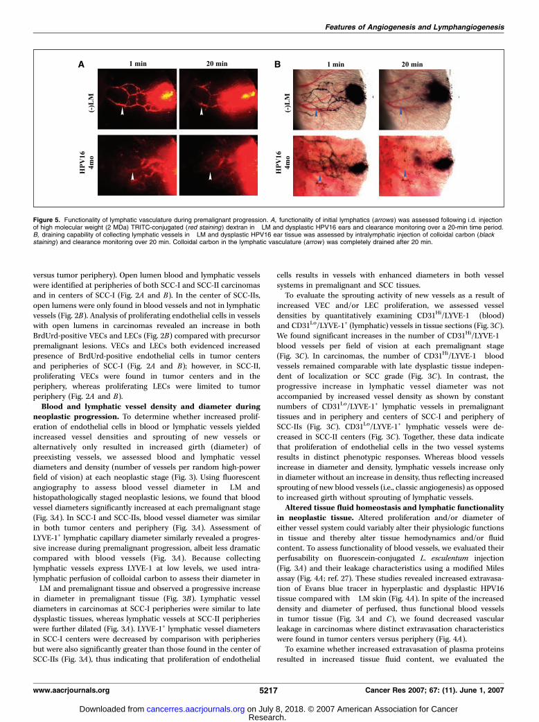
Figure 5. Functionality of lymphatic vasculature during premalignant progression. A, functionality of initial lymphatics (arrows ) was assessed following i.d. injectionof high molecular weight (2 MDa) TRITC-conjugated (red staining ) dextran in �LM and dysplastic HPV16 ears and clearance monitoring over a 20-min time period.B, draining capability of collecting lymphatic vessels in �LM and dysplastic HPV16 ear tissue was assessed by intralymphatic injection of colloidal carbon (blackstaining ) and clearance monitoring over 20 min. Colloidal carbon in the lymphatic vasculature (arrow ) was completely drained after 20 min.
Features of Angiogenesis and Lymphangiogenesis
www.aacrjournals.org 5217 Cancer Res 2007; 67: (11). June 1, 2007
Research. on July 8, 2018. © 2007 American Association for Cancercancerres.aacrjournals.org Downloaded from
relationship between blood vessel leakage (Fig. 4A) and lymphaticdrainage by determining tissue fluid amounts at each neoplasticstage (Fig. 4B) and found that tissue fluid in premalignant HPV16ear tissue was significantly higher than �LM tissue and furtherincreased in tumor peripheries, with the central regions of SCCshaving the highest fluid content (Fig. 4B). Because a major functionof lymphatics is to drain excess tissue fluid, one interpretation ofthese data would be that the drainage capacity of premalignantlymphatics had been exceeded due to lymphatic dysfunctionperhaps reflected by tissue fluid accumulation. To access thishypothesis, we further induced vascular leakage acutely by topicalapplication of mustard oil (compared with mineral oil) andassessed Evans blue dye leakage into dysplastic HPV16 ear skinversus �LM. As expected, mustard oil application further increasedleakage of plasma proteins out of blood vessels in both �LM andHPV16 skin, 16.5- and 3.5-fold, respectively (Fig. 4C); however, theseincreases resulted in similar accumulations of tissue fluid contentin �LM (8%) and dysplastic HPV16 (8%) tissue (Fig. 4D), indicatingthat clearance of tissue fluid by premalignant lymphatics is not atmaximum capacity. Thus, whereas lymphatics in acutely activatedhomeostatic tissue are more efficient at clearing excess tissue fluid,functional lymphatics in premalignant tissue maintain tissue fluiddynamics to a similar degree.
To directly assess lymphatic functionality in premalignant tissue,we i.d. injected high molecular weight, fluorescently labeleddextran (TRITC-dextran) tracer and monitored uptake anddrainage kinetics by lymphatic capillaries over time in �LM anddysplastic ear skin (Fig. 5A). Lymphatic capillaries were rapidlylabeled by tracer in both control and HPV16 ears, indicating rapiduptake. Twenty minutes following injection, tracer-containingcapillaries were detectable, indicating continuous lymph formation,thus showing their functional status.
Functionality of collecting lymphatic vessels in premalignanttissue was also evaluated following intralymphatic injection ofcolloidal carbon (Fig. 5B). Colloidal carbon is excluded bylymphatic capillaries during lymph formation due to its largemolecular weight.4 Direct injection of colloidal carbon into dermallymphatics of �LM and dysplastic ear skin revealed efficientdrainage of tracer from collecting lymphatic vessels in both tissueswithin 20 min (Fig. 5B), thus showing their similar functionality.Together, these analyses reveal functionality of lymphatic capillar-ies and collecting vessels in premalignant tissues based on theirability to efficiently clear tracer and fluid (Fig. 5A and B) throughtheir open lumina (Fig. 3A and B). Direct analysis of lymphaticfunctionality in tumor tissue was hampered by technical difficultiesdoing direct intralymphatic colloidal carbon injection as well asanalysis of interstitial drainage using smaller molecular weightdyes (colorimetric or fluorescent) readily taken up by functionallymphatics. Despite these failed attempts to analyze lymphaticfunctionality in tumor tissue, the implication that open luminalstatus is associated with vessel functionality in premalignant tissue,in combination with our assessment of tissue fluid content intumor centers versus tumor periphery (Fig. 4B), leads us toconclude that lymphatics with open lumina in premalignanttissue, at peripheries of SCCs and centers of grade 1 SCCs (Fig. 3C),retain functionality where they efficiently clear interstitial fluid,as opposed to tumor centers of less-differentiated SCCs where
lymphatic lumens are predominantly compressed and exhibitdiminished fluid drainage capacity (Fig. 3C, bottom right), acharacteristic feature of poorly differentiated carcinomas (28–30).
Discussion
Data presented herein indicate that blood and lymphaticvasculature undergo distinct physiologic and architectural alter-ations during de novo neoplastic progression accompanyingcarcinoma development. Whereas endothelial cells of each networkacquire proliferative capabilities as neoplasia ensues, resultantphenotypes of blood versus lymphatic vessels are distinct. Whereasblood vessels undergo classic angiogenic changes (e.g., increasedendothelial cell proliferation, vessel diameter, density, and leakage),lymphatic vessels evidence no alterations to support the notionthat sprouting of new vessels occurs. Instead, the increased level ofproliferating LECs present in lymphatic vessels enhances diameterof vessels that efficiently clear interstitial fluid in premalignanttissues and peripheries of carcinomas, but not in their centers,areas where interstitial fluid content remains high. Together, thesestudies show that neoplasia-associated angiogenesis, as opposedto lymphangiogenesis, is a distinct vascular process even wheninitiated by similar physiologic stimuli.Endothelial cell proliferation. Endothelial cells in quiescent
tissues divide approximately once every 2 to 3 years if unstimulated(31). During cancer development, however, it is well establishedthat local tissue levels of the proangiogenic factor vascularendothelial growth factor (VEGF)-A increase and, as a result,enhance proliferation of VECs and sprouting of new blood vesselsfrom preexisting vascular beds (5). In contrast, lymphangiogenesis(e.g., induction of LEC proliferation) in human cancers (32, 33)correlates instead with increased tissue levels of VEGF-C andVEGF-D (34–36). Accordingly, exogenous expression of VEGF-C orVEGF-D in experimental murine tumor xenograft models inducesLEC proliferation and subsequent lymphangiogenesis (37, 38),whereas exogenous VEGF-A expression induces not only anangiogenic response but also lymphangiogenesis in nontumor(24) and tumor tissue (39). It is not clear, however, if LEC responsesto increased VEGF-A levels represent a direct intrinsic responseof LECs or, instead, an indirect physiologic response resultingfrom increased tissue fluid due to increased leakage of plasmaproteins from newly formed immature angiogenic blood vessels.
In this study, we found evidence of angiogenesis as well aslymphangiogenesis in premalignant tissues of HPV16 mice and inemergent malignant carcinomas. Patterns of LEC proliferationvaried from that observed with VECs, thus indicating distinctresponses between the two endothelial cell subpopulations toproangiogenic and/or prolymphangiogenic growth factors presentin their microenvironment. Whereas VEGF-A mRNA levels havebeen reported to remain constant during premalignant angiogen-esis in a UV-induced murine skin carcinoma model (40), enhancedVEC proliferation in HPV16 mice is paralleled by progressiveincreases in VEGF-A mRNA expression (41) and protein levels(42). About LEC proliferation, our analyses indicate that, althoughLECs in premalignant tissue evidence enhanced proliferative statuscompared with homeostatic tissue, their proliferation does notsignificantly increase until malignant conversion occurs. Becausewe have observed increased mRNA levels of VEGF-C in fullymalignant carcinomas (data not shown), our interpretation ofthese data is that LECs respond modestly to increased VEGF-Alevels in premalignant tissue but more are responding to increased4 J. Nagy, personal communication.
Cancer Research
Cancer Res 2007; 67: (11). June 1, 2007 5218 www.aacrjournals.org
Research. on July 8, 2018. © 2007 American Association for Cancercancerres.aacrjournals.org Downloaded from

microenvironmental stress and need to clear fluid from premalig-nant tissue resultant from leaky immature angiogenic bloodvessels.Vascular sprouting. VEGF-A is a major proangiogenic factor
well documented to induce VEC proliferation and vascularsprouting (43). Sprouting of the lymphatic vasculature, however,depends on VEGF-C in experimental chick chorioallantoicmembrane assays, wound healing, and embryonic development(44–46). Recent studies in murine tumor models (transgenic andxenograft models) provide evidence that increased levels ofVEGF-C and/or VEGF-D promote tumor lymphangiogenesis,including lymphatic vessel sprouting (47–50). In contrast to theseexperimental tumor models, no increase in lymphatic vesseldensity was observed in HPV16 tissues, indicating that LECproliferation in a de novo tumor model lacking exogenousexpression of VEGF-C and/or VEGF-D does not result in lymphaticvessel sprouting but instead contributed to increased diameter(girth) of existing lymphatics to enable rapid clearance of tissuefluid resulting from increased leakage of plasma proteins fromangiogenic blood vessels.Tissue fluid dynamics in premalignant and SCC tissue. Blood
and lymphatic vessels are functionally interdependent vascularsystems (6) that together regulate tissue fluid dynamics andinfluence tissue homeostasis. In a UV-induced skin carcinogenesismodel, Hagendoorn et al. (40) revealed alterations in bloodvasculature, architectural changes, and impairment of lymphaticvessels along with increased IFP in premalignant tissues and thushypothesized that blood and lymphatic vasculature loose theirfunctional interdependence during premalignant progression. Ourdata, however, indicate that, during squamous carcinogenesis inHPV16 mice, the two vessel systems retain a functional interrela-tionship in premalignant tissue and in well-differentiated carcino-mas as evidenced by increased blood vessel leakage accompaniedby retained ability to clear fluid, albeit in tissue with higher tissuefluid content than normal.Lymphatic vessel functionality.We observed that intratumoral
lymphatic vessel functionality inversely correlates with SCC grade.In the published literature, there are contradicting reports on theexistence of intratumoral lymphatics in human (9, 10, 32, 33, 51)and experimental rodent (37, 38, 40, 49, 52) tumors, where onlyperipheral lymphatic structures are hypothesized to retain
functionality, whereas intratumoral lymphatic structures wererendered nonfunctional due to compression by rapidly proliferatingtumor cells (52). Similarly, we found functional lymphatic vesselsat tumor peripheries but also observed open lumen/functionallymphatic vessels in centers of low-grade SCCs. Our interpretationof these observations is that functional properties of higher-grade,less-differentiated carcinomas correlate with, and is effected by,limited lymphatic vessel functionality that, in turn, likely regulatestumor cell physiology and/or behavior.
In summary, by examining proliferation, diameter, density, andfunctionality of blood versus lymphatic vasculature during de novocancer development, we have revealed unique features of eachvasculature that illuminates their interdependency during prema-lignant progression as opposed to their independence in higher-grade carcinomas. Maintenance of the interdependence betweenblood and lymphatic systems during premalignancy and in low-grade carcinomas enables efficient fluid transport from leaky bloodvasculature via interstitia into draining, open lumen lymphaticvessels, which would result in minimizing IFP. The closed nature oflymphatic vessels in higher-grade carcinomas would exclude thisand support elevated IFPs common to less-differentiated carcino-mas. The implication of these findings supports the notion thatdelivery of anticancer therapeutics would best be realized inpremalignant tissue, in situ carcinomas, or carcinomas displayingwell-differentiated characteristics, where tissue hemodynamicsbetter support drug delivery.
Acknowledgments
Received 12/20/2006; revised 3/30/2007; accepted 4/9/2007.Grant support: Serono Foundation for the Advancement of Medical Science
(A. Eichten) and NIH grants CA72006, CA94168, and CA098075, Sandler Program inBasic Sciences, National Technology Center for Networks and Pathways grant U54RR020843, and Department of Defense Breast Cancer Research Program Era of HopeScholar Award BC051640 (L.M. Coussens).
The costs of publication of this article were defrayed in part by the payment of pagecharges. This article must therefore be hereby marked advertisement in accordancewith 18 U.S.C. Section 1734 solely to indicate this fact.
We thank Drs. K. de Visser, D. Soto, and N.E. Sounni for critical comments andsuggestions; Lidiya Korets, Aleksandr Rudik, and Nadia Milshteyn for technicalassistance in animal husbandry; Eva Soliven and the UCSF Comprehensive CancerCenter (CCC) Mouse Pathology Shared Resource for tissue sectioning; members ofthe UCSF CCC Laboratory for Cell Analysis for microscopic and cytometricassistance; and members of the Coussens laboratory for encouragement and criticaldiscussion.
References1. Folkman J. Angiogenesis in cancer, vascular, rheuma-toid and other disease. Nat Med 1995;1:27–31.2. Braverman IM, Sibley J. Role of the microcirculation inthe treatment and pathogenesis of psoriasis. J InvestDermatol 1982;78:12–7.3. Detmar M, Brown LF, Claffey KP, et al. Overexpressionof vascular permeability factor/vascular endothelialgrowth factor and its receptors in psoriasis. J Exp Med1994;180:1141–6.4. Folkman J, Watson K, Ingber D, Hanahan D. Inductionof angiogenesis during the transition from hyperplasiato neoplasia. Nature 1989;339:58–61.5. Bergers G, Benjamin LE. Angiogenesis: tumorigenesisand the angiogenic switch. Nat Rev Cancer 2003;3:401–10.6. Swartz MA. The physiology of the lymphatic system.Adv Drug Deliv Rev 2001;50:3–20.7. Heldin CH, Rubin K, Pietras K, Ostman A. Highinterstitial fluid pressure—an obstacle in cancer thera-py. Nat Rev Cancer 2004;4:806–13.8. Beasley NJ, Prevo R, Banerji S, et al. Intratumoral
lymphangiogenesis and lymph node metastasis in headand neck cancer. Cancer Res 2002;62:1315–20.9. Franchi A, Gallo O, Massi D, Baroni G, Santucci M.Tumor lymphangiogenesis in head and neck squamouscell carcinoma: a morphometric study with clinicalcorrelations. Cancer 2004;101:973–8.10. Giorgadze TA, Zhang PJ, Pasha T, et al. Lymphaticvessel density is significantly increased in melanoma.J Cutan Pathol 2004;31:672–7.11. Boucher Y, Brekken C, Netti PA, Baxter LT, Jain RK.Intratumoral infusion of fluid: estimation of hydraulicconductivity and implications for the delivery oftherapeutic agents. Br J Cancer 1998;78:1442–8.12. Padera T, Stoll B, Tooredman J, Capen D, di TomasoE, Jain R. Cancer cells compress intratumour vessels.Nature 2004;427:695.13. Coussens LM, Hanahan D, Arbeit JM. Geneticpredisposition and parameters of malignant progressionin K14-HPV16 transgenic mice. Am J Pathol 1996;149:1899–917.14. Smith-McCune K, Zhu Y-H, Hanahan D, Arbeit J.Angiogenesis and VEGF mRNA upregulation in progres-sive squamous carcinogenesis: cross-species compari-
son of human cervix and K14-HPV16 transgenic mice.Cancer Res 1997;57:1294–300.15. van Kempen LCL, Rhee JS, Dehne K, Lee J, EdwardsDR, Coussens LM. Epithelial carcinogenesis: dynamicinterplay between neoplastic cells and their microenvi-ronment. Differentiation 2002;70:610–23.16. Coussens LM, Raymond WW, Bergers G, et al.Inflammatory mast cells up-regulate angiogenesis dur-ing squamous epithelial carcinogenesis. Genes Dev 1999;13:1382–97.17. Coussens LM, Tinkle CL, Hanahan D, Werb Z. MMP-9supplied by bone marrow-derived cells contributes toskin carcinogenesis. Cell 2000;103:481–90.18. Junankar S, Eichten A, Kramer A, de Visser KE,Coussens LM. Analysis of immune cell infiltrates duringsquamous carcinoma development. J Invest Dermatol2006;126:36–43.19. Arbeit JM, Munger K, Howley PM, Hanahan D.Progressive squamous epithelial neoplasia in K14-human papillomavirus type 16 transgenic mice. J Virol1994;68:4358–68.20. Daniel D, Meyer-Morse N, Bergsland EK, Dehne K,Coussens LM, Hanahan D. Immune enhancement of
Features of Angiogenesis and Lymphangiogenesis
www.aacrjournals.org 5219 Cancer Res 2007; 67: (11). June 1, 2007
Research. on July 8, 2018. © 2007 American Association for Cancercancerres.aacrjournals.org Downloaded from

skin carcinogenesis by CD4+ T cells. J Exp Med 2003;197:1017–28.21. de Visser KE, Korets LV, Coussens LM. Earlyneoplastic progression is complement independent.Neoplasia 2004;6:768–76.22. Thurston G, Baluk P, Hirata A, McDonald DM.Permeability-related changes revealed at endothelialcell borders in inflamed venules by lectin binding. Am JPhysiol 1996;271:2547–62.23. Eichten A, Shen H-CJ, Coussens LM. Three-dimensional visualization of blood and lymphaticvasculature in tissue whole mounts using confocalmicroscopy. In: Robinson JP, editor. Current protocolsin cytometry. New Jersey: John Wiley & Sons, Inc.;2005. p. 12.5.1–12.5.11.24. Nagy JA, Vasile E, Feng D, et al. Vascular permeabilityfactor/vascular endothelial growth factor induces lym-phangiogenesis as well as angiogenesis. J Exp Med 2002;196:1497–506.25. DeLisser HM, Newman PJ, Albelda SM. Molecularand functional aspects of PECAM-1/CD31. ImmunolToday 1994;15:490–5.26. Cursiefen C, Chen L, Borges LP, et al. VEGF-Astimulates lymphangiogenesis and hemangiogenesis ininflammatory neovascularization via macrophage re-cruitment. J Clin Invest 2004;113:1040–50.27. Miles AA, Miles EM. Vascular reactions to histamine,histamine-liberator and leukotaxine in the skin ofguinea pigs. J Physiol 1952;118:228–57.28. Nathanson SD, Nelson L. Interstitial fluid pressure inbreast cancer, benign breast conditions, and breastparenchyma. Ann Surg Oncol 1994;1:333–8.29. Curti BD, Urba WJ, Alvord WG, et al. Interstitialpressure of subcutaneous nodules in melanoma andlymphoma patients: changes during treatment. CancerRes 1993;53:2204–7.30. Gutmann R, Leunig M, Feyh J, et al. Interstitialhypertension in head and neck tumors in patients:correlation with tumor size. Cancer Res 1992;52:1993–5.
31. Hirst DG, Denekamp J, Hobson B. Proliferationstudies of the endothelial and smooth muscle cells ofthe mouse mesentery after irradiation. Cell Tissue Kinet1980;13:91–104.32. Sipos B, Klapper W, Kruse ML, Kalthoff H, KerjaschkiD, Kloppel G. Expression of lymphangiogenic factorsand evidence of intratumoral lymphangiogenesis inpancreatic endocrine tumors. Am J Pathol 2004;165:1187–97.33. Koukourakis MI, Giatromanolaki A, Sivridis E, et al.LYVE-1 immunohistochemical assessment of lymphan-giogenesis in endometrial and lung cancer. J Clin Pathol2005;58:202–6.34. Karpanen T, Alitalo K. Lymphatic vessels as targets oftumor therapy? J Exp Med 2001;194:F37–42.35. Pepper MS. Lymphangiogenesis and tumor metasta-sis: myth or reality? Clin Cancer Res 2001;7:462–8.36. Achen MG, McColl BK, Stacker SA. Focus onlymphangiogenesis in tumor metastasis. Cancer Cell2005;7:121–7.37. Skobe M, Hawighorst T, Jackson DG, et al. Inductionof tumor lymphangiogenesis by VEGF-C promotesbreast cancer metastasis. Nat Med 2001;7:192–8.38. Karpanen T, Egeblad M, Karkkainen MJ, et al.Vascular endothelial growth factor C promotes tumorlymphangiogenesis and intralymphatic tumor growth.Cancer Res 2001;61:1786–90.39. Hirakawa S, Kodama S, Kunstfeld R, Kajiya K, BrownLF, Detmar M. VEGF-A induces tumor and sentinellymph node lymphangiogenesis and promotes lymphat-ic metastasis. J Exp Med 2005;201:1089–99.40. Hagendoorn J, Tong R, Fukumura D, et al. Onset ofabnormal blood and lymphatic vessel function andinterstitial hypertension in early stages of carcinogen-esis. Cancer Res 2006;66:3360–4.41. Smith-McCune K, Zhu YH, Hanahan D, Arbeit J.Cross-species comparison of angiogenesis during thepremalignant stages of squamous carcinogenesis in thehuman cervix and K14-HPV16 transgenic mice. CancerRes 1997;57:1294–300.
42. de Visser KE, Korets LV, Coussens LM. De novocarcinogenesis promoted by chronic inflammation is Blymphocyte dependent. Cancer Cell 2005;7:411–23.43. Leung DW, Cachianes G, Kuang WJ, Goeddel DV,Ferrara N. Vascular endothelial growth factor is asecreted angiogenic mitogen. Science 1989;246:1306–9.44. Oh SJ, Jeltsch MM, Birkenhager R, et al. VEGF andVEGF-C: specific induction of angiogenesis and lym-phangiogenesis in the differentiated avian chorioallan-toic membrane. Dev Biol 1997;188:96–109.45. Paavonen K, Puolakkainen P, Jussila L, Jahkola T,Alitalo K. Vascular endothelial growth factor receptor-3in lymphangiogenesis in wound healing. Am J Pathol2000;156:1499–504.46. Jeltsch M, Kaipainen A, Joukov V, et al. Hyperplasia oflymphatic vessels in VEGF-C transgenic mice. Science1997;276:1423–5.47. Mandriota SJ, Jussila L, Jeltsch M, et al. Vascularendothelial growth factor-C-mediated lymphangiogen-esis promotes tumour metastasis. EMBO J 2001;20:672–82.48. Stacker SA, Caesar C, Baldwin ME, et al. VEGF-Dpromotes the metastatic spread of tumor cells via thelymphatics. Nat Med 2001;7:186–91.49. Skobe M, Hamberg LM, Hawighorst T, et al.Concurrent induction of lymphangiogenesis, angiogen-esis, and macrophage recruitment by vascular endothe-lial growth factor-C in melanoma. Am J Pathol 2001;159:893–903.50. He Y, Kozaki K, Karpanen T, et al. Suppression oftumor lymphangiogenesis and lymph node metastasisby blocking vascular endothelial growth factor receptor3 signaling. J Natl Cancer Inst 2002;94:819–25.51. Williams CS, Leek RD, Robson AM, et al. Absenceof lymphangiogenesis and intratumoural lymph vesselsin human metastatic breast cancer. J Pathol 2003;200:195–206.52. Padera TP, Kadambi A, di Tomaso E, et al. Lymphaticmetastasis in the absence of functional intratumorlymphatics. Science 2002;296:1883–6.
Cancer Research
Cancer Res 2007; 67: (11). June 1, 2007 5220 www.aacrjournals.org
Research. on July 8, 2018. © 2007 American Association for Cancercancerres.aacrjournals.org Downloaded from
2007;67:5211-5220. Cancer Res Alexandra Eichten, William C. Hyun and Lisa M. Coussens
Tumor DevelopmentDe novoLymphangiogenesis Determine Their Functionality during Distinctive Features of Angiogenesis and
Updated version
http://cancerres.aacrjournals.org/content/67/11/5211
Access the most recent version of this article at:
Cited articles
http://cancerres.aacrjournals.org/content/67/11/5211.full#ref-list-1
This article cites 51 articles, 20 of which you can access for free at:
Citing articles
http://cancerres.aacrjournals.org/content/67/11/5211.full#related-urls
This article has been cited by 6 HighWire-hosted articles. Access the articles at:
E-mail alerts related to this article or journal.Sign up to receive free email-alerts
Subscriptions
Reprints and
To order reprints of this article or to subscribe to the journal, contact the AACR Publications
Permissions
Rightslink site. (CCC)Click on "Request Permissions" which will take you to the Copyright Clearance Center's
.http://cancerres.aacrjournals.org/content/67/11/5211To request permission to re-use all or part of this article, use this link
Research. on July 8, 2018. © 2007 American Association for Cancercancerres.aacrjournals.org Downloaded from