Embed Size (px)
Citation preview

Dissection of Porphyrin-Induced Conformational Dynamics in theHeme Biosynthesis Enzyme FerrochelataseAwuri P. Asuru, Mier An, and Laura S. Busenlehner*
Department of Chemistry, The University of Alabama, Tuscaloosa, Alabama 35487, United States
*S Supporting Information
ABSTRACT: Human ferrochelatase (EC 4.99.1.1) catalyzesthe insertion ferrous iron into protoporphyrin IX as the laststep in heme biosynthesis, an essential process to mostorganisms given the vast intracellular functions of heme. Evenwith multiple ferrochelatase structures available, the exactmechanism for iron insertion into porphyrin is still a matter fordebate. It is clear, however, that conformational dynamics areimportant for porphyrin substrate binding, initial chelation ofiron, insertion of iron into the macrocycle, and release ofprotoheme IX. In this work we characterize conformationaland dynamic changes in ferrochelatase associated with porphyrin binding using the substrate mesoporphyrin (MPIX) andbackbone amide hydrogen/deuterium exchange mass spectrometry (HDX-MS). In general, regions surrounding the active sitebecome more ordered from direct or indirect interactions with the porphyrin. Our results indicate that the lower lip of the activesite mouth is preorganized for efficient porphyrin binding, with little changes in backbone dynamics. The upper lip region has themost significant change in HDX behavior as it closes the active site. This movement excludes solvent from the porphyrin pocket,but leads to increased solvent access in other areas. A water lined path to the active site was observed, which may be the elusiveiron channel with final insertion via the M76/R164/Y165 side of the porphyrin. These results provide a rigorous view of theferrochelatase mechanism through the inclusion of dynamic information, reveal new structural areas for functional investigation,and offer new insight into a potential iron channel to the active site.
There is increasing evidence that protein dynamics arecritical components to enzyme catalyzed reaction
mechanisms.1,2 The information gleaned from three-dimen-sional structures of enzymes has led to a revolution with regardto specific protein conformations associated with bindingsubstrates, substrate analogues, transition state analogues, andproducts. However, the crystallographic structures of enzymesoften do not reveal the sometimes slight, yet crucial, dynamicchanges required for enzymatic processes. A rigorous measure-ment of protein dynamics is clearly needed to fully understandhow localized motions in enzymes correlate with specificcatalytic steps.3 This information is valuable for designingtherapeutic compounds that regulate the activity of keyenzymes involved in disease, as well.4 Amide hydrogen/deuterium exchange mass spectrometry (HDX-MS) providesa novel approach to define the role dynamics plays in enzyme-catalyzed reactions. The exchange rate of backbone amidehydrogens for deuterons is related to protein secondarystructure, solvent accessibility, and backbone flexibility.1 Thismakes amide hydrogens excellent reporters of protein structureand dynamics. It is especially useful for identifying functionalchanges in conformation when comparing two protein states(e.g., free enzyme vs substrate-bound enzyme). Thus, it isemerging as an attractive alternative or companion to otherstructural techniques due to the exceptional capability ofexamining both dynamic and structural components.
In this work, we report the structural and dynamic changesrelated to substrate binding for an important enzyme in hemebiosynthesis, human ferrochelatase (E.C. 4.99.1.1). Ferrochela-tase, the most extensively studied enzyme in the chelatasesubclass, catalyzes the last step in heme biosynthesis, theinsertion of ferrous iron (Fe2+) in protoporphyrin IX (PPIX) toform protoheme IX.5 The disease erythropoietic protophor-phyria (EPP), where the PPIX intermediate accumulates in redblood cells and causes painful light sensitivity, is caused bycharacterized mutations in the FECH gene;6,7 therefore,understanding the structure and mechanism of ferrochelataseis imperative for the development of treatments. Humanferrochelatase is an 84 kDa homodimer associated with theinner mitochondrial membrane, where the hydrophobic activesite “mouth” is directed toward the membrane layer and flankedby two stretches of residues called the upper and lower “lips”.8
The upper lips also serve as insertion points into themembrane.9 There are crystal structures of ferrochelatasesfrom several organisms including Bacillus subtilis, Bacillusanthracis, Saccharomyces cerevisiae, and Homo sapiens.8,10,11
This includes numerous structures with different metal ions,
Received: May 29, 2012Revised: August 14, 2012Published: August 16, 2012
Article
pubs.acs.org/biochemistry
© 2012 American Chemical Society 7116 dx.doi.org/10.1021/bi300704c | Biochemistry 2012, 51, 7116−7127

substrates, products, and inhibitors, as well as site-directedmutants.10,12−17
The ferrochelatase catalytic cycle includes PPIX substratebinding, Fe2+ binding and desolvation, deprotonation of twoPPIX pyrrole nitrogens, distortion of the PPIX macrocycleprior to Fe2+ insertion, and last, product release.18 Fromstructural analysis and other biochemical studies, it is apparentthat ferrochelatase must undergo conformational changes inorder to carry out these specific catalytic steps. However,sometimes these subtle changes in solution may not be easilydetected in crystallographic structures. If these changes indynamics can be identified, it would provide additionalmechanistic information about how ferrochelatase inserts ironinto PPIX. Knowledge of the ferrochelatase mechanism from astructural dynamics viewpoint is quite relevant for structure−activity based design of therapeutic compounds to regulate itsactivity as a treatment for EPP.There are several crystal structures of human ferrochelatase
with some form of porphyrin bound in the active site. ThePPIX-bound structure of E343K ferrochelatase by Medlock etal.20 indicates that the active site “mouth” closes around thesubstrate, resulting in the reorientation of key catalytic residuesand changes in hydrogen bonding within the PPIX bindingpocket.12,20 Structures with heme and metalated porphyrinsreveal that an unusual π-helix controls product release throughwinding and unwinding.13,20 Thus, ferrochelatase undergoeschanges in structure during the catalytic cycle and that controlof dynamics may be important for distortion of the porphyrinmacrocycle for iron insertion, chelation of iron, and release ofheme.There is still substantial debate as to how iron is acquired and
inserted into PPIX in human ferrochelatase and its homologues.To address this gap in knowledge, prior HDX-MS was used tomap conformational changes that occur when the cosubstrateFe2+ binds to ferrochelatase in the absence of porphyrinsubstrate.19 Those studies suggested that an iron-channel mayexist from the mitochondrial matrix surface of the enzyme tothe active site and supported a mechanism where the terminaliron acceptors before insertion are at or near R164 and Y165 onone face of the PPIX macrocycle. To gain a more completepicture of dynamic contributions to ferrochelatase catalysis, thechanges in structure and dynamics critical for porphyrinsubstrate binding must also be defined. If PPIX binding signalsiron uptake, then by comparison we can identify regions offerrochelatase that are responsive only to porphyrin, only toiron, and to both substrates. The regions that have overlappingconformational changes are likely to be involved in the catalyticmechanism.The research presented here uses HDX-MS to localize these
important structural changes upon binding the substrateanalogue mesoporphyrin IX (MPIX), which is structurallysimilar to PPIX (Chart 1). MPIX and is commonly used inenzyme activity assays because it is more stable and solublethan its native substrate PPIX.16,21 HDX-MS provides insightinto the coupling of PPIX binding with Fe2+ uptake, thusproviding a more detailed structural view of the ferrochelatasemechanism, which is still debated. A potential water-accessiblepathway from the surface of the protein to the active site wasobserved upon MPIX binding that could serve as an ironsubstrate channel, and thus HDX-MS has provided neededinformation about this key enzymatic process and opened newavenues for biochemical investigation.
■ MATERIALS AND METHODSMaterials. ACS grade HEPES, HPLC grade water, and
HPLC grade acetonitrile were obtained from EMD Chemicals(Billerica, MA), CHAPS from Applichem (St. Louis, MO),mesoporphyrin IX from Frontier (Logan, UT), deuteriumoxide 99.99% at. D from Acros Organics (Geel, Belgium),porcine pepsin (3200−4500 units/mg) from Sigma-Aldrich (St.Louis, MO), HPLC grade 2-propanol from Honeywell(Morristown, NJ), Ni-NTA Superflow affinity resin from 5Prime (Gaithersburg, MD), Circlegrow medium from Bio101(Vista, CA), and HB101 competent cells from Promega(Madison, WI). A Bruker HCTultra Discovery massspectrometer (Billerica, MA) was used with an Agilent 1100liquid chromatography system (Santa Clara, CA).
Ferrochelatase Expression and Purification. Escherichiacoli HB101 cells were transformed with pHisTF20E providedby Dr. Harry Dailey (University of Georgia) and plated onLuria−Bertani agar with 100 μg/mL ampicillin.9 A singlecolony was used to inoculate 2 L of Circlegrow medium thatwas supplemented with 50 μg/mL carbenicillin. Proteinexpression and purification are as described.19 Purifiedferrochelatase was dialyzed extensively against 4 L of chelexed25 mM HEPES (pH 7.4), 100 mM NaCl, 20 mM β-mercaptoethanol, and 0.1% CHAPS at 4 °C in nitric acidwashed glassware. The concentration of ferrochelatase wasestimated at 278 nm (ε278 = 46 910 M−1 cm−1) and thepercentage of bound [2Fe-2S] cluster was estimated by theabsorbance at 330 nm (ε330 = 12 000 M−1 cm−1).9 The ironcontent of purified free-ferrochelatase was measured by atomicabsorption spectroscopy and found to be 1.8 ± 0.1 mol iron permol ferrochelatase, as expected if each monomer contains the[2Fe-2S] cluster. UV−visible spectroscopy also confirmed thatthe extensively dialyzed, purified ferrochelatase contained lessthan 5% of porphyrin in the active site, as determined by loss ofthe characteristic Soret band.22 We refer to this preparation asthe free-enzyme. Previously we reported that CHAPS-solubilized ferrochelatase in the presence of reducing agentwith incubation under N2, as in HDX samples, retained the[2Fe-2S] cluster and ∼95% activity over 2 h at 25 °C.19
Hydrogen/Deuterium Exchange. The peptides thatresult from a pepsin digest of ferrochelatase were reported inref 19. The full peptide map has approximately 74% sequencecoverage by the peptides used reproducibly for H/D exchange(Figure S1). A fresh 1 mM mesoporphyrin IX (MPIX) stockwas prepared in 12 mM ammonium hydroxide and 1% CHAPSin chelexed ddiH2O. Approximately 1.2 mol equiv of MPIX wasadded to 20 μM ferrochelatase pretreated with 40 μM EDTA(pH 8.0) to chelate any residual iron. Samples of MPIX-bound
Chart 1
Biochemistry Article
dx.doi.org/10.1021/bi300704c | Biochemistry 2012, 51, 7116−71277117

ferrochelatase were aliquoted into thin-walled PCR tubes,wrapped in foil, and flash-frozen at −80 °C until HDX analysis.Deuterium exchange was initiated by the addition of 45 μL of
D2O into 5 μL of ∼20 μM free or MPIX-ferrochelatase at 23°C in a thin-walled PCR tube. The deuterium-protein sampleswere quickly purged with N2 gas for the longer time points,incubated at 23 °C, and exchange was quenched at specific timepoints (15 s−2 h) by 50 μL of quench buffer [0.1 M potassiumphosphate (pH 2.3) in H2O; 0 °C]. The sample wasimmediately transferred to an ice bath. Porcine pepsin in 10mM potassium phosphate, pH 7.0 was added to the sample (40μg) and the digestion proceeded for 5 min. The peptide digestswere separated by liquid chromatography on a 1 mm × 50 mmC18 reverse-phase column (Phenomenex, Torrence, CA)submerged in an ice water bath and the mass spectra werecollected, as described.19
The chromatographic retention profiles for each identifiedpeptide ion were extracted for all samples. The spectra withinthe elution peak were averaged to produce a compositespectrum for each peptide ion. The centroid (mt) of the givencomposite isotope envelope was calculated by MagTran 1.0beta 9 software23 and also by Sierra Analystics softwareHDExaminer (http://www.massspec.com/HDExaminer.html)with equivalent results. For multiply charged peptide ions, theisotopic envelopes from each charge state were averaged whenappropriate. The amount of deuterium incorporated at eachtime point was calculated using
=−
−
⎛⎝⎜
⎞⎠⎟D N
m mm m
t 0%
100% 0% (1)
where m0%, mt, and m100% are the centroid values of the peptidein the non-deuterated, the partially deuterated at time t, and thefully deuterated control samples, respectively.24 N is the totalnumber of exchangeable peptide amide protons less one for theN-terminal amide proton and any proline residues. The amountof deuterium incorporated in each peptide was averaged fromthree independent kinetic runs and plotted as a function of timein minutes. The resulting progress curve for each peptide wasfit using KaleidaGraph (Synergy Software) to single or doubleexponential equations as appropriate. All HDX-MS kinetictraces with fits are provided in Supporting Information availablefree of charge via the Internet at http://pubs.acs.org.
■ RESULTS
General Considerations. The kinetics of base catalyzed(OD−) deuterium exchange at backbone amide protons isdependent on a variety of factors.25,26 The first 15 s of a HDXtime course provides a rough estimate of the relative solventaccessibility of backbone amides as it is related to the secondaryand tertiary structure of the protein.27,28 HDX is useful forlocalizing ligand binding interactions through protection ofamide hydrogens in the ligand binding site.29,30 The specificamides protected by the ligand are identified after pepsindigestion and MS through a decrease in deuterium incorpo-ration as compared to the free protein. It should be noted thatsome large conformational changes outside a ligand binding sitemay bury previously exposed amides thereby also decreasingdeuterium incorporation, so a full characterization of H/Dexchange kinetics is required to help differentiate thesepossibilities. To determine the effect of substrate binding onferrochelatase, we compared the relative solvent accessibility ofthe iron-free enzyme to the MPIX-bound form after 15 sincubation in D2O. The changes in solvent accessibility with thecosubstrate Fe2+ have already been characterized.19
HDX Solvent Accessibility. A difference plot of thepercent deuterium incorporation in MPIX-ferrochelatase back-bone amides relative to free enzyme is shown in Figure 1A. Thecorresponding peptides are indicated on the structure of E343Kferrochelatase with PPIX bound,20 to which the HDX resultswill be compared (Figure 1B,C). Considerable protection byMPIX from deuterium exchange is observed for peptideresidues 93−100, 300−317, and 338−345, with minorprotection for peptides 74−89 and 260−269. These regionssurround the porphyrin binding site, as expected, with thegreatest protection observed for peptide 338−345. This peptideresides at the beginning of the π-helix13 and has residues withinvan der Waals distance to PPIX. Modest protection fromdeuterium exchange is shown for residues in the upper (93−100) and lower (300−317) lips of the porphyrin binding site.On the basis of the PPIX structure, the upper lip peptide 93−100 has several residues whose side chains are within 3.5 Å ofthe porphyrin. The lower lip peptide 300−317 is highlyconserved and located in a loop opposite the porphyrin bindingpocket as 93−100. Several residues within this peptide interactwith PPIX in the crystal structure. Two peptides, 74−89 and260−269, show minor decreases in solvent accessibility in
Figure 1. Difference in D2O accessibility to free-ferrochelatase backbone amides when MPIX is bound. (A) The percentage of deuterium lost orgained after a 15 s incubation in D2O was calculated for each pepsin-digested peptide. The cyan, light blue, and dark blue lines signify a greater than5, 15, and 25% decrease in deuterium incorporation, respectively. The pink, magenta, and red lines signify a greater than 5, 15, and 25% increase indeuterium incorporation, respectively. The color coded peptides from (A) are plotted on (B) the PPIX-ferrochelatase dimeric structure derived fromPBD 2QD120 and (C) one monomer subunit using PyMOL.32
Biochemistry Article
dx.doi.org/10.1021/bi300704c | Biochemistry 2012, 51, 7116−71277118

MPIX-bound ferrochelatase. The region spanning residues 74−89 is N-terminal to the upper lip that has significant protectionand contains two residues within interaction distance to PPIX.Peptide 260−263 is on the opposite face of the porphyrin andcontains H263, a key residue in the binding site that has a rolein the catalytic mechanism.31 In summary, the areas protectedby MPIX are within the immediate binding pocket.There are also regions of ferrochelatase that display an
increase in deuterium incorporation in the MPIX-bound formcompared to the free enzyme (Figure 1A−C). This likelydenotes a conformational change that exposes previouslyinaccessible amide protons to D2O. The largest changes insolvent accessibility occur for peptides 99−112 and 222−233.Peptide residues 99−112 are in helix2 above the porphyrinbinding pocket.20 As the major component to the upper lips,this region is thought to interact with the inner mitochondrialmembrane layer. It immediately follows peptide 93−100 thatshowed significant solvent protection by MPIX. Peptide 99−112 is farther away from the active site compared to 93−100and has no observed interactions with PPIX based on the
structure. Peptide 222−233 is over 11 Å away from thesubstrate binding site. This region may initially bind iron at theprotein surface.8 Minor increases in D2O access in the presenceof MPIX is noted for peptides 195−203 and 418−423. Peptide195−203 is close to the porphyrin site and may reside in aniron channel. The last area of increased solvent accessibility is atthe C-terminus, peptide 418−423. It is unclear what role thisregion may play in binding substrate porphyrins. The regionmust be important since many mutations that cause EPP arelocated in the dimer interface.6,7
Dynamics of MPIX Binding. In general, substrate bindingto an active site often results in decreased backbone dynamicmotions and rates of deuterium incorporation as compared tothe free enzyme.33 This can be attributed to additionalinteractions with residue side chains that constrain backbonedynamics, but it can also be the result of increased hydrogenbonding within backbone amides themselves (i.e., changes insecondary structure).34 For ferrochelatase we examined regionsof the enzyme where the rate of deuterium incorporation intobackbone amides was decreased upon MPIX binding,
Figure 2. Ferrochelatase peptides with decreased backbone dynamics after binding MPIX. The peptides identified as having a decrease in one ormore rate constant for exchange are mapped onto PPIX-ferrochelatase20 and are colored as follows: 74−89 (purple), 93−100 (red), 99−112 (darkgreen), 222−233 (pink), 260−269 (magenta), 277−291 (light green), 303−317 (cyan), 332−337 (blue), 338−345 (orange), and 350−356(yellow). PPIX is in green spheres and the [2Fe−S] cluster is in stick format. The structure on the right was rotated 180°. Figure created withPyMOL.32
Figure 3. HDX-MS rate profiles for selected ferrochelatase peptides. Shown are the kinetic traces for the number of deuterons incorporated intopeptides derived from free (black) and MPIX (red) ferrochelatase as a function of incubation time in D2O. The data are an average of 2−3 sets andwere fit to single or double exponential equations to obtain the rate constants for exchange (kn) and the corresponding number of deuterons in eachphase (Dn). The fitted parameters are given in Table 1. The numbers in parentheses are the total number of exchangeable amide protons for eachpeptide. (A) 93−100. (B) 99−112. (C) 74−89. (D) 303−317. (E) 260−269. (F) 332−337. (G) 338−345. (H) 222−233. (I) 277−291. (J) 350−356. The peptides are mapped on the monomeric structure in Figure 2.
Biochemistry Article
dx.doi.org/10.1021/bi300704c | Biochemistry 2012, 51, 7116−71277119

irrespective of solvent accessibility. This provides a morecomplete picture of the conformational changes associated withoccupation of substrate in the active site. HDX-MS kineticanalysis identifies 10 peptides with decreased backbonedynamics in the MPIX bound state. The peptides are mappedto the PPIX-ferrochelatase structure (Figure 2) that will serveas the reference structure for HDX results.20 Note thatdeuterium incorporation profiles that are unchanged withtime could indicate that conformational changes upon additionof MPIX were too fast to be observed (i.e., kex > 4 min−1) bythis method, especially if there is significant deuteriumincorporation before the first 15 s. Therefore, we cannot ruleout that there are key conformation and dynamic changes tothe catalytic mechanism that we cannot detect. Additionally, forsome peptides, amide proton exchange did not increase overthe 2 h incubation in D2O. With additional incubation, it maybe possible for those amides to eventually exchange. The slowerconformational changes in ferrochelatase structure reportedhere should be taken as a reflection of the global folding/unfolding motions all proteins undergo as reflected by theirthermodynamic stabilities.Active Site Dynamics. The majority of peptides surround
the porphyrin binding site, as expected. HDX rates for the twoupper lip peptides, 93−100 and 99−112, are decreased two-
and three-orders of magnitude, respectively (Figure 3A,B).Several amino acid side chains in peptide 93−100 are withininteraction distance to the porphyrin. With the additionalinteractions constraining loop flexibility, the overall rate ofdeuterium incorporation into the backbone is also decreased.Although peptide 99−112 has no direct contact with porphyrin,it is likely that decreased dynamics are the result of its locationin the upper lip. Other changes in dynamics faster than can bemeasured manually (kex > 4 min−1) cannot be ruled out,especially with the flat progress curve.Another peptide N-terminal to the upper lip, 74−89, has a
slight decrease in exchange kinetics and a reduction in overalldeuterium content upon MPIX binding (Figure 3C). Thispeptide contains an interaction with PPIX via M76 that couldconstrain dynamic motion.The lower lip of the active site, comprised by peptide 303−
317, does not show the same large reduction in HDX kineticswith MPIX that was observed for the upper lip peptides (Figure3D). This peptide is an extended loop opposite the upper lip ofthe porphyrin binding site and residues V305 and W110interact with PPIX.20 Exchange rates are generally faster inunstructured loops compared to areas with hydrogen bondedsecondary structure, so minor decreases in HDX kinetics evenwith interactions to PPIX are not unexpected.1 Near the lower
Table 1. HDX-MS Rate Constants and Amplitudes for Peptides from Figure 3a
peptide pre-exchangedb “fast” D1 k1 (min−1) “intermediate” D2 k2 (min−1)c “slow”
A: 93−100(7)free ∼2.7 2.0 (±0.5) 0.7 (±0.3) 2.4 (±0.2) ≤1 × 10⁴
MPIX ∼2 4.9 (±0.1) ≤1 × 10⁴
B: 99−112(11)free ∼3.8 7.2 (±0.4) 0.50 (±0.06)MPIX ∼8.1 2.85 (±0.09) ≤1 × 10⁴
C: 74−89(14)free ∼5.5 4.5 (±0.4) 0.22 (±0.05) 4.0 (±0.4) 2.3 (±0.9) × 103
MPIX ∼4.4 2.7 (±0.4) 0.5 (±0.1) 6.9 (±0.3) 1.5 (±0.6) × 103
D: 303−317 (11)free ∼6.8 4.2 (±0.5) 0.4 (±0.1) ∼4 ≤1 × 10⁴
MPIX ∼4.8 4.2 (±0.4) 0.5 (±0.1) 2.0 (±0.2) 6.1 (±0.3) × 103
E: 260−269 (8)free ∼1.6 0.9 (±0.1) 0.32 (±0.09) 5.5 (±0.1) 2.5 (±0.4) × 103
MPIX ∼1.4 6.64 (±0.06) 1.3 (±0.2) × 103
F: 332−337 (4)free ∼0 1.5 (±0.1) 0.24 (±0.06) 2.4 (±0.1) 1.3 (±0.7) × 103
MPIX ∼0.9 3.13 (±0.03) ≤1 × 10⁴
G: 338−345 (7)free ∼3.1 3.9 (±0.3) 0.45 (±0.08)MPIX ∼2.8 3.4 (±0.6) 2.9 (±0.6) 3.97 (±0.07) ≤1 × 10⁴
H: 222−233 (10)free ∼0 2.3 (±0.9) 1.7 (±0.9) 7.9 (±0.2) 1.9 (±0.6) × 103
MPIX ∼4.3 5.76 (±0.09) ≤1 × 10⁴
I: 277−291(14)free ∼5.5 4.2 (±0.4) 0.23 (±0.07) 4.3 (±0.4) 3 (±1) × 103
MPIX ∼6.3 7.7 (±0.2) 0.01 (±0.3)J: 350−356(6)
free ∼1.4 3.0 (±0.4) 0.81 (±0.09) 1.6 (±0.2) 5.0 (±0.2) × 103
MPIX ∼1.8 2.23 (±0.08) 0.041 (±0.007) ∼2 ≤1 × 10⁴
aParameters obtained from fitting the H/D-exchange kinetics of ferrochelatase peptides with and without MPIX (Figure 3A−J) according to a singleor double exponential expression. The number in parentheses is the total possible number of exchangeable amide hydrogens (N) for that peptide.The rates have been loosely grouped in to fast, intermediate, and slow exchange. bThe amount of exchange before the first time point (15 s) isestimated from the fit parameters and is assigned a rate of exchange >4 min−1. cThe amount of deuterium in the slowest phase was determined D2 =N − D1 − Dpre‑exchanged.
Biochemistry Article
dx.doi.org/10.1021/bi300704c | Biochemistry 2012, 51, 7116−71277120

lip on the same face of the porphyrin is peptide 260−269(Figure 3E). It has a 3-fold decrease in the rate of exchangewhen MPIX is bound, likely due to interactions between H263and PPIX. H263 sits atop the pyrrole nitrogens and is muchcloser to the porphyrin ring than residues within peptide 303−317. There is little change in D2O access to backbone amides,as well.Dynamics beyond the Active Site. There are three
peptides located outside the MPIX binding pocket withdecreased rates of deuterium incorporation, indicating thatbinding transmits conformational changes to other areas thatmay be functionally important. Peptide 222−233 is onepotential entry point for iron identified by previous H/Dexchange experiments,19 and a ferrochelatase crystal structure,8
peptide 277−291 is in helix11 at the dimerization interface, andpeptide 350−356 is in the middle of the π-helix13. Thedeuterium exchange rate for peptide 222−233 is too slow to beaccurately measured (Figure 3H), while that of peptide 277−291 is decreased ∼2-fold in the presence of MPIX (Figure 3I).The decrease for residues 222−233 may indicate that theputative iron entrance pathway responds to MPIX in the activesite to prepare for initial iron binding.19 Again, changes indynamics occurring faster than we can measure with the currenttechnique are feasible. For peptide 277−291, it is possible thatporphyrin binding causes a slight conformational change at thedimer interface (Figure 3I). Finally, peptide 350−356 shows a16-fold decrease in the rate in the presence of MPIX (Figure3J). This increased stability could be functionally significant forthe ferrochelatase mechanism since helix unwinding is involvedin product release.13,20
■ DISCUSSIONWhile vital structural information is gained from X-raycrystallography, a more complete view of protein functionmust include changes in dynamics and conformation. This isespecially true for enzymes, where these properties are oftenrelated to catalytic efficiency.2,4 Defining mobile and con-strained regions can help dissect the individual steps in acatalytic cycle. For example, it is necessary to understand whatregions are involved in substrate binding and how that affectsareas near the active site. But in many cases it is the change instructure or dynamics outside the immediate vicinity of theactive site than can yield additional information. These areasmay not have an obvious structural connection to the active siteand, as a result, can be overlooked by functional studies.We used HDX-MS to define functional and structural regions
of human ferrochelatase involved in binding mesoporphyrin IX,an analogue of the native substrate protoporphyrin IX.16 Theseresults are compared to those with the cosubstrate ferrousiron19 to give insight into how these two binding events may bestructurally linked. The role of active site dynamics toward themechanism and the potential paths for iron binding anddelivery to the bound porphyrin are discussed.A global HDX-MS analysis with intact ferrochelatase was
attempted to describe the overall change in deuteriumincorporation when MPIX binds at the active site. Unfortu-nately, the large size (84 kDa) of ferrochelatase solubilized withCHAPS detergent and the highly hydrophobic substrateporphyrin severely suppressed ion signals making globalHDX analysis difficult. To get a better feeling of globalbehavior, the number of amides exchanging in the fast,intermediate, and very slow phases were calculated from thekinetic fits to peptides spanning the ferrochelatase sequence. Of
the peptides identified, MPIX binding results in 10 additionalamides exchanging for deuterons in the fast time regime, a lossof 35 amides in the intermediate regime, and a gain of 24 in theslow regime (Table 2). This indicates that porphyrin substrate
causes an overall increase in solvent accessibility, but it alsostabilizes the global structure since the number of slowexchanging amides increases. Some of those amides mostcertainly were at the expense of intermediate exchangingprotons, which reflect the local dynamic changes after substratebinding. The HDX results suggest that MPIX does notdrastically change the overall secondary structural content butthat protein dynamics could play a substantive role in catalysis.
Porphyrin Binding Is Controlled by Reorientation ofthe Upper Lip. The most obvious changes in conformationidentified by HDX-MS occur around the ferrochelatase activesite, particularly around the upper lip. This is also observed inthe crystal structures with and without PPIX.20 The upper lip ofthe PPIX binding site mouth spans helix1 to helix3 (residues90−130). The loop peptide 93−100 connects helix1 to helix2and the adjacent peptide 99−112 is solely in helix2 (Figure 2).While these two upper lip peptides are stably folded anddynamically constrained (Figure 3A,B), they have differentsolvent accessibilities when MPIX is present (Figure 1A).MPIX binding decreases the amount of deuterium incorpo-ration for peptide 93−100, but increases for 99−112. This islikely due to interactions with PPIX for peptide 93−100 such asL98 and M99 that helps clamp the active site mouth around theporphyrin, decreasing the D2O accessibility of those amides(Figure 4, red). In contrast, peptide 99−112 has higher solventaccessibility initially, but no additional deuterium is incorpo-rated after that. While this decrease in exchange with MPIXindicates more stability toward the slower folding/unfoldingstates of ferrochelatase, it is also possible that the higher solventaccessibility for this peptide could reflect that MPIX increasesthe local dynamics that are faster than measurable by HDX-MS.Despite not directly interacting with the substrate, upper lipresidues in helix2 have a conformational response to MPIX.This is also discerned from the crystal structure alignments withand without PPIX20 (Figure 4, dark green). Compared to Fe2+-ferrochelatase, the rate profiles of peptides 93−100 and 99−112in the presence of iron are virtually identical to the free-enzyme.19 This indicates that the conformational response isexclusively due to porphyrin binding, which is likelycommunicated through residues 93−100.Another upper lip peptide, 116−120 in helix3 (Figure 4A,
brown), is completely solvent inaccessible with very stablehydrogen bonding in free-, MPIX-, and Fe2+-ferrochelatasesover a 2 h HDX time course (Figure 4B), despite showing
Table 2. Global HDX Analysis of CharacterizedFerrochelatase Peptidesa
number of exchanged amide protons (N = 194)
free MPIX difference
fastb 47.6 57.7 + 10.1intermediatec 104.5 69.2 −35.3slowd 41.7 65.9 +24.2
aThe number of exchanged protons for deuterons was obtained fromfits to the H/D-exchange kinetics of ferrochelatase peptides with andwithout MPIX as found in Supporting Information. bFast exchange, kex> 4 min−1. cIntermediate exchange, 4 min−1 < kex > 1 × 10⁻⁴ min−1.dSlow exchange, kex < 1 × 10⁻⁴ min−1.
Biochemistry Article
dx.doi.org/10.1021/bi300704c | Biochemistry 2012, 51, 7116−71277121
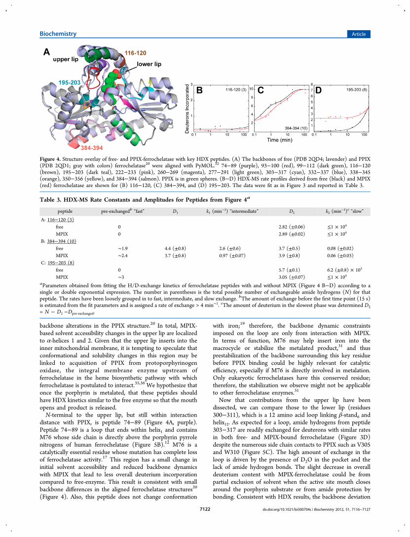
backbone alterations in the PPIX structure.20 In total, MPIX-based solvent accessibility changes in the upper lip are localizedto α-helices 1 and 2. Given that the upper lip inserts into theinner mitochondrial membrane, it is tempting to speculate thatconformational and solubility changes in this region may belinked to acquisition of PPIX from protoporphyrinogenoxidase, the integral membrane enzyme upstream offerrochelatase in the heme biosynthetic pathway with whichferrochelatase is postulated to interact.35,36 We hypothesize thatonce the porphyrin is metalated, that these peptides shouldhave HDX kinetics similar to the free enzyme so that the mouthopens and product is released.N-terminal to the upper lip, but still within interaction
distance with PPIX, is peptide 74−89 (Figure 4A, purple).Peptide 74−89 is a loop that ends within helix1 and containsM76 whose side chain is directly above the porphyrin pyrrolenitrogens of human ferrochelatase (Figure 5B).12 M76 is acatalytically essential residue whose mutation has complete lossof ferrochelatase activity.17 This region has a small change ininitial solvent accessibility and reduced backbone dynamicswith MPIX that lead to less overall deuterium incorporationcompared to free-enzyme. This result is consistent with smallbackbone differences in the aligned ferrochelatase structures20
(Figure 4). Also, this peptide does not change conformation
with iron;19 therefore, the backbone dynamic constraintsimposed on the loop are only from interaction with MPIX.In terms of function, M76 may help insert iron into themacrocycle or stabilize the metalated product,31 and thusprestabilization of the backbone surrounding this key residuebefore PPIX binding could be highly relevant for catalyticefficiency, especially if M76 is directly involved in metalation.Only eukaryotic ferrochelatases have this conserved residue;therefore, the stabilization we observe might not be applicableto other ferrochelatase enzymes.31
Now that contributions from the upper lip have beendissected, we can compare those to the lower lip (residues300−311), which is a 12 amino acid loop linking β-stand6 andhelix12. As expected for a loop, amide hydrogens from peptide303−317 are readily exchanged for deuterons with similar ratesin both free- and MPIX-bound ferrochelatase (Figure 3D)despite the numerous side chain contacts to PPIX such as V305and W310 (Figure 5C). The high amount of exchange in theloop is driven by the presence of D2O in the pocket and thelack of amide hydrogen bonds. The slight decrease in overalldeuterium content with MPIX-ferrochelatase could be frompartial exclusion of solvent when the active site mouth closesaround the porphyrin substrate or from amide protection bybonding. Consistent with HDX results, the backbone deviation
Figure 4. Structure overlay of free- and PPIX-ferrochelatase with key HDX peptides. (A) The backbones of free (PDB 2QD4; lavender) and PPIX(PDB 2QD1; gray with colors) ferrochelatase20 were aligned with PyMOL.32 74−89 (purple), 93−100 (red), 99−112 (dark green), 116−120(brown), 195−203 (dark teal), 222−233 (pink), 260−269 (magenta), 277−291 (light green), 303−317 (cyan), 332−337 (blue), 338−345(orange), 350−356 (yellow), and 384−394 (salmon). PPIX is in green spheres. (B−D) HDX-MS rate profiles derived from free (black) and MPIX(red) ferrochelatase are shown for (B) 116−120, (C) 384−394, and (D) 195−203. The data were fit as in Figure 3 and reported in Table 3.
Table 3. HDX-MS Rate Constants and Amplitudes for Peptides from Figure 4a
peptide pre-exchangedb “fast” D1 k1 (min−1) “intermediate” D2 k2 (min−1)c “slow”
A: 116−120 (3)free 0 2.82 (±0.06) ≤1 × 104
MPIX 0 2.89 (±0.02) ≤1 × 10⁴
B: 384−394 (10)free ∼1.9 4.4 (±0.8) 2.6 (±0.6) 3.7 (±0.5) 0.08 (±0.02)MPIX ∼2.4 3.7 (±0.8) 0.97 (±0.07) 3.9 (±0.8) 0.06 (±0.03)
C: 195−203 (8)free 0 5.7 (±0.1) 6.2 (±0.8) × 103
MPIX ∼3 3.05 (±0.07) ≤1 × 10⁴
aParameters obtained from fitting the H/D-exchange kinetics of ferrochelatase peptides with and without MPIX (Figure 4 B−D) according to asingle or double exponential expression. The number in parentheses is the total possible number of exchangeable amide hydrogens (N) for thatpeptide. The rates have been loosely grouped in to fast, intermediate, and slow exchange. bThe amount of exchange before the first time point (15 s)is estimated from the fit parameters and is assigned a rate of exchange > 4 min−1. cThe amount of deuterium in the slowest phase was determined D2= N − D1 −Dpre‑exchanged.
Biochemistry Article
dx.doi.org/10.1021/bi300704c | Biochemistry 2012, 51, 7116−71277122

within residues 303−317 of the free and PPIX alignedstructures is small but there is a noticeable shift closer to theporphyrin20 (Figure 4, cyan). HDX with Fe2+-ferrochelatase hasa similar kinetic profile to the free enzyme;19 therefore, we canassume the dynamic change in the lower lip is specific forporphyrin. Residues within 303−317 must be important to thecatalytic mechanism or structure of ferrochelatase since five arefully conserved and three are highly conserved amongeukaryotic and prokaryotic ferrochelatases.31 Random muta-genesis of the murine ferrochelatase lower lip (humannumbering 320−311) indicated that most substitutions increasethe Km for PPIX and that residues S303, G306, W310, andL311 were intolerant to most mutations.37 We propose that thelower lip is dynamically constrained even in the absence ofporphyrin substrate and that porphyrin access to the active siteis controlled only by reorientation of the upper lip to open andclose the pocket. It is interesting that ferrochelatase preorientsand stabilizes the lower lip in the free enzyme. A lack ofdynamics would lead to efficient substrate binding and/orporphyrin substrate orientation. Medlock and co-workers
suggested that the interactions between PPIX and hydrophobicresidues within 303−317 could be the trigger for closure of theupper lip.12 Our HDX-MS results are consistent with thathypothesis, where porphyrin binding does not requiresubstantial conformational change in the lower lip and is thusis not rate-limiting.
Residues Surrounding the Porphyrin Pocket Mediatethe Conformational Response. Turning to the bottom ofthe porphyrin binding pocket, we observed two adjacentpeptides that have a conformational response to MPIX,peptides 332−337 and 338−345. In free-ferrochelatase, peptide332−337 incorporates ∼50% deuterium over 2 h indicatingmoderate solvent accessibility and backbone dynamics in theabsence of substrate (Figure 3F). When MPIX binds, thebackbone dynamics are completely abrogated despite the smallincrease in D2O access within the first 15 s of deuteration. Theloss of backbone dynamics is probably due an interactionbetween F337 and porphyrin. Consistent with this, the F337Amutation increases the Km for PPIX about 2-fold.17 Thestructures of ferrochelatase clearly show the F337 benzyl ring in
Figure 5. MPIX-bound ferrochelatase solvent accessibility structural maps. (A) Peptides that have increased (pink) or decreased (cyan) D2Oaccessibility from HDX-MS are mapped to the monomeric ferrochelatase structure with PPIX bound (green spheres).20 Data were taken from Figure1. (B) Residues with increased deuterium access cluster on one face of the porphyrin and several key residues that could be involved in an ironchannel are indicated in stick format. Residues with an asterisk (*) are from peptides that have decreased backbone dynamics in the presence ofMPIX (Figure 3). (C) Residues with decreased deuterium access cluster on the lower lip side of the porphyrin (Figure 3). Key residues that could beinvolved in an iron channel are indicated in stick format. Residues with an asterisk (*) are from peptides that have decreased backbone dynamics inthe presence of MPIX. Figure created with PyMOL.32
Biochemistry Article
dx.doi.org/10.1021/bi300704c | Biochemistry 2012, 51, 7116−71277123

different positions with and without PPIX, but with littlechange in the backbone orientation20 (Figure 4, blue). It hasbeen postulated that the benzyl ring of F337 is a gate betweensolvent channels to and from the active site. HDX-MS suggeststhat the conformation of the backbone is significantly lessdynamic if MPIX is bound. If the backbone is more dynamic inthe free enzyme, MPIX binding to F337 would restrict itsmotion, allowing the side chain to change conformation tocontrol access of iron via a solvent channel to the active site.Thus the F337 interaction could be the sensor for porphyrinbinding site that triggers the iron uptake conformation.Residues 332−337 do not have a conformational response toFe2+, which lends more support for a functional role withPPIX.19
Adjacent to 332−337 is the π-helix13 peptide 338−345 thatresponds to MPIX binding with a striking decrease in backbonedeuterium access (Figure 3G). Several residue side chains arewithin interaction distance to the porphyrin including H341and I342, and there is a high level of amino acid conservationacross all ferrochelatases, especially for E34316 (Figure 5C).Mutations of D340 and E343 revealed that the acidic sidechains of these residues are required for activity.17,31 UnlikeMPIX, Fe2+ binding to free-ferrochelatase does not lead tochanges in solvent accessibility for peptide 338−345.19 Thus,this particular region is probably not involved in “sensing” iron,but instead MPIX through interaction with specific residues likeE343. Another π-helix13 peptide, 350−356, is farther down theπ-helix13 and has no change in solvent accessibility but has aslight decrease in dynamics (Figure 3J). Thus, only the residuesat the N-terminal end of the π-helix13 are critical for bindingMPIX and for the functional conformational response byconstraining dynamics that might facilitate premature release ofsubstrate instead of product.Localization of Porphyrin and Iron Responsive
Regions Gives Insight into the Ferrochelatase Mecha-nism. It is generally agreed that ferrochelatase has an orderedmechanism where PPIX binds first, followed by iron bindingand insertion into the macrocycle.38 The exact entrance routetaken by iron would greatly clarify the mechanism offerrochelatase since it could dictate from which face of theporphyrin it is inserted. We hypothesize that MPIX bindingshould be structurally linked to Fe2+ uptake, so we examinedregions that had higher solvent accessibility with MPIX bound.The rationale is that there must be a channel or path that theFe2+ takes to the active site and that channel must open inresponse to porphyrin substrate binding. There should also be adecrease in backbone dynamics to stabilize the channel and thechannel should become more restrictive to solvent closer to theactive site once hydrating waters are removed from the iron.39
Figure 5 shows peptides with a change in D2O accessibility andthe specific residues that might be important to the structureand mechanism of ferrochelatase. It is clear that peptides withincreased deuterium incorporation from solvent effects areclustered on one side of the porphyrin and lead to the surfaceof the enzyme (Figure 5A, pink). This is consistent with aconformational response after MPIX binding that links theactive site to a solvent accessible channel.We then compared the HDX results for MPIX and Fe2+ to
identify peptides that have a similar response to both substrates.There were four choices: peptides 99−112 in the upper lip,332−337 at the bottom of the active site, 222−233 at thesurface facing the matrix, and 195−203 that bridges the activesite and the [2Fe-2S] cluster. The upper lip peptide was ruled
out since there is no direct evidence that iron would be insertedfrom the membrane side of ferrochelatase. A recent reportdemonstrated that the mitochondrial iron importer mitoferrinforms a complex with ferrochelatase and it is proposed thatmitoferrin could deliver iron through the membrane.40 Peptide332−337 has a very small increase in solvent accessibility and isburied so this peptide was not considered further, at least forthe entrance of iron. It is possible that residues 332−337 are apart of the iron channel, but since it is much closer to PPIX itcould be more solvent restricted than other regions. This leavespeptides 222−233 and 195−203 as potential candidates forlining the solvent filled iron channel.Peptide 222−233 is located on the surface of ferrochelatase
and contains a few potential metal binding residues like D225,H230, and H231 (Figure 5B). It has a significant increase insolvent accessibility and decrease in backbone dynamics overthe times measured when MPIX binds (Figure 3H). As withpeptide 99−112, it is also possible that conformational changeswith MPIX resulting in increased deuterium incorporationcould occur before our first time point (15 s), but after whichno more deuterium is incorporated. Therefore, we arecomparing the much slower motions in this region with ouranalysis. An alignment of the free- and PPIX-bound structuresdoes not reveal backbone deviation in this region (Figure 4,pink), but HDX shows a dynamic response to MPIX.Interestingly, the HDX rate profile with MIPX bound isindistinguishable from that with Fe2+ bound;19 thus, porphyrinsubstrate binding results in a similar conformational change asFe2+ binding. This structural link to both substrates is a goodindication of an iron channel peptide. Combining the HDXresults for MPIX with that for Fe2+, we hypothesize thatresidues within 222−233 respond to MPIX binding to open theiron channel for efficient iron entry, thus exposing morebackbone amides to solvent. Previous crystal structuresobserved that Co2+ can coordinate the imidazole side chainof H231 along with the carboxylate side chain of D383.8 It hasbeen proposed that Fe2+ initially binds to H231 and D383before it is channeled to the postulated terminal ligands, R164and Y165, for insertion into the porphyrin macrocycle31
(Figure 5B). Unfortunately a peptide that includes D383 wasnot identified, but peptide 384−394 (Figure 4, salmon) is fairlysolvent accessible and dynamic in both free- and MPIX-ferrochelatase (Figure 4C). Like peptide 222−233, this peptidehas a small decrease in backbone flexibility with both MPIX andiron, again structurally linking this region to both substrates.The conformational link between MPIX and an iron channelmay explain why mutation of H231 to alanine, which is over 20Å away from the active site, increases the Km for Fe2+ but notfor MPIX. Other mutations like W227Y and D383A in thisregion also increase the Km for iron.31 It is tempting tospeculate that the H231A mutation may abrogate the Fe2+ andMPIX conformational response observed by HDX. There aresome reports that suggest that Fe2+ is delivered toferrochelatase by the iron chaperone frataxin.41,42 A recentdocking model showed that two aspartate residues of frataxincould be placed within 9 Å of H231 and D383 offerrochelatase.41 Thus, the change in conformation forferrochelatase around surface residues 222−233 could also berequired for efficient interaction with frataxin, but there is nodirect evidence that the interaction occurs in vivo.The other ferrochelatase peptide meeting the criteria of a
channel is 195−203 whose backbone amides are moreaccessible to D2O in both the MPIX (Figure 4D) and iron
Biochemistry Article
dx.doi.org/10.1021/bi300704c | Biochemistry 2012, 51, 7116−71277124

bound forms.19 This peptide spans the region between PPIXand the [2Fe-2S] cluster (Figure 4, dark teal). Side chain andbackbone atoms within peptide 195−203 (T198, S200, S201)participate in multiple hydrogen bonding interactions withsurrounding residues in the free-enzyme that rearrange in thePPIX-bound structure without significant changes in backboneconformation20 (Figure 4A). However, HDX with the Fe2+-bound enzyme indicates that peptide 195−203 has moreconformational flexibility than in the free- and MPIX states.This would imply that interactions with PPIX are needed tostabilize conformation and control iron insertion into theporphyrin. It also suggests that past this region, there should besolvent restriction to ensure that dehydrated iron is bound atthe terminal acceptors before insertion in the PPIX macrocycle.The alternate hypothesis delivers Fe2+ to the terminal ligands
H263 and E343 via an anionic surface on the π-helix1310,43
(Figure 5C). H263 would then serve as the metal donor residuefor insertion into PPIX. Visual inspection of the solventaccessibility of amides on this face of the porphyrin ring showsa general decrease around the MPIX binding site, with no largeincreases in deuteration (Figure 5C). Peptides that span the π-helix13 include 338−345 and 350−356 and both regions havedecreased dynamics when MPIX is bound (Figure 3G,J). Thus,the dynamic alterations we observe with MPIX were notapparent in the crystal structure (Figure 4, orange and yellow).Also the HDX kinetics for both π-helix13 peptides do notchange substantially with Fe2+ present,19 which is curious if thiswas the channel for iron. These results are consistent with arole in porphyrin substrate binding. Mutagenesis of π-helix13residues D340E, E343D, and H341C reduced activity without asignificant change in Km for either Fe2+ or PPIX, which shouldbe expected if they are involved in channeling iron to the activesite.31 Also since this face of the active site pocket is relativelyinaccessible to solvent with MPIX present, nonspecificchanneling of hydrated iron is also unlikely.17 If H263 is theterminal acceptor of iron before insertion in to PPIX, thesolvent accessibility of peptide 260−269 should be decreased sothat the iron is dehydrated properly and the dynamics shouldbe reduced in the MPIX bound state. This is, in fact, what isobserved from HDX (Figure 3E). The other possibility is thatD340, E343, and E347 may be involved in abstraction ofprotons from the pyrrole nitrogens prior to metalation,31,44
which would occur via the opposite face of the macrocycle asH263.20 This too would require a stable surface. Since H263 isa critical residue for ferrochelatase activity, the preorganizationof residues within 260−269 must be essential for its catalyticfunction, whether iron insertion or pyrrole extraction.
■ CONCLUSIONSHDX-MS indicates that porphyrin binding to ferrochelatasesignals dynamic changes in regions inside and outside thevicinity of the active site without large changes in the structure.When comparing the free- and PPIX-ferrochelatase alignedcrystal structures,20 the largest changes in conformation werealso verified by H/D exchange behavior of peptides in theseregions. However, new and potentially important dynamicregions of ferrochelatase have been identified by HDX-MS thatwarrant further structure−function investigation. These areashave not been functionally characterized as extensively as theregions around the active site, but they are clearly importantstructural contributors to the catalytic mechanism. Throughcomparison with previous HDX-MS experiments localizing thestructural response to iron substrate binding, insight into the
coupling of PPIX binding with Fe2+ uptake was illuminated,thus providing a more detailed conformational view of theferrochelatase mechanism. HDX-MS clearly shows that regionspreviously identified as responsive to iron also behave similarlyto porphyrin, and thus we have structurally linked iron andporphyrin binding for the first time. We propose that porphyrinsubstrate binding is propagated via conformational anddynamic changes that ultimately open a channel for ironuptake at the matrix-facing side of ferrochelatase, whilestabilizing the active site pocket where the porphyrin inbound. While HDX-MS cannot rule one pathway in or out formetal uptake and insertion, the results presented here providemore support to the highly debated mechanism whereby iron isinserted into PPIX from the M76/R164/Y165 side of themacrocycle.
■ ASSOCIATED CONTENT*S Supporting InformationThe pepsin digest peptide map of ferrochelatase (Figure S1)and all HDX-MS progress curves with fits for each peptide(Figure S2) are provided. This material is available free ofcharge via the Internet at http://pubs.acs.org.
■ AUTHOR INFORMATIONCorresponding Author*Phone, 205-348-0269. Fax, 205-348-9104. E-mail,[email protected] work was supported by the National Science FoundationAward No. 0845273 (to L.S.B.) and by the Ronald E. McNairPost-Baccalaureate Achievement Program (McNair Scholars)of The University of Alabama, U.S. Department of EducationTRIO Grant P217A030031 (to A.P.A.). The National ScienceFoundation CRIF program (CHE 0639003) is acknowledgedfor purchase of the mass spectrometer used in this study.NotesThe authors declare no competing financial interest.
■ ACKNOWLEDGMENTSThe authors would like to thank the Department of ChemistryMass Spectrometry Resource Facility manager, Dr. QiaoliLiang, for help with instrumentation and Dr. Harry Dailey(University of Georgia) for his gift of the ferrochelataseplasmid.
■ ABBREVIATIONS USEDHDX-MS, hydrogen/deuterium exchange mass spectrometry;β-ME, 2-mercaptoethanol; CHAPS, 3-[(3-cholamidopropyl)-dimethylammonio]-2-hydroxy-1-propanesulfonate; MPIX, mes-oporphyrin IX; PPIX, protoporphyrin IX; D2O, deuteriumoxide
■ REFERENCES(1) Busenlehner, L. S., and Armstrong, R. N. (2005) Insights intoenzyme structure and dynamics elucidated by amide H/D exchangemass spectrometry. Arch. Biochem. Biophys. 433, 34−46.(2) Eisenmesser, E. Z., Millet, O., Labeikovsky, W., Korzhnev, D. M.,Wolf-Watz, M., Bosco, D. A., Skalicky, J. J., Kay, L. E., and Kern, D.(2005) Intrinsic dynamics of an enzyme underlies catalysis. Nature438, 117−121.(3) Schramm, V. L. (2011) Enzymatic transition states, transition-state analogs, dynamics, thermodynamics, and lifetimes. Annu. Rev.Biochem. 80, 703−732.
Biochemistry Article
dx.doi.org/10.1021/bi300704c | Biochemistry 2012, 51, 7116−71277125

(4) Bakan, A., and Bahar, I. (2009) The intrinsic dynamics ofenzymes plays a dominant role in determining the structural changesinduced upon inhibitor binding. Proc. Natl. Acad. Sci. U.S.A. 106,14349−14354.(5) Dailey, H. A., Dailey, T. A. (2003) Ferrochelatase, in ThePorphyrin Handbook (Kadish, K. M., Smith, K. M., Guilard, R., Eds.)pp 93−121, Elsevier Science.(6) Sellers, V. M., Dailey, T. A., and Dailey, H. A. (1998)Examination of ferrochelatase mutations that cause erythropoieticprotoporphyria. Blood 91, 3980−3985.(7) Taketani, S., and Fujita, H. (1995) The ferrochelatase genestructure and molecular defects associated with erythropoieticprotoporphyria. J. Bioenerg. Biomembr. 27, 231−238.(8) Wu, C. K., Dailey, H. A., Rose, J. P., Burden, A., Sellers, V. M.,and Wang, B. C. (2001) The 2.0 Å structure of human ferrochelatase,the terminal enzyme of heme biosynthesis. Nat. Struct. Biol. 8, 156−160.(9) Burden, A. E., Wu, C., Dailey, T. A., Busch, J. L., Dhawan, I. K.,Rose, J. P., Wang, B., and Dailey, H. A. (1999) Human ferrochelatase:Crystallization, characterization of the [2Fe-2S] cluster and determi-nation that the enzyme is a homodimer. Biochim. Biophys. Acta 1435,191−197.(10) Karlberg, T., Lecerof, D., Gora, M., Silvegren, G., Labbe-Bois, R.,Hansson, M., and Al-Karadaghi, S. (2002) Metal binding toSaccharomyces cerevisiae ferrochelatase. Biochemistry 41, 13499−13506.(11) Al-Karadaghi, S., Hansson, M., Nikonov, S., Jonsson, B., andHederstedt, L. (1997) Crystal structure of ferrochelatase: The terminalenzyme in heme biosynthesis. Structure 5, 1501−1510.(12) Medlock, A., Swartz, L., Dailey, T. A., Dailey, H. A., andLanzilotta, W. N. (2007) Substrate interactions with humanferrochelatase. Proc. Natl. Acad. Sci. U. S. A. 104, 1789−1793.(13) Medlock, A. E., Carter, M., Dailey, T. A., Dailey, H. A., andLanzilotta, W. N. (2009) Product release rather than chelationdetermines metal specificity for ferrochelatase. J. Mol. Biol. 393, 308−319.(14) Karlberg, T., Hansson, M. D., Yengo, R. K., Johansson, R.,Thorvaldsen, H. O., Ferreira, G. C., Hansson, M., and Al-Karadaghi, S.(2008) Porphyrin binding and distortion and substrate specificity inthe ferrochelatase reaction: The role of active site residues. J. Mol. Biol.378, 1074−1083.(15) Lecerof, D., Fodje, M., Hansson, A., Hansson, M., and Al-Karadaghi, S. (2000) Structural and mechanistic basis of porphyrinmetallation by ferrochelatase. J. Mol. Biol. 297, 221−232.(16) Dailey, H. A., Dailey, T. A., Wu, C. K., Medlock, A. E., Wang, K.F., Rose, J. P., and Wang, B. C. (2000) Ferrochelatase at themillennium: Structures, mechanisms and [2Fe-2S] clusters. Cell. Mol.Life Sci. 57, 1909−1926.(17) Dailey, H. A., Wu, C. K., Horanyi, P., Medlock, A. E., Najahi-Missaoui, W., Burden, A. E., Dailey, T. A., and Rose, J. (2007) Alteredorientation of active site residues in variants of human ferrochelatase.Evidence for a hydrogen bond network involved in catalysis.Biochemistry 46, 7973−7979.(18) Hunter, G. A., Al-Karadaghi, S., and Ferreira, G. C. (2011)Ferrochelatase: The convergence of the porphyrin biosynthesis andiron transport pathways. J. Porphyr. Phthalocyanines 15, 350−356.(19) Asuru, A. P., and Busenlehner, L. S. (2011) Analysis of humanferrochelatase iron binding via amide hydrogen/deuterium exchangemass spectrometry. Intl. J. Mass Spectrom. 302, 76−84.(20) Medlock, A. E., Dailey, T. A., Ross, T. A., Dailey, H. A., andLanzilotta, W. N. (2007) A pi-helix switch selective for porphyrindeprotonation and product release in human ferrochelatase. J. Mol.Biol. 373, 1006−1016.(21) Okuda, M., Kohno, H., Furukawa, T., Tokunaga, R., andTaketani, S. (1994) Overexpression in Escherichia coli, and one-steppurification of the human recombinant ferrochelatase. Biochim.Biophys. Acta 1200, 123−128.(22) Dailey, H. A. (2002) Terminal steps of haem biosynthesis.Biochem. Soc. Trans. 30, 590−595.
(23) Zhang, Z., and Marshall, A. G. (1998) A universal algorithm forfast and automated charge state deconvolution of electrospray mass-to-charge ratio spectra. J. Am. Soc. Mass Spectrom. 9, 225−233.(24) Zhang, Z., and Smith, D. L. (1993) Determination of amidehydrogen exchange by mass spectrometry: A new tool for proteinstructure elucidation. Protein Sci. 2, 522−531.(25) Bai, Y., Milne, J. S., Mayne, L., and Englander, S. W. (1993)Primary structure effects on peptide group hydrogen exchange.Proteins 17, 75−86.(26) Molday, R. S., Englander, S. W., and Kallen, R. G. (1972)Primary structure effects on peptide group hydrogen exchange.Biochemistry 11, 150−158.(27) Dharmasiri, K., and Smith, D. L. (1996) Mass spectrometricdetermination of isotopic exchange rates of amide hydrogens locatedon the surfaces of proteins. Anal. Chem. 68, 2340−2344.(28) Smith, D. L., Deng, Y., and Zhang, Z. (1997) Probing the non-covalent structure of proteins by amide hydrogen exchange and massspectrometry. J. Mass Spectrom. 32, 135−146.(29) Mandell, J. G., Baerga-Ortiz, A., Croy, C. H., Falick, A. M.,Komives, E. A. (2005) Application of amide proton exchange massspectrometry for the study of protein-protein interactions. Curr. Protoc.Protein Sci. Chapter 20, Unit 20.9.(30) Busenlehner, L. S., Alander, J., Jegerscohld, C., Holm, P. J.,Bhakat, P., Hebert, H., Morgenstern, R., and Armstrong, R. N. (2007)Location of substrate binding sites within the integral membraneprotein microsomal glutathione transferase-1. Biochemistry 46, 2812−2822.(31) Sellers, V. M., Wu, C. K., Dailey, T. A., and Dailey, H. A. (2001)Human ferrochelatase: Characterization of substrate-iron binding andproton-abstracting residues. Biochemistry 40, 9821−9827.(32) Schrodinger, L. L. C. (2010) The PyMol molecular graphicssystem, version 1.3r1.(33) Engen, J. R., and Smith, D. L. (2001) Investigating proteinstructure and dynamics by hydrogen exchange MS. Anal. Chem. 73,256A−265A.(34) Chalmers, M. J., Busby, S. A., Pascal, B. D., West, G. M., andGriffin, P. R. (2011) Differential hydrogen/deuterium exchange massspectrometry analysis of protein-ligand interactions. Expert Rev.Proteomics 8, 43−59.(35) Ajioka, R. S., Phillips, J. D., and Kushner, J. P. (2006)Biosynthesis of heme in mammals. Biochim. Biophys. Acta 1763, 723−736.(36) Masoumi, A., Heinemann, I. U., Rohde, M., Koch, M., Jahn, M.,and Jahn, D. (2008) Complex formation between protoporphyrinogenIX oxidase and ferrochelatase during haem biosynthesis inThermosynechococcus elongatus. Microbiology 154, 3707−3714.(37) Shi, Z., and Ferreira, G. C. (2004) Probing the active site loopmotif of murine ferrochelatase by random mutagenesis. J. Biol. Chem.279, 19977−19986.(38) Davidson, R. E., Chesters, C. J., and Reid, J. D. (2009) Metal ionselectivity and substrate inhibition in the metal ion chelation catalyzedby human ferrochelatase. J. Biol. Chem. 284, 33795−33799.(39) Banci, L., Bertini, I., Cantini, F., D’Onofrio, M., and Viezzoli, M.S. (2002) Structure and dynamics of copper-free sod: The proteinbefore binding copper. Protein Sci. 11, 2479−2492.(40) Chen, W., Dailey, H. A., and Paw, B. H. (2010) Ferrochelataseforms an oligomeric complex with mitoferrin-1 and abcb10 forerythroid heme biosynthesis. Blood 116, 628−630.(41) Bencze, K. Z., Yoon, T., Millan-Pacheco, C., Bradley, P. B.,Pastor, N., Cowan, J. A., and Stemmler, T. L. (2007) Human frataxin:Iron and ferrochelatase binding surface. Chem. Commun. (Camb),1798−1800.(42) Yoon, T., and Cowan, J. A. (2004) Frataxin-mediated irondelivery to ferrochelatase in the final step of heme biosynthesis. J. Biol.Chem. 279, 25943−25946.(43) Hansson, M. D., Karlberg, T., Rahardja, M. A., Al-Karadaghi, S.,and Hansson, M. (2007) Amino acid residues His183 and Glu264 inBacillus subtilis ferrochelatase direct and facilitate the insertion of metalion into protoporphyrin ix. Biochemistry 46, 87−94.
Biochemistry Article
dx.doi.org/10.1021/bi300704c | Biochemistry 2012, 51, 7116−71277126

(44) Gora, M., Grzybowska, E., Rytka, J., and Labbe-Bois, R. (1996)Probing the active-site residues in Saccharomyces cerevisiae ferrochela-tase by directed mutagenesis. In vivo and in vitro analyses. J. Biol. Chem.271, 11810−11816.
Biochemistry Article
dx.doi.org/10.1021/bi300704c | Biochemistry 2012, 51, 7116−71277127