Embed Size (px)
Citation preview
Discovery and horizontal follow-up of an autoantibodysignature in human prostate cancerPaul J. Mintza, Anna Cecilia Rietzb,c, Marina Cardó-Vilab,c, Michael G. Ozawad, Eleonora Dondossolad, Kim-Anh Doe,Jeri Kima, Patricia Troncosof, Christopher J. Logothetisa, Richard L. Sidmang,1, Renata Pasqualinib,c,1,2,and Wadih Arapb,h,1,2
Departments of aGenitourinary Medical Oncology, eBiostatistics, and fPathology and dDavid H. Koch Center, The University of Texas M. D. AndersonCancer Center, Houston, TX 77030; gHarvard Medical School and Department of Neurology, Beth Israel Deaconess Medical Center, Boston, MA 02215;and bUniversity of New Mexico Cancer Center and Divisions of cMolecular Medicine, and hHematology and Medical Oncology, Department ofInternal Medicine, University of New Mexico School of Medicine, Albuquerque, NM 87131
Contributed by Richard L. Sidman, January 9, 2015 (sent for review December 10, 2014; reviewed by Otis Webb Brawley and Jorge Kalil)
In response to an urgent need for improved diagnostic and pre-dictive serum biomarkers for management of metastatic prostatecancer, we used phage display fingerprinting to analyze sequen-tially acquired serum samples from a patient with advancingprostate cancer. We identified a peptide ligand, CTFAGSSC, demon-strating an increased recovery frequency over time. Serum antibodyreactivity to this peptide epitope increased in the index patient, inparallel with development of deteriorating symptoms. The antigenmimicking the peptide epitope was identified as alpha-2–Heremans–Schmid glycoprotein, also known as fetuin-A. Metastatic prostatecancer cell lines and bone metastasis samples displayed robustfetuin-A expression, andwe demonstrated serum immune reactivityto fetuin-A with concomitant development of metastatic castrate-resistant disease in a large cohort of prostate cancer patients.Whereas fetuin-A is an established tumor antigen in several typesof cancer, including breast cancer, glioblastoma, and pancreas can-cer, this report is to our knowledge the first study implicating fetuin-A in prostate cancer and indicating that autoantibodies specific forfetuin-A show utility as a prognostic indicator for prostate cancerpatients prone to progress to metastatic disease.
cancer biomarker | peptide library | prostate cancer | phage display
Prostate cancer accounts for nearly 27,000 deaths annually,with end-stage bone metastases representing a leading cause
of morbidity and mortality (1). The introduction of diagnosticserum biomarkers into clinical practice, such as prostate-specificantigen (PSA), has greatly improved early detection of the dis-ease (2). However, the lack of reliable methods for prediction ofprogression beyond early-stage disease and the paucity oftreatment options for patients with bone metastasis results inmany patients with localized disease subjected to aggressivetreatment with sequelae including incontinence and impotence(3). Thus, identification of biomarkers to improve the accuracyof clinical assessment and stratification of patients needing con-servative versus aggressive treatment would constitute a majoradvance in the management of this disease.Antibodies specific for tumor-associated antigens are detectable
in the serum of cancer patients and have been studied as diagnosticand prognostic markers (4). Simultaneous quantification of auto-antibodies and PSA was proposed as a new approach to improvediagnosis and prognosis of prostate cancer (5). After autoanti-bodies against Huntingtin interacting protein-1 were identified inprostate cancer patients, combining serum reactivity with PSAvalues led to a screening discrimination with 97% specificity (6).Using combinatorial peptide phage libraries, we developed
a fingerprinting method based on targeting of circulating tumor-associated antibodies isolated from cancer patients (7, 8). Spe-cific autoantibodies and their cognate tumor-associated antigenshave been characterized, e.g., GRP78 for prostate cancer (9),HSP90 for ovarian cancer (8), ubiquilin 1 in lung cancer (10),and annexin XI-A in breast cancer (11). In addition, phage-based
screening approaches have been developed for high-throughputprofiling of immunogenic antigens for prostate cancer (12).In this study, we analyzed clinically annotated serum samples
obtained from an index patient at time points from his initialdiagnosis, presenting with androgen-dependent, localized pros-tate cancer, until his death with androgen-independent meta-static multifocal bone disease 7 y after the initial banked serumsample. A unique peptide, CTFAGSSC, was identified, for whichautologous serum IgG showed increasing reactivity. We identi-fied alpha-2–Heremans–Schmid glycoprotein (AHSG, also knownas fetuin-A) as the putative protein corresponding to the peptidemimic. We demonstrated increased serum antibody reactivity tofetuin-A during progression of disease in the index patient, as well asstrong serum reactivity in a large cohort of metastatic prostate cancerpatients. Reactivity to fetuin-A, identified years before the onset ofmetastatic disease in the index patient, indicates that serumantibodies constitute potential predictive biomarkers for meta-static prostate cancer and might facilitate relevant early treat-ment decisions.
ResultsCombinatorial Peptide Screening. Profiling of the humoral responseduring cancer progression can provide insights into changing
Significance
For the management of prostate cancer it has remained a sig-nificant clinical challenge to identify biomarkers that can beused as prognostic indicators to facilitate early treatmentdecisions and indicate patients at risk for castrate-resistantbone-metastatic prostate cancer in need of more aggressivetreatment. In this report, serum antibodies to alpha-2–Heremans–Schmidt glycoprotein (fetuin-A) were demonstrated to displayincreased reactivity with concomitant development of metastaticcastrate-resistant disease in a large cohort of prostate cancerpatients. Furthermore, metastatic prostate cancer cell lines andbone metastasis samples displayed robust fetuin-A expression. Toour knowledge, this is the first report to indicate that serumautoantibodies reactive to fetuin-A show utility as a prognosticindicator for prostate cancer patients at risk for progressing tometastatic disease.
Author contributions: P.J.M., M.G.O., E.D., R.L.S., R.P., and W.A. designed research; P.J.M.,M.C.-V., M.G.O., and E.D. performed research; A.C.R., K.-A.D., J. Kim, P.T., and C.J.L.analyzed data; and A.C.R., M.C.-V., K.-A.D., J. Kim, P.T., C.J.L., R.L.S., R.P., and W.A. wrotethe paper.
Reviewers: O.W.B., American Cancer Society; and J. Kalil, Butantan Institute and School ofMedicine, Sao Paulo.
The authors declare no conflict of interest.
Freely available online through the PNAS open access option.1To whom correspondence may be addressed. Email: [email protected],[email protected], or [email protected].
2R.P. and W.A. contributed equally to this work.
www.pnas.org/cgi/doi/10.1073/pnas.1500097112 PNAS | February 24, 2015 | vol. 112 | no. 8 | 2515–2520
MED
ICALSC
IENCE
S

antigenicity of a tumor and may identify serum autoantibodies ofdiagnostic and prognostic value. We applied a phage displayfingerprinting approach (7, 8) on isolated antibodies from se-quentially acquired serum samples from an index patient andperformed three rounds of selection for each time point. Relativephage recovery was very similar from serum obtained at initialdiagnosis of castrate-sensitive prostate adenocarcinoma (year 0)and serum obtained during androgen ablation treatment, inwhich PSA was undetectable (year 1) (Fig. 1A). An increase inrelative phage recovery was observed when the patient demon-strated elevated serum PSA and multifocal bone metastasis (year5) (Fig. 1A). Further increased phage recovery was evident in thesamples from year 7, when the patient presented with furtherelevated serum PSA concentration, increased number of bonelesions, and a biopsy confirmation of a right clavicle metastasis,consistent with prostate cancer metastasis (Fig. 1A).Sequence analysis of phage clones selected with patient IgG
from years 1, 5, and 7 demonstrated enrichment of a single pep-tide, CTFAGSSC, increasing in relative recovery frequency from27.5% in year 1 to 91% in year 7 (Fig. 1B). We also identifiedpeptide sequences unique for each serum collection date (Fig. 1B).Because the enrichment of CTFAGSSC phage was concom-
itant with development of advanced metastatic prostate cancer,we next sought to validate the selectivity and specificity of theCTFAGSSC peptide motif as a candidate marker of diseaseprogression. We evaluated binding of the isolated serum anti-bodies from the different time points to the CTFAGSSC pep-tide (Fig. 2A) and found an increase in reactivity against thepeptide over time, with the sample from year 7 producing themost robust binding. This trend mimicked the phage enrichmentprofile (Fig. 1A). Binding of year 7 antibodies to CTFAGSSCcould be inhibited in a concentration-dependent manner withpreincubation of the synthetic peptide CTFAGSSC, whereaspreincubation with a control peptide, CKDRFERC, did notinhibit binding (Fig. 2B). Immunostaining with autologous pu-rified antibodies on prostate cancer biopsies from the indexpatient showed reactivity (Fig. 2C, Left) that could be inhibitedby preincubation with the synthetic peptide CTFAGSSC (Fig.2C, Right). These data confirm the specificity of the isolatedantibodies for the peptide CTFAGSSC.
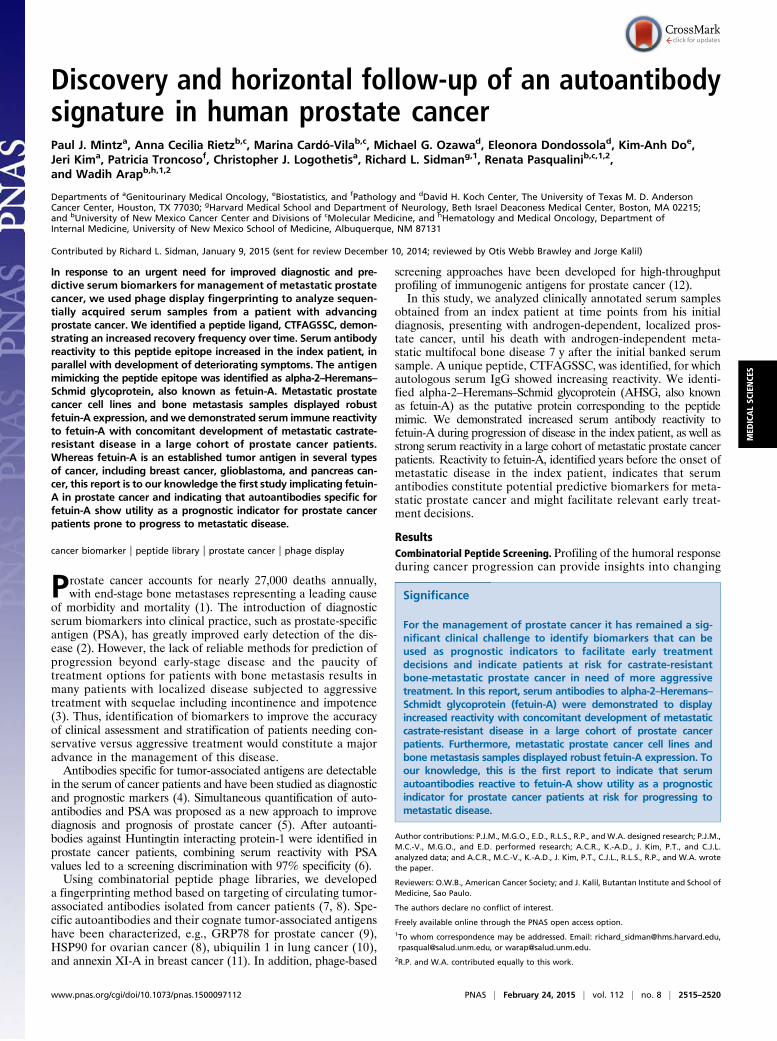
Fetuin-A Is the Antigen Mimicked by CTFAGSSC. After demonstrat-ing specific binding of CTFAGSSC to the antibodies presentduring metastatic disease, we next identified the antigen mim-icked by the peptide. Using the metastatic LNCaP prostate
cancer cell line, we performed 2D PAGE on whole-cell lysatesand subsequent Western blotting with purified antibodies fromyear 7 (Fig. 3A). A single band was identified by the antibodiesand was excised for protein sequence analysis by matrix-assistedlaser desorption/ionization–time-of-flight (MALDI-TOF) massspectrometry. Amalgamation of mass spectrometry data withresults from BLAST similarity searches of CTFAGSSC identi-fied AHSG, also known as fetuin-A, as the putative antigenrecognized by the antibodies.
Fig. 1. Fingerprinting the antibody repertoire with combinatorial phage display libraries. (A) Selection of peptide phage on isolated immunoglobulins fromsequentially acquired serum [year 0 (diagnosis), year 1, year 5, and year 7] from a prostate cancer patient. An increase in selectivity relative to the serum ofage-matched control men was observed over time. The third round of selection is shown, and bars represent mean values for phage-transducing units ± SEM,from triplicates. **P < 0.01, ***P < 0.001 by t test (two-tailed). (B) Peptide sequences identified from randomly selected phage clones from the third round ofselection on serum from year 1, 5, and 7 samples.
Fig. 2. CTFAGSSC peptide reactivity is acquired in metastatic prostate can-cer. (A) Immunoreactivity of isolated antibodies from the index patient tothe peptide sequence CTFAGSSC increased over time. (B) Specific inhibitionof antibody binding to immobilized peptide phage by the biotinylatedpeptide CTFAGSSC (black bars). No inhibition was seen with an unrelatedbiotinylated peptide (white bars) **P < 0.01, ***P < 0.001 by t test (two-tailed). (C) Immunoreactivity on prostate cancer biopsy samples from theindex patient with the autologous isolated antibodies (Ab, Left). Bindingwas inhibited by preincubation with the synthetic peptide CTFAGSSC (Right).Bars represent mean ± SEM, from triplicates.
2516 | www.pnas.org/cgi/doi/10.1073/pnas.1500097112 Mintz et al.
To confirm fetuin-A as the antigen, we first determined that theindex patient’s antibodies bound to fetuin-A and exhibited in-creasing reactivity over time (Fig. 3B). These data paralleled theinitial screening profile with temporally increased reactivity againstthe peptide CTFAGSSC. We next performed cross-inhibitionexperiments with isolated antibodies from the year 7 sample,polyclonal anti–fetuin-A antibodies, synthetic CTFAGSSC peptide,and purified fetuin-A (Fig. 3C). Binding of the isolated patientantibodies from year 7 to CTFAGSSC could be inhibited by pre-incubation with either fetuin-A or the cognate peptide. Similarresults were obtained with an anti–fetuin-A antibody.
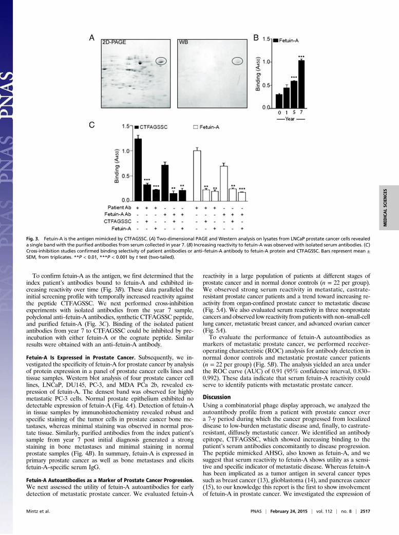
Fetuin-A Is Expressed in Prostate Cancer. Subsequently, we in-vestigated the specificity of fetuin-A for prostate cancer by analysisof protein expression in a panel of prostate cancer cells lines andtissue samples. Western blot analysis of four prostate cancer celllines, LNCaP, DU145, PC-3, and MDA PCa 2b, revealed ex-pression of fetuin-A. The densest band was observed for highlymetastatic PC-3 cells. Normal prostate epithelium exhibited nodetectable expression of fetuin-A (Fig. 4A). Detection of fetuin-Ain tissue samples by immunohistochemistry revealed robust andspecific staining of the tumor cells in prostate cancer bone me-tastases, whereas minimal staining was observed in normal pros-tate tissue. Similarly, purified antibodies from the index patient’ssample from year 7 post initial diagnosis generated a strongstaining in bone metastases and minimal staining in normalprostate samples (Fig. 4B). In summary, fetuin-A is expressed inprimary prostate cancer as well as bone metastases and elicitsfetuin-A–specific serum IgG.
Fetuin-A Autoantibodies as a Marker of Prostate Cancer Progression.We next assessed the utility of fetuin-A autoantibodies for earlydetection of metastatic prostate cancer. We evaluated fetuin-A
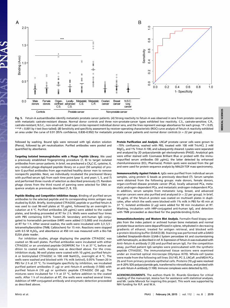
reactivity in a large population of patients at different stages ofprostate cancer and in normal donor controls (n = 22 per group).We observed strong serum reactivity in metastatic, castrate-resistant prostate cancer patients and a trend toward increasing re-activity from organ-confined prostate cancer to metastatic disease(Fig. 5A). We also evaluated serum reactivity in three nonprostatecancers and observed low reactivity frompatients with non–small-celllung cancer, metastatic breast cancer, and advanced ovarian cancer(Fig. 5A).To evaluate the performance of fetuin-A autoantibodies as
markers of metastatic prostate cancer, we performed receiver-operating characteristic (ROC) analysis for antibody detection innormal donor controls and metastatic prostate cancer patients(n = 22 per group) (Fig. 5B). The analysis yielded an area underthe ROC curve (AUC) of 0.91 (95% confidence interval, 0.830–0.992). These data indicate that serum fetuin-A reactivity couldserve to identify patients with metastatic prostate cancer.
DiscussionUsing a combinatorial phage display approach, we analyzed theautoantibody profile from a patient with prostate cancer overa 7-y period during which the cancer progressed from localizeddisease to low-burden metastatic disease and, finally, to castrate-resistant, diffusely metastatic cancer. We identified an antibodyepitope, CTFAGSSC, which showed increasing binding to thepatient’s serum antibodies concomitantly to disease progression.The peptide mimicked AHSG, also known as fetuin-A, and wesuggest that serum reactivity to fetuin-A shows utility as a sensi-tive and specific indicator of metastatic disease. Whereas fetuin-Ahas been implicated as a tumor antigen in several cancer typessuch as breast cancer (13), glioblastoma (14), and pancreas cancer(15), to our knowledge this report is the first to show involvementof fetuin-A in prostate cancer. We investigated the expression of
Fig. 3. Fetuin-A is the antigen mimicked by CTFAGSSC. (A) Two-dimensional PAGE and Western analysis on lysates from LNCaP prostate cancer cells revealeda single band with the purified antibodies from serum collected in year 7. (B) Increasing reactivity to fetuin-A was observed with isolated serum antibodies. (C)Cross-inhibition studies confirmed binding selectivity of patient antibodies or anti–fetuin-A antibody to fetuin-A protein and CTFAGSSC. Bars represent mean ±SEM, from triplicates. **P < 0.01, ***P < 0.001 by t test (two-tailed).
Mintz et al. PNAS | February 24, 2015 | vol. 112 | no. 8 | 2517
MED
ICALSC
IENCE
S
fetuin-A in a panel of prostate cancer cell lines and observedrobust expression of fetuin-A for the highly metastatic PC-3 cells,indicating that expression levels might correlate with metastaticpotential as found previously for angiogenic factors (16).Although microarray and serologic analysis of recombinant
cDNA expression libraries of human tumors with autologousserum have identified immunogenic molecules in various tumors,validation of putative antigens and biomarkers is often compli-cated by factors such as posttranslational modifications and dif-ferential cellular locations (17–19). Fingerprinting of enrichedserum antibodies with phage display offers a more direct com-binatorial screening for antibody epitopes and has identifiednumerous biologically relevant antigens (7–12). Moreover, stud-ies from other laboratories have indicated clinical relevance incancer diagnostics for antibodies against molecules such as osteo-pontin (20, 21), isoprostanes (22), and CXCL13 (23), among others.The concept of a global “autoantibody-ome” may apply, relatedto target discovery profiles for personalized therapeutics.Fetuin-A is a liver secretory protein that accumulates in bone
(24, 25). Fetuin-A also appears to be expressed in bone, astranscripts have been detected in chondrocytes and osteoblasts(26). Moreover, it has been suggested that fetuin-A is a trans-forming growth factor-β type II receptor mimic and acts as anantagonist of TGF-β and TGF-β–related bone morphogeneticprotein (BMP) activities (27). Multiple investigations of fetuin-A–deficient mice on various genetic backgrounds suggest thatfetuin-A has a role as a systemically acting inhibitor of ectopicbone formation (28–31). Bone lesions in prostate cancer arepredominantly osteoblastic and cause a relative increase in bonedeposition (32). In view of functions of fetuin-A as an inhibitorof bone morphogenetic proteins and ectopic bone formation, itis possible that anti–fetuin-A antibodies detected in the serumfrom metastatic castrate-resistant prostate cancer patients inthis study have a role in the bone-metastatic pathogenic process
by neutralization of fetuin-A in serum, thus precluding control ofbone deposition.This study supports the notion that profiling of the humoral
response could identify molecules indicating disease well beforeclinical manifestations. We have shown that autoantibodies againstfetuin-A, present in the serum of prostate cancer patients, con-stitute a putative early indicator of metastatic disease. Thecombination of the clinical history and development of theantibodies provides strong evidence that fetuin-A autoantibodiescould serve as a predictive biomarker for patients at risk fordeveloping metastases.
Materials and MethodsPatient Samples. Written informed consent was obtained from all subjectsand was approved by the Institutional Review Board (IRB) at the University ofTexas M. D. Anderson Cancer Center (UTMDACC). Tissue and serum sampleswere obtained from the UTMDACC Specialized Program of Research Excel-lence (SPORE) Tissue Resource and Pathology Core. Control samples wereobtained from blood-donor age-matched volunteers. The index patient hadserum banked in 1993 (defined as year 0). After response to chemical cas-tration, his disease was detected in bone 3 y after initially banking serum andcontinued to progress in this site until his death 7 y after the initial serumsample. Serum samples from years 0, 1, 5, and 7 were obtained. All bloodsamples were allowed to clot at room temperature, after which they werecentrifuged, separated into aliquots, and frozen.
Cell Culture and Reagents. DU145, PC-3, LNCaP, and MDA PCa 2b prostatecancer cells were purchased from the American Type Culture Collection(ATCC). Primary prostate epithelial cells were purchased from Cambrex.Synthetic peptides were produced by Genemed. Anti–fetuin-A antibody(M-17) was purchased from Santa Cruz Biotechnology.
Purification of Immunoglobulins. Biotinylated peptides were coated (50 μg/mL)on Neutravidin plates (Pierce) for 1 h at room temperature (RT). Wells wereblocked with PBS containing 4% (wt/vol) milk, 1% BSA, and 0.05% Triton X-100for 30 min at 37 °C, after which they were washed with PBS containing 0.05%Triton X-100. Serum antibodies (1:100) were incubated in wells for 2 h at RT,
Fig. 4. Fetuin-A is expressed in prostate cancer cells. (A) Western blot analysis of MDA PCa 2b, DU145, PC-3, and LNCaP metastatic prostate cancer cells dem-onstrated expression of fetuin-A. In contrast, no expression was detected in normal prostate epithelium (PE). (B) Anti–fetuin-A or purified serum antibodies from theindex patient revealed staining of prostate cancer bone metastases (Left), whereas minimal staining was observed in normal prostate tissue (Right).
2518 | www.pnas.org/cgi/doi/10.1073/pnas.1500097112 Mintz et al.
followed by washing. Bound IgGs were removed with IgG elution solution(Pierce), followed by pH neutralization. Purified antibodies were pooled andquantified by absorbance.
Targeting Isolated Immunoglobulins with a Phage Peptide Library. We useda previously established fingerprinting procedure (7, 8) to target isolatedantibodies from cancer patients. In brief, we precleared a CX6C (C, cysteine; X,any residue) phage-displayed peptide library on a pool (50 samples) of pro-tein G-purified antibodies from age-matched healthy donor men to removenonspecific peptides. Next, we individually incubated the precleared librarywith purified serum IgG from each time point (year 0, and years 1, 5, and 7)and performed three rounds of selection as described previously (7). Randomphage clones from the third round of panning were selected for DNA se-quence analysis as previously described (7, 8, 33).
Peptide Binding and Competitive Inhibition Assays. Binding of purified serumantibodies to the selected peptide and its corresponding mimic antigen wasstudied by ELISA. Briefly, biotinylated CTFAGSSC peptide or purified fetuin-Awas used to coat 96-well plates at 10 μg/mL, followed by an overnight in-cubation at 4 °C. Purified antibodies (20 μg/mL) were added to the coatedplates, and binding proceeded at RT for 2 h. Wells were washed four timeswith PBS containing 0.01% Tween-20. Secondary anti-human IgG conju-gated to horseradish peroxidase (HRP) were added (1:5,000) and incubatedat RT for 1 h. After several washes, the wells were incubated with 3,3′,5,5′-tetramethylbenzidine (TMB; Calbiochem) for 15 min. Reactions were stoppedwith 0.5 M H2SO4, and absorbance at 450 nm was measured with a Bio-TekELISA plate reader.
For inhibition studies, phage displaying the peptide CTFAGSSC wascoated on 96-well plates. Purified antibodies were incubated with eitherCTFAGSSC or an unrelated peptide CKDRFERC for 1 h at 37 °C, before ad-dition to coated wells. Analysis was as described above. For the cross-inhibition study, 96-well plates were coated with 10 μg/mL purified fetuin-A or biotinylated CTFAGSSC in 100 mM NaHCO3, overnight at 4 °C. Thewells were washed and blocked with 1% milk (wt/vol), 0.05% Tween-20 inPBS for 2 h at 37 °C. To investigate specificity by inhibition, we incubatedpurified patient antibodies (1 μg) or anti–fetuin-A antibody (1:500) withpurified fetuin-A (10 μg) or synthetic peptide CTFAGSSC (50 μg). Themixtures were incubated for 1 h at 37 °C, before addition to the coatedwells. After 1 h of incubation at RT, the wells were washed several times.Addition of HRP-conjugated antibody and enzymatic detection proceededas described above.
Protein Purification and Analysis. LNCaP prostate cancer cells were grown to∼70% confluence, washed with PBS, treated with 100 mM Tris·HCl, 2 mMMgCl2, and 1% Triton X-100, and subsequently sheared. Lysates were separatedand analyzed by 2D polyacrylamide gel electorphoresis (PAGE). Analytical gelswere either stained with Coomassie Brilliant Blue or probed with the immu-nopurified serum antibodies (50 μg/mL), the latter detected by enhancedchemiluminescence (ECL; Pharmacia). Protein spots were excised from the geland were used for protein sequence analysis by MALDI-TOF mass spectrometry.
Immunoreactivity Against Fetuin-A. IgGs were purified from individual serumsamples, using protein G beads as previously described (7). Serum sampleswere obtained from the following groups: male donors, female donors,organ-confined disease prostate cancer (PCa), locally advanced PCa, meta-static androgen-dependent PCa, and metastatic androgen-independent PCa.In addition, serum samples from metastatic lung, breast, and advancedovarian cancers were also purified and analyzed (n = 22 per group). In brief,10 μg/mL of the fetuin-A protein was coated on multiwell plates in dupli-cates, after which the wells were blocked with 1% milk in PBS for 45 min at37 °C. Isolated antibodies (2 μg) were added for 90 min incubation at RT.Washing, incubation with HRP-conjugated anti-human IgG, and detectionwith TMB proceeded as described for the peptide-binding ELISA.
Immunohistochemistry and Western Blot Analysis. Formalin-fixed biopsy sam-ples from the index patient or archived human bone metastasis and normalprostate tissue sectionswere deparaffinized in xylene, rehydrated in decreasinggradients of ethanol, treated for antigen retrieval, and blocked witha protein-blocking buffer (DAKO) (8). Staining was performed with aDAKOLabelled Streptavidin-Biotin (LSAB+) System peroxidase kit and counterstainingwith hematoxylin, as described in ref. 8. Expressionof fetuin-Awas evaluatedwithAnti–fetuin-A antibody (1:20) and purified serum IgG. For the competitionassay, purified patient IgG samples were preincubated with the syntheticpeptide CTFAGSSC. The immunostained tissue sections were examinedunder an inverted optical microscope (Olympus IX70). Whole-cell lysatesweremade from the following cell lines: DU145, PC-3, LNCaP, andMDA PCa2b and from primary prostate epithelial cells. Proteins (10 μg) were resolvedon4–20%SDS-polyacrylamidegels, transferred to nitrocellulose, and probedwithan anti–fetuin-A antibody (1:100). Immune complexes were detected by ECL.
ACKNOWLEDGMENTS. The authors thank Dr. Ricardo Giordano for criticalreading of the manuscript, Jessica Sun for assistance with statistical analyses,and Mr. Leslie Misrock for inspiring this project. This work was supported byNIH funding for R.P. and W.A.
Fig. 5. Fetuin-A autoantibodies identify metastatic prostate cancer patients. (A) Strong reactivity to fetuin-A was observed in sera from prostate cancer patientswith metastatic castrate-resistant disease. Normal donor controls and three non–prostate-cancer types exhibited low reactivity. C.S., castrate-sensitive; C.R.,castrate-resistant; N.S.C., non–small-cell. Small open circles represent individual donor sera, and the lines represent average absorbance for each group. *P < 0.05,***P < 0.001 by t test (two-tailed). (B) Sensitivity and specificity assessment by receiver-operating characteristic (ROC) curve analysis of fetuin-A reactivity exhibitedan area under the curve of 0.91 (95% confidence, 0.830–0.992) for metastatic prostate cancer patients and normal donor controls (n = 22 per group).
Mintz et al. PNAS | February 24, 2015 | vol. 112 | no. 8 | 2519
MED
ICALSC
IENCE
S
1. Rubin MA (2008) Targeted therapy of cancer: New roles for pathologists—prostatecancer. Mod Pathol 21(Suppl 2):S44–S55.
2. Brawer MK, et al. (1992) Screening for prostatic carcinoma with prostate specificantigen. J Urol 147(3 Pt 2):841–845.
3. Schover LR, et al. (2002) The use of treatments for erectile dysfunction among sur-vivors of prostate carcinoma. Cancer 95(11):2397–2407.
4. Madrid FF, Maroun MC (2011) Serologic laboratory findings in malignancy. Rheum DisClin North Am 37(4):507–525.
5. Xie C, et al. (2011) A novel multiplex assay combining autoantibodies plus PSA haspotential implications for classification of prostate cancer from non-malignant cases.J Transl Med 9(43):1–11.
6. Bradley SV, et al. (2005) Serum antibodies to huntingtin interacting protein-1: A newblood test for prostate cancer. Cancer Res 65(10):4126–4133.
7. Mintz PJ, et al. (2003) Fingerprinting the circulating repertoire of antibodies fromcancer patients. Nat Biotechnol 21(1):57–63.
8. Vidal CI, et al. (2004) An HSP90-mimic peptide revealed by fingerprinting the pool ofantibodies from ovarian cancer patients. Oncogene 23(55):8859–8867.
9. Arap MA, et al. (2004) Cell surface expression of the stress response chaperone GRP78enables tumor targeting by circulating ligands. Cancer Cell 6(3):275–284.
10. Chen G, et al. (2007) Autoantibody profiles reveal ubiquilin 1 as a humoral immuneresponse target in lung adenocarcinoma. Cancer Res 67(7):3461–3467.
11. Fernández-Madrid F, et al. (2004) Autoantibodies to Annexin XI-A and other auto-antigens in the Diagnosis of breast cancer. Cancer Res 64(15):5089–5096.
12. Wang X, et al. (2005) Autoantibody signatures in prostate cancer. N Engl J Med353(12):1224–1235.
13. Dowling P, et al. (2012) Analysis of acute-phase proteins, AHSG, C3, CLI, HP and SAA,reveals distinctive expression patterns associated with breast, colorectal and lungcancer. Int J Cancer 131(4):911–923.
14. Petrik V, et al. (2008) Serum alpha 2-HS glycoprotein predicts survival in patients withglioblastoma. Clin Chem 54(4):713–722.
15. Chen J, et al. (2013) Profiling the potential tumor markers of pancreatic ductal ade-nocarcinoma using 2D-DIGE and MALDI-TOF-MS: Up-regulation of Complement C3and alpha-2-HS-glycoprotein. Pancreatology 13(3):290–297.
16. Aalinkeel R, et al. (2004) Gene expression of angiogenic factors correlates withmetastatic potential of prostate cancer cells. Cancer Res 64(15):5311–5321.
17. Jia HL, et al. (2007) Gene expression profiling reveals potential biomarkers of humanhepatocellular carcinoma. Clin Cancer Res 13(4):1133–1139.
18. Liu R, et al. (2007) The prognostic role of a gene signature from tumorigenic breast-cancer cells. N Engl J Med 356(3):217–226.
19. Zhang R, Pan X, Huang Z, Weber GF, Zhang G (2011) Osteopontin enhances the ex-pression and activity of MMP-2 via the SDF-1/CXCR4 axis in hepatocellular carcinomacell lines. PLoS ONE 6(8):e23831.
20. Tilli TM, et al. (2012) Expression analysis of osteopontin mRNA splice variants inprostate cancer and benign prostatic hyperplasia. Exp Mol Pathol 92(1):13–19.
21. Milne GL, Dai Q, Roberts LJ 2nd (2014) The isoprostanes—25 years later. BiochimBiophys Acta, 10.1016/j.bbalip.2014.10.007.
22. Ammirante M, Shalapour S, Kang Y, Jamieson CA, Karin M (2014) Tissue injury andhypoxia promote malignant progression of prostate cancer by inducing CXCL13 ex-pression in tumor myofibroblasts. Proc Natl Acad Sci USA 111(41):14776–14781.
23. Scanlan MJ, Gure AO, Jungbluth AA, Old LJ, Chen YT (2002) Cancer/testis antigens: Anexpanding family of targets for cancer immunotherapy. Immunol Rev 188(1):22–32.
24. Pedersen KO (1944) Fetuin, a new globulin isolated from serum. Nature 154:575.25. Triffitt JT, Gebauer U, Ashton BA, Owen ME, Reynolds JJ (1976) Origin of plasma
alpha2HS-glycoprotein and its accumulation in bone. Nature 262(5565):226–227.26. Yang F, et al. (1991) Alpha 2-HS-glycoprotein: Expression in chondrocytes and aug-
mentation of alkaline phosphatase and phospholipase A2 activity. Bone 12(1):7–15.27. Demetriou M, Binkert C, Sukhu B, Tenenbaum HC, Dennis JW (1996) Fetuin/alpha2-HS
glycoprotein is a transforming growth factor-beta type II receptor mimic and cytokineantagonist. J Biol Chem 271(22):12755–12761.
28. Szweras M, et al. (2002) alpha 2-HS glycoprotein/fetuin, a transforming growth factor-beta/bone morphogenetic protein antagonist, regulates postnatal bone growth andremodeling. J Biol Chem 277(22):19991–19997.
29. Schafer C, et al. (2003) The serum protein alpha 2-Heremans-Schmid glycoprotein/fetuin-A is a systemically acting inhibitor of ectopic calcification. J Clin Invest 112(3):357–366.
30. Rittenberg B, et al. (2005) Regulation of BMP-induced ectopic bone formation byAhsg. J Orthop Res 23(3):653–662.
31. Seto J, et al. (2012) Accelerated growth plate mineralization and foreshortenedproximal limb bones in fetuin-A knockout mice. PLoS One 7(10):e47338.
32. Berruti A, et al. (1999) Differential patterns of bone turnover in relation to bone painand disease extent in bone in cancer patients with skeletal metastases. Clin Chem45(8 Pt 1):1240–1247.
33. Scott JK, Smith GP (1990) Searching for peptide ligands with an epitope library.Science 249(4967):386–390.
2520 | www.pnas.org/cgi/doi/10.1073/pnas.1500097112 Mintz et al.