Embed Size (px)
Citation preview
Different fixation methods in anterior cruciate ligament reconstruction
Extraction drilling versus compaction by serial dilation
PhD thesis
Ole Gade Sørensen
Faculty of Health Sciences University of Aarhus
2009


Different fixation methods in anterior cruciate ligament reconstruction
Extraction drilling versus compaction by serial dilation
PhD thesis
Ole Gade Sørensen
Faculty of Health Sciences University of Aarhus

Supervisors
Kjeld Søballe, Professor, MD, DMSc (Principal supervisor)
Department of Orthopaedics
Aarhus University Hospital
Denmark
Bent Wulff Jakobsen, MD
Eira Private Hospital
Science Center Skejby
Denmark
Søren Kold, MD, PhD
Orthopaedic Division
Aalborg University Hospital
Denmark
Torben Bæk Hansen, MD, PhD
Department of Orthopaedics
Hospital Unit West
Denmark
Evaluation committee
Jon Karlsson, Professor, MD, DMSc
Department of Orthopaedics
Sahlgrenska University Hospital
Sweden
Per Hølmich, MD
Orthopaedic Division
Amager Hospital
Denmark
Ole Simonsen, MD, DMSc (Chairman of the committee)
Orthopaedic Division
Aalborg University Hospital
Denmark

I
Preface This thesis is based on studies performed during my employment as a medical
doctor and researcher at the Department of Orthopaedics, Aarhus University
Hospital and the Department of Orthopaedics, Hospital Unit West during the period
2004-2009. The studies were carried out at both locations.
I am deeply indebted to a number of people who made this work possible
I would like to thank the health care personnel who attributed to the studies at
Aarhus University Hospital and Hospital Unit West.
I would like to express my sincere gratitude to my supervisors Kjeld Søballe, Bent
Wulff Jakobsen, Søren Kold, and Torben Bæk Hansen who all gave invaluable
advice, constructive criticism, and support throughout the process.
A special thank to Torben Bæk Hansen, who provided excellent working conditions.
I have always valued his loyal support and belief in me. He has been a mentor, not
only as a researcher, but also in my education towards becoming an orthopaedic
surgeon.
Furthermore, I wish to thank my fellow researchers Jesper Schønneman, Maiken
Stilling, and Kristian Larsen for participating in many good discussions and
facilitating a constructive research environment.
I am especially indebted to my colleague and dear friend Kristian Larsen who has
been a true source of inspiration. He has made invaluable contributions to the
development of ideas, methods, and interpretations of the outcomes in this thesis. No
matter how big the obstacles have been, he has always been able to see a way
through it, due to his outstanding methodological knowledge and his positive way of
being.

II
I also owe thanks to Rikke Mørup and Lone Løvgren Andersen for their assistance
with the planning of patient examinations, and to Rasmus Larsen for his skilful
assistance in radiostereometric analysis.
Finally, I want to thank my wife Lone and my children Thea and Aksel for their
everlasting patience, tolerance, and support.
Ole Gade Sørensen
Holstebro, October 2009

III
This thesis is based on the following papers:
I. Serial dilation versus extraction drilling in Anterior Cruciate Ligament
reconstruction: a biomechanical study. Knee Surg Sports Traumatol
Arthrosc. 2009 Sep 26. [Epub ahead of print]
II. The combination of radiostereometric analysis and the Telos stress Device
results in poor precision for knee laxity measurements after anterior
cruciate ligament reconstruction. Manuscript preparation.
III. Serial Dilation reduces Graft Slippage compared to Extraction Drilling in
Anterior Cruciate Ligament Reconstruction: a Randomized Controlled
Trial using Radiostereometric Analysis. Manuscript preparation.

IV
Abbreviations ACL: Anterior cruciate ligament
ANOVA: Analysis of variance
A-P: Anterior – posterior
BA plot: Bland-Altman plot
BMD: Bone mineral density
BPTB: Bone-patella-tendon-bone
C: Celsius
Cm: Centimetre
DFA: Distal fixation arm
Group EXDR: The extraction drilling group
Group SEDI: The serially dilated group
IKDC: International Knee Documentation Committee
Kp: Kilo pond
Mm: Millimetre
N: Newton
NSP: New standardized protocol
OFP: Original firm protocol
PFA: Proximal fixation arm
RCI: Round cannulated interference
RCT: Randomized controlled trial
RSA: Radio stereometric analysis
SA: Stress arm
SD: Standard deviation
TSD: Telos Stress Device

V

VI
Contents 1. English summary ............................................................................................................ 1 2. Danish summary............................................................................................................. 5 3. Introduction..................................................................................................................... 9 4. Aim of the thesis ........................................................................................................... 15 5. Design ............................................................................................................................ 17 6. Materials & methods..................................................................................................... 19
Ethical issues ................................................................................................................. 19 Outcomes....................................................................................................................... 33 Statistical analysis ......................................................................................................... 34
7. Results............................................................................................................................ 37 Patient characteristics ................................................................................................... 37 Results............................................................................................................................ 39
8. Discussion...................................................................................................................... 47 Key findings .................................................................................................................. 47 Comparison with relevant findings from other studies ............................................ 50 Limitations/Generalizability ....................................................................................... 50
9. Conclusion..................................................................................................................... 55 10. Perspectives and future research ............................................................................... 57 11. References .................................................................................................................... 59 Appendices........................................................................................................................ 66

VII

1
1. English summary
Introduction: The hamstring tendon graft has become increasingly popular in
anterior cruciate ligament (ACL) reconstruction because of low donor-site morbidity.
However, the tibial fixation is considered difficult, partly because of low tibial
mineral bone density. Therefore, we tested whether preparation of the tibial tunnel
with compaction by serial dilation provided a stronger anchorage of the graft-
fixation-device complex compared with traditional extraction drilling of the tibial
tunnel.
Prior to and during these investigations we became aware that the knee laxity
measurements using the Telos Stress Device (TSD) and radio stereometric analysis
(RSA) were difficult to reproduce. We therefore designed a new standardized
protocol (NSP) on how to apply the TSD aimed at ensuring (1) a reliable positioning
of the TSD on the patients` extremity that would result in (2) precise knee laxity
measurements.
Matherials and methods
Study 1: In 20 bovine tibiae, the bone tunnels were created with either extraction
drilling (group EXDR) or compaction by serial dilation (group SEDI). Twenty bovine
digital extensor tendons were fixated in the bone tunnel with an Intrafix device. The
graft-fixation-device complexes were mounted in a hydraulic test machine. The
fixation strength was evaluated after cyclic loading.
Study 2: Part study 1: One investigator followed the official company instructions on
how to apply the TSD. Another investigator followed the NSP. The TSD was applied
to the knee of 30 healthy persons. Double measurements were carried out. The
position of the stress arms of the TSD was marked following each measurement. The
reliability of each protocol was calculated as the difference in length between the first
and second markings.
Part study 2: The NSP for the TSD was then used in a clinical study. Thirty-five
patients underwent ACL reconstruction. Double measurements of knee laxity by
RSA were performed at a 3-month follow-up.
2
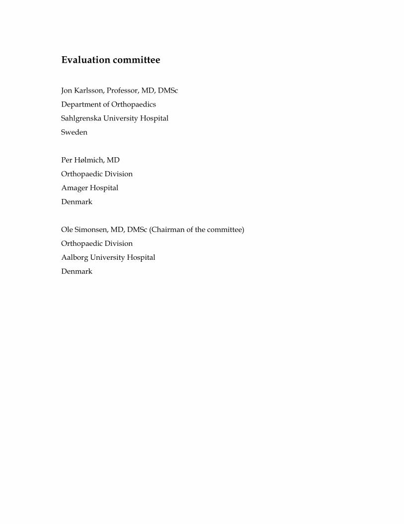
Study 3: Forty patients (22 males and 18 females) undergoing ACL reconstruction
were randomized to either extraction drilling (group EXDR) or compaction by serial
dilation (group SEDI) of the tibial tunnel. The hamstring graft was anchored with a
Retrobutton® and a supplementary interference screw (Arthrex®) in the femur and a
Delta interference screw (Arthrex®) in the tibia. Tantalum beads were placed in both
the proximal part of the tibia and distal part of the femur. Beads were placed in the
hamstring graft at the fixation sites as well. RSA was performed postoperatively and
again after 6, 12, and 24 weeks. The ACL reconstructed knee was stressed with a
TSD. Migration of the tantalum markers in the graft was measured in reference to the
bone markers in the tibia and femur. Knee laxity was assessed at every follow-up by
measuring the relation of the tibial bone markers to the femoral bone markers in both
the anterior and the posterior stress positions.
Results
Study 1: The difference between group SEDI and group EXDR ranged from a mean
slippage of 0 mm at 70-220 N, to a mean slippage of 0.1 mm at 70-520 N. We found
no significant difference in slippage of the graft-fixation-device complex after 1600
cycles.
Study 2: Part study 1: Using the NSP for TSD positioning, the prediction interval at
the marking sites ranged from ±0.4 mm - ±1.1 mm. Following the company
instructions, the prediction interval ranged from ±0.8 mm - ±3.9 mm depending on
marking site. Thus, the precision of positioning the stress arms of the TSD was
improved at all marking sites using the NSP compared with the original company
instructions. Part study 2: The double measurements of the knee laxity in the clinical
study using the NSP resulted in a mean difference of 0.0 mm and a prediction
interval of ±5.2 mm.
Study 3: Six patients (3 males and 3 females) were excluded during follow-up, which
resulted in 17 patients in group EXDR (mean age: 32.5 years (range: 20 – 50)) and 17
patients in group SEDI (mean age: 32.0 years (range: 20 – 49)). The mean migration of
the graft at the tibial fixation site after 3 months was 1.3 (SD 0.6) mm, in group EXDR
and 0.8 (SD 0.5) mm in group (P = 0.02). The knee laxity after 3 months was 13.0 (SD
4.0) mm in group EXDR and 10.9 (SD 3.1) mm in group SEDI (P = 0.09).

3
Conclusion:
Study 1 failed to show a significant difference between group SEDI and group EXDR.
In contrast in study 3 we found a significantly smaller mean migration of the
hamstring graft at the tibial fixation site in group SEDI compared with group EXDR.
No significant difference in stress radiographic knee laxity was found between the
two groups.
Even though the NSP improved the positioning of the TSD on the patients’
extremities, the combination of the TSD and RSA was not able to provide acceptable
knee laxity measurements in a clinical setting compared with published results
regarding other devices on the market.

4

5
2. Danish summary
Introduktion
Brugen af hamstringsener som graft ved rekonstruktion af forreste korsbånd er
blevet tiltagende populært grundet få bivirkninger fra donorstedet. Det kan dog
være problematisk at fiksere det nye korsbånd i skinnebenet. En af grundene til dette
er den lave knogledensitet i den øvre del af skinnebenet. Når man borer
knoglekanalen i skinnebenet fjerner man normalt det knoglevæv, der svarer til
graftens diameter (konventionel teknik). Alternativt kan man bore op til en mindre
diameter og gradvist presse den resterende del af knoglevævet ud i borekanalens
væg og dermed nå den samme diameter af borekanalen men med mere knoglevæv
bevaret i borekanalens periferi (seriel dilatation). Vi ville teste om seriel dilatation
kunne bidrage til en stærkere fiksering af hamstringgraften i skinnebenet i forhold
til den konventionelle metode.
Vi havde forinden opdaget, at det var svært at reproducere målinger af knæløsheden
når man brugte en kombination af et Telos apparatur (TSD) og stereo-røntgen (RSA).
Vi lavede derfor en ny standardiseret protokol (NSP) for selve påsætningen af TSD
på patientens ben. Vi ønskede at undersøge (1) om en NSP kunne medføre en mere
præcis påsætning af TSD på patientens ben i forhold til firmaets oprindelige protokol
(OFP) og (2) om brugen af NSP ville føre til mere præcise målinger af knæløsheden
ved brug af TSD og RSA.
Materialer og metoder
Studie 1: Knoglekanalen i 20 kalveskinneben blev enten tildannet med den
konventionelle teknik eller ved seriel dilatation. Tyve kalvesener blev fikseret i hvert
deres skinneben. Kalveknogle-senekomplekset blev herefter fastspændt i en
hydraulisk test maskine. Fiksationsstyrken blev målt efter cykliske test.
Studie 2: Delstudie 1: En undersøger fulgte OFP i påsætningen af TSD. En anden
undersøger fulgte NSP. TSD blev påsat 30 personer. Der blev foretaget
dobbeltmålinger. Positionen af TSD`s stressarme blev markeret efter hver påsætning.
Hver protokols præcision blev beregnet som forskellen i længden mellem den første
og anden markering.

6
Delstudie 2: NSP til påsætningen af TSD blev brugt i et klinisk studie. 35 patienter fik
foretaget forreste korsbåndsrekonstruktion. Præcisionen for knæløshedsmålingerne
blev målt efter dobbeltmålinger ved hjælp af RSA.
Studie3: Fyrre patienter (22 mænd og 18 kvinder), som fik foretaget forreste
korsbåndsrekonstruktion, blev randomiseret til udboring af knoglekanalen i
skinnebenet med enten konventionel teknik eller seriel dilatation. Der blev placeret
tantalumkugler i den nedre del af lårbenet og den øvre del af skinnebenet. Ligeledes
blev der placeret tantalumkugler i graften svarende til fiksationsstederne. Der blev
foretaget RSA efter 7-10 dage postoperativt og igen efter 6, 12 og 24 uger.
Migrationen af tantalumkuglerne i hamstringgraften kunne beregnes i forhold til
knoglemarkørerne i både skinneben og lårben. Løsheden af knæet kunne beregnes
ved at sammenligne knoglemarkørernes position skinneben og lårben, når knæet var
stresset med TSD i forreste og bagerste position.
Results:
Studie 1: Forskellen i middel migration af graften ved skinnebenfiksationen mellem
den serielt dilaterede gruppe og den gruppen, hvor konventionel teknik var brugt,
spændte fra 0 mm ved 70 – 220 Newton til 0.1 mm ved 70 – 520 Newton. Vi fandt
ingen signifikant forskel af middel migrationen mellem de to grupper efter 1600
cycli.
Studie 2: Delstudie 1: Præcisionen ved påsætningen af TSD blev forbedret ved alle
markeringspunkter, når NSP blev brugt set i forhold til OFP.
Delstudie 2: Dobbeltbestemmelserne af selve knæløsheden ved brug af NSP og RSA
resulterede i en middelforskel mellem 1. og 2. måling på 0.0 mm med en præcision
(prædiktionsinterval) på ±5.2 mm.
Studie 3: Seks patienter (3 mænd og 3 kvinder) blev ekskluderet i løbet af
opfølgningsperioden, hvilket resulterede i 17 patienter i den serielt dilaterede gruppe
(middel alder: 32.0 år (spændvidde 20 - 50)) og 17 patienter i den gruppe, hvor
konventionel teknik blev brugt (middel alder: 32.5 år (spændvidde 20 - 49)). Graftens
middel migration ved skinnebensfiksationen var henholdsvis 0.8 (SD 0.5) mm og 1.3
(SD 0.6) mm i de to grupper efter tre måneder (P = 0.02). Løsheden af knæet i de to

7
grupper efter 3 måneder måltes til henholdsvis 10.9 (SD 3.1) mm og 13.0 (SD 4.0) mm
(P = 0.09).
Konklusion
I studie 1 fandt vi ikke en signifikant forskel i graftens migration mellem den serielt
dilaterede gruppe og den gruppe, hvor konventionel teknik var brugt. I modsætning
hertil viste studie 3, at den serielt dilaterede gruppe havde signifikant reduceret
migration af graften ved fiksationen i skinnebenet efter tre måneder set i forhold til
gruppen, hvor konventionel teknik var brugt. Vi fandt ingen signifikant forskel i
knæløshed mellem de to grupper.
NSP var i stand til markant at forbedre præcisionen af påsætningen af TSD på
patientens ben. Det var dog ikke tilstrækkeligt til at kombinationen af TSD og RSA
kunne give en acceptabel præcision af knæløshedsmålingerne, når man
sammenligner med de publicerede resultater af præcisionen af andre
måleinstrumenter på markedet.

8

9
3. Introduction
Approximately 2500 primary anterior cruciate ligament (ACL) reconstruction are
performed every year in Denmark . Today, early postoperative motion and weight
bearing after anterior cruciate ligament (ACL) reconstruction are normally accepted
[2,54,62,64,74]. Therefore, stress on the graft cannot be avoided, before osteo-
integration of the tendons has occurred. Forces up to 450 Newtons (N) may act upon
the cruciate ligament in the rehabilitation period [30,52,65]. A strong anchorage of the
graft is therefore essential to avoid slippage of the graft at the fixation sites, and
thereby cause increased laxity of the knee.
Previously the bone-patella-tendon-bone (BPTB) graft was very popular in cruciate
ligament reconstruction, but its use has diminished, probably due to donor-site
morbidity [11,45] . On the other hand, harvest of the semitendinosus- and gracilis
tendons is well accepted [69,80]. Therefore the hamstringgraft has become
increasingly popular in ACL reconstruction and has been shown to be equivalent to
the BPTB graft [9,18,35]. In Denmark, the hamstring graft is used in approximately
70% of all ACL reconstructions [1].
A number of different fixation devices have been used to secure the hamstringgraft
at the tibial fixation site. Extra-cortical devices, such as washers, have provided a
high fixation strength [7], but problems with bungee-cord effect and wind shield-
wiper effect have been seen[34,68,76]. This resulted in the use of fixation devices with
a juxta-articular fixation, such as interference screws. In addition Ishisbashi et al. [33]
showed increased knee stability after use of joint-near fixation devices compared
with extra cortical devices. Weiler et al. [77] also showed, that direct fixation of the
graft in the bone tunnel, as provided by an interference screw, will minimize the
micromotion of the graft in bone tunnel and probably enhance a direct tendon-to-
bone healing.
10
Especially the tibial fixation of the semitendinosus-gracilis graft is considered
problematic, partly because of the bone mineral density of the tibia is less than in the
femur resulting in potentially insufficient stability of fixation implants placed in the
tibial tunnel. [8]
.
Compaction of the bone tunnel by serial dilation
A tunnel preparation technique that compacts the periphery of the tibia tunnel by
serial dilation could provide a stronger anchorage of the graft than does traditional
extraction drilling of the tibia tunnel.
The use of compaction by serial dilation in ACL reconstruction is inherited mainly
from the research on hip implants. Green et al. [25] used a canine model to show that
compaction could improve early fixation stiffness and strength of porous-coated
implants. Histological examinations showed that compaction resulted in increased
bone density at the implant surface. Kold et al. [48-50] were able to show the same
benefits of compaction for implants with other surfaces. Because compaction by
serial dilation tends to preserve cancellous bone material instead of removing it, as
seen in conventional drilling, they suggested that the improvement of the early
fixation strength is a result of both larger bone volume in the proximity of the
implant and compressive forces of the compacted bone also known as the “spring-
back effect” [47]. Both results minimize the gap between bone and implant surface
and produce an increase in bone-implant friction.
To our knowledge, Johnson et al. [36] were the first to report the use of serial dilators.
They tested the difference in fixation strength between metal interference screws and
bioabsorbable interference screws in ACL reconstruction. They compacted the
femoral bone tunnel by serial dilation, but did not speculate on whether the
compaction could enhance the fixation strength.
Cain et al. [10] followed with a study using human cadaveric knee specimens. The
tibial tunnel was serially dilated, and the graft was anchored in both tibial and
femoral specimens. This entire construct was secured in a test machine that was able

11
to load the construct with a translatoric force. Ultimate failure load was recorded.
With seven specimens in each group, Cain et al. [10] were able to show a significant
difference in favour of the serial dilated group compared with the extraction drilled
group.
Rittmeister et al. [63] also used a human cadaveric model. Cyclical loading tests were
performed. The force applied to the graft was increased after each cycle. Loads at
different permanent displacements of the grafts were recorded. A comparison
between serial tunnel dilation and extraction drilling was carried out in 14 pairs of
specimens (half secured with a 7 millimetre (mm) RCI screw, and half secured with a
9 mm RCI screw). The results were pooled. The serial dilated group showed higher
loads at all permanent displacements, but the differences were not significant.
Nurmi et al. [56] were not able to show any positive effect of compaction by serial
dilation compared with extraction drilling. In a human cadaveric set-up, 21 pairs of
tibia were submitted to cyclical loading. Displacement of the graft was measured
after various numbers of cycles and a single-cycle load-to-failure test was finally
performed.
When we started our own studies, only findings [10,56,63] regarding serial dilation
of the tibial tunnel in ACL reconstruction using hamstring grafts had been published.
Dunkin et al. [17] illuminated the issue further. Their results are in line with the
study of Nurmi et al. Dunkin used a porcine model. Twenty specimens underwent
either serial dilation or extraction drilling. The bone volume in the periphery of the
bone tunnel was measured. The serially dilated group showed significantly higher
bone volumes compared with the non-dilated group, but no difference in initial
fixation strength could be detected. Instead, they found a correlation between
decrease in fixation strength and screw divergence.
Recently Gokce et al. [24] reported a clinical retrospective study. They investigated
the influence of compaction by serial dilation on tunnel widening. In total, 44
patients were enrolled (21 in the intervention group and 23 in the control group).

12
Tibial tunnel enlargement was found to be significantly higher in the control group
compared with the intervention group, indicating that serial dilation of the tibial
tunnel could protect against tunnel widening. No significant difference in
postoperative Lysholm Scores and IKDC-scores was found.
Dargel et al. [15] investigated the use of serial dilators at the femoral fixation site
when a BPTB graft was used. In a porcine setup they compared three ways of
preparing the femoral bone canal; (1) extraction drilling to 9 mm, (2) extraction
drilling to 8 mm followed by serial dilation to 9 mm, and (3) extraction drilling to 6.5
mm followed by serial dilation to 9 mm. Surprisingly they found, that group 2 had
significantly increased initial fixation strength compared with both groups 1 and 3.
The springback effect was largest in group 3.
Finally, studies on compaction of the bone tunnel by stepped routers (not serial
dilators) have been published [55,57]. No effect on initial fixation strength was
found.
In summery, the conclusions of the studies regarding fixation strength after serial
dilation of the bone tunnel in reconstruction of the ACL are contradictive, and no
consensus has yet been reached. Apart from Gokce et al. [24] all studies are
biomechanical studies and carried out with fresh-frozen materials. This means that it
is possible to reflect on the differences in initial fixation strength, but impossible to
conclude anything about long-term results of serial dilation. Keeping in mind that
proper osseointegration of the hamstring graft probably has not occurred before 6 –
12 weeks after surgery, in-vivo studies are necessary to illuminate potential benefits
of serial dilation in ACL reconstruction.
Knee laxity measurements
Anterior-posterior knee laxity measurements have traditionally been used to
diagnose cruciate ligament rupture, and to evaluate the outcome after cruciate
ligament reconstruction. In the search for an accurate and precise method, several
different devices have been used.

13
The KT-1000 arthrometer (MEDmetric corp. San Diego, CA, USA) [13,14,51] is
probably the most widely used device [3,28,29,61,72,73,79] for non-invasive knee
laxity measurements. Varying results have been reported regarding the precision of
the device. Steiner et al. [72] found a precision of approximately 4.2 mm (± 2
standard deviations (SD) of the mean between the first and second measurements),
whereas Torzilli et al. [73] reported the precision to be approximately 2 mm. Another
device for non-invasive knee laxity measurements is the Rolimeter knee-tester
(Aircast Europa, Neubeuern, Germany), which has been used in several studies
[4,23,27,53,59]. The reliability of the device is found to be comparable to the KT-1000
arthrometer [4,23]. The combination of a stress device and radiography (stress
radiography) is another established knee laxity measurement technique. The use of
the Telos Stress Device (TSD) in stress radiography is regarded by many to be the
gold standard for evaluation of posterior cruciate insufficiency [42,66,71]. The
intratester and intertester reliability is reported by Staubli [70]. They used one set of
radiographs on each patient. To our knowledge, the precision of stress radiography
in combination with the TSD following double measurements has not been reported.
Radio stereometric analysis (RSA)
RSA was originally developed by Selvik et al. [67]. Because of its high accuracy of 1
mm, RSA has mainly been used to determine the migration of arthroplasty
components over time in relation to bone. RSA is an invasive method that relies on
implantation of tantalum beads. The calculation of the migration is based on a set of
radiographs, with the patient in relation to a calibration box. The calibration box and
computer software convert the 2-dimensional radiographs into a 3-dimensional
coordinate system. A set of radiographs is defined as reference, and the relation of
two rigid bodies can then be calculated at each follow-up. Because of the high
accuracy, RSA should have the potential to provide a precise measure of the knee
laxity.
RSA and knee laxity measurements
Several studies have used the RSA in combination with a stress device
[21,22,31,32,37-41,43]. Kærholm et al. [44] and Friden et al. [21] used custom-made

14
stress devices and reported precisions (±2 SD) of 1.6 mm and 2.2 mm, respectively.
Fleming et al. [19,20] also used a custom-made pneumatic load device in combination
with RSA. They tested the accuracy and repeatability in five goat knees [20], and
found a good repeatability after repeated measurements. They later published a
clinical study, in which they compared the knee laxity obtained by RSA, planar stress
radiography, and the KT-1000 arthrometer in 15 patients. No precision after double
measurements was reported in the clinical study [19]. Khan et al. [46] used TSD and
RSA in six patients. They found a precision (±2 SD) of 1.9 mm. They used the
original firm protocol on how to apply the TSD on the extremity of the patient. Our
hypothesis was that this protocol could be improved, which potentially could result
in a higher precision in knee laxity measurements.

15
4. Aim of the thesis
The overall aim in this thesis was to compare the fixation strength and thus the
migration of the hamstring graft at the tibial fixation site after conventional
extraction drilling or compaction by serial dilation of the tibial tunnel after ACL
reconstruction. Prior to and during these investigations we became aware that knee
laxity measurements using the TSD and RSA were difficult to reproduce. This led to
further methodological considerations regarding the usefulness of the TSD combined
with RSA (study II).
The individual studies in this thesis had the following aims:
Study I
To compare the initial fixation strength between extraction drilling and serial dilation
of the tibial bone tunnel after cyclic loading of the hamstring graft.
Study II
Part study 1: whether a new standardized protocol would lead to a more precise
positioning of the Telos Stress Device compared with the original firm protocol.
Part study 2: whether a more precise positioning of the stress arms of the Telos Stress
Device would result in more precise A-P knee laxity results in a clinical study using
radio stereometric analysis (part study 2).
Study III
To evaluate whether compaction by serial dilation of the tibial bone tunnel compared
with extraction drilling could reduce the migration of the hamstring graft at the tibial
fixation site,

16

17
5. Design
Study I
Prospective paired biomechanical randomized study using a bovine set-up.
Study II
Part study 1 and part study 2: Both part studies were performed as reliability
(precision) studies after double measurements.
Study III
Prospective, randomized clinical trial using radio stereometric analysis.

18

19
6. Materials & methods
Ethical issues
Study I
The calf tibiae were obtained from a local slaughterhouse. There were no ethical
considerations in this project.
Study II
In the first part of this study, we examined the precision of the application of the TSD
on the patients` lower extremities. Only healthy individuals participated. No
approval from the local ethics committee was needed.
The second part of the study examined the precision of knee laxity measurements.
The data were retrieved from study III. For ethical considerations, please see below.
Study III
The study was approved by the Regional Committee of Biomedical Research Ethics
(record number 20060158). Informed and written consent were obtained from all
patients. The ethical standards of the Regional Committee of Biomedical Research
Ethics were in accordance with the Helsinki Declaration of 1995. The study was
registered with the Danish Data Protection Agency (record number 2006 – 41 – 7247).

20
Materials/patients and intervention
Study I
Ten pairs of bovine tibiae and 20 bovine digital extensor tendons were used. The
bovine tendons are shown to have similar properties compared with human
hamstring grafts [16] . The calves were aged 34 weeks ± 2 weeks. The fresh tibiae and
tendons were stripped of soft tissue and fresh frozen at -20˚ C in sealed plastic bags.
Before freezing, the diameter of the tendons was measured with a graft sizer with
increments of 0.5 mm, and only tendons with a diameter of 10 mm were accepted.
Twelve hours before use, the tibiae and tendons were defrosted at room temperature.
This type of preservation does not affect the properties of the material[60]
Fig. 1: Two methods used to create the tibial bone tunnel. Left, 10 mm cannulated
drill for extraction drilling; right, serial dilators (8-10 mm)(Smith &Nephew) with
0.5 mm increments.
A paired design was used. For each pair, one tibia was prepared with serial drilling
and the other was prepared with extraction drilling. The tip of an ACL tibial drill
guide was placed at the ACL footprint at the centre of the tibial plateau. Guide angle
and length were set to 45˚ and 50 mm, respectively, and a guide wire was passed
along, following the drill guide. In group 1, conventional extraction drilling was

21
performed, leaving a drill hole with a diameter of 10 mm. In group 2, both the intra-
articular cortex and the antero-medial cortex were pre-drilled to 10 mm because of
the thickness of the bovine cortex. Then a bone tunnel of 8 mm in diameter was
created using extraction drilling. Subsequently, the tunnel diameter was compacted
by stepwise serial dilation ending up with a tunnel diameter of 10 mm (Fig. 1).
The tendons were split in half in a natural cleavage. The tendons were then folded at
the middle leaving a quadrupled graft. Each strand was marked, resulting in a
looped intra-articular portion of 3 centimetres (cm) and a tibial portion of at least 5
cm. A running baseball suture (Ethibond Excel 2-0® Jonhson & Johnson, Langhorne,
PA, USA) was applied to each leg at the tibial portion of the graft.
The tibia was then secured in a custom made fixation device on a MTS servo-
hydraulic test machine (MTS Systems Corp, Eden Prairie, MN, USA) (Fig. 2). During
fixation of the graft, the tibia was turned so that the anteromedial opening faced the
surgeon. The prepared graft was pulled through the bone tunnel, and a 3 cm broad
ruler was temporary placed in the loop, making sure that the length of the looped
end was the same in all specimens. The sutures were tied together two and two and
passed around an ACL Tie Tensioner (DePuy Mitek, Raynham, MA, USA). The graft
was secured with a 9-11 mm Intrafix tibial fastener (DePuy Mitek). This fixation
device is used in other comparative biomechanical studies, for example [12], and is
shown to provide a strong fixation of the hamstring graft. During fixation of the
graft, we used full tension (15.9 kg (approximately 156 N)) on the ACL Tie Tensioner
(DePuy Mitek), in order to achieve the same tension in the graft during graft fixation.
Subsequently we turned the tibia-graft complex 180 degrees. A crossbar (diameter 9
mm) was passed through the looped end, mimicking the femoral fixation. The bone
tunnel was aligned parallel to the loading axis in a “worst case scenario” setup (Fig.
2).

22
Fig. 2: Tibia-graft complex mounted in the servo-hydraulic MTS test machine.
Loading procedure
The pretension was set to 70 N [5]. The graft was cyclic preconditioned 10 times
between 70 N and 120 N. Thousand cyclic loads of 70 N – 220 N were then applied at
a cross-head speed of 80 cycles per minute. The load was then increased 50 N
following every 100 cycles, ending with loads between 70 N and 520 N. This load
protocol should reflect the forces on the ACL during walking and jogging [30,65].
Cross-bar position was recorded at the first peak load. Displacement of the graft was
then measured as the displacement of the cross-bar at peak load immediately before
every load increase.
Study II
Part study 1: The precision of the new standardized protocol compared with the
original firm protocol for TSD positioning
The original firm protocol (OFP)
The guidelines of the OFP are as follows. For anterior stress of the tibia: (1)
Positioning of the patient as shown in Fig. 3. Knee flexion angle of 10˚- 20˚, slight
turning-out rotation of the lower leg through “stable lateral position”. (2) The
pressure device should lie approximately 7 cm below the hollow of the knee, reading
of the pressure device: 15 kp. With freshly injured muscular athlete: possibly 20 kp.

23
Fig 3: Anterior stress of the tibia using Fig 4: Posterior stress of the tibia using
the TSD. the TSD.
For posterior stress of the tibia: (4) Positioning of the patient as shown in Fig. 4, knee
flexion angle of 10˚- 20˚. (5) The pressure device should be placed at the tuberositas
tibia level. (6) Reading of the pressure device: 15 kp.
The new standardized protocol (NSP)
The TSD is assembled as shown in Fig. 5 (please note that the extension arm is not
used). As seen, the TSD has a proximal fixation arm (PFA), a stress arm (SA), and a
distal fixation arm (DFA).
Fig 5: The TSD, when assembled.
(1) The patient is placed in a supine position. (2) A cushion is placed under the knee.
The cushion is replaced until the flexion angle of the knee is 20˚ measured with a
goniometer. (3) The proximal part of the patella is marked with a transverse line. (4)
Another mark is made two cm proximally and parallel to the first line. This second
Marking site 1 A1
Marking site 5
Marking site 3
Marking site 4
Marking site 2
Marking site 6
24
mark (marking site 1) represents the position of PFA during anterior stress of the
tibia. Care was taken to gently support the distal part of the patella during marking
to avoid displacement of the patella (Fig 6).
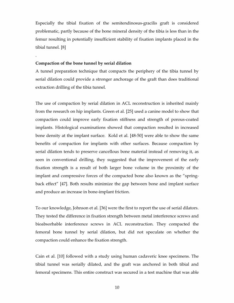
Fig 6: The patella is supported before marking to avoid displacement.
5) The proximal part of the tuberositas tibiae is palpated and marked (Fig. 7). This
line represents the position of the SA during posterior stress of the tibia.
Fig 7: Palpation and marking of the tibial tuberosity.
(6) The patient is placed in an upright position with 15 cm between the medial parts
of both heels. (7) The distance between marking site 1 and the floor is measured with
a ruler and 2 cm are added. (8) The same distance from the floor is measured on the
posterior side of the extremity and another marking line (marking site 4) is drawn.
This marking represents the position of the PFA during posterior stress of the tibia.
Adding 2 cm is necessary to prevent the metal in the PFA from shadowing the
femoral tantalum beads during RSA. (9) The distance between marking site 2 and the
floor is measured, and the same distance is marked on the posterior side of the lower
leg. This marking (marking site 5) represents the position of the SA during anterior

25
stress of the tibia. (10) The distance between marking site 2 and the floor is multiplied
by 0.70. Using this distance a transverse line is drawn on the anterior (marking site 3)
and posterior (marking site 6) aspect of the lower limb (Fig 8). Marking site 3
represents the position of the DFA during anterior stress of the tibia and marking site
6 represents the position of the DFA during posterior stress of the tibia.
Fig. 8: Marking of the distal marking site on the posterior aspect of the tibia.
(11) The patient is placed in lateral position. When anterior stress is needed, the TSD
is applied using the marking sites, as described above (Fig 3). The tibia is cycled 3
times with a force between 0 – 15 kp before a force of 15 kp is maintained. (12) When
posterior stress is needed, the TSD is turned around (Fig. 4). The tibia is cycled 3
times with a force between 0 – 10 kp before a force of 10 kp is maintained.
Precision of the protocols
In the scientific literature reliability, precision, and reproducibility are used in
different contexts. In this study, we use the term precision defined as the prediction
interval (±1.96 SD) of mean difference between the first and second measurements
[6].
The study took place at the Department of Orthopaedics, Hospital Unit West,
Denmark from September 2006 to January 2007. One investigator used the OFP on
how to apply the TSD and another investigator used the NSP. Thirty healthy
individuals were included. For each investigator we defined a learning period of 30
measurements, which was followed by a test period on the same 30 individuals.

26
After application of the TSD, the positions of the stress arms were marked
perpendicularly to the leg. To avoid mixing of the marking sites, the final marking
was performed on the other side of the stress arm in relation to the marking site used
for placement of the stress arms. The final markings were labelled (marking sites A-
F) and were the making sites used for analysis. Double examinations were carried
out during both the learning period and the test period. A pen visible only in UV
light was used as a marker at the first measurement, which left the second marking
unbiased by the first. The first and second measurements were separated by a break
which allowed the patient to walk around in the examination room. The length in
millimeters between the first and second marks at each stress position was measured.
Due to practical reasons the anterior stress tests (marking site A,E,C) were performed
by two investigators on 30 persons, and the posterior stress tests (marking site D,B,F)
were carried out by two other investigators on 30 persons. The investigators had the
same qualifications, and they had not used the TSD prior to this study.
Part study 2: Precision of the knee laxity measurements in a clinical RSA study
using the NSP in the application of the TSD.
The data are retrieved from the double measurements performed at the third follow-
up in study III. The patient characteristics, insertion of the bony tantalum markers,
the RSA setup, and the knee laxity calculations are given below (study III)
Study III
The study was carried out at the Institute of Sportstraumatology, Department of
Orthopaedics, University Hospital of Aarhus, Denmark, and Institute of
Sportstraumatology, Department of Orthopaedics, Hospital Unit West, Denmark.
From March 2007 to April 2009, 40 patients with an ACL deficient knee were
enrolled. All patients were between 18 and 50 years of age. Patients were operated
with singlebundle hamstring graft ACL reconstruction. Patients with multiligament
injuries and patients with repairable meniscal lesion, which would alter the degree of
mobilization postoperatively, were excluded. Pregnancy discovered before surgery
and in the follow-up period was an exclusion criterion as well. Because of the serial

27
dilation process, hamstring grafts with a diameter of 7 mm or less after graft
preparation were excluded.
The hamstring graft was harvested through an oblique incision at the pes anserinus.
The semitendinosus and gracilis tendons were folded, resulting in a four-stranded
graft. With a pen the graft was divided into three portions. The femoral region
measured 2.5 cm, the intraarticular region measured 3 cm, and the tibial region
measured 3.5 cm (Fig 9).
Fig. 9: Looped semitendinosus tendon divided into three regions: femoral region (0-
2.5 cm), intra-articular region (2.5-5.5 cm,) and tibial region (5.5-9 cm).
A running baseball suture (Ethibond Excel 2-0® Jonhson & Johnson, Langhorne, PA,
USA) was applied to each strand at the tibial portion of the graft. The diameter of the
graft was measured with a graft sizer (Smith & Nephew, Andover, MA, USA).
At this point during surgery, the 40 patients were randomized to either extraction
drilling (group EXDR) or compaction by serial dilation (group SEDI) of the tibial
tunnel, leaving 20 patients in both groups. The randomization was performed by a
nurse who was not otherwise involved in the study. Non-transparent envelopes were
used. We stratified on gender by drawing a red envelope for female patients and a
blue envelope for male patients.
Ligament remnants from the torn ACL were removed, and a notch plasty was
performed if necessary. A tibial guide was used to place a 2.4-mm guide wire at the
anterior half of the footprint of the native ACL. In group EXDR, conventional
extraction drilling of the tibial tunnel was performed, leaving a drill hole with the
same diameter as the graft. In group SEDI, the antero-medial cortex was predrilled to

28
graft diameter to prevent cortical fracture. Then a bone tunnel 2 mm smaller than the
graft diameter was created by using extraction drilling. Subsequently, the tibial
tunnel was compacted by stepwise serial dilation (Smith & Nephew, Andover, MA,
USA) (Fig 1) producing a tunnel diameter the same size as the graft diameter.
With 90 degrees flexion of the knee and use of a femur guide, the femoral tunnel was
drilled. To ensure an anatomical placement at the femoral footprint, it was optional
for the surgeon to drill the femoral canal from either the antero-medial portal or
through the tibial tunnel. If the tibial bone canal was used, the femoral drill was
advanced through the tibial tunnel without drilling, in order not to enlarge the tibial
tunnel or remove compacted bony material. A Retrobutton (Arthrex, Naples, FL,
USA) was used as fixation in the femur supplemented with a 23-mm interference
screw (Arthrex, Naples, FL, USA) (same diameter as the graft), to obtain a joint-near
fixation. In the tibia, the graft was secured with a 35-mm Delta interference screw
(Arthrex, Naples, FL, USA) with a diameter of +1 mm compared with the graft
diameter. The tibial graft was fixated with a knee flexion of approximately 10 degrees
and equal tension of all four graft ends.
All ACL reconstructions were performed by senior surgeons, specialized in
sportstraumatology. All patients were discharged on the day of surgery. Weight
bearing using crutches was allowed from day 1. A rehabilitation program was
planned for every patient and physiotherapy started approximately 14 days after
surgery.
Insertion of tantalum markers
In the graft, all the tantalum markers were placed in the semitendinosus tendon. In
total, four beads were placed in the tibial portion of the distal part of the tendon, and
three beads were placed in the femoral part of the tendon. For marker insertion, we
used a spinal needle of 1.3 x 88 mm (Braun, Melsungen, Germany). The spinal needle
was introduced into the tendon and advanced approximately 1 cm (Fig 10).

29
Fig 10: The spinal needle is introduced into the semitendinosus tendon.
The stent of the needle was then removed, and a 0.8-mm tantalum bead was
introduced into the lumen of the needle. The stent was then reinserted and the spinal
needle removed. This procedure was copied for every graft marker insertion.
Because of the tapered shape of the Delta screw, we attended not to place tantalum
markers within 1 cm of the joint in the tibial region of the graft in order not to place
markers in non-fixated graft material.
Five tantalum markers (1.0 mm) were placed in both the femur and the tibia (three
markers in medial femoral condyle, two markers in the lateral femoral condyle, three
markers in the lateral tibial condyle, and two markers in the medial tibial condyle).
With each condyle, the first marker was placed approximately 2 cm from the joint
line and the second marker was placed a further 1.5 cm away from the joint. In the
medial femoral condyle and lateral tibial condyle, we added another marker 1.5 cm
behind the first marker at the same distance from the joint. The aim of this protocol
was to provide an even distribution of markers in all patients. All bony markers were
introduced with a 1.0-tantalum bead-insertion instrument, called a kulkanon
(Wennbergs Finmek, Gunnilse, Sweden) (Fig 11).

30
Fig 11: Insertion of 1.0- mm tantalum marker in the medial femoral condyle using a
kulkanon.
The beads in the medial tibial condyle could be inserted through the oblique incision.
With the three remaining condyles, 2-mm stab skin incisions were used.
RSA setup
The RSA setup described by Khan et al. [46] was used for all examinations. The
patient was placed in lateral position. The TSD was applied following our own
standardized protocol. The tibia of the patient was aligned in the proximal-distal
direction. Beneath the patient, a calibration box (large calibration box, Medis, Leiden,
the Netherlands) with two radiographic plates (uniplanar technique) was placed.
Two synchronized ceiling-fixed roentgen tubes (Arco-Ceil/Medira; Santax Medico,
Odense, Denmark) were used, resulting in two crossing beams of 40 degrees (Fig 12).
The exposure was set to 90 kV and 10 mAs. An anterior stress of 15 kiloponds (kp)
(approximately 150 N) and a posterior stress of 10 kp (approximately 100 N) were
applied by use of the TSD. A set of images was taken at both anterior and posterior
stress positions of the tibia. All stereo images were fully digitized (FCR Profect CS;
Fujifilm (Aarhus University Hospital), and AGFA CR75.0; Agfafilm (Hospital Unit
West)).

31
Fig. 12: RSA setup.
Analysis of all stereo images was performed twice by two different observers with
the software Model Based RSA version 3.02 (Medis, Leiden, the Netherlands). A
discrepancy of the results led to a third analysis performed by the two observers
working together, and an outcome was agreed upon. The upper limit for mean error
body fitting (stable markers used for migration analysis) was 0.5 mm.
RSA was performed 7-10 days following the ACL reconstruction and again 6, 12, and
24 weeks postoperatively. At the third follow-up, double examinations were
performed in order to calculate the precision of the setup. The mean condition
number (dispersion of the bone markers in the tibia) was 33.3 (SD 9.2, range 17.4 –
59.6)
Each tibial and femoral graft marker was labelled independently. The 3-dimensional
position of each graft marker in relation to the bony markers in the tibia and femur
(marked with red circles in Fig. 13), could be assessed at each follow-up. Only RSA
images in the anterior stress position were used for migration calculations. We used
the first follow-up (7-10 days) as reference and calculated the 3-dimensional x, y, z

32
migration values of each graft marker at 6, 12, and 24 weeks. The total migration of
the graft at each follow-up was then calculated using the formula:
Total migration = (x2 + y2 + z2)0.5
The graft marker with the largest migration in the tibia and femur was used for
analysis at each follow-up, resulting in a worst case scenario. Only tibial markers
migrating with a positive y-value and femoral markers migrating with a negative y-
value were considered for analysis. In the tibia, only markers inside the tibial tunnel
were used for analysis.
The knee laxity at each follow-up was calculated as the 3-dimensional movement of
the tibial bone markers (red circles in the tibia in Fig. 13) in relation to femoral bone
markers (red circles in the femur in Fig 13) from the posterior stress position to the
anterior stress position of the knee. The total knee laxity at each follow-up was
calculated according to the formula:
Total knee laxity = (x2 + y2 + z2)0.5
The difference in knee laxity (∆ knee laxity) from the first follow-up (reference) to 6,
12 and 24 weeks was calculated as well.

33
Fig. 13: Example of the marker distribution after RSA. Red circles represent the bony
markers in the tibia and femur. The tibial graft markers are labelled independently
(orange, pink, light blue, and purple circles (1 -4)). The femoral graft markers are
labelled independently as well (orange, pink, and light blue circles (1 – 3)). The green
(control markers) and yellow (fiducial markers) markers are incorporated into the
calibration box beneath the patient.
Outcomes
Study I
Endpoint was graft displacement at different numbers of cycles and loads.
Study II
Part study 1: Endpoint was precision at each marking site after application of the
TSD using the NSP and the OFP.
Part study 2: Endpoint was precision of the knee laxity measurements at the third
follow-up (study III) using the TSD and RSA.
Study III
The migration (slippage) of the graft in the tibial tunnel was the primary endpoint of
this study. In preparation of the study, a difference of 1 mm between the extraction

34
drilling group and the serial dilated group was decided to have clinical importance.
Khan et al. [46] found a SD of the slippage to be approximately 1 mm. Using a power
of 0.80 and defining P value <0.05, we needed approximately 17 patients in each
group (Stata 9.0, StataCorp LP, Texas, USA). Therefore 20 patients were randomized
to each group. Laxity of the knee and slippage of the graft in the femoral tunnel were
regarded as secondary endpoints
Statistical analysis
Study I
All analyses were performed using Stata 9.0 ( StataCorp LP, Texas, USA). Difference
in displacement of the graft was analyzed with repeated-measures analysis of
variance (ANOVA). We also compared the displacement at each load with a Student
t-test to investigate the development of displacement as a function of time and higher
load cycles. Finally, we analyzed difference in standard deviation between group 1
and group 2 by using Pitmann´s test of variance. P values <0.05 were considered
significant.
Study II
Part study 1: The mean distance and prediction interval were calculated at each
marking site at the final positions of the position bars and stress bar. Prior to the
study, we defined a prediction interval smaller than ±10 mm as acceptable.
Part study 2: The mean difference and prediction interval of both the knee laxity and
the X, Y, Z rotation of the distal femur in relation to the tibia between the first and
second measurements were calculated. The knee laxity results were visualized in a
Bland-Altman plot. Given the already reported precision by Khan et al. [46] and
Fleming et al. [20], we defined a prediction interval of ±1.5 mm to be acceptable prior
to the study.

35
Study III
All analyses were performed using Stata 9.0 (StataCorp LP, Texas, USA). The
significance level was set at P < 0.05. All data were tested for normal distribution
using tests for skewness and curtosis. A Student´s t-test was used for normally
distributed data, and a non-parametric test (Mann-Whitney) was used for non-
normally distributed data. Migration of the graft inside the tibial tunnel at 12 weeks
was adjusted for age, gender, and hospital, with use of an ordinal least square
regression analysis.

36

37
7. Results
Patient characteristics
Study II
Part study 1: In total 60 persons were included in the study. Thirty persons (21
females, 9 males) were allocated to the anterior stress test (mean age 25 (SD 2.7, range
20 - 31)) and another 30 (14 females, 16 males) persons were allocated to the posterior
stress test (mean age 24 (SD 2.5, range 20 -29)).
Part study 2: Forty patients were enrolled. In total 5 persons (2 females, 3 males)
were excluded during follow-up, which left 35 persons (16 females, 19 males) (mean
age 32 years) for double measurements of the knee laxity (SD 8.6 (range 20 – 50).
Study III

38
Fig. 14: Flow diagram of the patients in study III
There was no significant difference in age (P = 0.53) or gender (P = 0.40) between the
patients allocated for randomization and the group of nonconsenters. There was no
significant difference in gender (P = 0.28) or age (P = 0.06) between the patients
allocated for randomization and the group excluded on criteria. Even though a
significant P value was not reached, the group excluded on criteria tended to be
younger compared with the patients allocated for randomization. This can probably
be explained by the fact, that mainly younger patients with repairable meniscal
injuries were excluded.

39
Double measurements were performed at the third follow-up. The precision of the
migration measurements (defined as the prediction interval (± 1.96 SD) of mean
difference between the 1st and 2nd measurement) was found to be 0.16 mm. The
precision of the knee laxity measurements was 5.2 mm.
Table 1. Patient characteristics at baseline for 40 patients in two randomization groups
Group Extraction drilling Serial dilation
ACL reconstructions (n) 20 20
Female / male 9 / 11 9 / 11
Age, mean (SD), range 32.5 (9.43), 20 – 50 32.0 (8.13), 20 – 47
Surgery performed in University Hospital
of Aarhus/ Hospital Unit West 16/4 15/5
Time from injury to surgery (SD) (months) 60.1 (97.5) 44.5 (80.1)
Time of 1st follow-up, mean (SD) (days) 8.8 (1.0) 8.6 (1.2)
Patients used for analysis at 1st follow-up (n) 20 18
Time of 2nd follow-up, mean (SD) (weeks) 6.17 (0.2) 6.1 (0.3)
Patients used for analysis at 2nd follow-up (n) 17 14
Time of 3rd follow-up, mean (SD) (weeks) 12.1 (0.4) 12.2 (0.5)
Patients used for analysis at 3rd follow-up (n) 17 17
Time of 4th follow-up, mean (SD) (weeks) 24.3 (0.5) 24.2 (0.5)
Patients used for analysis at 4th follow-up (n) 11 8
Meniscal injury (n) 5 7
Cartilage lesion >1 cm² (n) 3 0
(SD): standard deviation, (n): Number
Results
Study I
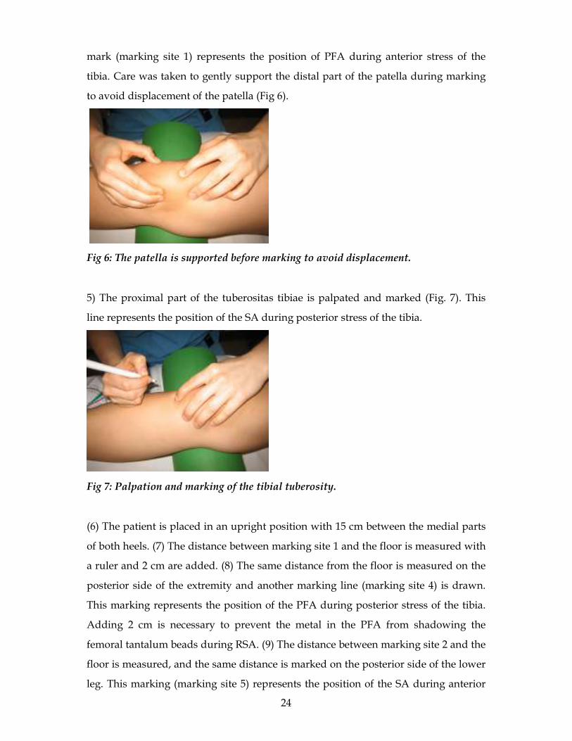
None of the specimens failed during the cyclic loading tests. Repeated-measures
analysis of variance did not show a significant difference between the two groups.
We compared the displacement in the two groups at each load (see table 2). No
40
statistically significant difference between the extraction drilling group and the serial
dilation group was found.
Table 2: Displacement of the graft in millimetre (mm) as a result of increasing load and number of
cycles
________________________________________________________________________________________
Number of cycles/load* Extraction drilling Serial dilation
Mean (SD) Mean (SD) P-value** P-value***
_______________________________________________________________________________________
1000/70-220 N 0.6 (0.2) 0.6 (0.1) 0.90 0.09
1100/70-270 N 0.7 (0.3) 0.7 (0.1) 0.83 0.09
1200/70-320 N 0.9 (0.3) 0.9 (0.2) 0.77 0.10
1300/70-370 N 1.0 (0.4) 1.0 (0.2) 0.70 0.12
1400/70-420 N 1.2 (0.4) 1.2 (0.2) 0.64 0.12
1500/70-470 N 1.4 (0.5) 1.3 (0.3) 0.58 0.12
1600/70-520 N 1.6 (0.6) 1.5 (0.3) 0.54 0.10
*Load is given in Newton (N). ** marked P values describe the differences between extraction drilling
and compaction by serial dilation (Student´s t-test). Standard deviations (SD) are shown in brackets.
*** marked P values describe the differences in standard deviation between the two groups (Pittman´s
test).
Study II
Part study 1
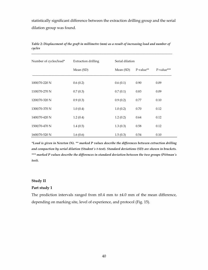
The prediction intervals ranged from ±0.4 mm to ±4.0 mm of the mean difference,
depending on marking site, level of experience, and protocol (Fig. 15).

41
Fig. 15: Mean differences in centimetre between the first and second mark at the six
marking sites are presented with coloured dots and the prediction intervals are
shown with error bars.

42
Part study 2
We found a mean difference in knee laxity of 0.0 mm and a prediction interval of ±5.2
mm. The rotation of the femur in relation to the tibia around the X, Y, Z axes resulted
in mean differences and prediction intervals of 0.6˚(±5.2˚), 0.3˚(±8.0˚), and –2.5˚(±15˚).
For visualization the data is plotted in a Bland Altman plot (Fig. 16).
Fig. 16: Bland Altman plot. The absolute differences between the 1st and 2nd knee
laxity measurements are plotted on the Y-axis against the average of the same
measurements on the X-axis. The green line shows the mean differences between 35
persons. The dotted red lines show the upper and lower limits of agreement (95%
prediction interval).
Study III
Fig. 17 describes the migration of the tibial graft markers inside the tibial tunnel as a
function of time. At all times the migration in group EXDR was larger than in group
SEDI. At 12 weeks, the difference was significant (P = 0.02). At 6 weeks no significant
difference was found (P = 0.12). Borderline significance was reached at 24 weeks (P =
0.06). The results from the adjusted analysis did not differ from the results found in
the univariate analysis.

43
Fig. 17: Migration of the tantalum markers inside the tibial tunnel at 6, 12, and 24
weeks after surgery. Standard deviations are shown with error bars.
Fig. 18 shows the migration of the graft in the femoral tunnel. There was no
statistically significant difference detected between the two groups after 6, 12, and 24
weeks (P = 0.83, P = 0.69, and P = 0.18, respectively).
Fig. 18: Migration of the tantalum markers inside the femoral tunnel at 6, 12, and 24
weeks after surgery. Standard deviations are shown with error bars.

44
Fig. 19 shows the development in absolute knee laxity in the first 24 weeks after
ACL-reconstruction. No significant difference was found at the first follow-up and at
6, 12, and 24 weeks (P = 0.67, P = 0.79, P = 0.09 and P = 0.34, respectively).
Fig. 19: Knee laxity at 7-10 days and 6, 12, and 24 weeks after surgery. Standard
deviations are shown with error bars.
No statistical difference in ∆ knee laxity between the extraction drilling group and
the serial dilation group was found (P = 0.67, P = 0.14, P = 0.17 after 6, 12, and 24
weeks, respectively) (Fig.20).
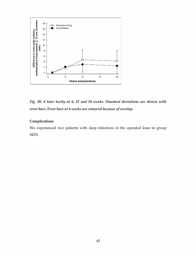
45
Fig. 20: ∆ knee laxity at 6, 12 and 24 weeks. Standard deviations are shown with
error bars. Error bars at 6 weeks are removed because of overlap.
Complications
We experienced two patients with deep infections in the operated knee in group
SEDI.

46

47
8. Discussion
Key findings
Study I
In this study we found no significant difference between compaction by serial
dilation and extraction drilling. In addition we investigated the development in
difference between the two groups as a result of increasing loads and cycles. Again
no significant difference between the two groups could be detected (Table 2).
Looking at Table 2, the standard deviations seem to be smaller in the dilated group
than in the extraction drilling group, which could indicate that compaction by serial
dilation produces a more uniform fixation than does extraction drilling. No
significant difference between the standard deviations in the two groups was found
(Table 2).
Study II
The results of this study show that it is possible to increase the precision of the TSD
positioning by an optimized protocol (aim 1). Regarding marking site B (the tibial
tuberosity), both protocols had an acceptable precision during the “experienced”
period (Fig. 15). At all marking sites the NSP had a superior precision compared with
the OFP. At all marking sites except one (marking site D), the NSP produced an
acceptable precision. In contrast, the original protocol was only able to produce an
acceptable precision at a single marking site (marking site B). At almost all marking
sites, some practice before using the TSD is beneficial.
Looking at the manufactures directions in the original protocol, there is absolutely no
guidance on how to position the proximal and distal stress arms. As a result, the
precision is very poor at marking sites A, C, D, and F. At marking sites A and C, the
prediction interval ranges by approximately ±4 cm. This means that the distance
between the position arms can differ up to 8 cm from one investigation to another,
which subsequently must result in a potential difference in the measured knee laxity.

48
Even though the TSD was precisely applied on the patients’ extremities, the precision
of the A-P knee translation measurements using RSA in our study was very
disappointing, with a precision of more than ±5 mm (aim 2). Khan et al. [46] reported
a precision of ±1.9 mm on six patients with a setup similar to ours. Fleming et al. [20]
reported a higher precision. They published the translation data obtained in five
goats. Using Fleming et al.’s data, we tried to calculate the precision, defined as ±2
SD. This resulted in a precision of ±1.77 mm, which is far more precise compared
with our data.
The results indicate that we were not able to control the rotation of the tibia in
relation to the femur. The rotation around the Z-axis (flexion/extension of the knee)
is especially problematic, with a precision of ±15˚. We inspected the data for the
persons with the largest differences in knee laxity measurements (the outliers in Fig.
16). We found that all persons had a substantial difference in Z-rotation of the knee.
When we apply the TSD, we measure the flexion degree of the knee before force was
applied. The knee tended to extend and flex, when posterior and anterior force,
respectively, were applied to the knee. For some reason, we could not control the
magnitude of this extension/flexion movement, even though we started out with the
same degree of flexion. In future studies, we recommend measuring knee flexion
after force appliance.
Study III
To our knowledge, we are the first to evaluate potential benefits of compaction by
serial dilation after ACL reconstruction in a prospective clinical randomized trial. At
the 3 months follow-up, we found a significantly reduced graft migration at the tibial
fixation site (P = 0.02) in group SEDI compared with group EXDR. At the 6 months
follow-up, we recorded a marginally significant difference (P = 0.06). Since several
patients were excluded from the last follow-up a substantial risk of a type II error
exists for this result. The value of the 6 month follow-up data is thus questionable. In
the power calculation, we defined a difference of 1 mm between the two groups to be
clinically relevant. The mean difference between the two groups was 0.5 mm in
favour of group SEDI (Fig. 17). The graft is introduced to the knee joint at an angle of

49
approximately 50 – 60 degrees in relation to the tibial plateau, which must mean, that
a migration at the tibial fixation site of 0.5 mm results in an increased knee laxity of
less than 0.5 mm. Thus, even though we found a significant difference in graft
migration between the two groups, the clinical relevance is debatable.
In Fig. 17 and Fig. 18 the mean migration of the hamstring graft at the femoral
fixation site is approximately the same as seen at the tibial fixation site. This finding
is surprising, since we theorized that tibial fixation of the hamstring graft is more
demanding than femoral fixation. In addition, we have used a hybrid femoral
fixation, which has been shown to be superior to cortical button fixation or
interference screw fixation alone [58]. Migration at the femoral fixation site is related
to knee laxity [26] and if compaction of the bone tunnel by serial dilators is able to
produce the same effect on the femoral migration as that seen at the tibial fixation
site, a clinically relevant reduction of total migration might be obtained. Further
studies are needed to elucidate this question.
Knee laxity measurements revealed no differences between group SEDI and group
EXDR. Knee laxity increased in both groups from the postoperative measurement to
3 months follow-up. From 3 to 6 months the knee laxity was unchanged or slightly
decreased. The fluctuation of the knee laxity cannot be explained merely by
migration at the fixation site. Other factors could have influenced the results. Despite
a 7- 10 days wait before the first measurement, the patients may still have been
affected by the surgery, and therefore not able to fully relax due to postoperative
pain. This would result in increased muscle tension and an underestimation of the
knee laxity. The slight decrease in knee laxity we observed from the third to the
fourth follow-up could be a result of the knee stabilizing exercises provided by the
rehabilitation. Looking at the standard deviations of the knee translation
measurements, the fluctuation seen in Figs. 19 and 20 could merely be coincidental.
50
Comparison with relevant findings from other studies
As mentioned in the introduction, we are not the first to investigate a possible
beneficial effect of serial dilation in ACL reconstructions using hamstring grafts. The
results of Cain et al. [10] were in line with the findings in our RCT (study 3). In
contrast, other biomechanical studies could not show a significant difference by
using serial dilation at the fixation sites of the hamstring graft [17,56,63]. Looking at
the results from Rittmeister et al. [63], it seems as if the difference between the serial
dilated specimens and their non-dilated controls is highest in the two sub-groups
secured with 7-mm RCI screws. The standard deviations of the sub-groups were not
published, which makes it impossible for us to calculate whether the mean difference
in load at a given permanent displacement is significantly different in the dilated
versus the non- dilated sub-group. The study of Nurmi et al. [56] had one major
limitation. Nine specimens failed during the cyclical loading test. These specimens
were excluded from the data analysis. Six of these failures were in the extraction
drilling group and only three were in the serial dilated group. A different outcome of
the study might have resulted if the failures had been included in the analysis. To
our knowledge, only Gokce et al. [24] have studied the use of compaction by serial
dilators in a clinical study. Forty-four patients were enrolled in a retrospective study.
They found a protective effect of serial dilation on tunnel enlargement. No significant
difference in postoperative Lysholm scores and IKDC scores was found. If that had
been the aim of the study, one could suspect that the study was underpowered.
Limitations/Generalizability
Study I
A bovine set-up was used in this study. Weiler et al.[78] described the BMD in the
proximal part of the bovine tibia. They stated that a calf tibia of approximately 24
weeks of age has the same BMD as that of tibiae in young adults. In Denmark, we do

51
not slaughter calves this young. Our animals were between 32 and 36 weeks of age.
This means that our animals weighed more, and consequently had a higher BMD in
the proximal tibia. Brand et al. [8] showed that BMD is related to fixation strength.
The dislocation in both our groups might be underestimated because of the high
BMD, and from a clinical point of view, we might expect a greater migration of the
tendon in the tibial tunnel in patients undergoing ACL reconstruction.
Amis et al. [12] performed a creep test of the tendons in a study with a bovine set-up
quite similar to that used in this study. They tested five tendons to quantify the creep
(irreversible stretch) and calculated the average creep of these tendons. The average
creep was then subtracted from the measured displacement of the graft, giving the
slippage of the graft. When a constant is subtracted from the displacement in both
groups, one ends up with a smaller mean slippage in both groups. However, the
mean difference, the P value, and the standard deviation of the test will be the same.
Because of that, we did not perform creep tests in this study.
We assume that it takes 6-12 weeks before a proper in-growth of the tendon to the
bone at the tunnel entrance has occurred. A strong fixation is necessary during this
period to prevent slippage of the graft at the fixation site. We tested our fixation-
device complexes with 1600 cycles with varying loads. The number of cycles and
loads approximate the degree of mobilization within the first 7-14 days after surgery.
In addition, we used cadaveric animal material, which means that we tested the
initial fixation strength of serial dilation versus extraction drilling. Therefore, based
on this study and other studies with comparable designs, we are not able to predict
how compaction by serial dilation will perform on a long-term basis. To answer that
question, we need prospective clinical randomized trials with at least 3 months of
follow-up (study III).
In Table 2, there is a steady decrease in P values with an increase in load and number
of cycles. A significant difference was not reached at any time, but the development
is very interesting from a clinical perspective, because the patients perform much
more than 1600 steps during the part of the postoperative period in which the in-

52
healing of the tendon to the bone has not been completed, and migration of the
tendon at the fixation sites is still a possibility. Again, study III should provide the
answer to this question.
Study II
We could have chosen to examine the persons in a random order, minimizing the
risk that the examiner could recognize patient details when performing double
measurements. In this study, there were only a few minutes between the first and
second measurements for the same person, which gives the investigator the
opportunity to remember some of the details of the patient’s knee, resulting in better
reliability. We wanted to correlate the results found in this study with the precision
of knee laxity measurements found in the clinical TSD-RSA study, in which we only
made one or two examinations per day, which necessitated a short time interval
between the first and second measurements. Therefore the same time interval was
used in the TSD study.
One major problem with the TSD is that people feel uncomfortable when it is
applied. They are not able to fully relax. Especially the position of the “healthy” leg is
difficult. Some patients actually experience the TSD application as a painful
experience [42]. This potentially results in a higher degree of muscles tension during
the measurements, which could result in underestimated knee laxity measurements.
In a pilot study, we discovered that use of the extension arm (Fig. 3) contributed to
the aching. In the NSP, we did not use the extension arm. No patient complained of
pain, but we were not able to fully remove the feeling of discomfort.
Another problem is the plasticity of the skin. The stress applied with the TSD allows
the skin to move several cm in relation to the underlying bone. Even though we can
place the stress arms in approximately the same position on the skin, we cannot be
sure that the relation of the stress arms to the femoral and tibial bone can be
repeated. This could result in a different anterior-posterior translation potential of
the knee from measurement to measurement and a reduced knee laxity measurement
precision.

53
We used a ruler to measure the distance from the floor to the marking sites. Use of a
laser pointer could, perhaps, improve the positioning of the TSD and thereby lower
the prediction interval of the knee laxity.
Study III
Only ten patients in group EXDR and eight patients in group SEDI could be analyzed
at the last follow up for different reasons (Fig 14). In group EXDR only one patient
had a negative tibial marker migration on the Y-axis, whereas eight patients in group
SEDI had the same problem and had to be excluded from further analysis. The
question is why did we see this difference? A negative marker migration must result
from loose markers. One explanation for the negative migration could be that the
osseointegration of the graft has ended, and the transformation of tendon to bone
provides a random movement of the graft markers. If this is right, one could
speculate that the difference between the two groups must mean that serial dilation
of the tibial tunnel results in a faster in-healing of the tendon to bone compared with
extraction drilling. Another explanation could be that the migration of the graft
markers could be reversibly influenced by the stress provided by the TSD. The
position of the graft markers at the postoperative measurements would then be
registered too close to the knee joint. As the influence of the TSD diminish over time
as a result of the ongoing osseointegration, the migration of the markers at later
follow-ups could be detected as negative on the Y-axis of the coordinate system,
merely because of a “false” measurement of marker position at baseline. This could
explain some of the negative marker migration seen, but it cannot explain the
difference between the serially dilated group and the extraction drilling group at the
fourth follow-up. To detect whether or not the stress from the TSD cause marker
migration, comparative studies with stressed and unstressed knees are necessary.
From 16 April to 15 July 2008 Danish hospital personnel were on strike. As a result,
we were not able to perform elective procedures during this period. Therefore four
patients (three patients in group EXDR and one patient in group SEDI) had their

54
fourth follow-up postponed. Given the RSA guidelines in Valstar et al. [75], these
four patients were excluded from analysis (Fig 14).
As explained above, RSA measurements were not performed until 7-10 days after
surgery because of pain and swelling of the knee, which could result in increased
muscle tonus during the stress test. Therefore we cannot say anything about the
migration at the fixation site during the first week after surgery. The TSD is not able
to provide precise knee laxity measurements, and our opinion is that it can be left out
in future studies. Thus the first follow-up (not stressed) could then be made very
shortly after surgery, and would therefore also include migration during the first
week.
Another limitation of the study is the interpretation of marker migration in patients
with only one or two markers available for analysis. We placed four markers at the
tibial fixation site and three markers at the femoral fixation site. Markers that
migrated in the wrong direction, indicating that they were loose, were excluded from
analysis. One marker could be placed in the graft, but outside the tibial tunnel, in
which case the marker was excluded from analysis as well. Only one or two tibia
markers would then be available for analysis, which makes it impossible to test for
loose markers, as described by Khan et al. [46]. In these cases, we cannot be certain
whether the marker migration is representative for the actual graft migration or a
result of a random movement of a loose marker. In future studies we recommend
that at least five or six markers be inserted at each fixation site to insure that testing
for loose graft markers is possible.
I a short time period we experienced three patients with deep infections in the
operated knee after arthroscopic knee surgery at the same hospital. Two of the
infections was seen after ACL reconstruction and were both enrolled in this study
and randomized to group SEDI. The last infection resulted after a knee arthroscopy.
Two infections in 40 patients is a high incidence, but we believe it was coincidental
and not a result of the use of tantalum beads or the serial dilation procedure. Of
course this problem calls for awareness in future studies.

55
9. Conclusion
Studies I and III
The biomechanical test in this thesis failed to show a significant difference in initial
fixation strength between serial dilation and extraction drilling of the tibia bone
tunnel in ACL reconstruction. The number of cycles and loads used in study I
resembled the degree of mobilization within the first 7-14 days after surgery. Based
on study I alone, we cannot say anything about long term results after serial dilation.
To overcome that problem we carried out study III. This study showed that
compaction by serial dilation of the tibial tunnel significantly reduced the migration
of the hamstring graft at the tibial fixation site compared with conventional
extraction drilling. The mean difference between the two groups at 3-month follow-
up was 0.5 mm, which must be interpreted as a difference with limited clinical
impact. The mean migration of the hamstring graft at the femoral fixation site was
larger than expected. If serial dilation at the femoral fixation site could produce the
same reduction in graft migration as seen at the tibial fixation site, the use of serial
dilation of the bone tunnels in ACL reconstruction could be clinically beneficial for
the patient. We were not able to show any significant difference in knee laxity
between the serially dilated group and the extraction drilling group. The use of
tantalum markers inserted into the hamstring graft and evaluation of fixation site
graft migration using RSA in clinical studies is still new and some methodological
considerations are still needed.
Study II
This study showed that even though we were able to position the TSD with a good
precision, the combination of the TSD and RSA failed to provide knee laxity
measurements with a acceptable precision. A fast and non invasive method, such as
the KT-1000 arthrometer, is a more precise device for knee laxity measurements and
it provides results almost comparable to most RSA results. RSA is very resource
demanding and invasive, and its role in clinical A-P translation measurements seems
to be limited at the present time. RSA has a high potential due to a high accuracy, but
a stress device that can produce reliable joint translation results is needed.

56

57
10. Perspectives and future research
There are some methodological questions remaining that still need attention.
(1) It is extremely important to investigate whether the measured migration of the
graft markers at the fixation sites truly reflects the migration of the graft or just a
random migration because of loose markers. This could be done by placing five or six
markers at each fixation site, and then measure the distance between the graft
markers at each follow-up. A marker with a migration pattern different from the
other graft markers should be considered loose and excluded from the analysis.
(2) Does the stress provided by the TSD cause reversible marker migration? A
comparative study with stressed and unstressed knees is necessary.
(3) What is the magnitude of the migration of the graft at the fixation sites during the
first 7-10 days?
These questions call for further studies.
The use of tantalum markers inserted into the hamstring graft that allow evaluation
of fixation site-graft migration using RSA in clinical studies has a great potential. In
fact every fixation method introduced on the market could be tested using this
method, including a potential benefit of serial dilation of the femoral bone tunnel. As
mentioned above, some methodological challenges have to be overcome first.
We still believe that RSA can play a part in future knee laxity measurements. At the
moment we do not have a stress device which can provide a precise load application
to the knee. The search for a better stress device is ongoing. A device that could
provide precise anterior-posterior knee translation measurements and precise
rotational instability measurements would be preferable.

58

59
11. References
1. Dansk korsbåndsregister - Årsrapport 2008 (Danish ACL Registry 2008). Ref Type: Report
2. Andersson D, Samuelsson K, Karlsson J (2009) Treatment of anterior cruciate ligament injuries with special reference to surgical technique and rehabilitation: an assessment of randomized controlled trials. Arthroscopy 25:653-685
3. Bach BR, Jr., Warren RF, Flynn WM, Kroll M, Wickiewiecz TL (1990) Arthrometric evaluation of knees that have a torn anterior cruciate ligament. J Bone Joint Surg Am 72:1299-1306
4. Balasch H, Schiller M, Friebel H, Hoffmann F (1999) Evaluation of anterior knee joint instability with the Rolimeter. A test in comparison with manual assessment and measuring with the KT-1000 arthrometer. Knee Surg Sports Traumatol Arthrosc 7:204-208
5. Beynnon BD, Amis AA (1998) In vitro testing protocols for the cruciate ligaments and ligament reconstructions. Knee Surg Sports Traumatol Arthrosc 6 Suppl 1:S70-S76
6. Bland JM, Altman DG (1986) Statistical methods for assessing agreement between two methods of clinical measurement. Lancet 1:307-310
7. Brand J, Jr., Weiler A, Caborn DN, Brown CH, Jr., Johnson DL (2000) Graft fixation in cruciate ligament reconstruction. Am J Sports Med 28:761-774
8. Brand JC, Jr., Pienkowski D, Steenlage E, Hamilton D, Johnson DL, Caborn DN (2000) Interference screw fixation strength of a quadrupled hamstring tendon graft is directly related to bone mineral density and insertion torque. Am J Sports Med 28:705-710
9. Brandsson S, Karlsson J, Sward L, Kartus J, Eriksson BI, Karrholm J (2002) Kinematics and laxity of the knee joint after anterior cruciate ligament reconstruction: pre- and postoperative radiostereometric studies. Am J Sports Med 30:361-367
10. Cain EL, Phillips BB, Charlebois SJ, Azar FM (2005) Effect of tibial tunnel dilation on pullout strength of semitendinosus-gracilis graft in anterior cruciate ligament reconstruction. Orthopedics 28:779-783
11. Christen B, Jakob RP (1992) Fractures associated with patellar ligament grafts in cruciate ligament surgery. J Bone Joint Surg Br 74:617-619

60
12. Coleridge SD, Amis AA (2004) A comparison of five tibial-fixation systems in hamstring-graft anterior cruciate ligament reconstruction. Knee Surg Sports Traumatol Arthrosc 12:391-397
13. Daniel DM, Malcom LL, Losse G, Stone ML, Sachs R, Burks R (1985) Instrumented measurement of anterior laxity of the knee. J Bone Joint Surg Am 67:720-726
14. Daniel DM, Stone ML, Sachs R, Malcom L (1985) Instrumented measurement of anterior knee laxity in patients with acute anterior cruciate ligament disruption. Am J Sports Med 13:401-407
15. Dargel J, Schmidt-Wiethoff R, Bruggemann GP, Koebke J (2007) The effect of bone tunnel dilation versus extraction drilling on the initial fixation strength of press-fit anterior cruciate ligament reconstruction. Arch Orthop Trauma Surg 127:801-807
16. Donahue TL, Gregersen C, Hull ML, Howell SM (2001) Comparison of viscoelastic, structural, and material properties of double-looped anterior cruciate ligament grafts made from bovine digital extensor and human hamstring tendons. J Biomech Eng 123:162-169
17. Dunkin BS, Nyland J, Duffee AR, Brunelli JA, Burden R, Caborn D (2007) Soft tissue tendon graft fixation in serially dilated or extraction-drilled tibial tunnels: a porcine model study using high-resolution quantitative computerized tomography. Am J Sports Med 35:448-457
18. Ejerhed L, Kartus J, Sernert N, Kohler K, Karlsson J (2003) Patellar tendon or semitendinosus tendon autografts for anterior cruciate ligament reconstruction? A prospective randomized study with a two-year follow-up. Am J Sports Med 31:19-25
19. Fleming BC, Brattbakk B, Peura GD, Badger GJ, Beynnon BD (2002) Measurement of anterior-posterior knee laxity: a comparison of three techniques. J Orthop Res 20:421-426
20. Fleming BC, Peura GD, Abate JA, Beynnon BD (2001) Accuracy and repeatability of Roentgen stereophotogrammetric analysis (RSA) for measuring knee laxity in longitudinal studies. J Biomech 34:1355-1359
21. Friden T, Ryd L, Lindstrand A (1992) Laxity and graft fixation after reconstruction of the anterior cruciate ligament. A roentgen stereophotogrammetric analysis of 11 patients. Acta Orthop Scand 63:80-84
22. Friden T, Sommerlath K, Egund N, Gillquist J, Ryd L, Lindstrand A (1992) Instability after anterior cruciate ligament rupture. Measurements of sagittal laxity compared in 11 cases. Acta Orthop Scand 63:593-598
23. Ganko A, Engebretsen L, Ozer H (2000) The rolimeter: a new arthrometer compared with the KT-1000. Knee Surg Sports Traumatol Arthrosc 8:36-39

61
24. Gokce A, Beyzadeoglu T, Ozyer F, Bekler H, Erdogan F (2009) Does bone impaction technique reduce tunnel enlargement in ACL reconstruction? Int Orthop 33:407-412
25. Green JR, Nemzek JA, Arnoczky SP, Johnson LL, Balas MS (1999) The effect of bone compaction on early fixation of porous-coated implants. J Arthroplasty 14:91-97
26. Grover D, Thompson D, Hull ML, Howell SM (2006) Empirical relationship between lengthening an anterior cruciate ligament graft and increases in knee anterior laxity: a human cadaveric study. J Biomech Eng 128:969-972
27. Hatcher J, Hatcher A, Arbuthnot J, McNicholas M (2005) An investigation to examine the inter-tester and intra-tester reliability of the Rolimeter knee tester, and its sensitivity in identifying knee joint laxity. J Orthop Res 23:1399-1403
28. Highgenboten CL, Jackson A, Meske NB (1989) Genucom, KT-1000, and Stryker knee laxity measuring device comparisons. Device reproducibility and interdevice comparison in asymptomatic subjects. Am J Sports Med 17:743-746
29. Highgenboten CL, Jackson AW, Jansson KA, Meske NB (1992) KT-1000 arthrometer: conscious and unconscious test results using 15, 20, and 30 pounds of force. Am J Sports Med 20:450-454
30. Holden JP, Grood ES, Korvick DL, Cummings JF, Butler DL, Bylski-Austrow DI (1994) In vivo forces in the anterior cruciate ligament: direct measurements during walking and trotting in a quadruped. J Biomech 27:517-526
31. Isberg J, Faxen E, Brandsson S, Eriksson BI, Karrholm J, Karlsson J (2006) Early active extension after anterior cruciate ligament reconstruction does not result in increased laxity of the knee. Knee Surg Sports Traumatol Arthrosc 14:1108-1115
32. Isberg J, Faxen E, Brandsson S, Eriksson BI, Karrholm J, Karlsson J (2006) KT-1000 records smaller side-to-side differences than radiostereometric analysis before and after an ACL reconstruction. Knee Surg Sports Traumatol Arthrosc 14:529-535
33. Ishibashi Y, Rudy TW, Livesay GA, Stone JD, Fu FH, Woo SL (1997) The effect of anterior cruciate ligament graft fixation site at the tibia on knee stability: evaluation using a robotic testing system. Arthroscopy 13:177-182
34. Jansson KA, Harilainen A, Sandelin J, Karjalainen PT, Aronen HJ, Tallroth K (1999) Bone tunnel enlargement after anterior cruciate ligament reconstruction with the hamstring autograft and endobutton fixation technique. A clinical, radiographic and magnetic resonance imaging study with 2 years follow-up. Knee Surg Sports Traumatol Arthrosc 7:290-295
35. Jansson KA, Linko E, Sandelin J, Harilainen A (2003) A prospective randomized study of patellar versus hamstring tendon autografts for anterior cruciate ligament reconstruction. Am J Sports Med 31:12-18
62
36. Johnson LL, vanDyk GE (1996) Metal and biodegradable interference screws: comparison of failure strength. Arthroscopy 12:452-456
37. Jonsson H, Elmqvist LG, Karrholm J, Fugl-Meyer A (1992) Lengthening of anterior cruciate ligament graft. Roentgen stereophotogrammetry of 32 cases 2 years after repair. Acta Orthop Scand 63:587-592
38. Jonsson H, Karrholm J, Elmqvist LG (1993) Laxity after cruciate ligament injury in 94 knees. The KT-1000 arthrometer versus roentgen stereophotogrammetry. Acta Orthop Scand 64:567-570
39. Jonsson H, Riklund-Ahlstrom K, Lind J (2004) Positive pivot shift after ACL reconstruction predicts later osteoarthrosis: 63 patients followed 5-9 years after surgery. Acta Orthop Scand 75:594-599
40. Jorn LP, Friden T, Ryd L, Lindstrand A (1997) Persistent stability 3 years after reconstruction of the anterior cruciate ligament. A radiostereometric analysis (RSA) of 20 patients. Acta Orthop Scand 68:427-429
41. Jorn LP, Friden T, Ryd L, Lindstrand A (1998) Simultaneous measurements of sagittal knee laxity with an external device and radiostereometric analysis. J Bone Joint Surg Br 80:169-172
42. Jung TM, Reinhardt C, Scheffler SU, Weiler A (2006) Stress radiography to measure posterior cruciate ligament insufficiency: a comparison of five different techniques. Knee Surg Sports Traumatol Arthrosc 14:1116-1121
43. Karrholm J, Selvik G, Elmqvist LG, Hansson LI (1988) Active knee motion after cruciate ligament rupture. Stereoradiography. Acta Orthop Scand 59:158-164
44. Karrholm J, Selvik G, Elmqvist LG, Hansson LI, Jonsson H (1988) Three-dimensional instability of the anterior cruciate deficient knee. J Bone Joint Surg Br 70:777-783
45. Kartus J, Stener S, Lindahl S, Engstrom B, Eriksson BI, Karlsson J (1997) Factors affecting donor-site morbidity after anterior cruciate ligament reconstruction using bone-patellar tendon-bone autografts. Knee Surg Sports Traumatol Arthrosc 5:222-228
46. Khan R, Konyves A, Rama KR, Thomas R, Amis AA (2006) RSA can measure ACL graft stretching and migration: development of a new method. Clin Orthop Relat Res 448:139-145
47. Kold S, Bechtold JE, Ding M, Chareancholvanich K, Rahbek O, Soballe K (2003) Compacted cancellous bone has a spring-back effect. Acta Orthop Scand 74:591-595
48. Kold S, Rahbek O, Toft M, Ding M, Overgaard S, Soballe K (2005) Bone compaction enhances implant fixation in a canine gap model. J Orthop Res 23:824-830

63
49. Kold S, Rahbek O, Vestermark M, Overgaard S, Soballe K (2005) Bone compaction enhances fixation of weightbearing titanium implants. Clin Orthop Relat Res :138-144
50. Kold S, Rahbek O, Vestermark M, Overgaard S, Soballe K (2006) Bone compaction enhances fixation of weight-bearing hydroxyapatite-coated implants. J Arthroplasty 21:263-270
51. Malcom LL, Daniel DM, Stone ML, Sachs R (1985) The measurement of anterior knee laxity after ACL reconstructive surgery. Clin Orthop Relat Res :35-41
52. Markolf KL, Slauterbeck JR, Armstrong KL, Shapiro MS, Finerman GA (1997) A biomechanical study of replacement of the posterior cruciate ligament with a graft. Part II: Forces in the graft compared with forces in the intact ligament. J Bone Joint Surg Am 79:381-386
53. Muellner T, Bugge W, Johansen S, Holtan C, Engebretsen L (2001) Inter- and intratester comparison of the Rolimeter knee tester: effect of tester's experience and the examination technique. Knee Surg Sports Traumatol Arthrosc 9:302-306
54. Noyes FR, Mangine RE, Barber S (1987) Early knee motion after open and arthroscopic anterior cruciate ligament reconstruction. Am J Sports Med 15:149-160
55. Nurmi JT, Jarvinen TL, Kannus P, Sievanen H, Toukosalo J, Jarvinen M (2002) Compaction versus extraction drilling for fixation of the hamstring tendon graft in anterior cruciate ligament reconstruction. Am J Sports Med 30:167-173
56. Nurmi JT, Kannus P, Sievanen H, Jarvela T, Jarvinen M, Jarvinen TL (2004) Interference screw fixation of soft tissue grafts in anterior cruciate ligament reconstruction: part 1: effect of tunnel compaction by serial dilators versus extraction drilling on the initial fixation strength. Am J Sports Med 32:411-417
57. Nurmi JT, Kannus P, Sievanen H, Jarvinen M, Jarvinen TL (2003) Compaction drilling does not increase the initial fixation strength of the hamstring tendon graft in anterior cruciate ligament reconstruction in a cadaver model. Am J Sports Med 31:353-358
58. Oh YH, Namkoong S, Strauss EJ, Ishak C, Hecker AT, Jazrawi LM, Rosen J (2006) Hybrid femoral fixation of soft-tissue grafts in anterior cruciate ligament reconstruction using the EndoButton CL and bioabsorbable interference screws: a biomechanical study. Arthroscopy 22:1218-1224
59. Papandreou MG, Antonogiannakis E, Karabalis C, Karliaftis K (2005) Inter-rater reliability of Rolimeter measurements between anterior cruciate ligament injured and normal contra lateral knees. Knee Surg Sports Traumatol Arthrosc 13:592-597

64
60. Pelker RR, Friedlaender GE, Markham TC, Panjabi MM, Moen CJ (1984) Effects of freezing and freeze-drying on the biomechanical properties of rat bone. J Orthop Res 1:405-411
61. Rangger C, Daniel DM, Stone ML, Kaufman K (1993) Diagnosis of an ACL disruption with KT-1000 arthrometer measurements. Knee Surg Sports Traumatol Arthrosc 1:60-66
62. Richmond JC, Gladstone J, MacGillivray J (1991) Continuous passive motion after arthroscopically assisted anterior cruciate ligament reconstruction: comparison of short- versus long-term use. Arthroscopy 7:39-44
63. Rittmeister ME, Noble PC, Bocell JR, Jr., Alexander JW, Conditt MA, Kohl HW, III (2001) Interactive effects of tunnel dilation on the mechanical properties of hamstring grafts fixed in the tibia with interference screws. Knee Surg Sports Traumatol Arthrosc 9:267-271
64. Rosen MA, Jackson DW, Atwell EA (1992) The efficacy of continuous passive motion in the rehabilitation of anterior cruciate ligament reconstructions. Am J Sports Med 20:122-127
65. Rupp S, Hopf T, Hess T, Seil R, Kohn DM (1999) Resulting tensile forces in the human bone-patellar tendon-bone graft: direct force measurement in vitro. Arthroscopy 15:179-184
66. Schulz MS, Russe K, Lampakis G, Strobel MJ (2005) Reliability of stress radiography for evaluation of posterior knee laxity. Am J Sports Med 33:502-506
67. Selvik G (1989) Roentgen stereophotogrammetry. A method for the study of the kinematics of the skeletal system. Acta Orthop Scand Suppl 232:1-51
68. Simonian PT, Erickson MS, Larson RV, O'kane JW (2000) Tunnel expansion after hamstring anterior cruciate ligament reconstruction with 1-incision EndoButton femoral fixation. Arthroscopy 16:707-714
69. Simonian PT, Harrison SD, Cooley VJ, Escabedo EM, Deneka DA, Larson RV (1997) Assessment of morbidity of semitendinosus and gracilis tendon harvest for ACL reconstruction. Am J Knee Surg 10:54-59
70. Staubli HU, Jakob RP (1991) Anterior knee motion analysis. Measurement and simultaneous radiography. Am J Sports Med 19:172-177
71. Staubli HU, Noesberger B, Jakob RP (1992) Stressradiography of the knee. Cruciate ligament function studied in 138 patients. Acta Orthop Scand Suppl 249:1-27
72. Steiner ME, Brown C, Zarins B, Brownstein B, Koval PS, Stone P (1990) Measurement of anterior-posterior displacement of the knee. A comparison of the results with instrumented devices and with clinical examination. J Bone Joint Surg Am 72:1307-1315

65
73. Torzilli PA, Panariello RA, Forbes A, Santner TJ, Warren RF (1991) Measurement reproducibility of two commercial knee test devices. J Orthop Res 9:730-737
74. Tyler TF, McHugh MP, Gleim GW, Nicholas SJ (1998) The effect of immediate weightbearing after anterior cruciate ligament reconstruction. Clin Orthop Relat Res :141-148
75. Valstar ER, Gill R, Ryd L, Flivik G, Borlin N, Karrholm J (2005) Guidelines for standardization of radiostereometry (RSA) of implants. Acta Orthop 76:563-572
76. Webster KE, Feller JA, Hameister KA (2001) Bone tunnel enlargement following anterior cruciate ligament reconstruction: a randomised comparison of hamstring and patellar tendon grafts with 2-year follow-up. Knee Surg Sports Traumatol Arthrosc 9:86-91
77. Weiler A, Hoffmann RF, Bail HJ, Rehm O, Sudkamp NP (2002) Tendon healing in a bone tunnel. Part II: Histologic analysis after biodegradable interference fit fixation in a model of anterior cruciate ligament reconstruction in sheep. Arthroscopy 18:124-135
78. Weiler A, Windhagen HJ, Raschke MJ, Laumeyer A, Hoffmann RF (1998) Biodegradable interference screw fixation exhibits pull-out force and stiffness similar to titanium screws. Am J Sports Med 26:119-126
79. Wroble RR, Van Ginkel LA, Grood ES, Noyes FR, Shaffer BL (1990) Repeatability of the KT-1000 arthrometer in a normal population. Am J Sports Med 18:396-399
80. Yasuda K, Tsujino J, Ohkoshi Y, Tanabe Y, Kaneda K (1995) Graft site morbidity with autogenous semitendinosus and gracilis tendons. Am J Sports Med 23:706-714
66
Appendices
THESES FROM THE ORTHOPAEDIC RESEARCH GROUP
PhD and Doctoral Theses from the Orthopaedic Research Group,
www.OrthoResearch.dk,
University Hospital of Aarhus, Denmark
PhD Theses
1. In vivo and vitro stimulation of bone formation with local growth factors
Martin Lind, January 1996
www.OrthoResearch.dk
2. Gene delivery to articular cartilage
Michael Ulrich-Vinther, September 2002
www.OrthoResearch.dk
3. The influence of hydroxyapatite coating on the peri-implant migration of
polyethylene particles
Ole Rahbek, October 2002
www.OrthoResearch.dk
4. Surgical technique's influence on femoral fracture risk and implant fixation.
Compaction versus conventional bone removing techniques
Søren Kold, January 2003
www.OrthoResearch.dk
5. Stimulation and substitution of bone allograft around non-cemented implants
Thomas Bo Jensen, October 2003
www.OrthoResearch.dk
6. The influence of RGD peptide surface modification on the fixation of orthopaedic
implants
Brian Elmengaard, December 2004
www.OrthoResearch.dk
7. Biological response to wear debris after total hip arthroplasty using different
bearing materials
Marianne Nygaard, June 2005
www.OrthoResearch.dk
8. DEXA-scanning in description of bone remodeling and osteolysis around

67
cementless acetabular cups
Mogens Berg Laursen, November 2005
www.OrthoResearch.dk
9. Studies based on the Danish Hip Arthroplasty Registry
Alma B. Pedersen, 2006
www.OrthoResearch.dk
10. Reaming procedure and migration of the uncemented acetabular component in
total hip replacement
Thomas Baad-Hansen, February 2007
www.OrthoResearch.dk
11. On the longevity of cemented hip prosthesis and the influence on implant design
Mette Ørskov Sjøland, April 2007
www.OrthoResearch.dk
12. Combination of TGF-β1 and IGF-1 in a biodegradable coating. The effect on
implant fixation and osseointegration and designing a new in vivo model for
testing the osteogenic effect of micro-structures in vivo
Anders Lamberg, June 2007
www.OrthoResearch.dk
13. Evaluation of Bernese periacetabular osteotomy; Prospective studies examining
projected load-bearing area, bone density, cartilage thickness and migration
Inger Mechlenburg, August 2007
Acta Orthopaedica (Suppl 329) 2008;79
14. Rehabilitation of patients aged over 65 years after total hip replacement - based
on patients’ health status
Britta Hørdam, February 2008
www.OrthoResearch.dk
15. Efficacy, effectiveness, and efficiency of accelerated perioperative care and
rehabilitation intervention after hip and knee arthroplasty
Kristian Larsen, May 2008
www.OrthoResearch.dk
16. Rehabilitation outcome after total hip replacement; prospective randomized
studies evaluating two different postoperative regimes and two different types of
implants
Mette Krintel Petersen, June 2008
www.OrthoResearch.dk

68
17. CoCrMo alloy, in vitro and in vivo studies
Stig Storgaard Jakobsen, June 2008
www.OrthoResearch.dk
18. Adjuvant therapies of bone graft around non-cemented experimental orthopaedic
implants. Stereological methods and experiments in dogs
Jørgen Baas, July 2008
Acta Orthopaedica (Suppl 330) 2008;79
19. The Influence of Local Bisphosphonate Treatment on Implant Fixation
Thomas Vestergaard Jakobsen, December 2008
www.OrthoResearch.dk
20. Surgical Advances in Periacetabular Osteotomy for Treatment of Hip Dysplasia in
Adults
Anders Troelsen, March 2009
Acta Orthopaedica (Suppl 332) 2009;80
21. Polyethylene Wear Analysis. Experimental and Clinical Studies in Total Hip
Arthroplasty.
Maiken Stilling, June 2009
www.OrthoResearch.dk
22. Step-by-step development of a novel orthopaedic biomaterial: A
nanotechnological approach.
Thomas H.L. Jensen, September 2009
www.OrthoResearch.dk
23. Osteoclastic bone resorption in chronic osteomyelitis
Kirill Gromov, November 2009
www.OrthoResearch.dk
24. Use of medications and the risk of revision after primary total hip
arthroplasty
Theis Thillemann, December 2009
www.OrthoResearch.dk
Doctoral Theses
1. Hydroxyapatite ceramic coating for bone implant fixation. Mechanical and
histological studies in dogs
Kjeld Søballe, 1993
Acta Orthop Scand (Suppl 255) 1993;54

69
2. Growth factor stimulation of bone healing. Effects on osteoblasts, osteomies, and
implants fixation
Martin Lind, October 1998
Acta Orthop Scand (Suppl 283) 1998;69
3. Calcium phosphate coatings for fixation of bone implants. Evaluated
mechanically and histologically by stereological methods
Søren Overgaard, 2000
Acta Orthop Scand (Suppl 297) 2000;71
4. Adult hip dysplasia and osteoarthritis. Studies in radiology and clinical
epidemiology
Steffen Jacobsen, December 2006
Acta Orthopaedica (Suppl 324) 2006;77
5. Gene therapy methods in bone and joint disorders. Evaluation of the adeno-
associated virus vector in experimental models of articular cartilage disorders,
periprosthetic osteolysis and bone healing
Michael Ulrich-Vinther, March 2007
Acta Orthopaedica (Suppl 325) 2007;78


71

72