Embed Size (px)
Citation preview
KNEE
Posterior cruciate ligament tears: functionaland postoperative rehabilitation
Casey M. Pierce • Luke O’Brien • Laurie Wohlt Griffin •
Robert F. LaPrade
Received: 8 November 2011 / Accepted: 12 March 2012
� Springer-Verlag 2012
Abstract
Purpose Historically, the results of posterior cruciate
ligament (PCL) reconstructions are not as favourable as
anterior cruciate ligament (ACL) reconstructions, and it is
well recognized that nonoperative treatment and postop-
erative rehabilitation for PCL injuries must be altered
compared to those for ACL injuries. The purpose of this
article was to review current peer-reviewed PCL rehabili-
tation programmes and to recommend a nonoperative and
postoperative programme based on basic science and
published outcomes studies.
Methods To discover the current practices being used to
rehabilitate PCL injuries, we conducted a search of Pub-
Med with the terms ‘‘posterior cruciate ligament’’ and
‘‘rehabilitation’’ from 1983 to 2011. All articles within the
reference lists of these articles were also examined to
determine their rehabilitation programmes.
Results A review of peer-reviewed PCL rehabilitation
protocols revealed that the treatment of PCL injuries depends
on the timing and degree of the injury. Rehabilitation should
focus on progressive weight bearing, preventing posterior
tibial subluxation and strengthening of the quadriceps mus-
cles. General principles of proper PCL rehabilitation,
whether nonoperative or postoperative, should include early
immobilization (when necessary), prone passive range of
motion to prevent placing undue stress on grafts or healing
tissue, and progression of rehabilitation based on biome-
chanical, clinical, and basic science research.
Conclusions An optimal set of guidelines for the non-
operative or postoperative management of PCL injuries has
not yet been defined or agreed upon. Based on the current
review study, suggested guidelines are proposed.
Level of evidence IV.
Keywords PCL �Treatment �Nonoperative rehabilitation �Postoperative rehabilitation � Guidelines
Introduction
Unlike the anterior cruciate ligament (ACL), the posterior
cruciate ligament (PCL) has an intrinsic ability to heal and
may regain continuity following an injury; however, in a
PCL-deficient knee, gravity and the forces on the joint
from the hamstring muscles can potentially cause the tibia
to be positioned in a posteriorly subluxed location relative
to the femur [17, 25, 29, 44–47, 49]. Healing of the PCL in
an elongated position can lead to chronic instability and
disability [46]. The use of a cylindrical cast, which applies
an anterior drawer force, has demonstrated that placing the
PCL in a properly reduced position, with less posterior sag,
allows for improved healing [26]. Patients who have
undergone surgical reconstruction of the PCL commonly
report residual posterior laxity, especially following treat-
ment of chronic tears [2, 27, 43, 54].
Numerous studies have investigated rehabilitation pro-
tocols for patients following ACL reconstruction, but
unfortunately the same cannot be said for patients with a
C. M. Pierce
Steadman Philippon Research Institute,
Vail, CO, USA
L. O’Brien � L. W. Griffin
Howard Head Sports Medicine Center,
Vail, CO, USA
R. F. LaPrade (&)
The Steadman Clinic, 181 W. Meadow Drive,
Suite 400, Vail, CO 81657, USA
e-mail: [email protected]
123
Knee Surg Sports Traumatol Arthrosc
DOI 10.1007/s00167-012-1970-1
PCL injury. This is likely due to the higher incidence of
ACL versus PCL injuries each year and the fact that PCL
tears are less frequently operated on compared to ACL
tears, resulting in more research into ACL rehabilitation
protocols [16, 18]. New investigations into PCL rehabili-
tation protocols could potentially help to improve out-
comes following PCL reconstruction and nonoperative
rehabilitation.
Materials and methods
A systematic search of the literature was conducted
utilizing a PubMed MEDLINE database (PubMed) key-
word search with the keywords ‘‘posterior cruciate liga-
ment’’ and ‘‘rehabilitation’’ (http://www.ncbi.nlm.nih.gov/
pubmed). The articles were categorized by the degree of
injury and the type of treatment employed: operative versus
nonoperative. In addition, all of the articles within the
reference lists of these articles were examined to determine
their rehabilitation programmes and outcomes. The bio-
mechanical properties of the PCL were also investigated
through a literature search to determine the limits and
characteristics that should be considered when developing
an optimal PCL rehabilitation programme. The authors’
current practices and recommendations for treating and
rehabilitating patients with PCL injuries were also
reviewed.
Results
A search performed of the English literature in PubMed
yielded 242 results when searching for ‘‘posterior cruciate
ligament’’ and ‘‘rehabilitation’’. Of the search results, 69 of
the 242 articles mentioned or described aspects of reha-
bilitation protocols and exercises focused specifically on
the treatment and rehabilitation of PCL injuries. Of these
69 articles, 33 were found to describe their PCL rehabili-
tation protocols and are described here. Of the 242 articles,
none were outcome studies comparing different aspects of
PCL rehabilitation.
Treatment/rehabilitation
The treatment of PCL injuries depends on the timing of the
injury, degree of injury, the patient’s complaints, and the
patient’s demands/level of activity [13, 29]. Isolated grade
I and II tears (Table 1) and non-displaced PCL bony
avulsions with a small fragment and grade I/II laxity should
reportedly be initially treated nonoperatively [3, 17, 29].
Rehabilitation should focus on progressive weight bearing
and emphasize quadriceps strengthening, while protecting
the healing ligament or graft [8, 21]. The historical treat-
ment for grade III isolated tears is an area of disagreement,
but most reports note that the knee should be immobilized
in extension for between 2 and 4 weeks to prevent pos-
terior tibial subluxation and decrease tension on the
anterolateral bundle of the PCL [6, 7, 14, 21, 33]. Further
rehabilitation then progresses from that point, but not all
patients recover and a large per cent have been reported to
eventually require reconstruction of the PCL [5, 26, 47].
Rehabilitation protocols for both nonoperative and
operative treatment of PCL injuries are generally divided
into specific time-related and objective findings-related
treatment phases. In general, phase I in both groups
incorporated oedema/effusion control, knee motion within
limits which have been reported to not stress the PCL, and
reactivation of the quadriceps musculature, which was
followed for the initial 4–6 weeks. Phase II, followed for
the next 4–6 weeks, strove for regaining full knee range of
motion, light low-impact strengthening activities, and
avoidance of knee effusions. Further phases are progressed
according to regaining strength, endurance, and agility with
progressive advancement based on both functional testing
and objective examination or stress radiographic outcomes.
There is a lack of quality studies investigating the
effects that different rehabilitation protocols have on PCL
treatment outcomes [29, 35, 55]. Rehabilitation of PCL
injuries, whether following surgery or as part of a nonop-
erative protocol, focuses specifically on quadriceps
strengthening and recovery of proprioception [21, 29, 35].
Studies have reported that patients with PCL injuries who
regained greater quadriceps strength obtained better func-
tional outcomes [40, 50]. The ultimate goal of rehabilita-
tion protocols should be to re-establish a firm end point on
the posterior drawer examination with minimal patient-
reported instability. Patients should be allowed to return to
sports-related activities once they have painless active
range of motion and adequate return of quadriceps strength
[29, 45]. Because PCL reconstructions typically do not
yield as improved objective stability results compared to
ACL reconstructions, it has been advocated that postop-
erative rehabilitation should be more conservative than
ACL rehabilitation [15, 27].
Nonoperative treatment of PCL tears
An optimal set of guidelines for nonoperative management
of PCL injuries has not yet been defined or agreed upon
[19, 41]. However, all nonsurgical treatment modalities
rely on physical therapy and temporary bracing or immo-
bilization to restore normal tibiofemoral positioning [5, 6,
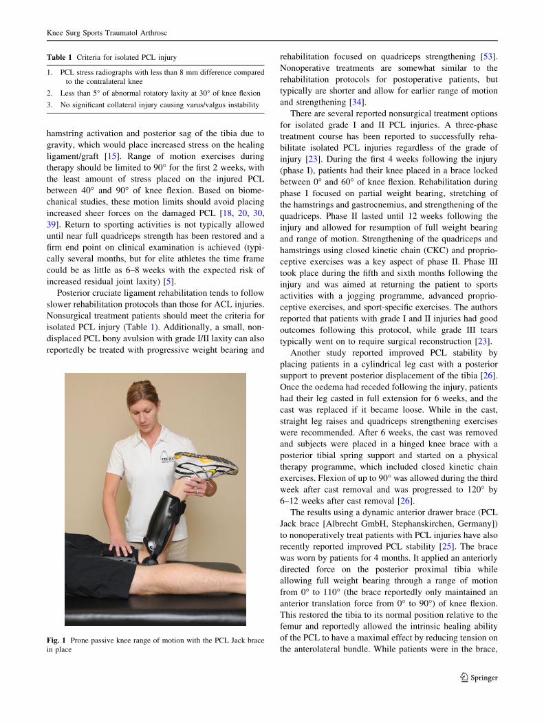
17, 21, 23, 25, 26, 47]. Knee range of motion exercises
should be performed only in the prone position (Fig. 1) for
the first week or weeks following injury/surgery to prevent
Knee Surg Sports Traumatol Arthrosc
123
hamstring activation and posterior sag of the tibia due to
gravity, which would place increased stress on the healing
ligament/graft [15]. Range of motion exercises during
therapy should be limited to 90� for the first 2 weeks, with
the least amount of stress placed on the injured PCL
between 40� and 90� of knee flexion. Based on biome-
chanical studies, these motion limits should avoid placing
increased sheer forces on the damaged PCL [18, 20, 30,
39]. Return to sporting activities is not typically allowed
until near full quadriceps strength has been restored and a
firm end point on clinical examination is achieved (typi-
cally several months, but for elite athletes the time frame
could be as little as 6–8 weeks with the expected risk of
increased residual joint laxity) [5].
Posterior cruciate ligament rehabilitation tends to follow
slower rehabilitation protocols than those for ACL injuries.
Nonsurgical treatment patients should meet the criteria for
isolated PCL injury (Table 1). Additionally, a small, non-
displaced PCL bony avulsion with grade I/II laxity can also
reportedly be treated with progressive weight bearing and
rehabilitation focused on quadriceps strengthening [53].
Nonoperative treatments are somewhat similar to the
rehabilitation protocols for postoperative patients, but
typically are shorter and allow for earlier range of motion
and strengthening [34].
There are several reported nonsurgical treatment options
for isolated grade I and II PCL injuries. A three-phase
treatment course has been reported to successfully reha-
bilitate isolated PCL injuries regardless of the grade of
injury [23]. During the first 4 weeks following the injury
(phase I), patients had their knee placed in a brace locked
between 0� and 60� of knee flexion. Rehabilitation during
phase I focused on partial weight bearing, stretching of
the hamstrings and gastrocnemius, and strengthening of the
quadriceps. Phase II lasted until 12 weeks following the
injury and allowed for resumption of full weight bearing
and range of motion. Strengthening of the quadriceps and
hamstrings using closed kinetic chain (CKC) and proprio-
ceptive exercises was a key aspect of phase II. Phase III
took place during the fifth and sixth months following the
injury and was aimed at returning the patient to sports
activities with a jogging programme, advanced proprio-
ceptive exercises, and sport-specific exercises. The authors
reported that patients with grade I and II injuries had good
outcomes following this protocol, while grade III tears
typically went on to require surgical reconstruction [23].
Another study reported improved PCL stability by
placing patients in a cylindrical leg cast with a posterior
support to prevent posterior displacement of the tibia [26].
Once the oedema had receded following the injury, patients
had their leg casted in full extension for 6 weeks, and the
cast was replaced if it became loose. While in the cast,
straight leg raises and quadriceps strengthening exercises
were recommended. After 6 weeks, the cast was removed
and subjects were placed in a hinged knee brace with a
posterior tibial spring support and started on a physical
therapy programme, which included closed kinetic chain
exercises. Flexion of up to 90� was allowed during the third
week after cast removal and was progressed to 120� by
6–12 weeks after cast removal [26].
The results using a dynamic anterior drawer brace (PCL
Jack brace [Albrecht GmbH, Stephanskirchen, Germany])
to nonoperatively treat patients with PCL injuries have also
recently reported improved PCL stability [25]. The brace
was worn by patients for 4 months. It applied an anteriorly
directed force on the posterior proximal tibia while
allowing full weight bearing through a range of motion
from 0� to 110� (the brace reportedly only maintained an
anterior translation force from 0� to 90�) of knee flexion.
This restored the tibia to its normal position relative to the
femur and reportedly allowed the intrinsic healing ability
of the PCL to have a maximal effect by reducing tension on
the anterolateral bundle. While patients were in the brace,
Table 1 Criteria for isolated PCL injury
1. PCL stress radiographs with less than 8 mm difference compared
to the contralateral knee
2. Less than 5� of abnormal rotatory laxity at 30� of knee flexion
3. No significant collateral injury causing varus/valgus instability
Fig. 1 Prone passive knee range of motion with the PCL Jack brace
in place
Knee Surg Sports Traumatol Arthrosc
123
they were only allowed to remove it to shower and when in
the prone position. At the end of the 4 months, the brace
was removed and physical therapy was initiated to help
regain strength and mobility. Follow-up assessment
showed improvement on radiological evaluation using
bilateral Puddu views and bilateral lateral views in 70� of
flexion and hamstring contraction (from 8.5 to 8.1 mm of
posterior sag to 3.2 and 3.1 mm of posterior sag, respec-
tively). Return to sport-related activities was allowed at
6 months [25].
A suggested nonoperative rehabilitation protocol for
PCL injuries based on a compilation of these studies, with
further stepwise details based on the authors’ clinical
practice, is outlined in Table 2. Patients who fail conser-
vative treatment should be reassessed and considered for
surgical repair or reconstruction. Chronic isolated or
combined PCL injuries may require serial PCL stress
radiographs to objectively gauge injury progression and
dictate treatment modifications [22, 24].
Postoperative rehabilitation of PCL reconstructions
Rehabilitation protocols following repairs and reconstruc-
tion of the PCL have not been clearly outlined [42]. Fol-
lowing surgery, patients are typically placed on a strict
rehabilitation schedule that focuses on quadriceps
strengthening and restoring full range of motion without
stressing the graft [53]. It is very important that patients be
advised that their compliance in the postoperative reha-
bilitation programme is essential to their ultimate outcome.
Depending on the severity of the original injury and the
structures that are concurrently reconstructed or repaired,
rehabilitation protocols will vary somewhat; however, the
approach to rehabilitation should be more conservative
compared to techniques used following ACL reconstruc-
tion [15]. Rehabilitation exercises which cause posterior
tibial translation (e.g. seated leg curls) should be discour-
aged [53]. Regardless of the postoperative treatment pro-
gramme, the joint should be immobilized in extension
immediately after surgery to keep the knee reduced and
curtail posterior tibial sag due to gravity and the hamstring
forces while the graft heals [21, 38].
One reported rehabilitation protocol, which has been
widely accepted and implemented, recommended keeping
the knee locked in a long leg brace in full extension for
3–6 weeks [15, 28]. Patients were kept non-weight bearing
on crutches until the brace was unlocked between postop-
erative weeks 4–6. Progressive range of motion was started
during week 4 and weight bearing with a 25% increase in
body weight per week was initiated during postoperative
week 7. Crutches were not discontinued until the patient
had sufficient quadriceps strength and control for unas-
sisted ambulation. Open kinetic chain (OKC) quadriceps
exercises from 45� to 0� of knee flexion began at week 11,
but OKC resisted knee flexion was not started until
6 months postoperatively. Return to athletics was allowed
between 6 and 9 months following surgery, depending on
return of strength, range of motion, and proprioceptive
skills [15].
Another report utilized a PCL Jack brace starting on day
3 and kept patients non-weight bearing on crutches for
6 weeks following surgery [48]. Range of motion exercises
were initiated on postoperative day 1 and emphasized
prone knee flexion from 0� to 90� and quadriceps
strengthening. Patients were advised to avoid isolated
hamstring exercises until at least 6 weeks postoperatively
to prevent undue stress on the healing repair/graft due to
potential posterior subluxation of the tibia [6]. Partial
weight bearing and hamstring strengthening exercises were
started at 6 weeks postoperatively in addition to leg presses
to a maximum of 70� of knee flexion and riding a stationary
bike. Strengthening exercises included both closed and
open kinetic chain exercises accompanied by functional
training and stability exercises. At 12 weeks postopera-
tively, patients were allowed to begin using low-impact
knee exercises as well as a pool programme [48]. The
authors evaluated the patients at 6 months postoperatively,
both clinically and using posterior knee stress radiographs,
to objectively determine the amount of PCL healing. If
there had been adequate healing of the reconstruction/
repair (\2 mm of increased posterior translation on PCL
stress radiographs compared to the contralateral knee), the
PCL Jack brace was discontinued. Patients were then
allowed to begin a running progression programme, side-
to-side exercises, and proprioceptive exercises. Between 9
and 12 months postoperatively, patients underwent func-
tional testing, which included balance, strength, and
endurance testing to gauge their ability to resume full
activities and sports [48].
A study conducted on patients following a single-bundle
PCL reconstruction used a ‘‘non-aggressive’’ rehabilitation
protocol and reported significant improvements in sub-
jective laxity at an average of 30 months postoperatively
[42]. The protocol was based on bracing to temporarily
reduce shear forces due to gravity and hamstring contrac-
tion, strength training in the quadriceps, and early and
progressive increases in weight bearing. For 45 days fol-
lowing surgery, patients were kept immobilized in a brace
locked in extension, which utilized a foam cushion behind
the tibia to prevent strain on the graft. Minimal weight
bearing on crutches was allowed on day 1, but progressive
weight bearing was not allowed until day 10 and crutches
were required until day 45. Passive flexion was limited to
60� for the first 2 weeks following surgery and was grad-
ually progressed to 95� by day 45 and to 120� by day 90.
Exercises to strengthen the gastrocnemius and quadriceps
Knee Surg Sports Traumatol Arthrosc
123
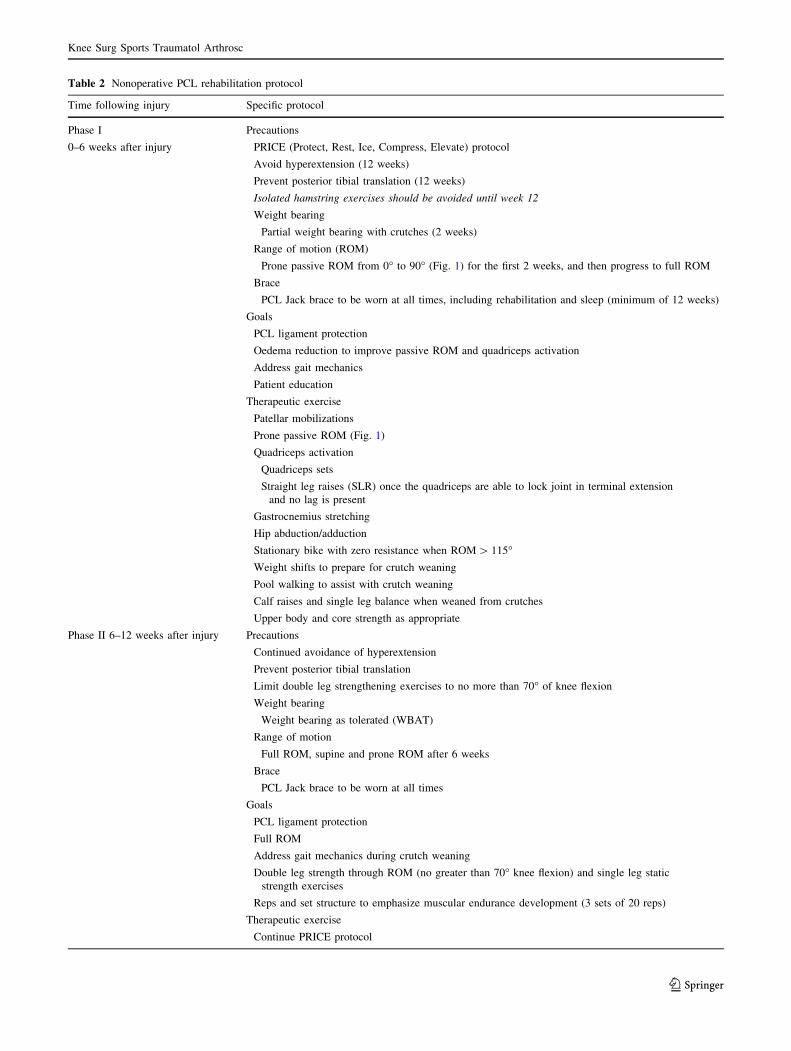
Table 2 Nonoperative PCL rehabilitation protocol
Time following injury Specific protocol
Phase I
0–6 weeks after injury
Precautions
PRICE (Protect, Rest, Ice, Compress, Elevate) protocol
Avoid hyperextension (12 weeks)
Prevent posterior tibial translation (12 weeks)
Isolated hamstring exercises should be avoided until week 12
Weight bearing
Partial weight bearing with crutches (2 weeks)
Range of motion (ROM)
Prone passive ROM from 0� to 90� (Fig. 1) for the first 2 weeks, and then progress to full ROM
Brace
PCL Jack brace to be worn at all times, including rehabilitation and sleep (minimum of 12 weeks)
Goals
PCL ligament protection
Oedema reduction to improve passive ROM and quadriceps activation
Address gait mechanics
Patient education
Therapeutic exercise
Patellar mobilizations
Prone passive ROM (Fig. 1)
Quadriceps activation
Quadriceps sets
Straight leg raises (SLR) once the quadriceps are able to lock joint in terminal extension
and no lag is present
Gastrocnemius stretching
Hip abduction/adduction
Stationary bike with zero resistance when ROM [ 115�Weight shifts to prepare for crutch weaning
Pool walking to assist with crutch weaning
Calf raises and single leg balance when weaned from crutches
Upper body and core strength as appropriate
Phase II 6–12 weeks after injury Precautions
Continued avoidance of hyperextension
Prevent posterior tibial translation
Limit double leg strengthening exercises to no more than 70� of knee flexion
Weight bearing
Weight bearing as tolerated (WBAT)
Range of motion
Full ROM, supine and prone ROM after 6 weeks
Brace
PCL Jack brace to be worn at all times
Goals
PCL ligament protection
Full ROM
Address gait mechanics during crutch weaning
Double leg strength through ROM (no greater than 70� knee flexion) and single leg static
strength exercises
Reps and set structure to emphasize muscular endurance development (3 sets of 20 reps)
Therapeutic exercise
Continue PRICE protocol
Knee Surg Sports Traumatol Arthrosc
123
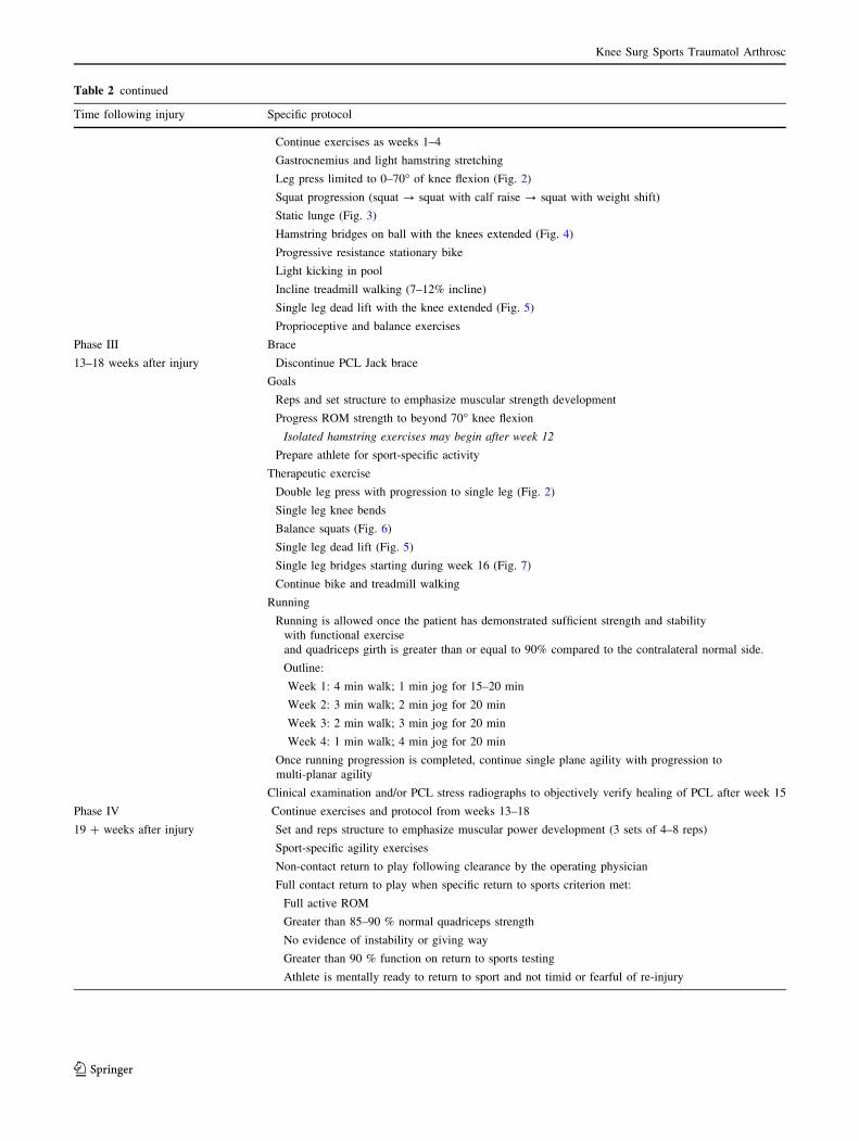
Table 2 continued
Time following injury Specific protocol
Continue exercises as weeks 1–4
Gastrocnemius and light hamstring stretching
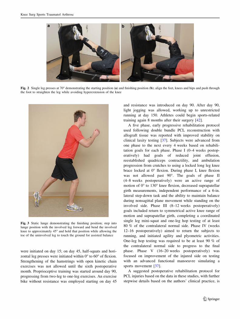
Leg press limited to 0–70� of knee flexion (Fig. 2)
Squat progression (squat ? squat with calf raise ? squat with weight shift)
Static lunge (Fig. 3)
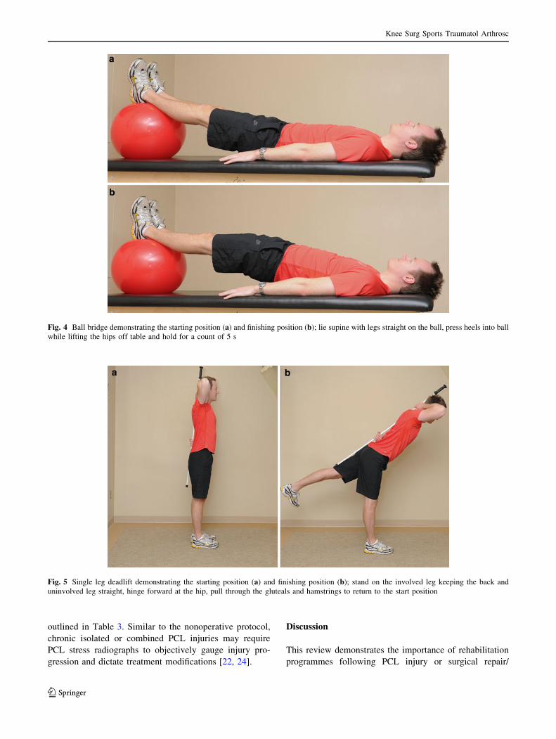
Hamstring bridges on ball with the knees extended (Fig. 4)
Progressive resistance stationary bike
Light kicking in pool
Incline treadmill walking (7–12% incline)
Single leg dead lift with the knee extended (Fig. 5)
Proprioceptive and balance exercises
Phase III
13–18 weeks after injury
Brace
Discontinue PCL Jack brace
Goals
Reps and set structure to emphasize muscular strength development
Progress ROM strength to beyond 70� knee flexion
Isolated hamstring exercises may begin after week 12
Prepare athlete for sport-specific activity
Therapeutic exercise
Double leg press with progression to single leg (Fig. 2)
Single leg knee bends
Balance squats (Fig. 6)
Single leg dead lift (Fig. 5)
Single leg bridges starting during week 16 (Fig. 7)
Continue bike and treadmill walking
Running
Running is allowed once the patient has demonstrated sufficient strength and stability
with functional exercise
and quadriceps girth is greater than or equal to 90% compared to the contralateral normal side.
Outline:
Week 1: 4 min walk; 1 min jog for 15–20 min
Week 2: 3 min walk; 2 min jog for 20 min
Week 3: 2 min walk; 3 min jog for 20 min
Week 4: 1 min walk; 4 min jog for 20 min
Once running progression is completed, continue single plane agility with progression to
multi-planar agility
Clinical examination and/or PCL stress radiographs to objectively verify healing of PCL after week 15
Phase IV
19 ? weeks after injury
Continue exercises and protocol from weeks 13–18
Set and reps structure to emphasize muscular power development (3 sets of 4–8 reps)
Sport-specific agility exercises
Non-contact return to play following clearance by the operating physician
Full contact return to play when specific return to sports criterion met:
Full active ROM
Greater than 85–90 % normal quadriceps strength
No evidence of instability or giving way
Greater than 90 % function on return to sports testing
Athlete is mentally ready to return to sport and not timid or fearful of re-injury
Knee Surg Sports Traumatol Arthrosc
123
were initiated on day 15; on day 45, half-squats and hori-
zontal leg presses were initiated within 0� to 60� of flexion.
Strengthening of the hamstrings with open kinetic chain
exercises was not allowed until the sixth postoperative
month. Proprioceptive training was started around day 90,
progressing from two-leg to one-leg exercises. An exercise
bike without resistance was employed starting on day 45
and resistance was introduced on day 90. After day 90,
light jogging was allowed, working up to unrestricted
running at day 150. Athletes could begin sports-related
training again 8 months after their surgery [42].
A five phase, early progressive rehabilitation protocol
used following double bundle PCL reconstruction with
allograft tissue was reported with improved stability on
clinical laxity testing [37]. Subjects were advanced from
one phase to the next every 4 weeks based on rehabili-
tation goals for each phase. Phase I (0–4 weeks postop-
eratively) had goals of reduced joint effusion,
reestablished quadriceps contractility, and ambulation
progression from crutches to using a locked long leg knee
brace locked at 0� flexion. During phase I, knee flexion
was not allowed past 90�. The goals of phase II
(4–8 weeks postoperatively) were an active range of
motion of 0� to 130� knee flexion, decreased suprapatellar
girth measurements, independent performance of a 6-in.
lateral step-down task and the ability to maintain balance
during nonsagittal plane movement while standing on the
involved side. Phase III (8–12 weeks postoperatively)
goals included return to symmetrical active knee range of
motion and suprapatellar girth, completing a coordinated
single leg mini-squat and one-leg hop testing of at least
80 % of the contralateral normal side. Phase IV (weeks
12–16 postoperatively) aimed to return the subjects to
running, and initiated agility and plyometric activities.
One-leg hop testing was required to be at least 90 % of
the contralateral normal side to progress to the final
phase. Phase V (16–20 weeks postoperatively) was
focused on improvement of the injured side on testing
with an advanced functional manoeuvre simulating a
sports movement [37].
A suggested postoperative rehabilitation protocol for
PCL injuries based on the data in these studies, with further
stepwise details based on the authors’ clinical practice, is
Fig. 2 Single leg presses at 70� demonstrating the starting position (a) and finishing position (b); align the feet, knees and hips and push through
the foot to straighten the leg while avoiding hyperextension of the knee
Fig. 3 Static lunge demonstrating the finishing position; step into
lunge position with the involved leg forward and bend the involved
knee to approximately 45� and hold that position while allowing the
toe of the uninvolved leg to touch the ground for assisted balance
Knee Surg Sports Traumatol Arthrosc
123
outlined in Table 3. Similar to the nonoperative protocol,
chronic isolated or combined PCL injuries may require
PCL stress radiographs to objectively gauge injury pro-
gression and dictate treatment modifications [22, 24].
Discussion
This review demonstrates the importance of rehabilitation
programmes following PCL injury or surgical repair/
Fig. 4 Ball bridge demonstrating the starting position (a) and finishing position (b); lie supine with legs straight on the ball, press heels into ball
while lifting the hips off table and hold for a count of 5 s
Fig. 5 Single leg deadlift demonstrating the starting position (a) and finishing position (b); stand on the involved leg keeping the back and
uninvolved leg straight, hinge forward at the hip, pull through the gluteals and hamstrings to return to the start position
Knee Surg Sports Traumatol Arthrosc
123
reconstruction as a key factor in influencing ligament or
graft healing and improving patient outcomes. Current
practices and reports of PCL rehabilitation programmes do
not show continuity and typically do not address the
majority of important aspects related to successful PCL
rehabilitation. This is especially important because PCL
injuries occur more often than initially thought, and sur-
gical reconstructions of the PCL are increasing [32]. As
such, rehabilitation programmes merit further attention and
agreement in the medical community.
Regardless of the treatment modality selected for a PCL
injury, specific rehabilitation exercises are shared among
treatment protocols. Unlike ACL rehabilitation protocols,
initially keeping a patient non-weight bearing immediately
following PCL reconstruction is important to prevent strain
on the graft, because the PCL is the primary static stabilizer
of the knee [52].
Biomechanical studies have elucidated the sheer forces
that act on the PCL during normal knee movements and
following injury to the PCL [9, 17, 25, 29, 44–47, 49, 52].
During normal knee motion, the PCL is a primary static
stabilizer preventing posterior translation of the femur over
the tibia, and tension in the PCL changes with varying
degrees of knee flexion [32, 52]. Sheer forces on the PCL
have reported to increase at 30� of flexion and to be
greatest at flexion angles higher than 50�–60� [9, 36].
Following PCL injury/reconstruction, isokinetic extension
exercises at less than 70� of knee flexion have been
reported to be safe and will not overstress the ligament/
graft. Isokinetic flexion exercises and deep flexion squats,
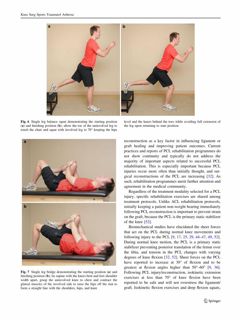
Fig. 6 Single leg balance squat demonstrating the starting position
(a) and finishing position (b); allow the toe of the uninvolved leg to
touch the chair and squat with involved leg to 70� keeping the hips
level and the knees behind the toes while avoiding full extension of
the leg upon returning to start position
Fig. 7 Single leg bridge demonstrating the starting position (a) and
finishing position (b); lie supine with the knees bent and feet shoulder
width apart, grasp the uninvolved knee to chest and contract the
gluteal muscles of the involved side to raise the hips off the mat to
form a straight line with the shoulders, hips, and knee
Knee Surg Sports Traumatol Arthrosc
123
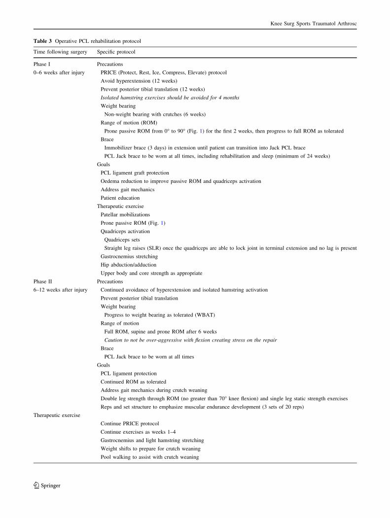
Table 3 Operative PCL rehabilitation protocol
Time following surgery Specific protocol
Phase I
0–6 weeks after injury
Precautions
PRICE (Protect, Rest, Ice, Compress, Elevate) protocol
Avoid hyperextension (12 weeks)
Prevent posterior tibial translation (12 weeks)
Isolated hamstring exercises should be avoided for 4 months
Weight bearing
Non-weight bearing with crutches (6 weeks)
Range of motion (ROM)
Prone passive ROM from 0� to 90� (Fig. 1) for the first 2 weeks, then progress to full ROM as tolerated
Brace
Immobilizer brace (3 days) in extension until patient can transition into Jack PCL brace
PCL Jack brace to be worn at all times, including rehabilitation and sleep (minimum of 24 weeks)
Goals
PCL ligament graft protection
Oedema reduction to improve passive ROM and quadriceps activation
Address gait mechanics
Patient education
Therapeutic exercise
Patellar mobilizations
Prone passive ROM (Fig. 1)
Quadriceps activation
Quadriceps sets
Straight leg raises (SLR) once the quadriceps are able to lock joint in terminal extension and no lag is present
Gastrocnemius stretching
Hip abduction/adduction
Upper body and core strength as appropriate
Phase II Precautions
6–12 weeks after injury Continued avoidance of hyperextension and isolated hamstring activation
Prevent posterior tibial translation
Weight bearing
Progress to weight bearing as tolerated (WBAT)
Range of motion
Full ROM, supine and prone ROM after 6 weeks
Caution to not be over-aggressive with flexion creating stress on the repair
Brace
PCL Jack brace to be worn at all times
Goals
PCL ligament protection
Continued ROM as tolerated
Address gait mechanics during crutch weaning
Double leg strength through ROM (no greater than 70� knee flexion) and single leg static strength exercises
Reps and set structure to emphasize muscular endurance development (3 sets of 20 reps)
Therapeutic exercise
Continue PRICE protocol
Continue exercises as weeks 1–4
Gastrocnemius and light hamstring stretching
Weight shifts to prepare for crutch weaning
Pool walking to assist with crutch weaning
Knee Surg Sports Traumatol Arthrosc
123
Table 3 continued
Time following surgery Specific protocol
Squat progression (squat ? squat with calf raise ? squat with weight shift)
Double leg press (0–70� knee flexion)
Hamstring bridges on ball with the knees extended (Fig. 4)
Stationary bike with zero resistance when ROM [ 115�Light kicking in pool
Phase III
13–18 weeks after injury
Precautions
Patient to remain in Jack PCL brace for all activities
Full weight bearing in Jack PCL brace
Full passive ROM
Avoid isolated hamstring exercise until week 16
Goals
Joint protection
Address gait mechanics
Progressive weight-bearing strength, including progressive hamstring strengthening
Can progress leg press and knee bends past 70� knee flexion after 16 weeks
Therapeutic exercise
Continue as in previous stages
Double leg press 0–70� with progression to single leg (Fig. 2)
Balance squats (Fig. 6)
Squat progression
Single leg bridges starting during week 16 (Fig. 7)
Proprioceptive and balance exercises
Progress stationary bike resistance and duration
Phase IV
19–24 weeks after injury
Precautions
Patient to remain in Jack PCL brace for all activities
Goals
Continue to build strength, and single leg endurance for all lower extremity musculature
with increasing emphasis to developing power
Therapeutic exercise
Continue OKC and CKC strength and endurance work with progressive weight
Initiate initial sport-specific drills near end of this phase
Clinical examination and/or PCL stress radiographs to objectively verify healing of PCL after week 24
Phase V
25–36 weeks after injury
Goals
Patient education and return to activity progressions
Patients can be weaned out of the Jack brace starting at 24 weeks if they are ready
Therapeutic exercise
Initiate absorption activities
Continue strength and endurance exercises, and OKC for quadriceps and hamstrings
Straight line jogging progression:
Outline:
Week 1: 4 min walk; 1 min jog for 15–20 min
Week 2: 3 min walk; 2 min jog for 20 min
Week 3: 2 min walk; 3 min jog for 20 min
Week 4: 1 min walk; 4 min jog for 20 min
Once running progression is completed, continue single plane agility with progression to multi-planar agility
Sport-specific drills
Knee Surg Sports Traumatol Arthrosc
123
on the other hand, should not be attempted until sufficient
time has passed to allow for healing of the injured ligament
or reconstruction graft [9, 51].
In addition, further strain is placed on the PCL during
active contraction of the hamstring muscles [49]. A proper
rehabilitation programme should minimize these forces
during PCL rehabilitation to allow for successful graft/
ligament healing. This is readily accomplished by keeping
the knee immobilized using an anterior directed drawer
force and by not allowing active isolated hamstring exer-
cises until an appropriate time during rehabilitation
(12 weeks after starting a nonoperative rehabilitation pro-
gramme and 24 weeks following surgery). Because graft
healing in PCL reconstructions has been reported to take
nearly twice as long compared to ACL reconstructions, it
has been reported that keeping PCL reconstruction patients
non-weight bearing for 6 weeks is necessary to allow
for adequate graft healing and revascularization to occur
[1, 4, 21].
Eccentric weakness of the quadriceps and hamstrings
has been reported as major factors that need to be
addressed following PCL injuries [31]. This suggests that
eccentric strengthening, including open and closed kinetic
chain exercises, should be a vital part of any therapy. Open
and closed kinetic chain exercises are the foundation of
PCL rehabilitation protocols; however, OKC exercises
should only be used with limited flexion angles until the
ligament/graft has had adequate time to heal [36].
Open kinetic chain exercises are able to isolate single
muscle groups for strengthening, which makes them
especially important in the early weeks following PCL
injury or surgery [36]. However, OKC exercises that acti-
vate the hamstrings should be avoided in the initial phases
of PCL rehabilitation, because studies have reported that
they can stretch out grafts or cause further injury to the
already damaged ligament [29, 30].
Closed kinetic chain exercises are unable to isolate a
single muscle group because they activate antagonistic
muscle groups across multiple joints [30]. They can also
produce increased shear forces on the healing ligament. For
these reasons, CKC exercises should be initially avoided
while OKC exercises are used to strengthen the quadriceps
during the early stages of rehabilitation [56].
Closed chain exercises, including squats and leg presses
(Fig. 2), are ideal for strengthening the quadriceps and gluteal
muscles [30]. It has been reported that the eccentric squat is an
excellent exercise to increase quadriceps strength during any
form of lower extremity rehabilitation [32]. Strengthening the
quadriceps is especially important in PCL rehabilitation,
because the quadriceps secondarily contribute to anteropos-
terior stability with the PCL, and, as previously stated,
patients with improved quadriceps strength typically achieve
significantly better outcomes following PCL injury [32].
Escamilla et al. [10] favoured leg presses with a narrow stance
over squats during the initial phases of PCL rehabilitation.
This is because squats generate greater PCL tensile forces
than leg presses over varying knee flexion angles. Once the
quadriceps strength of the injured side is great than or equal to
90 % compared to the uninjured side, the patient can begin a
progression of running activities [53].
Reports have suggested that therapists and physicians
should use caution when allowing patients to begin forward
and side lunge exercises in the rehabilitation process, due
to the high forces on the PCL that are generated by these
exercises [11]. Lower knee flexion angles and a shorter
stride lunge should be used when starting such exercises,
because they have been reported to generate the least force
on the PCL [12].
The limitations of this study are that it is a review article
and does not have any outcome data to support the rec-
ommendations made. The studies which were reviewed all
came from the English-based literature and reports pub-
lished in other languages were not considered. This
review clearly demonstrates that there is a paucity of
peer-reviewed data comparing suggested forms of PCL
rehabilitation and the impact they have on patient out-
comes. Therefore, future research is needed to investigate
and establish an accepted protocol for PCL rehabilitation.
Based on these reports, the studies reviewed above,
and the author’s clinical experience, recommended
postoperative and nonoperative programme for patients
following PCL injury are presented in Tables 2 and 3,
respectively.
Conclusions
An optimal set of guidelines for nonoperative or post-
operative management of PCL injuries has not yet been
defined or agreed upon. There is a lack of peer-reviewed
publications comparing the subjective and objective out-
comes of both postoperative PCL rehabilitation and
nonoperative treatment programmes. Future studies need
to define outcomes for various PCL rehabilitation pro-
grammes to allow practitioners to agree on and imple-
ment the most effective protocols to improve patient
outcomes.
Acknowledgments This research was supported by the Steadman
Philippon Research Institute, which is a 501(c)(3) non-profit institu-
tion supported financially by private donations and corporate support
from the following entities: Smith & Nephew Endoscopy, Arthrex,
Inc., Siemens Medical Solutions USA, Inc., OrthoRehab, ConMed
Linvatec, Ossur Americas, Small Bone Innovations, Inc., and Opedix.
One of the authors is a paid consultant for Arthrex.
Conflict of interest None.
Knee Surg Sports Traumatol Arthrosc
123
References
1. Bellelli A, Adriani E, Margheritini F, Camillieri G, Della Rocca
C, Mariani PP (1999) Synovial healing in reconstructed cruciate
ligaments. Our personal experience compared in single inter-
ventions and combined reconstructions. Radiol Med 98:454–461
2. Bergfeld JA, Graham SM, Parker RD, Valdevit AD, Kambic HE
(2005) A biomechanical comparison of posterior cruciate liga-
ment reconstructions using single- and double-bundle tibial inlay
techniques. Am J Sports Med 33:976–981
3. Miller RH, Azar FM (2007) Knee Injuries. In: Canale ST, Beaty
JH (eds) Campbell’s operative orthopedics, 11th edn. Elsevier,
Maryland Heights, pp 2552–2565
4. Clancy WG Jr, Narechania RG, Rosenberg TD, Gmeiner JG,
Wisnefske DD, Lange TA (1981) Anterior and posterior cruciate
ligament reconstruction in rhesus monkeys. J Bone Joint Surg
Am 63:1270–1284
5. Colvin AC, Meislin RJ (2009) Posterior cruciate ligament injuries
in the athlete: diagnosis and treatment. Bull NYU Hosp Jt Dis
67:45–51
6. Cosgarea AJ, Jay PR (2001) Posterior cruciate ligament injuries:
evaluation and management. J Am Acad Orthop Surg 9:297–307
7. Dandy DJ, Pusey RJ (1982) The long-term results of unrepaired
tears of the posterior cruciate ligament. J Bone Joint Surg Br
64:92–94
8. Edson CJ, Fanelli GC, Beck JD (2010) Postoperative rehabilita-
tion of the posterior cruciate ligament. Sports Med Arthrosc
18:275–279
9. Escamilla RF (2001) Knee biomechanics of the dynamic squat
exercise. Med Sci Sports Exerc 33:127–141
10. Escamilla RF, Fleisig GS, Zheng N, Lander JE, Barrentine SW,
Andrews JR, Bergemann BW, Moorman CT (2001) Effects of
technique variations on knee biomechanics during the squat and
leg press. Med Sci Sports Exerc 33:1552–1566
11. Escamilla RF, Zheng N, MacLeod TD, Imamura R, Edwards
WB, Hreljac A, Fleisig GS, Wilk KE, Moorman CT, Paulos L,
Andrews JR (2010) Cruciate ligament tensile forces during the
forward and side lunge. Clin Biomech 25:213–221
12. Escamilla RF, Zheng N, Macleod TD, Imamura R, Edwards WB,
Hreljac A, Fleisig GS, Wilk KE, Moorman CT 3rd, Paulos L,
Andrews JR (2010) Cruciate ligament forces between short-step and
long-step forward lunge. Med Sci Sports Exerc 42:1932–1942
13. Fanelli GC (1993) Posterior cruciate ligament injuries in trauma
patients. Arthroscopy 9:291–294
14. Fanelli GC, Boyd JL, Heckler MW (2009) How I manage
posterior cruciate ligament injuries. Oper Tech Sports Med
17:175–193
15. Fanelli GC (2008) Posterior cruciate ligament rehabilitation: how
slow should we go? Arthroscopy 24:234–235
16. Fanelli GC, Edson CJ (1995) Posterior cruciate ligament injuries
in trauma patients: part II. Arthroscopy 11:526–529
17. Fowler PJ, Messieh SS (1987) Isolated posterior cruciate liga-
ment injuries in athletes. Am J Sports Med 15:553–557
18. Fox RJ, Harner CD, Sakane M, Carlin GJ, Woo SLY (1998)
Determination of the in situ forces in the human posterior cruciate
ligament using robotic technology a cadaveric study. Am J Sports
Med 26:395–401
19. Grassmayr MJ, Parker DA, Coolican MR, Vanwanseele B (2008)
Posterior cruciate ligament deficiency: biomechanical and bio-
logical consequences and the outcomes of conservative treatment.
A systematic review. J Sci Med Sport 11:433–443
20. Grood ES, Stowers SF, Noyes FR (1988) Limits of movement in
the human knee: effect of sectioning the posterior cruciate liga-
ment and posterolateral structures. J Bone Joint Surg Am
70A:88–97
21. Harner CD, Hoher J (1998) Evaluation and treatment of posterior
cruciate ligament injuries. Am J Sports Med 26:471–482
22. Hewett TE, Noyes FR, Lee MD (1997) Diagnosis of complete
and partial posterior cruciate ligament ruptures. Stress radiogra-
phy compared with KT-1000 arthrometer and posterior drawer
testing. Am J Sports Med 25:648–655
23. Ittivej K, Prompaet S, Rojanasthien S (2005) Factors influencing
the treatment of posterior cruciate ligament injury. J Med Assoc
Thai 88(Supp 5):S84–S88
24. Jackman T, LaPrade RF, Pontinen T, Lender PA (2008) Intra-
observer and interobserver reliability of the kneeling technique of
stress radiography for the evaluation of posterior knee laxity. Am
J Sports Med 36:1571–1576
25. Jacobi M, Reischl N, Wahl P, Gautier E, Jakob RP (2010) Acute
isolated injury of the posterior cruciate ligament treated by a
dynamic anterior drawer brace. J Bone Joint Surg Br
92:1381–1384
26. Jung YB, Tae SK, Lee YS, Jung HJ, Nam CH, Park SJ (2008)
Active non-operative treatment of acute isolated posterior cruci-
ate ligament injury with cylinder cast immobilization. Knee Surg
Sports Traumatol Arthrosc 16:729–733
27. Lenschow S, Zantop T, Weimann A, Lemburg T, Raschke M,
Strobel M, Petersen W (2006) Joint kinematics and in situ forces
after single bundle PCL reconstruction: a graft placed at the
center of the femoral attachment does not restore normal pos-
terior laxity. Arch Orthop Trauma Surg 126:253–259
28. Levy BA, Boyd JL, Stuart MJ (2011) Surgical treatment of acute
and chronic anterior and posterior cruciate ligament and lateral
side injuries of the knee. Sports Med Arthrosc Rev 19:110–
119
29. Lopez-Vidriero E, Simon DA, Johnson DH (2010) Initial evalu-
ation of posterior cruciate ligament injuries: history, physical
examination, imaging studies, surgical and nonsurgical indica-
tions. Sports Med Arthrosc 18:230–237
30. Lutz GE, Palmitier RA, An KN, Chao EY (1993) Comparison of
tibiofemoral joint forces during open-kinetic-chain and closed
kinetic-chain exercises. J Bone Joint Surg Am 75:732–739
31. MacLean CL, Taunton JE, Clement DB, Regan W (1999)
Eccentric and concentric isokinetic moment characteristics in the
quadriceps and hamstrings of the chronic isolated posterior cru-
ciate ligament injured knee. Br J Sports Med 33:405–408
32. MacLean CL, Taunton JE, Clement DB, Regan WD, Stanish WD
(1999) Eccentric kinetic chain exercise as a conservative means
of functionally rehabilitating chronic isolated insufficiency of the
posterior cruciate ligament. Clin J Sport Med 9:142–150
33. Margheritini F, Rihn J, Musahl V, Mariani PP, Harner C (2002)
Posterior cruciate ligament injuries in the athlete: an anatomical,
biomechanical and clinical review. Sports Med 32:393–408
34. Markey KL (1991) Functional rehabilitation of the cruciate-
deficient knee. Sports Med 12:407–417
35. Matava MJ, Ellis E, Gruber B (2009) Surgical treatment of
posterior cruciate ligament tears: an evolving technique. J Am
Acad Orthop Surg 17:435–446
36. Mesfar W, Shirazi-Adl A (2008) Knee joint biomechanics in
open-kinetic-chain flexion exercises. Clin Biomech 23:477–482
37. Nyland J, Hester P, Caborn DN (2002) Double-bundle posterior
cruciate ligament reconstruction with allograft tissue: 2-year
postoperative outcomes. Knee Surg Sports Traumatol Arthrosc
10:274–279
38. Ogata K, McCarthy JA (1992) Measurements of length and
tension patterns during reconstruction of the posterior cruciate
ligament. Am J Sports Med 20:351–355
39. Pandy MG, Shelburne KB (1997) Dependence of cruciate-liga-
ment loading on muscle forces and external load. J Biomech
30:1015–1024
Knee Surg Sports Traumatol Arthrosc
123
40. Parolie JM, Bergfeld JA (1986) Long-term results of nonopera-
tive treatment of isolated posterior cruciate ligament injuries in
the athlete. Am J Sports Med 14:35–38
41. Petrigliano FA, McAllister DR (2006) Isolated posterior cruciate
ligament injuries of the knee. Sports Med Arthrosc 14:206–212
42. Quelard B, Sonnery-Cottet B, Zayni R, Badet R, Fournier Y,
Hager JP, Chambat P (2010) Isolated posterior cruciate ligament
reconstruction: is non-aggressive rehabilitation the right proto-
col? Orthop Traumatol Surg Res 96:256–262
43. Sekiya JK, West RV, Ong BC, Irrgang JJ, Fu FH, Harner CD
(2005) Clinical outcomes after isolated arthroscopic single-bun-
dle posterior cruciate ligament reconstruction. Arthroscopy
21:1042–1050
44. Shelbourne KD, Davis TJ, Patel DV (1999) The natural history of
acute, isolated, nonoperatively treated posterior cruciate ligament
injuries: a prospective study. Am J Sports Med 27:276–283
45. Shelbourne KD, Gray T (2002) Natural history of acute posterior
cruciate ligament tears. J Knee Surg 15:103–107
46. Shelbourne KD, Jennings RW, Vahey TN (1999) Magnetic res-
onance imaging of posterior cruciate ligament injuries: assess-
ment of healing. Am J Knee Surg 12:209–213
47. Shelbourne KD, Muthukaruppan Y (2005) Subjective results of
nonoperatively treated, acute, isolated posterior cruciate ligament
injuries. Arthroscopy 21:457–461
48. Spiridonov SI, Slinkard NJ, LaPrade RF (2011) Isolated and
combined grade III PCL tears treated with double bundle
reconstructions using an endoscopic femoral graft placement:
operative technique and clinical outcomes. J Bone Joint Surg Am
93:1773–1780
49. Strobel MJ, Weiler A, Schulz MS, Russe K, Eichhorn HJ (2002)
Fixed posterior subluxation in posterior cruciate ligament-defi-
cient knees: diagnosis and treatment of a new clinical sign. Am J
Sports Med 30:32–38
50. Torg JS, Barton TM, Pavlov H, Stine R (1989) Natural history of
the posterior cruciate ligament-deficient knee. Clin Orthop Relat
Res 246:208–216
51. Toutoungi DE, Lu TW, Leardini A, Catani F, O’Connor JJ (2000)
Cruciate ligament forces in the human knee during rehabilitation
exercises. Clin Biomech 15:176–187
52. Van Dommelon BA, Fowler PJ (1989) Anatomy of the posterior
cruciate ligament. A review. Am J Sports Med 17:24–29
53. Veltri DM, Warren RF (1993) Isolated and combined posterior
cruciate ligament injuries. J Am Acad Orthop Surg 1:67–75
54. Veltri DM, Warren RF, Silver G (1993) Complications in pos-
terior cruciate ligament surgery. Oper Techn Sport Med
1:154–158
55. Watsend AM, Oestad TM, Jakobsen RB, Engebretsen L (2009)
Clinical studies on posterior cruciate ligament tears have weak
design. Knee Surg Sports Traumatol Arthrosc 17:140–149
56. Wilk KE (1994) Rehabilitation of isolated and combined pos-
terior cruciate ligament injuries. Clin Sports Med 13:649–677
Knee Surg Sports Traumatol Arthrosc
123